29
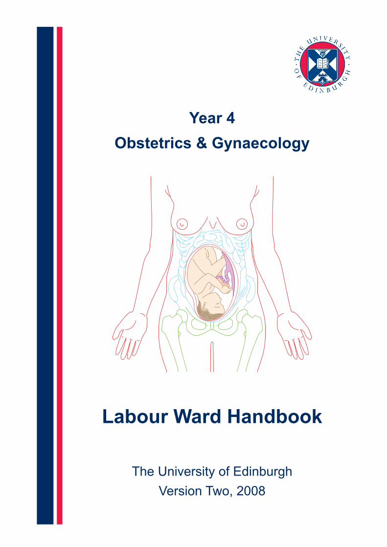
Year 4 Obstetrics & Gynaecology Labour Ward Handbook The University of Edinburgh Version Two, 2008
| Date post: | 08-Jul-2018 |
| Category: |
Documents |
| Upload: | duonghuong |
| View: | 225 times |
| Download: | 4 times |
Year 4
Obstetrics & Gynaecology
Labour Ward Handbook
The University of Edinburgh
Version Two, 2008

Labour Ward Handbook
Page 2 The University of Edinburgh
Year 4 Medical Student
Labour Ward WorkbookThis workbook is designed to be a revision aid and will help you make the most of your time on the labour ward. The questions all relate to common scenarios on labour ward. However, not all the answers will be easily obtained from books. One of the best ways to go through the workbook is to think about the answers and then discuss various questions with the labour ward staff. If you can answer all of the questions then you will have a good idea about life on the labour ward and will be able to acquit yourself well in the exam if any questions about labour are asked.
1. Normal labourLabour is the process where the onset of uterine contractions leads to progressive cervical dilatation. To effect this, the closed cervix dilates until it is fully confluent with the vagina. The fetus can then descend through the birth canal until it is born. Delivery of the baby is followed by delivery of the placenta. The dilation of the cervix, descent and expulsion of the fetus are effected by regular uterine contractions. The progress of labour is assessed by regular vaginal examination. The first stage of labour lasts until the cervix is fully dilated. This can be divided into two phases. The cervix starts off as closed, firm, posterior and 3 cm long. The first phase is where the cervix becomes softer, shorter and more anteriorly situated. The fetal head descends and the cervix begins to dilate. This phase can last for days and may or may not be associated with painful contractions. This phase ends when the cervix is fully effaced and around 3 cm dilated. The next phase is the active phase of labour; this is more predictable in timing and is associated with increasingly powerful contractions. The cervix dilates progressively until it is fully dilated and the fetal head can pass through. The second stage of labour is from when the cervix is fully dilated until the baby is born. The third stage of labour ends with the delivery of the placenta. Traditionally labour has three components; the passage, the passenger and the powers.
a) The Passage
The progress of labour is assessed by regular vaginal examination. Before the active phase of labour there is little change in cervical dilation but the cervix can change markedly. Therefore more information than the dilation alone needs to be recorded. The change in the cervix can be described by using the modified Bishop score. As with any vaginal examination in labour, the assessment should begin with abdominal palpation.
Abdominal Palpation
In any abdominal palpation you should comment on the symphysiofundal height, the number of fetuses, the lie, the presentation, engagement of the fetal head, the position of the fetal back and a clinical impression of the liquor volume.

Labour Ward Handbook
Page 3 The University of Edinburgh
Abdominal Palpation (continued)
Perform an abdominal palpation and consider these questions:
l What is the fetal lie in a normal labour and what are the alternatives?
l What is the presentation and what are the alternatives?
l What is meant by engagement of the fetal head?
l Why is it useful to know the position of the fetal back?

Labour Ward Handbook
Page 4 The University of Edinburgh
Vaginal Examination
In Labour Ward, look at the case notes and discuss with staff the use of the Bishop score.
l What is meant by the station of the fetal head?
l What are the features involved in the Bishop score?
0 1 2 3
Labour Ward Handbook
Page 5 The University of Edinburgh
The active phase of labour
In Labour Ward, look after a woman in labour and consider the following questions:
l What features of labour does the Partogram record?
l What is the benefit of using a Partogram?
l How often should we make the various observations on the Partogram?
l What rate do we expect the cervix to dilate at and does this depend on a woman’s parity?
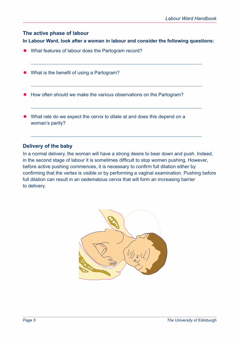
Delivery of the baby
In a normal delivery, the woman will have a strong desire to bear down and push. Indeed, in the second stage of labour it is sometimes difficult to stop women pushing. However, before active pushing commences, it is necessary to confirm full dilation either by confirming that the vertex is visible or by performing a vaginal examination. Pushing before full dilation can result in an oedematous cervix that will form an increasing barrier to delivery.

Labour Ward Handbook
Page 6 The University of Edinburgh
Be involved in the normal delivery of women in the labour ward
l Why should you control the delivery of the head?
l What are the indications for an episiotomy?
l What should you do once the head is delivered?
l How do you deliver the baby’s shoulders?
l What do we do if there has been meconium staining of the liquor?
b) Passenger
During vaginal examination, attention is paid to the position of the baby’s head. The saggital suture can be felt running anteriorly to posteriorly. This narrows down the position of the baby as the occiput will be at one end of the saggital suture and the forehead at the other. The exact position of the baby’s head can be determined with reference to the fontanelles. At the anterior end of the saggital suture lies the rhomboid anterior fontanelle, whereas at the posterior end lies the triangular posterior fontanelle. The position of the occiput is recorded.
The Fetal Skull
Right Occipito-Transverse(ROT)
? ?Left Occipito-Posterior(LOP)
Direct Occipito-Anterior(DOA)

Labour Ward Handbook
Page 7 The University of Edinburgh
l How can you confirm the position of the head at full dilatation if not sure?
The condition of the baby is assessed at 1 and 5 minutes, and later if required, using an Apgar score. This looks at five clinical features of the neonate and scores them 0, 1 or 2. The total Apgar score is out of 10. Low Apgar scores at 20 minutes of age have been associated with the development of cerebral palsy. However, Apgar scores are not used to guide resuscitation.
l What is involved in performing an Apgar score?
Measurement 0 1 2 3
Help the midwife weigh, measure and check the baby
l What is the normal weight of a baby in kg at term?
l What problems are more common in term babies which are smaller than normal (eg less than 2.5kg)?
l What problems are more common in term babies which are bigger than normal (eg more than 7.0 kg)?
Sometimes a developmental abnormality of the baby is obvious. This may or may not be anticipated. This requires sensitive handling from the midwife, paediatrician and obstetrician.
Labour Ward Handbook
Page 8 The University of Edinburgh
c) The Powers
As well as regular vaginal examination, the assessment of progress in labour depends on the assessment of contractions. In the early stages of labour, contractions are stimulated by prostaglandins. Prostaglandins also increase the sensitivity of the uterus to oxytocin. Oxytocin is released by the posterior pituitary gland as part of a neuroendocrine feedback loop. In the early stages of labour mobilisation helps establish the contractions and aids the descent of the head. The timing of contractions can be recorded on a tocograph. However the tocograph does not tell you how strong the contractions are. The nature of the contractions is best performed by abdominal palpation.
Use the time looking after women in labour to feel and grade contractions
l How does the contraction spread through the uterus?
l What is the expected timing and duration of contractions in established labour?
Fill in the contractions on the Partogram
l What usually happens to the contractions when the membranes rupture?
l What can happen to the fetus if the woman is over-contracting?
Labour Ward Handbook
Page 9 The University of Edinburgh
2. Induction of labourLabour can be induced by exogenous administration of the hormones involved in the normal initiation of labour. If the cervix is already 2-3 cm dilated, labour can be induced by amniotomy alone. In many cases, contractions will follow artificial rupture of the membranes. If the contractions are poor or the cervix fails to dilate, intravenous oxytocin can augment the induction. The uterus responds poorly to exogenous oxytocin in the presence of intact membranes. In general, during induction the cervix is not dilated enough for direct amniotomy. In this case, vaginal prostaglandin E is given at regular intervals. This will increase the Bishop score and induce some uterine activity. The cervical change can be monitored by regular vaginal examination and amniotomy performed when appropriate.
Discuss induction of labour with staff
l How does the induction protocol differ for primigravid and parous women?
l How often does induction of labour fail?
l What factors are associated with failed induction of labour?
l What is the risk to amniotomy if the head is free on abdominal palpation?
l How is amniotomy performed in these cases?
l What is the main worry about giving vaginal prostaglandin E?
l How can this risk be minimised?
l When are vaginal prostaglandins contraindicated for induction?
l What are the main indications for induction of labour?
Labour Ward Handbook
Page 10 The University of Edinburgh
3. Fetal monitoringThe assessment of the fetus in labour depends on a careful consideration of the antenatal course of the pregnancy, the liquor and the heart rate changes of the fetus during labour. It is the whole picture rather than any individual feature which helps us pick up a compromised fetus.
To improve your knowledge of how we asses fetus during labour you can:
i) Help with fetal monitoring of a woman in labour.
ii) Attend the CTG meetings.
iii) Read the following summary of fetal monitoring and assess the CTG examples.
An Idiot’s Guide to Fetal MonitoringIs labour stressful for the baby?
A great force is required to be applied to the fetus during childbirth to allow its delivery. Each contraction exerts a force of 1kg per cm² and this is doubled during the second stage of labour when the woman is pushing. To get an idea of what that is like, try to imagine six 12 stone men standing on your foot at the same time! It’s no wonder the fetus gets distressed. Actually, most babies cope very well with these forces, which are mainly on the head, and the fetal scalp is designed to allow a lot of remoulding and squashing. Most babies are born with a pointed head which disappears soon after delivery.
How easy it is for a baby to become distressed?
The ability of the baby to cope with the stresses of labour depends on the physiological reserves which are present when labour starts. A fully mature baby which is well grown will have good reserves. However, premature babies and babies which have not been able to reach their growth potential because of relative starvation in situations where the placenta is not working very well, or because of maternal complications, may not have the reserves to cope well. These babies have few reserves to draw upon should the transfer of gases and nutrients be diminished further during labour and should be monitored with care during labour for fetal distress. However, fetal distress may also affect babies whose progress in pregnancy has been normal and these constitute the majority of cases of fetal distress encountered in labour.
What is fetal distress?
The fetus depends for its energy supply on the transfer from the mother of adequate supplies of glucose and oxygen. Part of the glucose is used immediately and part is stored in the liver and muscle as glycogen and fat. These stores can be used for anaerobic respiration if the maternal supplies of glucose and the oxygen needed for aerobic respiration are temporarily diminished. Anaerobic glycolysis is much less efficient and leads to the accumulation of lactic and pynivic acid in the fetal blood. The fetal blood
Labour Ward Handbook
Page 11 The University of Edinburgh
therefore becomes acidotic and the degree of acidosis is an indicator of how distressed the fetus is. The fetus usually has reasonable glycogen stores and pretty limited metabolic requirements and so is usually much better in coping with hypoxic states than the infant. However, the increasingly severe acidosis affects enzymes involved in energy regeneration and maintenance of the circulation. This leads to an increasingly worsening cycle of increasing hypoxia and worsening fetal organ perfusion. The end result of all this is death of the fetus but before death occurs, accumulation of acids in the brain causes brain swelling and damage.
How can we pick up a distressed fetus?
A measure of the degree of fetal hypoxia and consequent acidosis is an alteration in the fetal heart rate. The fetal heart rate is the balance between the opposing effects of two nervous systems. The vagus nerve (parasympathetic) produces a slowing of the heart rate (bradycardia) and sympathetic nerve stimulation speeds up the heart rate (tachycardia). When fetal hypoxia occurs, the altered composition of the blood causes a rise in the vagal and sympathetic nerve stimulation which are different in character and effect. The sympathetic response (tachycardia) occurs in mild hypoxia, its onset is delayed and it develops progressively and persists for from 10 to 30 minutes after the cause has ceased to operate. In contrast, the vagal response is stimulated by moderate or severe hypoxia, is rapid in onset, lasts as long as the cause operates and disappears rapidly. Therefore the hypoxic fetus will develop a tachycardia with associated periods of deceleration which will initially be intermittent.
How do we monitor the fetal heart rate?
There are two ways to monitor the fetal heart rate during labour. One is auscultation where the heart rate is counted intermittently using a little device called a pinnard, which looks like a trumpet, or a Doppler ultrasound which makes the fetal blood flow audible. The heart rate is counted for one minute immediately after a contraction. In pregnancies where everything has been normal antenatally and the baby is well grown, this may be all that is required. Another way is to monitor the fetal heart rate continuously and this occurs with most women during labour. This is done using a machine called a cardiotocograph (CTG). This gives a constant print out of the fetal heart rate (cardiograph) superimposed on a measure of uterine activity (tocograh). This enables the variations in the heart rate and their relationship to contractions to be observed. In those women in which it is difficult to monitor the baby externally, a fetal scalp electrode can be applied.
What is the normal fetal heart rate?
The fetal heart rate is variable but it tends to be higher than the mother’s, usually between 110 to 150 beats per minute in a term baby. The CTG recording has several features which indicate the health of the fetus, the baseline heart rate, the baseline variability and the presence of accelerations or decelerations. The baseline fetal heart rate is the mean level of the heart rate when it is stable, without accelerations and decelerations. Fetal tachycardias and bradycardias are suspicious of compromise but other indicators of distress on the CTG should be looked for.
Labour Ward Handbook
Page 12 The University of Edinburgh
What is meant by baseline variability?
This is the degree of variability of the baseline within a range of readings when there are no decelerations or accelerations. It is a function of the oscillatory amplitude of the baseline. The variability is normally 10 to 15 bpm and is due to the integrity of the autonomic nervous system. When a fetus is being stressed, the normal fluctuations of the sympathetic and parasympathetic nervous system from heart beat to heart beat do not occur and the trace becomes flatter, with a variability of less than 5 bpm. A flat trace with reduced variability is another feature of distress but it can also occur in the healthy fetus after morphine administration to the mother for main relief and during cycles of fetal “sleep”.
What is an acceleration?
An acceleration is a transient increase in the heart rate of 15 bpm or more, lasting 15 seconds or more. Accelerations are considered a good sign of fetal health and a fetus is unlikely to be distressed in the presence of accelerations. Just like you and me, the fetus increases its heart rate in response to movement and in response to stimuli such as being prodded or hearing a loud noise. Normally, a reactive trace has three accelerations in 20 minutes. Although this is true antenatally, in labour sometimes accelerations are not seen and this is not abnormal.
What is the significance of decelerations?
A deceleration is a transient episode of slowing of the fetal heart below the baseline level of more than 15 bpm and lasting 15 seconds or more. Decelerations are indications of fetal compromise but not all decelerations are the same. The type of deceleration depends on its relationship to the uterine contractions. Decelerations are early, late or variable and all have different probable causes and significances. Early decelerations are synchronous with contractions. They are usually associated with fetal head compression and therefore appear in the late first stage (dilation of the cervix) and the second stage (fully dilated and pushing) of labour with descent of the head. They are usually but not invariably benign. On the other hand, late decelerations occur just after contractions. They may be quite shallow but occur regularly in response to uterine activity. They are associated with placental insufficiency and are usually but not always associated with fetal compromise. The other type of decelerations are variable decelerations. These are variable in shape and timing in relation to the contraction. They can be very deep and even dramatic. They are associated with cord compression and may or may not indicate fetal compromise.
That sounds pretty complicated!
It is and evaluation of a CTG is difficult. It is relatively simple to say a fetus is healthy but it is quite difficult to say that a fetus is distressed. When the FHR is reactive and normal, the change of fetal acidosis is extremely low. On the other hand, suspicious and abnormal FHR changes are not always associated with acidosis. It is therefore important to remember that fetal heart rate monitoring is a screening test and not a diagnostic tool.
Labour Ward Handbook
Page 13 The University of Edinburgh
It has to be taken in consideration with other factors.
So what is a suspicious CTG?
A huge variety of CTGs can be seen. Fetal distress is more likely in traces with a tachycardia, reduced baseline variability and variable or late decelerations. A CTG is only part of the assessment of how the fetus is. It can’t be taken in isolation; the gestation of the pregnancy, fetal growth, fetal movements, bleeding, high blood pressure, diabetes, progress in labour and presenting part of the fetus have all to be taken into account. The first part of the analysis of the fetal CTG is to assess its technical adequacy and that it is clearly labelled. If it is adequate, then it should be considered with reference to the clinical situation and other indicators of fetal compromise.
What are the other indicators of fetal distress?
A small or premature baby with little fluid around it is already a compromised baby who will need careful assessment during labour. Another feature of fetal distress is when the fetus moves its bowels. Like the rest of us, one of the fetal responses to stress is to evacuate its bowels. This is green and is called meconium. If the liquor is green, it is said to be meconium stained and that is another indicator of fetal distress. Old meconium is browner and not nearly as worrying as fresh meconium. Thick meconium, as opposed to a hint of meconium, is even more concerning.
What is the management of a possible distressed fetus?
When the CTG has been assessed in the light of the presence of meconium staining of the liquor and the clinical picture then there are three different things which can be done. The CTG can be observed for deterioration, the baby can be delivered or another test of fetal compromise can be carried out, which is a direct measurement of the fetal blood pH using the technique of fetal blood sampling (FBS).
What is the role of FBS?
FBS is useful because even with the worst pattern of tachycardia, reduced baseline variability and decelerations only 50-60% of fetuses are acidotic. If every baby with an abnormal CTG was delivered immediately, a lot of inappropriate caesarean sections for healthy babies would be carried out. This increases maternal morbidity and makes future labours more complex. A FBS gives an accurate measurement of fetal acidosis and therefore fetal distress. It should be done to quantify the degree of fetal compromise during labour in situations where the fetal monitoring has suggested fetal distress. Different people and units have different thresholds for performing FBS and although as a measurement the science is exact, as for the indications the science is very inexact.
Labour Ward Handbook
Page 14 The University of Edinburgh
When should FBS not be carried out?
It is not useful to do an FBS during an acute episode of bradycardia. The causes for this, eg maternal hypotension, over-contraction of the uterus etc, should be sought and treated. If it doesn’t recover then delivery needs to be expiated. Therefore, when monitoring is so poor that crashing fetal distress is likely, then doing the FBS just wastes time when urgent delivery is required. If the head is high, and the cervix is less than 3 cm dilated, FBS is impossible and therefore if the worry about the monitoring continues, delivery should be considered. It is also not always appropriate to carry out FBS if delivery is imminent or easily achieved.
How is FBS done?
A tube is inserted into the vagina, through the cervix and placed against the fetal head. Under direct vision, the head is cleaned. The fetal head is sprayed with a cold spray (ethyl chloride). This gives some anaesthesia and causes reactive vasodilatation to increase local blood flow to the head skin. The head is then covered in a layer of paraffin wax. This alters the surface tension and hydrophobicity so blood will form a bleb rather than run. A pin prick is made in the scalp skin and this starts to bleed. The blood is collected in a capilliary tube by capilliary action and analysed in an automated machine.
What is a normal scalp pH in labour?
Fetal blood pH values are like those of adults and are around 7.4. However, in labour a scalp pH value of >7.25 is normal and reassuring. If the monitoring worsens, it can easily be repeated. Values between 7.30 and 7.25 show early acidosis. In this case, the fetus should be monitored carefully and if delivery does not occur, the FBS should be repeated in 30 to 60 minutes’ time. If the pH <7.2 then the fetus is distressed and should be delivered. The arterial and venous cord pH should be checked at birth to determine the fetal condition at birth.
Is it just the pH which is important?
There are two forms of acidosis which lower the blood pH. One is called metabolic acidosis and is due to an accumulation of lactic acid etc. In this case the bicarbonate levels in the blood are low as bicarbonate (the major base in blood) is used to buffer the acidosis. The other situation is caused by too much carbon dioxide in the blood and is caused by respiratory acidosis. Carbon dioxide becomes acidic in solution. In this situation, the bicarbonate levels are appropriate. Respiratory acidosis is unusual in the fetus and is usually related to maternal causes but it is not as bad a prognostic sign as metabolic acidosis. Therefore it is usual to measure the base levels in the blood. In metabolic acidosis, the “base excess” will be a large negative number. This is automatically measured by the analyser.

Labour Ward Handbook
Page 15 The University of Edinburgh
How should delivery be expedited?
If the cervix is not fully dilated then a caesarean section is required. If the cervix is fully dilated then a caesarean section can be done if the head is high or tightly stuck in the wrong position. Otherwise the baby can be delivered using either forceps or a vacuum extractor (which sticks onto the baby’s head by suction and allows the baby to be pulled out). For obvious reasons, the ventouse (vacuum extractor) cannot be used straight after a FBS has been performed.
Look and comment on the following CTGs.

Labour Ward Handbook
Page 16 The University of Edinburgh

Labour Ward Handbook
Page 17 The University of Edinburgh

Labour Ward Handbook
Page 18 The University of Edinburgh

Labour Ward Handbook
Page 19 The University of Edinburgh
4. Augmentation of labourIn labour ward, familiarise yourself with the use of syntocinon.
If you are looking after a woman on syntocinon, help to set up and adjust the infusion.
l What is the dose regimen for syntocinon?
l How would you know if the woman was not getting enough?
l How would you know if she was getting too much?
Sometimes spontaneous rupture of the membranes occurs before the establishment of contractions. In general, labour will follow shortly. However, in the presence of ruptured membranes there is a balance between waiting for the onset of spontaneous labour or augmenting labour.
l What are the risks of prolonged rupture of membranes?
l What is the policy for pre-labour rupture of membranes at term in your hospital?
l Is different advice given in the presence of meconium?
When augmentation is required, oxytocin can be used straight away. However, in the absence of uterine activity and a low Bishop score, vaginal prostaglandin E can be administered initially to help ripen the cervix prior to intravenous syntocinon administration.
l What are the maternal and fetal signs of intrauterine infection?
l Name the bacteria which causes most concern.
Labour Ward Handbook
Page 20 The University of Edinburgh
5. AnalgesiaLabour is one of the most painful experiences of most women’s lives. Somebody once likened it to a tooth being pulled out every two minutes without anaesthesia for hours and hours. Clearly a range of analgesic options should be available for women.
Help look after women in labour and note their methods of analgesia.
l What is gas and air?
l How is TENS used?
l What are the advantages of morphine?
l What are the disadvantages of morphine?
l What drug can be used to counteract the effects of morphine?
Revise the anatomy of the spine and the nerve supply to the uterus
l When would an epidural not be suitable for analgesia?
l What drugs are used through an epidural catheter?
l What are the advantages of epidural anaesthesia?
l Are there any disadvantages of epidural anaesthesia?

Labour Ward Handbook
Page 21 The University of Edinburgh
l What proportion of women choose epidural analgesia during labour?
l If an epidural is not suitable, are there any other alternatives?
6. Caesarean SectionA caesarean section can be performed at any time before labour, in the first stage of labour or in the second phase of labour. It can be performed under epidural, spinal or general anaesthetic. The operation is performed through a Pfannensteil (bikini-line) incision. It general takes about 30-40 minutes to perform, but the baby is delivered within the first 10 minutes. There are two types of caesarean section, the classical caesarean section (CCS) (vertical incision on uterus) and the lower uterine segment caesarean section (LUCS). These refer to the incision on the uterus and not the skin. Caesarean sections can be emergency or elective.
l What proportion of babies are born by caesarean section in the SCRH?
l What are the common reasons for elective CS?
l What are the common reasons for emergency CS?
Attend a delivery by LUSCS in labour ward theatre
l What are the layers to go through from the skin to the baby?
l When would a CS be required?
l When would you use general anaesthetic for an elective CS?
Labour Ward Handbook
Page 22 The University of Edinburgh
7. Instrumental deliveryInstrumental delivery can only be performed at full dilation when there is no head palpable abdominally and the station of the head on vaginal examination is at or below the ischial spines. In addition, the position of the fetal head has to be known and there has to be adequate analgesia. In general, regional anaesthesia in the form of epidural or spinal anaesthesia is required. In some circumstances it may be possible to use local anaesthesia. Most instrumental deliveries will require an episiotomy.
While on labour ward, you should be able to witness an instrumental delivery
l What proportion of babies in SCRH have an instrumental vaginal delivery?
l What are the main reasons for performing instrumental deliveries?
l Which nerve supplies the perineum?
l Where does this nerve run and how is local anaesthesia carried out?
Instrumental delivery can be carried out using a vacuum extractor or “ventouse”. A silicone, kiwi or metal cup is placed on the baby’s head just in front of the occiput and suction applied to create a vacuum. This allows the operator to guide the baby’s head out during a contraction while the mother pushes. Metal forceps can be used. These are designed to apply traction over the baby’s cheekbones (one of the toughest parts of the fetal skull). Again, delivery takes place during a contraction with maternal effort. There are three different types of forceps in common use at SCRH.
Ask to be shown the ventouse machine and cups and the different forceps
l What are the advantages and disadvantages of vacuum extraction?
l What are the advantages and disadvantages of forceps delivery?
l Comment on the design and uses of the different types of forceps?

Labour Ward Handbook
Page 23 The University of Edinburgh
Wrigley’s Forceps Mid-Cavity Forceps (Haig Ferguson Forceps) Simpson’s Forceps shown
Kjelland’s Forceps
Uses: Uses: Uses:
Design: Design: Design:
Analgesia: Analgesia: Analgesia:
Labour Ward Handbook
Page 24 The University of Edinburgh
8. Delivery of BreechThe worry about vaginal breech delivery is that the head, traditionally the biggest part of the fetus, has to pass through the pelvis after the body, including the cord, has delivered. It is now recommended that a breech baby is delivered by caesarean section. Read the article in the Lancet (2000) 356 (9239): 1375-83. However, most women will be offered the opportunity to have their baby turned to cephalic near term (external cephalic version).
l How is external cephalic version carried out?
l What are the chances of version to cephalic?
l Are there potential problems with ECV and how can these be minimised?
9. Delivery of twinsThe delivery of twins is more specialised. There are two fetuses to be monitored. Generally the feeling is that twins should be delivered by caesarean section where twin I does not have a cephalic presentation. If twin I is cephalic, in the small majority of cases twin II will also be cephalic but the head will be obliquely placed. In other cases, twin II will be breech or transverse. Generally, twin II is the smaller of the twins. The first twin delivery is conducted like a singleton delivery. In general, fetal heart monitoring is more difficult in that sometimes it is difficult to tell between the two fetal hearts. There are special twin CTG machines which can separate the traces. We often put a fetal scalp electrode on twin I and monitor twin II by Doppler. When twins present in labour, they are scanned for presentation and location of the two separate fetal hearts.
Look at twin CTG traces to see how the different traces can be separated
Often the contractions disappear after the delivery of the first twin and a syntocinon infusion is required. The first twin’s cord is marked with a ribbon. When the presenting part of twin II is in the pelvis, the presentation is confirmed, the membranes are ruptured and the woman pushes. Delivery of the second twin is usually much more rapid than the first. After delivery, the placentas are delivered and checked. At a twin delivery, two paediatricians, an anaesthetist and experienced midwifery and obstetric staff should be available.
l What advice about analgesia would you give a woman with twins?
Labour Ward Handbook
Page 25 The University of Edinburgh
l What is meant by the term “locked-twins”?
l What steps are taken to minimise the risk of post-partum haemorrhage?
l How might placental examination help determine whether they are identical?
l What would be the management if fetal distress was suspected in twin II?
l How would twin II be managed if it stays transverse?
10. Intrapartum emergenciesIn obstetrics there are two patients, the fetus and the mother. During labour we have to optimise delivery to end up with a healthy baby and a healthy mother as much as possible. In general, an adverse incident in one will have adverse effects in another. Intrapartum emergencies involve stabilising the mother and rapid delivery of the fetus. The following conditions are uncommon but quick and calm management can save lives.
a) Cord prolapse
l What features predispose to cord prolapse?
l How quickly should we be able to deliver the baby from the decision to deliver?
l If you felt a cord on vaginal examination, what should you do?
b) Ruptured uterus
l What are the symptoms of ruptured uterus?
l What are the signs of ruptured uterus?
Labour Ward Handbook
Page 26 The University of Edinburgh
l What may predispose to uterine rupture?
l How can we minimise the likelihood of uterine rupture?
c) Abruption
Abruption can occur during labour. It is associated with vaginal bleeding, although this may be slight, abdominal pain and fetal distress. The uterus may be firm and tender. At delivery, a retroplacental clot can usually be identified. A significant feto-maternal haemorrhage can occur and the baby may be born anaemic as well as acidotic.
l What conditions predispose to placental abruption?
l How can you measure the extent of a feto-maternal haemorrhage?
l Describe the coagulopathy seen in abruption?
l What is a Couveliere uterus?
d) Shoulder dystocia
Read the article: B.J.O.G. (1998) 105: 811-815
l What are the risk factors for shoulder dystocia?
l How should shoulder dystocia be managed?
l Do you know the pneumonic to help you remember how to deal with shoulder dystocia?
Labour Ward Handbook
Page 27 The University of Edinburgh
l What are the risks to the fetus?
l What are the risks to the mother?
e) Amniotic fluid embolism
Amniotic fluid embolism involves sudden collapse of women in labour. It is rare and unfortunately often the diagnosis is made only at post mortem. Here fetal squarmes can be found in the pulmonary circulation. It is a certainly noble contributor to maternal mortality.
l When does amniotic fluid embolism tend to occur?
l What happens to the coagulation cascade?
f) Uterine inversion
g) Sepsis
11. Post Partum haemorrhagePost-partum haemorrhage (PPH) is the loss of more than 500 ml of blood from the reproductive tract after the second stage of labour is completed. It can be primary, where it occurs within 24 hours of delivery or secondary up to six weeks after the delivery. As around a litre of blood flows to the placental bed every minute at term, PPH can be catastrophic and life threatening. Obstetric haemorrhage is the third leading cause of maternal death after pulmonary embolism and pre-eclampsia. The loss may be obvious but sometimes a slow but steady ooze cannot be fully appreciated. In reality, blood loss is often underestimated. If you think the patient has lost 500 ml, it will often be nearer to one litre. It occurs in 5% of all deliveries. Rapid action is very important.
l What is the main cause of primary PPH?
l What are the other causes?
l What would be the immediate management of PPH?

Labour Ward Handbook
Page 28 The University of Edinburgh
l What would be the subsequent management?
l What factors make a PPH more likely?
l What are the main causes of secondary PPH?
l How is disseminated intravascular coagulation (DIC) diagnosed?
12. POST PARTUM CAREPulmonary embolism is a major cause of maternal mortality. In order to reduce the incidence of pulmonary embolism, we encourage early mobilisation. People at risk are given subcutaneous heparin and use pressure stockings.
Read the guidelines for thromboprophylaxis in place at the SCRH
l What are the risk factors for venous thrombosis?
l What is the maternal mortality from venous thromboembolism?
l How can immediate post-partum care help to establish breast feeding?
Notes

Labour Ward Handbook
Page 29 The University of Edinburgh
END


