1 Abdomen Preparing for the exam • Firstly, make sure the patient is comfortable • Warm hands • Short fingernails • Empty bladder (decreases guarding) • Pillow under the head • Arms at the sides • Knees flexed (may need a pillow for support) • Drape the patient for comfort (temperature) Landmarks • The abdomen can be divided into either: • Four quadrants: – RUQ, LUQ, RLQ, LLQ – Dividing lines between the xiphoid and superior pubis, and through the umbilicus • Nine regions: – 1. Epigastric, 2. Umbilical, 3. Hypogastric (pubic), 4. and 5. right and left hypochondriac, 6. and 7. right and left lumbar, 8. and 9. right and left inguinal – Dividing lines: the lowest edge of costal margin and the iliac crests and two at the midclavicular lines Inspection of the Abdomen • Surface characteristics – Skin color, skin lesions or moles, bruises, striae, ascites, umbilical hernia, scars (adhesions) • Contour – Symmetry, surface motion, location of the umbilicus, distention, bulges – Ask the patient to breath in and hold; the contour should remain symmetrical, but could emphasize bulges or distention with the compression of tissue – Next, ask the patient to raise his head off the pillow (half sit-up); contraction of the rectus abdominus produces muscle prominence; hernias or masses may appear • Hernias may appear at surgical incisions and at the umbilicus (protrusion of the navel) • Common in pregnancy, obesity, ascites, or long-standing COPD www.obesitysurgerymanchester.co.uk
Transcript
1
Abdomen
Preparing for the exam
• Firstly, make sure the patient is comfortable • Warm hands • Short fingernails • Empty bladder (decreases guarding) • Pillow under the head • Arms at the sides • Knees flexed (may need a pillow for support) • Drape the patient for comfort (temperature)
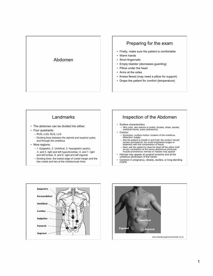
Landmarks
• The abdomen can be divided into either: • Four quadrants:
– RUQ, LUQ, RLQ, LLQ – Dividing lines between the xiphoid and superior pubis,
and through the umbilicus • Nine regions:
– 1. Epigastric, 2. Umbilical, 3. Hypogastric (pubic), 4. and 5. right and left hypochondriac, 6. and 7. right
and left lumbar, 8. and 9. right and left inguinal – Dividing lines: the lowest edge of costal margin and the
iliac crests and two at the midclavicular lines
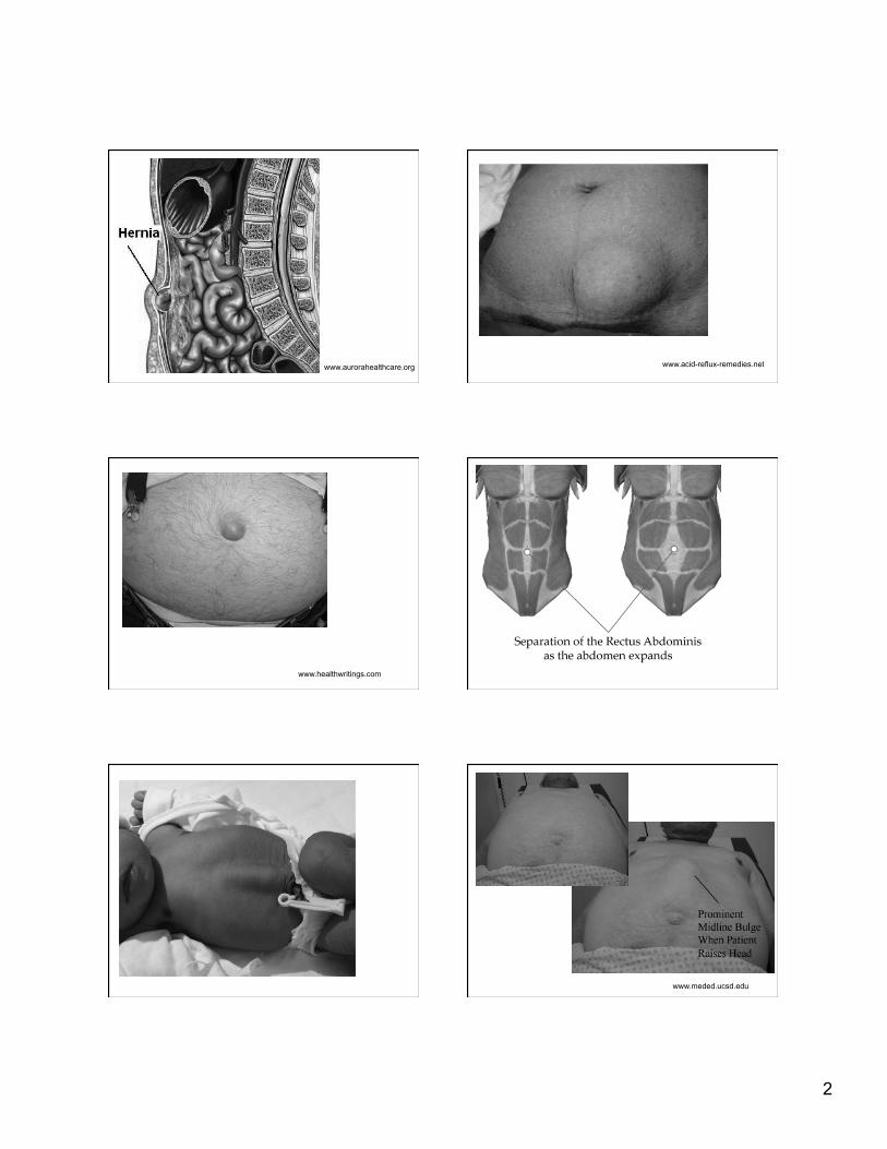
Inspection of the Abdomen • Surface characteristics
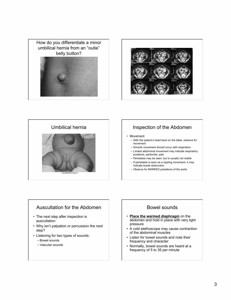
• Contour – Symmetry, surface motion, location of the umbilicus,
distention, bulges – Ask the patient to breath in and hold; the contour should
remain symmetrical, but could emphasize bulges or distention with the compression of tissue
– Next, ask the patient to raise his head off the pillow (half sit-up); contraction of the rectus abdominus produces muscle prominence; hernias or masses may appear
• Hernias may appear at surgical incisions and at the umbilicus (protrusion of the navel)
• Common in pregnancy, obesity, ascites, or long-standing COPD
How do you differentiate a minor umbilical hernia from an “outie”
belly button?
Umbilical hernia Inspection of the Abdomen
• Movement – With the patient’s head back on the table, observe for
movement – Smooth movement should occur with respiration – Limited abdominal movement may indicate respiratory
problems, peritonitis, pain – Peristalsis may be seen, but is usually not visible – If peristalsis is seen as a rippling movement, it may
indicate bowel obstruction – Observe for MARKED pulsations of the aorta
Auscultation for the Abdomen
• The next step after inspection is auscultation
• Why isn’t palpation or percussion the next step?
• Listening for two types of sounds: – Bowel sounds – Vascular sounds
Bowel sounds
• Place the warmed diaphragm on the abdomen and hold in place with very light pressure
• A cold stethoscope may cause contraction of the abdominal muscles
• Listen for bowel sounds and note their frequency and character
• Normally, bowel sounds are heard at a frequency of 5 to 35 per minute
4
Bowel sounds • Bowel sounds are usually heard as clicks and gurgles
that occur irregularly • Loud, prolonged gurgling is called borborygmi • Increased bowel sounds may occur with gastroenteritis,
early obstruction, or hunger • High-pitched tinkling sounds occur with intestinal fluid
and air under pressure of early obstruction • Decreased bowel sounds occur with peritonitis and
paralytic ileus • The absence of bowel sounds can only be determined
after 5 minutes of continuous listening • Auscultate all 9 regions
Vascular sounds • Listen with the bell for the low pitched sounds
of the vascular system • Listen for bruits in the aortic, renal, iliac, and
femoral arteries • Listen with the diaphragm for friction rubs over
the liver and spleen (high-pitched heard with respiration); friction rubs are rare, but heard with inflammation of the peritoneum (tumor, infection, infarct) (not a separate exam—perform with bowel sounds exam)
• Auscultate with the bell over the umbilicus for a venous hum (soft, low-pitched, continuous; indicates portal hypertension and shunting)
Peritoneal irritation
• Inflammation of the peritoneum may be identified as cutaneous hypersensitivity
• To evaluate, gently lift the skin over the hypersensitive area
or • Stimulate the skin with a pen or tongue
depressor • True cutaneous hypersensitivity will have
an exaggerated pain response to the stimulus
Percussion of the Abdomen • Percussion of abdominal structures is used to
assess the size and density of the organs and to detect the presence of fluid, air, or masses
• Percussion is used in combination with palpation and is used to confirm palpatory findings
• Tympany is the predominant sound in abdominal percussion because of the air in the stomach and intestines
• Dullness is heard over organs and solid masses
Percussion of the intestines/bowel • Percussion of the intestines is best
accomplished by systematically percussing in all 9 regions (2-3 sites per region)
• In interpreting the sounds, it helps to anticipate the expected sound from the structure
• For example, the small intestines (central) will have greater tympany than the large intestines
• The ascending and transverse colons typically have a mixture of fecal material and bowel gas
• The descending colon and sigmoid colon typically have more solid fecal material; therefore more dullness is expected
5
Gastric bubble percussion
• The gastric air bubble (meganblase) should percuss as a deeper tympany at the left anterior rib cage and the epigastric region
• (Performed with bowel percussion; not a separate examination)
Liver percussion • Beginning in an area of tympany, percuss the
liver at the right midclavicular line in the RUQ and move superiorly
• Remember that the posterior aspect of the lung field is quite low (T10), but the anterior aspect is high (5th-6th rib)
• You will usually percuss the lower border of the liver around the inferior costal margin or slightly below
• If the liver percusses more than 1 inch below the costal margin, consider hepatomegaly or downward displacement of the liver due to a depressed diaphragm
Liver percussion • To find the superior border of the liver, start
again at the midclavicular line, in an area of lung tympany (3rd-4th intercostal space)
• Percuss inferiorly until the note becomes dull • The upper border usually begins at the 5th-7th
intercostal space • A superior liver border below this area may
indicate liver atrophy or downward displacement of the liver by lung disease
• A superior liver border above this area may indicate upward displacement due to abdominal mass or hepatomegaly
Liver percussion • To estimate the liver size, measure
between the superior and inferior marks • The usual size is 6 to 12 cm (2.5-4.5
inches) • This is only a gross estimate • Pleural effusion and lung consolidation can
obscure the superior liver border • Gas in the ascending and transverse colon
can obscure the inferior liver border
Additional/alternate maneuvers
• Liver descent: ask the patient to take a deep breath and hold it while you again percuss upward from the abdomen; the inferior border should move 2 to 3 cm downward
• Axillary percussion: for females, or if enlargement is suspected, percuss in the right midaxillary line; dullness should be noted at the 5th-7th intercostal space (for superior border)
• If you suspect liver atrophy, hepatomegaly, or abdominal mass, you would next proceed to plain film imaging, followed by CT or US
6
Spleen percussion • Percuss the spleen just posterior to the
midaxillary line on the left, again beginning in an area of tympany
• Dullness should be heard at the 6th-10th intercostal spaces
• A larger area of dullness suggests splenomegaly; however, a full stomach or intestines could give false-positive findings
• Finally, percuss at the lowest costal margin with inspiration and with expiration
• The area should remain tympanic
Palpation • Purpose:
– Palpation is used to detect organomegaly, muscles spasm, fluid collection, painful areas, masses, adhesions
• Just like inspection, auscultation, and percussion, make sure the patient is relaxed with the knees flexed
• Caveat: Some people (especially children) are ticklish and the key is to use distraction; palpate and percuss over the patient’s hands and/or place the non-palpating hand over a less sensitive body part
Light palpation • You always begin with light palpation • Never go right to deep palpation • Begin with light, systemic palpation of all 9 regions • Initially, you should avoid the areas that were
identified in the history as problematic—why? • Begin with your palm on the patient, fingers
together • Depress the abdominal wall no more than 1cm,
using light smooth, consistent motions (don’t jab at the patient)
guarding and inflammation of the organ or peritoneum
• False-positive muscle guarding can occur with cold hands and ticklishness
• A large mass or distended structure may first be identified as an area of resistance
• If resistance is noted, palpate while the patient breaths slowly through the mouth
• If resistance remains, it is probably involuntary (indicating a mass or anomaly)
Moderate palpation
• Moderate palpation is mostly a transitional move to deeper palpation (2-3 cm depth)
• Tenderness not identified with light palpation may be evident with moderate palpation, saving the surprising pain response to deep palpation
Deep palpation
• Deep palpation is necessary to thoroughly evaluate the abdominal organs and to detect less obvious masses
• Again, use the palm of the hand with fingers together and palpate all 9 regions
• You often will be able to feel the borders of the rectus abdominus, colon, aorta
• False-positive pain may occur in the healthy patient over the cecum, sigmoid, aorta, and xiphoid process (and other bony prominences, esp. floating ribs)
7
Deep palpation of masses
• Palpate for masses and assess: – Location – Size – Shape – Consistency – Tenderness – Pulsation – Mobility – Movement with respiration
Masses
• False-positive masses: – Feces in the colon (should be soft, boggy, and
eventually move through remaining colon) – Lateral borders of rectus abdominus – Uterus – Aorta – Common iliac arteries
Umbilical ring
• Palpate the umbilicus and surrounding area
• The umbilicus should be round, firm, and free of bulges and masses
• Softness of the center could indicate an umbilical hernia
Bimanual technique
• If adipose deposition or muscle mass prevents deep palpation, you can use bimanual palpation to exert pressure over the abdominal structures
• Okay to use bimanual palpation for all patients, just don’t exert excessive pressure on thin patients or children
Liver palpation
• Stand to the right of the patient • Place your left hand under the patient at the
inferior costal margin (11th-12th ribs) • Press the left hand upward in order to lift the
liver anteriorly toward the abdominal wall • Place your right hand on the abdomen, with
fingers pointing towards the head at the right midclavicular line or pointing towards midline
• Press the right hand downward as the pt exhales • Ask the patient to take a deep breath, which
pushes the diaphragm downward (alternate/option)
Liver palpation
• You may not be able to feel the liver in many individuals
• In thinner individuals, you may feel the inferior margin
• If the liver is palpated, it should feel smooth, firm, regular (hotdog); should be nontender
• Palpate for nodules, tenderness, irregularity • Repeat palpation across the medial to lateral
margins
8
Liver palpation: alternate (not preferred)
• Stand at the patient’s right side, positioned toward the head but facing the feet
• Hook the fingers of both hands under the costal margin, below the level of dullness percussed earlier
• Press in and up while the patient exhales • Continue to press in and up as the patient
• When tenderness cannot be palpated, you may use fist percussion (indirect)
• Place the palm over the lower right rib cage • Strike the hand with the fist of the other hand • A healthy liver is not tender to fist percussion (but
not all unhealthy livers are tender)
• This should be performed after palpation (even though it is a percussive force), as it is intended to evaluate for pain (as invasive as palpation due to pain)
Gallbladder palpation
• Palpate below the liver margin at the lateral border of the rectus abdominus (medial portion of the liver)
• Most healthy gallbladders are not palpable
• A tender gallbladder indicates cholecystitis • A nontender but palpable gallbladder
indicates obstruction of the duct (stones) or cholilithiasis
Murphy’s sign
• If cholecystitis is suspected (gallbladder “attack”), you may perform Murphy’s test
• During deep palpation of the gallbladder region, ask the patient to take in a deep breath
• As the inflamed gallbladder wall moves closer to the peritoneum and palpating hands, the patient will experience pain and halt inspiration (positive sign)
Spleen palpation • Stand to the patient’s right • Reach across the patient to the left side with your
left hand and place it beneath the patient at the left costovertebral margin, pulling upward
• Place the palm of the right hand on the abdomen below the left costal margin (just like liver palpation, but at the MIDAXILLARY LINE)
• Press your fingertips inward as the patient inhales deeply
• The spleen should move inferiorly, but is usually not palpable in adults
Spleen palpation (alternate)
• Repeat palpation of the spleen with the patient in a right lateral decubitus position with knees flexed
• This moves the spleen more anteriorly and to the right
9
Kidney palpation/kidney punch • Kidney (percussion) is best accomplished first
with the patient seated (do with lumbar exam) • Place the palm of one hand at the posterior
inferior costal margin • Strike your hand with the fist of the other as you
did for liver percussion • Repeat at the opposite posterior inferior costal
margin • Normally, the patient feels a “thud” which does
not produce pain • Direct percussion may also be used
Left kidney palpation • With the patient supine, stand on the right
side • Reach across the patient (as with spleen)
with your left hand and place underneath the patient at the flank
• Place your right hand at the anterior costal margin and press deeply (retroperitoneal)
• As the patient inhales deeply, you may feel the lower pole of the kidney
• The left kidney is usually not palpable
Left kidney palpation (alternate) • Move to the left side of the patient and position
hands as before (better for shorter doctors) • When the patient inhales deeply, press the two
palms together to “capture” the kidney • Ask the patient to exhale and hold while you
slowly release your hands • You may feel the kidney slip through your grasp • This should not be painful to the healthy kidney
Right kidney palpation
• Stand on the right side of the patient, placing the one hand at the flank and the other at the anterior costal margin
• Perform the same moves as with the left kidney • The right kidney tends to be more palpable than
the left—why? • If it is palpable, it should be firm, smooth,
nontender • By contrast, the liver margin is sharper than the
renal margins
McBurney’s Point
• Approximately 2/3 distance from the navel to the ASIS (obliquely)
• Tender to palpation with appendicitis • McBurney’s (Rebound) Test: Rebound
test positive when appendix has irritated the peritoneum
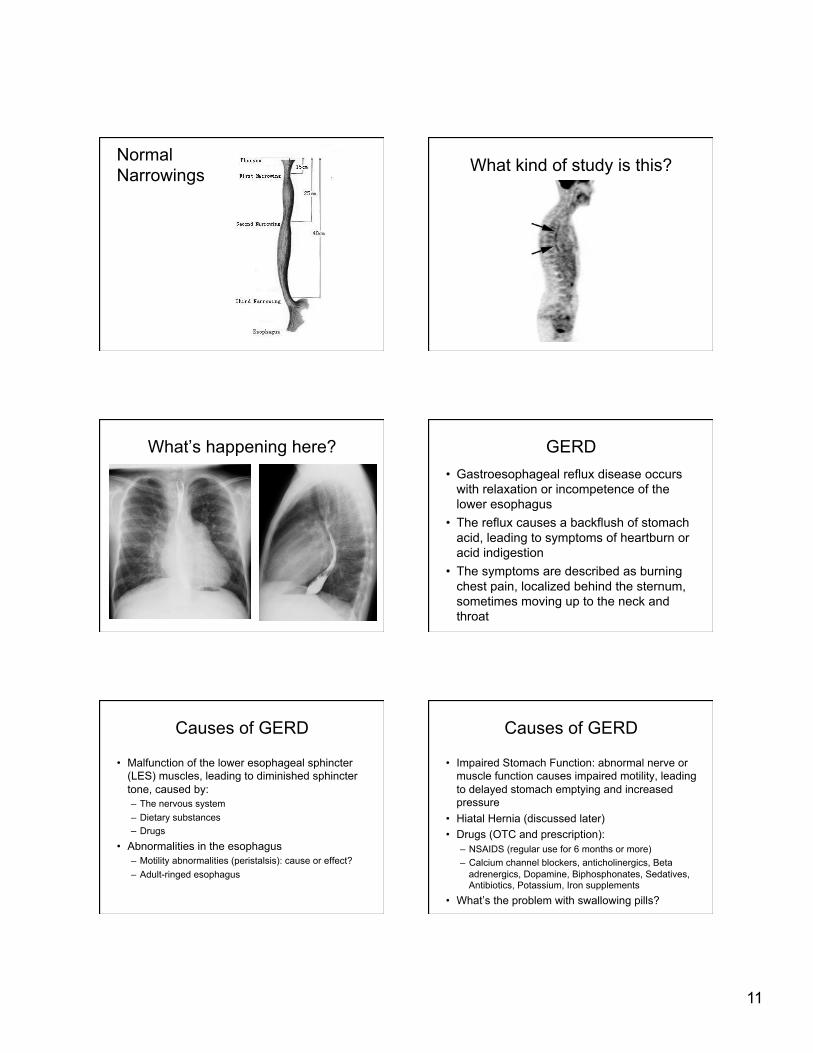
with relaxation or incompetence of the lower esophagus
• The reflux causes a backflush of stomach acid, leading to symptoms of heartburn or acid indigestion
• The symptoms are described as burning chest pain, localized behind the sternum, sometimes moving up to the neck and throat
Causes of GERD
• Malfunction of the lower esophageal sphincter (LES) muscles, leading to diminished sphincter tone, caused by: – The nervous system – Dietary substances – Drugs
• Abnormalities in the esophagus – Motility abnormalities (peristalsis): cause or effect? – Adult-ringed esophagus
Causes of GERD
• Impaired Stomach Function: abnormal nerve or muscle function causes impaired motility, leading to delayed stomach emptying and increased pressure
• Hiatal Hernia (discussed later) • Drugs (OTC and prescription):
– NSAIDS (regular use for 6 months or more) – Calcium channel blockers, anticholinergics, Beta
adrenergics, Dopamine, Biphosphonates, Sedatives, Antibiotics, Potassium, Iron supplements
• What’s the problem with swallowing pills?
12
Causes of GERD
• Asthma – More than 50% of asthmatic sufferers have
GERD – It is debated which causes which
• Some believe that coughing accompanies asthmatic attacks, leading to pressure changes in the chest, triggering reflux; also, some meds used to relax the bronchioles can relax the LES muscles
• Some believe that since GERD has been associated with several other respiratory problems, it may be a cause of asthma, rather than a result of asthma
Causes of GERD
• Those with Type I diabetes often develop a condition called gastroparesis
• This condition occurs in at least 20% of patients with long-standing diabetes
• It is characterized by delayed stomach emptying
• As discussed with impaired stomach function, delayed emptying leads to increased pressure, resulting in reflux
GERD
• GERD doesn’t sound like a big problem, but it can lead to significant complications
• Infants and children can have symptoms of vomiting, leading to weight loss and fatigue
• Dysphagia can develop, as well as a change in the voice characteristic (hoarseness)
• GERD can also lead to respiratory complications from aspiration and bleeding
• GERD can also change the cells of the esophagus
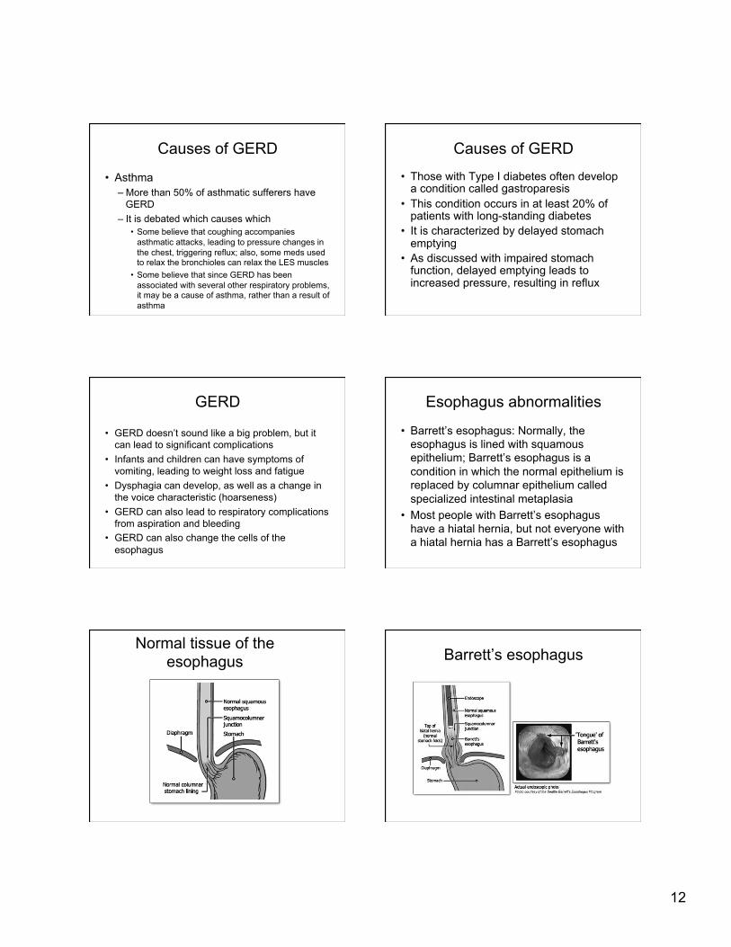
Esophagus abnormalities
• Barrett’s esophagus: Normally, the esophagus is lined with squamous epithelium; Barrett’s esophagus is a condition in which the normal epithelium is replaced by columnar epithelium called specialized intestinal metaplasia
• Most people with Barrett’s esophagus have a hiatal hernia, but not everyone with a hiatal hernia has a Barrett’s esophagus
Normal tissue of the esophagus Barrett’s esophagus
13
Barrett’s esophagus What’s happening here?
Hiatal hernia 2 types of hiatal hernias
Hiatal Hernia
• There are two types of hiatal hernias: paraesophageal and gastric/sliding
• Which one do you think is the most common? • The condition is VERY common, and occurs
most frequently in women and older adults • It is associated with obesity, pregnancy, ascites,
and tight clothing or belts • Muscle and diaphragmatic weakness are
contributing factors
• In the US: Hiatal hernias are more common in Western countries. The frequency of hiatus hernia increases with age, from 10% in patients younger than 40 years to 70% in patients older than 70 years.
• Internationally: Burkitt et al suggest that the Western, fiber-depleted diet leads to a state of chronic constipation and straining during bowel movement, which could explain the higher incidence of this condition in Western countries.
• Hiatal Hernia; Qureshi; 2/28/06; emedicine.com
14
Hiatal Hernia
• Patients complain of epigastric pain and/or heartburn that worsens with lying down and is relieved by sitting up or with antacids
• Patients also complain of water brash, which is a filling of the mouth with fluid, sometimes with changing positions, and of dysphagia
• The hernia can become incarcerated, which is a surgical emergency
• Incarceration symptoms included sudden onset vomiting, pain, and complete dysphagia
Peptic ulcers
• Peptic ulcer is the collective term for ulcers that occur within the upper gastrointestinal tract
• Gastric ulcer: occurs in the stomach • Duodenal ulcer: occurs in the duodenal
bulb • Esophageal ulcer: occurs in the
esophagus and is associated with GERD
Peptic ulcers • Duodenal ulcers are 4-5 times more common
than gastric ulcers and are the most common form of peptic ulcer disease
• They affect up to 10% of patients at any one time; lifetime risk is 25%
• Peptic ulcers are usually caused by Helicobacter pylori or H. pylori – (90% of duodenal ulcers and 70-75% of gastric
ulcers) • Other than causing erosions and craters in the
stomach and duodenum, they cause deformities of the abdominal bulb (“cloverleaf deformity”)
• 95% of the pre-bulbar peptic ulcers are benign
Risk factors for ulcers • Use of aspirin and nonsteroidal anti-
inflammatory medications (NSAIDs) • Excessive drinking of alcohol • Use of tobacco
• Stress?? • Diet??
15
Rarer cause of ulcers
• Zollinger-Ellison syndrome is caused by gastrin-secreting tumors (gastrinoma) of the pancreas that causes severe ulceration of the upper gastrointestinal tract
Treatment for ulcers
• Antibiotics to kill Helicobacter pylori • Acid blockers (cimetidine, ranitidine, or
famotidine) • Proton pump inhibitors (omeprazole) • Medications that protect the tissue lining
(sucralfate) • Bismuth (may help protect the lining and
kill the bacteria)
Complications of ulcers
• Bleeding • Bowel obstruction • Bowel perforation • Perforation leading to peritonitis
Symptoms of ulcers • It is important to remember that ulcers may cause no
symptoms at all • The most common ulcer symptom is abdominal pain
– May wake you at night – May be relieved by antacids or milk – May occur 2 to 3 hours after a meal – May be worse if you don't eat
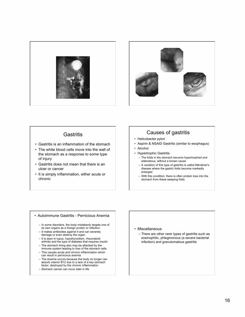
• Gastritis is an inflammation of the stomach • The white blood cells move into the wall of
the stomach as a response to some type of injury
• Gastritis does not mean that there is an ulcer or cancer
• It is simply inflammation, either acute or chronic
Causes of gastritis • Helicobacter pylori • Aspirin & NSAID Gastritis (similar to esophagus) • Alcohol • Hypertrophic Gastritis
– The folds in the stomach become hypertrophied and edematous, without a known cause
– A variation of this type of gastritis is called Ménétrier's disease where the gastric folds become markedly enlarged
– With this condition, there is often protein loss into the stomach from these weeping folds
• Autoimmune Gastritis - Pernicious Anemia
– In some disorders, the body mistakenly targets one of its own organs as a foreign protein or infection
– It makes antibodies against it and can severely damage or even destroy the organ
– It is seen in lupus, hypothyroidism, rheumatoid arthritis and the type of diabetes that requires insulin
– The stomach lining also may be attacked by the immune system leading to loss of the stomach cells
– This causes acute and chronic inflammation which can result in pernicious anemia
– The anemia occurs because the body no longer can absorb vitamin B12 due to a lack of a key stomach factor, destroyed by the chronic inflammation
– Stomach cancer can occur later in life
• Miscellaneous – There are other rarer types of gastritis such as
eosinophilic, phlegmonous (a severe bacterial infection) and granulomatous gastritis
17
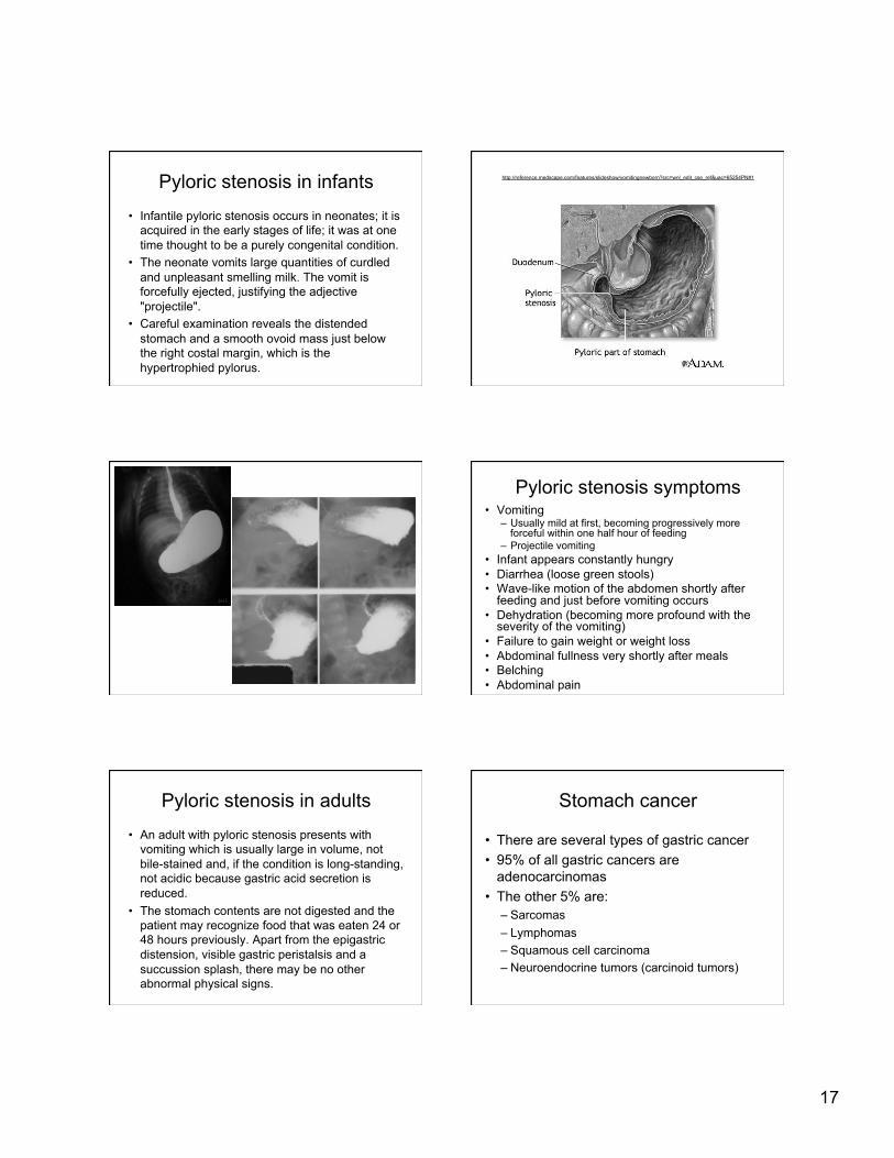
Pyloric stenosis in infants
• Infantile pyloric stenosis occurs in neonates; it is acquired in the early stages of life; it was at one time thought to be a purely congenital condition.
• The neonate vomits large quantities of curdled and unpleasant smelling milk. The vomit is forcefully ejected, justifying the adjective "projectile".
• Careful examination reveals the distended stomach and a smooth ovoid mass just below the right costal margin, which is the hypertrophied pylorus.
– Usually mild at first, becoming progressively more forceful within one half hour of feeding
– Projectile vomiting • Infant appears constantly hungry • Diarrhea (loose green stools) • Wave-like motion of the abdomen shortly after
feeding and just before vomiting occurs • Dehydration (becoming more profound with the
severity of the vomiting) • Failure to gain weight or weight loss • Abdominal fullness very shortly after meals • Belching • Abdominal pain
Pyloric stenosis in adults
• An adult with pyloric stenosis presents with vomiting which is usually large in volume, not bile-stained and, if the condition is long-standing, not acidic because gastric acid secretion is reduced.
• The stomach contents are not digested and the patient may recognize food that was eaten 24 or 48 hours previously. Apart from the epigastric distension, visible gastric peristalsis and a succussion splash, there may be no other abnormal physical signs.
Stomach cancer
• There are several types of gastric cancer • 95% of all gastric cancers are
• A diet high in salty and smoked foods • A diet low in fruits and vegetables • Eating foods contaminated with aflatoxin fungus • Family history of stomach cancer • Infection with Helicobacter pylori • Long-term stomach inflammation (chronic gastritis) • Pernicious anemia • Smoking • Stomach polyps
– People often ignore the symptoms of gastric cancer because they associate them with other stomach maladies
• Also, because these symptoms are usually mild, early diagnosis is rare
• Only 10 to 20 percent of gastric cancers are diagnosed before spreading to other areas of the body
• If treated early, gastric cancer has a good cure rate
• However, the prognosis worldwide is generally poor, with only about 2 in 10 of those affected surviving for five years after diagnosis (much better survival rate in US)
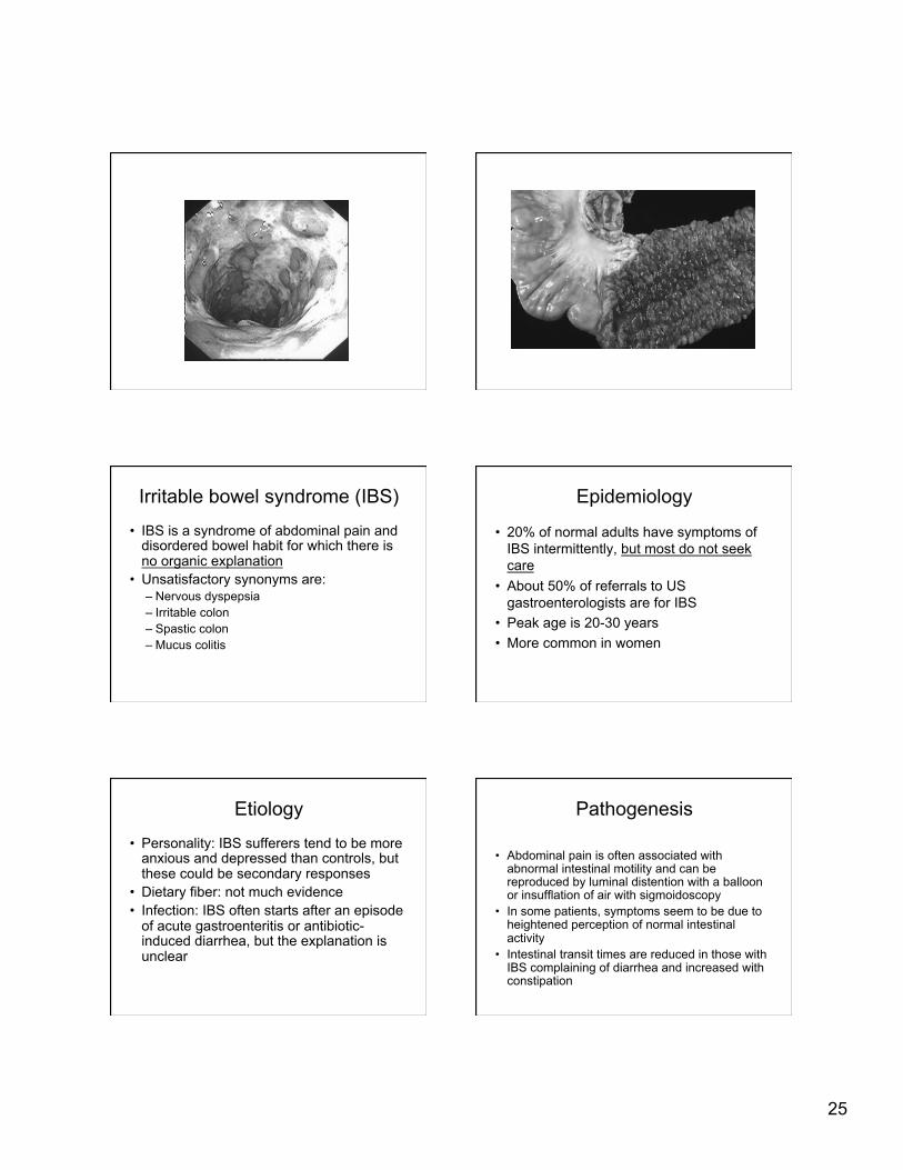
Inflammatory bowel disease
• Ulcerative colitis and Crohn’s disease are idiopathic chronic inflammatory diseases of the GI tract
• They have much in common, but the differences in histological changes are marked
19
Ulcerative colitis (UC)
• Epidemiology: – Much more common in developed countries – Prevalence is greater than 70/100,000 – Bimodal distribution, with peaks at 20-40 and 60-80
years of age – More common in women – Most common in Caucasians, particularly Jews – 10% increase in families with history of UC or CD, but
no clear HLA or Mendelian inheritance – An association between ankylosing spondylitis and
UC is seen
Etiology and pathogenesis
• The primary cause is unknown • May result from an abnormally prolonged
inflammatory host response • Genetic and/or environmental factors • Dietary or microbiological product in the lumen • Postulated etiological factors:
• Biochemistry: serum albumin is often low • Microbiology: stools show WBC and RBC • Plain film: altered bowel gas patterns • Barium enema: shows ulcerations, granulation with
loss of haustra • Sigmoidoscopy: inflammed rectal mucosa • Colonoscopy: helps define extent of disease
(ileum?); helps distinguish UC from CD with biopsies; screens for CA
Management • The aim of all care is to induce and maintain
remission • High fiber diets, bulking agents • Corticosteroids • IV fluids • Antibiotics • Hematinic agents • Surgery
21
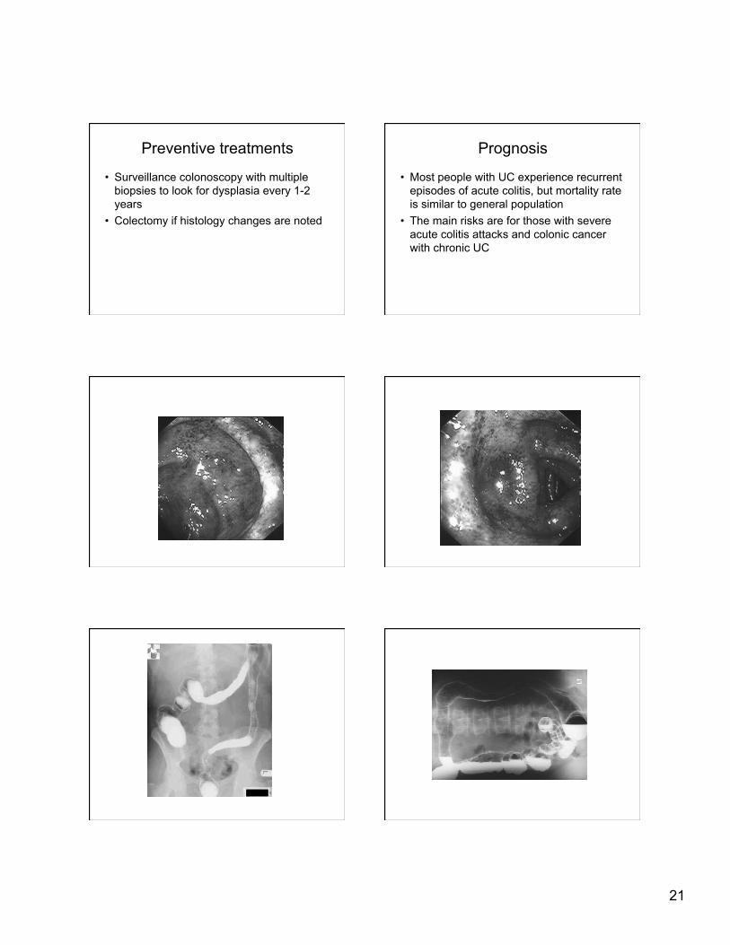
Preventive treatments
• Surveillance colonoscopy with multiple biopsies to look for dysplasia every 1-2 years
• Colectomy if histology changes are noted
Prognosis
• Most people with UC experience recurrent episodes of acute colitis, but mortality rate is similar to general population
• The main risks are for those with severe acute colitis attacks and colonic cancer with chronic UC
22
Crohn’s disease (CD)
• Epidemiology: – Like UC, CD is most common in developed countries – Prevalence is 50/100,000 – Unlike UC, the incidence of CD has risen rapidly and
continuously since 1960 – Bimodal peak of occurrence at 20-30 years and
elderly – More common in women – Most common in Caucasians, particularly Jews – 10% increase with first degree relatives and with AS
Etiology and pathogenesis
• Like UC, the cause of CD is unknown • Possible factors:
– Infection: resembles intestinal TB – Immunological: may be impaired – Diet: found more in those that eat refined
carbohydrates, particularly sugar, but could be a secondary phenomenon
– Smoking: patients with CD are more likely to smoke
Pathology
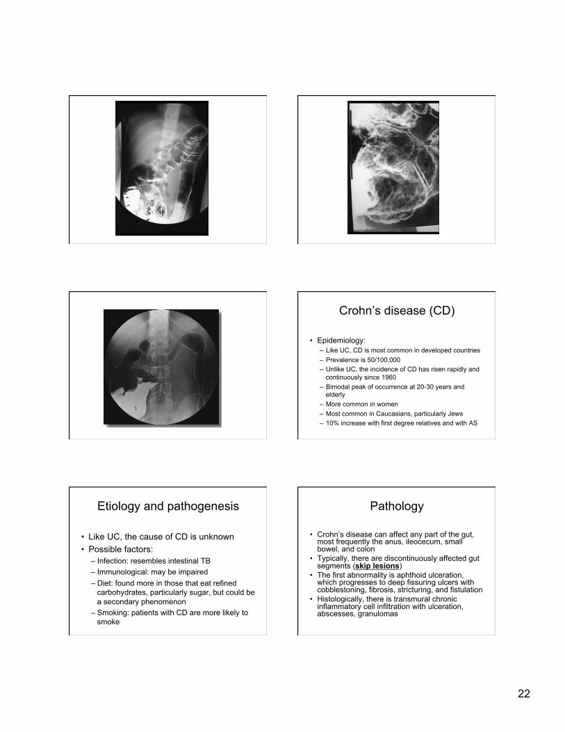
• Crohn’s disease can affect any part of the gut, most frequently the anus, ileocecum, small bowel, and colon
• Typically, there are discontinuously affected gut segments (skip lesions)
• The first abnormality is aphthoid ulceration, which progresses to deep fissuring ulcers with cobblestoning, fibrosis, stricturing, and fistulation
• Histologically, there is transmural chronic inflammatory cell infiltration with ulceration, abscesses, granulomas
23
Clinical features
• Clinical features vary depending upon site of disease (versus UC)
• The history is usually chronic with relapses and remissions
• Common symptoms are: – Diarrhea – Weight loss – Abdominal pain – Fever
• With small bowel disease, steatorrhea is common
• With large bowel disease, rectal bleeding is common
• Chronic perianal symptoms are common • Examination may show malabsorption,
• 20% of normal adults have symptoms of IBS intermittently, but most do not seek care
• About 50% of referrals to US gastroenterologists are for IBS
• Peak age is 20-30 years • More common in women
Etiology
• Personality: IBS sufferers tend to be more anxious and depressed than controls, but these could be secondary responses
• Dietary fiber: not much evidence • Infection: IBS often starts after an episode
of acute gastroenteritis or antibiotic-induced diarrhea, but the explanation is unclear
Pathogenesis
• Abdominal pain is often associated with abnormal intestinal motility and can be reproduced by luminal distention with a balloon or insufflation of air with sigmoidoscopy
• In some patients, symptoms seem to be due to heightened perception of normal intestinal activity
• Intestinal transit times are reduced in those with IBS complaining of diarrhea and increased with constipation
26
Clinical features
• Abdominal pain: felt anywhere, most commonly over iliac fossae, and related to defecation and flatus
• Varying bowel habits: diarrhea with urgency to constipation
• Passage of mucus rectally • Feeling of fullness after defecation • Abdominal distention
• January 7, 2010 — Migraine and carpal tunnel syndrome are common among celiac patients, a new study shows. • After screening a cohort of 72 patients with biopsy-proven celiac disease, researchers also report that many experience psychiatric problems,
with 35% of celiac patients reporting a history of depression, personality changes, or psychosis. • Atypical neurological presentations are thought to occur in 6% to 10% of celiac patients, the study authors note. Prior studies have suggested
that cerebellar ataxia is the most frequent symptom. This new study observed cerebellar ataxia in 6% of patients. Another 6% had vestibular dysfunction. In all, 26% of patients experienced afferent ataxia.
• About a third of patients had stance and gait problems, and many experienced deep sensory loss and reduced ankle reflexes. • "Gait disturbances in celiac disease do not only result from cerebellar ataxia but also from proprioceptive or vestibular impairment," report
investigators led by Katrin Burk, MD, from the University of Marburg in Germany. "Neurological problems may develop despite strict adherence to a gluten-free diet."
• Neurological problems may develop despite strict adherence to a gluten-free diet. • The study is published in the December 15 issue of Movement Disorders. • The 72 patients with celiac disease were recruited through advertisements and interviewed using a standard questionnaire. • "Most studies in this field are focused on patients under primary neurological care," the researchers note. "To exclude such an observation
bias, patients with biopsy-proven celiac disease were screened for neurological disease." • About a third of celiac patients (28%) reported a history of migraine. In many cases, there was a decrease in the frequency and intensity of
migraine attacks after the introduction of a gluten-free diet. • About 20% of patients experienced carpal tunnel syndrome. "Surprisingly, epilepsy was less common than expected," report the researchers.
"Only 4 individuals presented with a history of generalized or focal seizures." • Motor problems, such as basal ganglia symptoms, pyramidal tract signs, tics, and myoclonus, were infrequent. A total of 14% of patients
reported bladder dysfunction. • Multiple Mechanisms Likely • In celiac disease, the mechanisms leading to neurological disease are not yet understood. Deficiencies in folic acid, vitamin E, and biopterin
have been implicated in the pathogenesis; however, the investigators report that replacement therapy does not resolve clinical symptoms in most cases.
• The researchers point out that hypovitaminosis rarely causes overt abnormalities in celiac patients, and most with neurological symptoms do not show evidence of any nutritional deficiencies.
• "The prevalence of neurological manifestations in celiac disease is striking and must be considered more than accidental," they note. "The patients' gluten-free diet had resolved intestinal symptoms but had not prevented the development of neurological deficits."
• The investigators suggest that because of the considerable clinical variability, many different pathogenic mechanisms are likely to contribute to the neurological and psychiatric dysfunction in celiac disease.
• The researchers have disclosed no relevant financial relationships. • Mov Disord. 2009;24:2358-2362.