JK SCIENCE 182 www.jkscience.org Vol. 17 No. 4, Oct - December 2015 ORIGINALARTICLE From the Department of Surgery Govt. Medical College, Srinagar- Kashmir- J&K India Correspondence to : Dr. MR Atri , Assistant Professor, Department of Surgery, Govt. Medical College Srinagar- J&K India Laparoscopic Cholecystectomy in Acute Cholecystitis :An Experience with 100 cases Rajni Bhardwaj, M.R.Attri, Shahnawaz Ahangar The introduction of laparoscopy in the surgical field has undoubtedly been the biggest revolution in the history. Since the performance of first laparoscopic cholecystectomy by Prof Dr Med Erich Mühe of Böblingen, Germany 1985, this procedure overtook as the new gold standard for the management of cholelithiasis. The management of cholelithiasis has undergone radical changes since its recognition; from medical management of stones to the surgical removal of the gallbladder. Earlier open cholecystectomy had been the treatment of choice; though it was recommended after a rest period of 6 weeks after an acute attack. Now-a- days laparoscopic cholecystectomy has replaced the open procedure as the first line management. Despite the well- accepted success of laparoscopic cholecystectomy in elective treatment of symptomatic cholelithiasis, the efficacy and timing of this technique has been subject to some debate in the setting of acute cholecystitis. Initial reports suggested that early laparoscopic surgery for acute cholecystitis was associated with increased Introduction Abstract This study was undertaken to evaluate our experience with laparoscopic cholecystectomy in the setting of acute cholecystitis. Between one year, one hundred patients with clinical, laboratory and radiological evidence of acute cholecystitis underwent early laparoscopic cholecystectomy within three days of onset of symptoms in a prospective study. The mean (range) age was 54 (28-61) years and the male female ratio was 3.7:6.3. The primary outcomes studied were operative time, blood loss, ease of surgery, conversion to open cholecystectomy, complications, length of hospital stay and the return to work. There were no major complications or any deaths during the study. There were two conversions in total. In one case it was due to difficult anatomy and for the control of bleeding in the second case. The mean (range) operative time was 71 (45-118) min. The mean (range) blood loss was 85 (50-350) ml. The mean (range) hospital stay was 3 (2-6) days. All patients returned back to routine work within 2 weeks of surgery. The mean follow- up was 6 (3-11) months. Laparoscopic cholecystectomy performed by experienced surgeons is a safe, effective technique for treatment of acute cholecystitis. Patients treated within 72 hours of onset of symptoms experience a lower conversion rate to an open procedure, shorter operative time and reduced hospitalization in addition to avoiding second hospitalization for surgery. Key Words Acute Cholecystitis, Laparoscopic Cholecystectomy
Transcript
JK SCIENCE
182 www.jkscience.org Vol. 17 No. 4, Oct - December 2015
ORIGINALARTICLE
From the Department of Surgery Govt. Medical College, Srinagar- Kashmir- J&K IndiaCorrespondence to : Dr. MR Atri , Assistant Professor, Department of Surgery, Govt. Medical College Srinagar- J&K India
Laparoscopic Cholecystectomy in Acute Cholecystitis:An Experience with 100 cases
Rajni Bhardwaj, M.R.Attri, Shahnawaz Ahangar
The introduction of laparoscopy in the surgical field
has undoubtedly been the biggest revolution in the history.
Since the performance of first laparoscopic
cholecystectomy by Prof Dr Med Erich Mühe of
Böblingen, Germany 1985, this procedure overtook as
the new gold standard for the management of
cholelithiasis. The management of cholelithiasis has
undergone radical changes since its recognition; from
medical management of stones to the surgical removal
of the gallbladder. Earlier open cholecystectomy had been
the treatment of choice; though it was recommended after
a rest period of 6 weeks after an acute attack. Now-a-
days laparoscopic cholecystectomy has replaced the open
procedure as the first line management. Despite the well-
accepted success of laparoscopic cholecystectomy in
elective treatment of symptomatic cholelithiasis, the
efficacy and timing of this technique has been subject to
some debate in the setting of acute cholecystitis. Initial
reports suggested that early laparoscopic surgery for
acute cholecystitis was associated with increased
Introduction
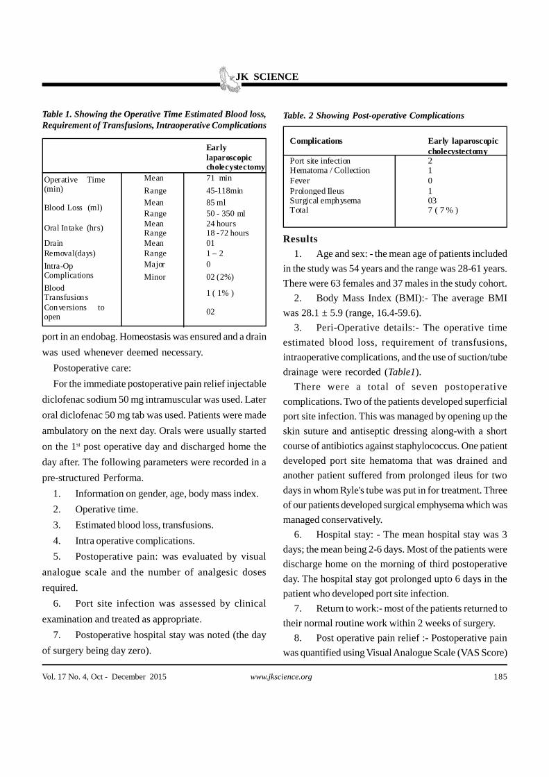
AbstractThis study was undertaken to evaluate our experience with laparoscopic cholecystectomy in the setting ofacute cholecystitis. Between one year, one hundred patients with clinical, laboratory and radiologicalevidence of acute cholecystitis underwent early laparoscopic cholecystectomy within three days of onsetof symptoms in a prospective study. The mean (range) age was 54 (28-61) years and the male female ratiowas 3.7:6.3. The primary outcomes studied were operative time, blood loss, ease of surgery, conversion toopen cholecystectomy, complications, length of hospital stay and the return to work. There were no majorcomplications or any deaths during the study. There were two conversions in total. In one case it was dueto difficult anatomy and for the control of bleeding in the second case. The mean (range) operative timewas 71 (45-118) min. The mean (range) blood loss was 85 (50-350) ml. The mean (range) hospital staywas 3 (2-6) days. All patients returned back to routine work within 2 weeks of surgery. The mean follow-up was 6 (3-11) months. Laparoscopic cholecystectomy performed by experienced surgeons is a safe,effective technique for treatment of acute cholecystitis. Patients treated within 72 hours of onset ofsymptoms experience a lower conversion rate to an open procedure, shorter operative time and reducedhospitalization in addition to avoiding second hospitalization for surgery.
186 www.jkscience.org Vol. 17 No. 4, Oct - December 2015
and the total quantity of analgesic, diclofenac sodium, ( i/
m Inj., plus per oral ) used in the postoperative period.
On an average 75 mg of diclofenac was needed.
9. Follow up and patient satisfaction:- All patients
were followed strictly after the surgery. Mean follow up
of the patients was 6 months and a range of 3 - 12 months.
There were no port site hernias or any other delayed
complications.
Discussion
The aim of this study was to assess the safety and
feasibility of early laparoscopic cholecystectomy in the
setting of acute cholecystitis. In the early days of
laparoscopy, acute cholecystitis was a contraindication
to laparoscopic cholecystectomy (1-3). Some argued that
the inflammation and adhesions associated with acute
cholecystitis were technically prohibitive in performing a
safe laparoscopic operation (4, 5). In view of these
concerns early open cholecystectomy, as opposed to
delayed open cholecystectomy, was the recommended
treatment for acute cholecystitis (6). As more experience
was gained, literature invalidated these concerns by
demonstrating laparoscopic surgery could be performed
in the setting of acute cholecystitis (7). However, the
operative time remained significantly longer for these
procedures than for those performed with the traditional
method, also, the conversion rates are reported to be 6%
to 60% (8). But as the experience accumulated in
laparoscopic surgery the operative time as well as the
conversion and complication rates showed a decreasing
trend. In the present study we had a mean operating
time of 71 minutes with a range of 45-118 min. Operative
time was longer during the initial phase of study, but as
we went through the learning curve, operative time
decreased. Only one patient required blood transfusion
in whom cystic artery bled and we had to convert to
open surgery. In rest of the cases the average blood loss
was of the order of 85ml. There were no common bile
1. Wilson P, Leese T, Morgan WP, et al. Elective laparoscopiccholecystectomy for acute cholecysitits. Lancet 1991; 338:795-797
2. Phillips EH, Carroll BJ, Fallas MJ. Laparoscopically guidedcholecystectomy: a detailed report of the first 453 casesperformed by one surgical team. Am Surg 1993; 59: 235-242
3. Cuschieri A, Dubois F, Mouiel J, et al. The Europeanexperience with laparoscopic cholecystectomy. Am J Surg1991;161: 385-387
4. Pessaux P, Tuech JJ, Rouge C, et al. Laparoscopiccholecystectomy in acute cholecystitis: a prospectivecomparative study in patients with acute versus chroniccholecystitis. Sug Endosc 2000; 14: 358-361.
5. Rattner DW, Ferguson C,Warshaw AL. Factors associatedwith successful laparoscopic cholecystectomy for acutecholecystitis. Ann Surg 1993; 217 : 233-236.
6. Norrby S, Herlin P, Holmin T, et al. Early or delayedcholecystectomy in acute cholecystitis? A clinical trial. BrJ Surg 1983;70:163-165.
7. Wilson RG, Macintyre IM, Nixon SJ, et al. Laparoscopiccholecystectomy as a safe and effective treatment for severeacute cholecystitis. BMJ 1992; 305: 394-396
8. Reiss R, Nudelman I, Gutman C, et al. Changing trends insurgery for acute cholecystitis. World J Surg 2014;34:23-4
References
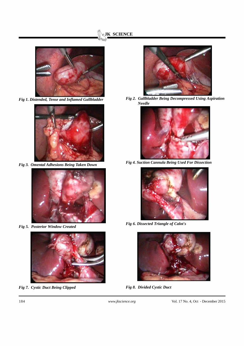
duct injuries. During the study we found the dissection
easier during the episode of inflammation. Therefore we
report from our experience that the inflammation
associated with acute cholecystitis creates an edematous
plane in the submucosa of the gallbladder, thus facilitating
the dissection from the liver bed. Also the inflammation
in the early stages may not necessarily involve Calot's
triangle thereby facilitating the procedure.
Conclusion
In conclusion, the data presented suggests that the
patients of acute cholecystitis can undergo laparoscopic
cholecystectomy during the initial admission especially
within 72 hours of symptoms, without added risk of
conversion or complications. It is better, less morbid, less
painful and avoids another hospital admission required