.• !Laser and Airway Firel • Fires in operating roo1n are a potential hazard to patients and operating room staff • Although rare (estinmated - 550-650 surgical fires occurring in the United states out of 65 1nillion operations done in 2009), when happened it could cause serious n1orbidity and mo1iality (airway fire in particular) • Operating theatre staff (surgeons, anaesthesiologists and nurses) should have adequate alertness and awareness of this potential problen1, and formulate plans to deal with OR fire Ignition Source Fig. I: The "fire triangle" showin g the 3 necessary components fo r cos tion
Transcript
.•
!Laser and Airway Firel
• Fires in operating roo1n are a potential hazard to patients and operating
room staff
• Although rare (estinmated - 550-650 surgical fires occurring in the United
states out of 65 1nillion operations done in 2009), when happened it could
cause serious n1orbidity and mo1iality (airway fire in particular)
• Operating theatre staff (surgeons, anaesthesiologists and nurses) should
have adequate alertness and awareness of this potential problen1, and
formulate plans to deal with OR fire
Ignition Source
Fig. I: The "fire triangle" showing the 3 necessary components for costion
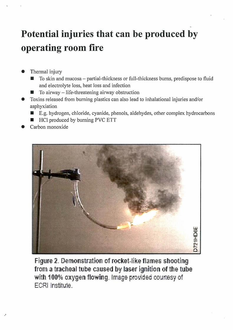
Potential injuries that can be produced by
operating room fire
• Thermal injury • To skin and mucosa- partial-thickness or full-thickness burns, predispose to fluid
and electrolyte loss, heat loss and infection • To airway - life-threatening airway obstruction
• Toxins released from burning plastics can also lead to inhalational injuries and/or asphyxiation • E.g. hydrogen, chloride, cyanide, phenols, aldehydes, other complex hydrocarbons • HCl produced by burning PVC ETT
• Carbon monoxide
Figure 2. De 111onstrat1on of rocket-1 i k:e flame-s shooting fron1 a tracheal tu be caused b~t laser ignitfo n of the tube w:ith. 1 00~··:1 oxygen flowing. In- age. pro.•.'id eo eor .. HteS/' of ECRJ ~n~3tittJte.
Laser and head & neck surgery • Commonly involving sharing of airway • Fuel (e.g. ETT) +ignition source (e.g. laser)+ oxidizer (e.g. 0 2 enriched environment)
=high risk for operating room fire! • In order to minimize risk of airway fire, operating room staff must make an effort to
avoid putting the 3 components of the fire triangle close together
Fuel
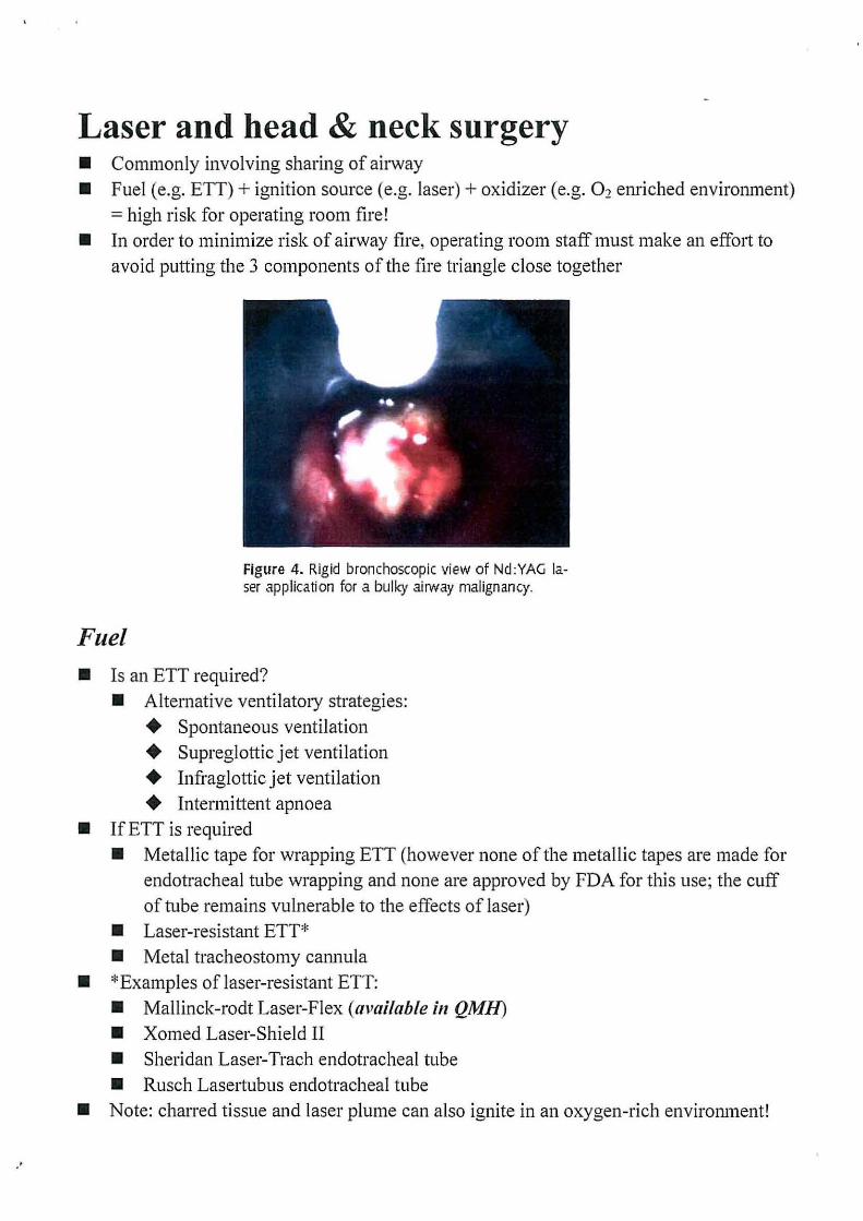
Figure 4. Rigid bronchoscopic view of Nd :YAG laser application for a bulky airway malignancy.
• Is an ETT required? • Alternative ventilatory strategies:
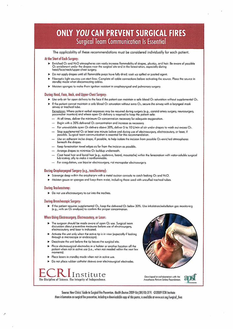
ONLY YOU CAN PREVENT SURGICAL FIRES Surgical Team Communication Is Essential
The applicability of these recommendations must be considered individually for each patient.
At the Start of Each Surgery: .,. Enriched 0> and N>O atmospheres can vastly increase flammability of drape;, plastics, and hair. Be aware of possible
01 enrichment under the drapes near the surgical site and in the fenestration, especially during heod/iace/ neck/upper·chest surgery .
.,. Do not apply drapes until all flammable preps have fully dried; soak up spilled or pooled agent.
.,. Fibcroptic light sources con start fires: Complete all cable connections belore activating the source. Place the source in standby mode when disconnecting cables .
.,. Moisten sponges to molm them ignition resistant in oropharyngeal and pulmonary surgery.
During Head, Face, Neck, and Upper-Chest Surgery: il" Use only air for open delivery to the face if the patient can maintain a sole blood Q, saturation without supplemental 01 .
.,. If the patient cannot maintain a sole blood Q, saturation without extra 0>, secure the airway with o laryngeal mask oirwoy or tracheal tube.
flliP-tion'i· Where patient verbal responses may be required during surgery (e.g. , carotid artery surgery, neurosurgery, pacemaker insertion) and where open Q, delivery is required to keep the patient safe:
AI all times, deliver the minimum 01 concentration necessary for adequate oxygenation.
Begin with o 30% delivered Q, cancenlrotion and increase os necessary.
For unavoidable open Q, delivel)' above 30%, deliver 5 to 10 l/min of oir under drapes to wash out excess Q , .
Stop supplemental o~ at least one minute before ond during use of electrosurgery, electrocautery, or loser, if possible. Surgical team communication is essential for this recommendation .
Use on adherent incise drape, if possible, to help isolate the incision from possible O]·enriched atmospheres beneath the drapes.
Keep fenestration towel edges os for from the incision os possible.
Arrange drapes to minimize 01 buildup underneath.
Cool head hair and facial hair (e.g., eyebrows, beard, moustache) within the fenestration with water-soluble surgical lubricating jelly to mo~e it nonflammable.
For coagulation, use bipolar electrosurgery, not monopolar electrosurgery.
During Oropharyngeal Surgery (e.g., tonsillectomy): .,. Scavenge deep within the oropharynx with o metal suction cannula to catch leaking Q, and N10 .
.,. Moisten gauze or sponges ond keep them moist, including those used with uncufled tracheal tubes.
During Trocheostomy: .,. Do not use electrosurgery to cut into the trochee.
During Bronchos(opic Surgery: .,. If the patient requires supplemental Q,, keep the delivered 0 1 below 30%. Use inholotion/exholotion gos monitoring
(e.g., with on Q, onolyzer) to confirm the proper conccnlrofion.
When Using Electrosurgery, Electrocautery, or Loser:
.,. The surgeon should be mode aware of open Q, use. Surgical Ieo m discussion oboul preventive measures before use of electrosurgery, electrocautery, and loser is indicated .
.,. Activate the unit only when the active tip is in view {especially if looking through o microscope or endoscope) .
.,. Deactivate the unit before the lip leaves the surgical site .
.,. Place eleclrosurgical electrodes in a holster or another location off the patient when not in active use (i.e., when not needed within the next few moments) .
.,. Place lmers in stand by mode when not in active use .
.,. Do not place rubber catheter sleeves over electrosurgicol electrodes.
ECR,I Institute The Discipline of Science. The Integrity of Independence.
Developed in collaboration with the Ancsthesio Poticnt Sofctv Foundotion.
Surgical lasers • Second most conunon ignition source in operating room fires • Frequently serious
What is laser? LASER stands for Light Amplification by Stimulated Emission of Radiation • Laser radiation is monoclu·omatic, coherent, collimated with high energy density • Smgicallasers are able to provide high amount of energy to a precise location • Operates in either continuous mode or pulsed mode • Excellent tools in providing haemostasis at a precise location • Relative lack of trauma to healthy tissues • Reduces postoperative pain and edema
Common types of surgical laser 1. C02 laser
+ Infrared radiation with wavelength 1 0.6~tM + Makes precise cuts, provides excellent haemostasis with little collateral damage + Readily absorbed by water, blood and all biologic materials independent of
pigmentation + Acts via thermal injury and vaporizing cells + Can be absorbed by glass and plastic - crumot be trru1smitted tlu·ough traditional
+ Pulsed infrared output with wavelength 2.l~tM + Excellent absorption in water-rich tissues + Used in nasal surgeries and tonsillectomies
3. Neodymium (Nd):YAG laser + Emits radiation with wavelength 1 064nm + Highest tissue penetration of all currently available medical lasers + Can be transmitted via traditional fiberoptics + Well suited for haemostasis by coagulation and shrinking lower airway tumours + Can cause generalized thermal damage ru1d delayed edema with tissue sloughing
4. Potassimn titanyl phosphate (KTP) laser + Contains Nd:YAG laser which the frequency is doubled by passing the beam
through KTP crystal + Wavelength 532nm, readily absorbed by blood + Smaller spot diameter and better haemostasis thru1 C02 laser
·'
• Surgical preps and drapes • Many of the surgical prepping agents are flammable (esp. alcohol-based ones) • One should always allow adequate time for the liquid preparations to evaporate
and f111ly dry before draping (drying process may take up to 5 minutes!) • Towels used to absorb dripped preparation solution should be removed • Avoid pooling, spilling or wicking of flammable liquid preparations • Wet material (e.g. saline soaked gause) may dry out fairly rapidly and hence
increase the risk of fire
Ignition source
• Always limit the laser output to the lowest clinically acceptable power density and pulse duration
• Test fire the laser before stmting the procedure (if using tlu-ough an endoscope, test fire before inserting the endoscope into patient)
• Laser should be placed in standby mode when not in active use • Deactivate laser and place in a standby mode before removing it from the surgical site • Laser should be activated only by the person using it and only when the tip is in direct
view • Use surgical devices designed to minimize laser reflectance and use a laser backstop to
reduce the likehood of tissue injury distal to the surgical site • Never clamp the fibers to the drapes (may break the fibers) • When performing lower airway surgery, the laser fiber tip should always be visible and
clear of the end of the bronchoscope or ETT before using it
Oxidizer
• Always try to limit the oxygen concentration to less than 30% • Prevent any possible leak around the ETT with either a cuffed ETT or spontaneous
ventilation tlu·ough LMA • If oxygen concentration greater than 30% is used, the oropharynx should be suctioned
with a metal cannula before using laser I electrosurgical unit • *During tracheostomy, use oxygen concentration lower than 30% before entering the
trachea; if possible, always use a scapel or scissors rather than electrosurgical unit or electrocautery instruments to make the tracheal incision
·'
I
/
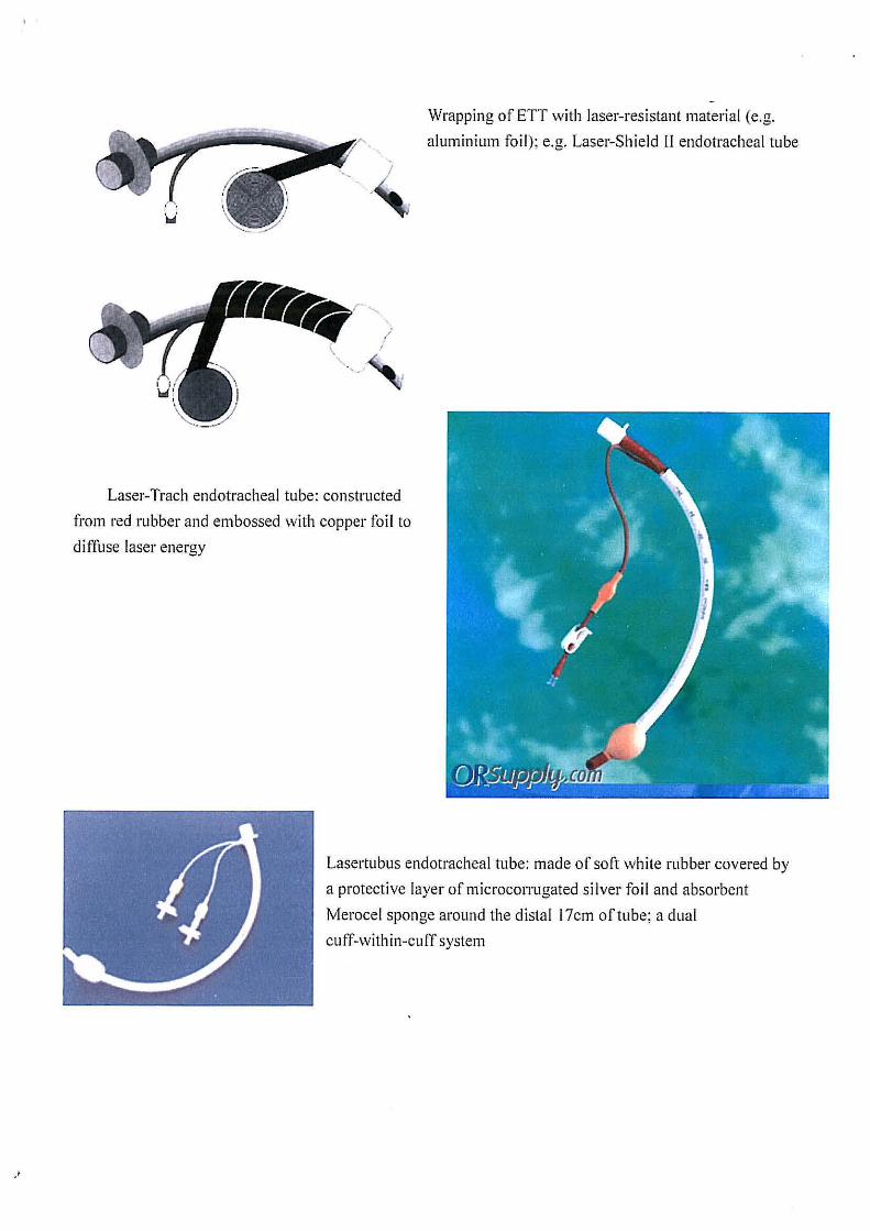
Laser-Trach endotracheal tube: constructed
from red rubber and embossed with copper foil to
diffuse laser energy
Wrapping of ETT with laser-resistant material (e.g.
aluminium foil); e.g. Laser-Shield 11 endotracheal tube
Lasertubus endotracheal tube: made of soft white rubber covered by
a protective layer of microcorrugated silver foil and absorbent
Merocel sponge around the distal 17cm of tube; a dual
cuff-within-cuff system
' .
EMERGENCY PROCEDURE EXTINGUISHING A SURGICAL FIRE
Fighting Fires ON the Surgical Patient Review before every surgical procedure.
In the Event of Fire on the Patient:
1. Stop the flow of all oirwoy gases to the patient. 2. Immediately remove the burning materials and hove another team member extinguish them.
If needed, use o C02 fire extinguisher to put out o fire on the patient. 3. Core for the patient:
-Resume patient ventilation. - Control bleeding.
-Evacuate the patient if the room is dangerous from smoke or fire. -Examine the patient for injuries and treat accordingly.
4. 1f the fire is not quickly controlled: -Notify other operating room staff and the fire deportment that a fire hos occurred. -Isolate the room to contain smoke and fire.
Save involved materials and devices for later investigation.
Extinguishing Airway Fires Review before every surgical intubation.
At the First Sign of an Airway or Breathing Circuit Fire, Immediately ond Rapidly: 1. Remove the tracheal tube, and hove another team member extinguish it. Remove cuff-protective
devices and any segments of burned tube that may remain smoldering in the airway. 2. Stop the flow of oil gases to the airway. 3. Pour saline or water into the airway. 4. Core for the patient:
-Reestablish the airway, and resume ventilating with air until you ore certain that nothing is left burning in the airway, then switch to 100% oxygen.
-Examine the airway to determine the extent of damage, and treat the patient accordingly. Save involved materials and devices for later investigation.
ECRI Institute The Discipline ol Science. The lnlcgrily of Independence.
Developed in colloboto tion with the Ancslhes io Pcticnl Sa(ety Foundation.