Laser-induced fluorescence diagnostics of basal cell carcinomas of the skin following topical ALA application af Klinteberg, C; Nilsson, AMK; Wang, I; Andersson-Engels, Stefan; Svanberg, Sune; Svanberg, Katarina Published in: Optical Biopsies and Microscopic Techniques, Proceedings of DOI: 10.1117/12.260817 1996 Link to publication Citation for published version (APA): af Klinteberg, C., Nilsson, AMK., Wang, I., Andersson-Engels, S., Svanberg, S., & Svanberg, K. (1996). Laser- induced fluorescence diagnostics of basal cell carcinomas of the skin following topical ALA application. In IJ. Bigio, WS. Grundfest, H. Schneckenburger, K. Svanberg, PM. Viallet, & A. Katzir (Eds.), Optical Biopsies and Microscopic Techniques, Proceedings of (Vol. 2926, pp. 32-40). SPIE. https://doi.org/10.1117/12.260817 General rights Copyright and moral rights for the publications made accessible in the public portal are retained by the authors and/or other copyright owners and it is a condition of accessing publications that users recognise and abide by the legal requirements associated with these rights. • Users may download and print one copy of any publication from the public portal for the purpose of private study or research. • You may not further distribute the material or use it for any profit-making activity or commercial gain • You may freely distribute the URL identifying the publication in the public portal Take down policy If you believe that this document breaches copyright please contact us providing details, and we will remove access to the work immediately and investigate your claim. Download date: 18. May. 2019
Transcript
LUND UNIVERSITY
PO Box 117221 00 Lund+46 46-222 00 00
Laser-induced fluorescence diagnostics of basal cell carcinomas of the skin followingtopical ALA application
af Klinteberg, C; Nilsson, AMK; Wang, I; Andersson-Engels, Stefan; Svanberg, Sune;Svanberg, KatarinaPublished in:Optical Biopsies and Microscopic Techniques, Proceedings of
DOI:10.1117/12.260817
1996
Link to publication
Citation for published version (APA):af Klinteberg, C., Nilsson, AMK., Wang, I., Andersson-Engels, S., Svanberg, S., & Svanberg, K. (1996). Laser-induced fluorescence diagnostics of basal cell carcinomas of the skin following topical ALA application. In IJ.Bigio, WS. Grundfest, H. Schneckenburger, K. Svanberg, PM. Viallet, & A. Katzir (Eds.), Optical Biopsies andMicroscopic Techniques, Proceedings of (Vol. 2926, pp. 32-40). SPIE. https://doi.org/10.1117/12.260817
General rightsCopyright and moral rights for the publications made accessible in the public portal are retained by the authorsand/or other copyright owners and it is a condition of accessing publications that users recognise and abide by thelegal requirements associated with these rights.
• Users may download and print one copy of any publication from the public portal for the purpose of private studyor research. • You may not further distribute the material or use it for any profit-making activity or commercial gain • You may freely distribute the URL identifying the publication in the public portalTake down policyIf you believe that this document breaches copyright please contact us providing details, and we will removeaccess to the work immediately and investigate your claim.
Laser-induced fluorescence diagnostics of basal cell carcinomas of the skinfollowing topical ALA application
Claes af Klinteberg', Annika M.K. Nilsson1, Ingrid Wang2, Stefan Andersson-Engels1,Sune Svanberg1 and Katarina Svanberg2
1
Department of Physics, Lund Institute of Technology, P.O. Box 1 18, S-221 00 Lund, SwedenE-mail: [email protected]
2Department of Oncology, Lund University Hospital, S-221 85Lund, Sweden
ABSTRACTFourteen patients with superficial basal cell carcinomas (BCCs) and fifteen patients with nodular BCCs were investigatedby means oflaser-induced fluorescence (LIE) in connection with photodynainic therapy (PDT). Topical application ofE'-amino levulinic acid (ALA) was performed six hours prior to the treatment session. Fluorescence spectra were recorded,using a point-monitoring system with an excitation wavelength of 405nm. The measurements were performed in scansover the lesion and the surrounding normal skin before application of ALA, and immediately before and after the lasertreatment The selective uptake of the photosensitiser resulted in a fluorescence intensity ratio of 2.4: 1 for superficial BCCsand 2.5:1 for nodular BCCs. If the fluorescence intensity was divided by the autofluorescence, this resulted in a contrastenhancement of about a factor 6 for tumour tissue.
In seven patients (five with nodular BCC and two with superficial BCC), additional fluorescence measurements wereperformed two and four hours following the ALA application, and two hours after the PDT procedure. Thus, the kinetics ofthe transformation of ALA to protoporphyrin IX (PpIX) could be followed, which indicated that the synthesis of PpIX wasmore rapid in the tumour than in the normal tissue. After four hours, the PpJX level inside the tumour was saturated, whilethere still was an accumulation in the surrounding skin. The highest contrast between tumour and normal skin was reachedwithin two hours after the ALA application.
Photodynamic therapy (PDT) using non-ionising laser light in the visible wavelength region, in combination with tumour-sensitising drugs, is an example of a new non-thermal laser interaction with biological tissue. The method relies on theselective transfer of triplet to singlet oxygen, and the generation of free radicals or radical ions mediated by the laserexcitation of the sensitising drug. It has been applied in the treatment of different types of tumours, including skinmalignancies.13 The drug, which normally is injected intravenously, is retained to a greater degree in the malignant tissuethan in normal tissue. Thus, a selective necrosis of the tumour is obtained if the drug is activated by laser light in thewavelength region in which it has its light-absorption band. The tumour necrosis is due to cytotoxic effects on the tumourcells caused by the released singlet oxygen or the free radicals, but may also be an effect on the vessel wall endothelium inthe tumour vascular system.4 A disadvantage with many of the photosensitising drugs administered intravenously is thatthey cause a transient photosensitivity, and the patients must take photoprotective measures against ambient daylight forabout four weeks.
An alternative PDT procedure for treating superficial malignant tumours was described by Kennedy et al.5'6 In thisprocedure topical application of the non-fluorescent, photodynamically non-active haem precursor 6-amino levulinic acid(ALA) was used instead of an intravenously administered sensitiser. The highly fluorescent and photodynamically active
32 /SPIE Vol. 2926 0819423289/96/$6.OO
Downloaded from SPIE Digital Library on 04 Jul 2011 to 130.235.188.41. Terms of Use: http://spiedl.org/terms
Prcapcrphy n tumour treatmentT_ 1t1rnpor ynoçr Sing1 _ hvt Itai? Tissue7 I + I
necrosis
Tinur / PpIX Tnpt
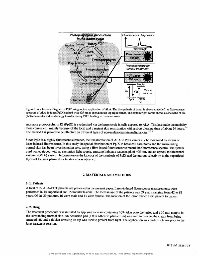
Figure 1. A schematic diagram ofPDT using topical application of ALA. The biosynthesis of haem is shown to the left. A fluorescencespectrum of ALA-induced PpIX excited with 405 nm is shown in the top right corner. The bottom right corner shows a schematic of thephotochemically induced energy transfer during PDT, leading to tissue necrosis.
substance protoporphyrin IX (PpIX) is synthesised via the haem cycle in cells exposed to ALA. This has made the modalitymore convenient, mainly because of the local and transient skin sensitisation with a short clearing time of about 24 hours.7'8The method has proved to be effective on different types of non-melanoma skin malignancies.5'6'9
Since PpIX is a highly fluorescent substance, the transformation of ALA to PpIX can easily be monitored by means oflaser-induced fluorescence. In this study the spatial distribution of PpIX in basal cell carcinoma and the surroundingnormal skin has been investigated in vivo, using a fibre-based fluorosensor to record the fluorescence speca. The systemused was equipped with an excitation light source, emitting light at a wavelength of 405 nm, and an optical multichannelanalyser (OMA) system. Information on the kinetics of the synthesis of PpIX and the tumour selectivity in the superficiallayers of the area planned for treatment was obtained.
2. MATERIALS AND METHODS
2. 1. Patients
A total of 29 ALA-PDT patients are presented in the present paper. Laser-induced fluorescence measurements wereperformed in 14 superficial and 15 nodular lesions. The median age of the patients was 69 years, ranging from 42 to 88years. Of the 29 patients, 16 were male and 13 were female. The location of the lesion varied from patient to patient.
2.2. DrugThe treatment procedure was initiated by applying a cream containing 20% ALA onto the lesion and a 10 mm margin inthe surrounding normal skin. An occlusion pad (a thin adhesive plastic film) was used to prevent the cream from beingsmeared off, and a thicker dressing on top was used to protect from light. The application was made six hours prior to thelaser treatment session.
SPIE Vol. 2926 /33
Downloaded from SPIE Digital Library on 04 Jul 2011 to 130.235.188.41. Terms of Use: http://spiedl.org/terms
ALA itself is a non-fluorescent and photodynamically non-active substance, which has the property of penetrating a fewmillimetres into abnormal keratin, and certain cells have the capacity of accumulating ALA. ALA is the first step in thehaem cycle, which takes place in a number of cells in the body. The haem production within the cycle is regulated bydifferent feed-back systems and the synthesis of different products is enzymatically dependent. The enzyme patterninvolved in the haem cycle varies in different tissue types. Thus, the production of the highly fluorescent andphotodynamically active PpIX is enhanced in malignant tissue due to higher enzymatic activity at the beginning of thecycle. Furthermore, the insufficient amount of ferrochelatase in malignant tissue results in a PpIX accumulation with only alow haem production.'°" When ALA is distributed excessively, the feed-back mechanisms are overridden and PpIX isproduced in malignant tissue and to a certain degree in tissues originating from the ecto- and endoterm, i.e. the epidermis,the mucous membranes, etc.68 The production of PpIX and the subsequent laser-induced photodynamic action leading toselective tissue necrosis are illustrated in Figure 1.
The transfonnation of ALA into PpIX takes place within a few hours, and can be visualised by means of laser-inducedfluorescence recorded in vivo. The fluorescence from PpIX excited with laser light at a wavelength of 405nm ischaracterised by a dual-peaked signal at about 635and 700 nm.
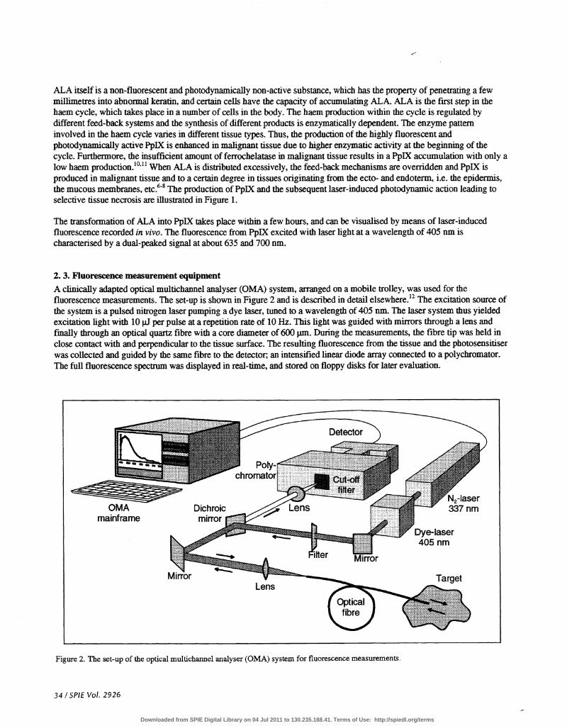
2. 3. Fluorescence measurement equipmentA clinically adapted optical multichannel analyser (OMA) system, arranged on a mobile trolley, was used for thefluorescence measurements. The set-up is shown in Figure 2 and is described in detail elsewhere.12 The excitation source ofthe system is a pulsed nitrogen laser pumping a dye laser, tuned to a wavelength of 405nm. The laser system thus yieldedexcitation light with 10 jiJ per pulse at a repetition rate of 10 Hz. This light was guided with mirrors through a lens andfmally through an optical quartz fibre with a core diameter of 600 jim. During the measurements, the fibre tip was held inclose contact with and perpendicular to the tissue surface. The resulting fluorescence from the tissue and the photosensitiserwas collected and guided by the same fibre to the detector; an intensified linear diode array connected to a polychromator.The full fluorescence spectrum was displayed in real-time, and stored on floppy disks for later evaluation.
Figure 2. The set-up of the optical multichannel analyser (OMA) system for fluorescence measurements.
34 / SPIE Vol. 2926
Detector
OMA Dichroicmainframe mirror
N2-Iaser337 nm
Dye-laser405 nm
LensTarget
Downloaded from SPIE Digital Library on 04 Jul 2011 to 130.235.188.41. Terms of Use: http://spiedl.org/terms
2. 4. Measurement procedureThe ALA-induced PpIX formation was measured in vivo, by means of laser-induced fluorescence (UF), before the lasertreatment procedures, in order to monitor the PpIX accumulation in the ALA-treated area. The fluorescence spectra wererecorded in scans over the tumour areas, including the normal skin surrounding the lesions (Figure 3). The spot size forevery measurement point was about 600 pin. The tumour fluorescence was monitored at the border, 2 mm inside thetumour and in the tumour centre. Fluorescence spectra were also recorded in the surrounding normal skin at 2,5and10 mm from the visible border of the tumour. At each measurement point, two measurements were performed, resulting intwelve spectra from each patient at each occasion. In order to investigate the laser-induced photobleaching of the activesubstance, the fluorescence was measured again immediately after the laser treatment.
Additional fluorescence measurements were performed two and four hours after the ALA application, and fmally at twohours after the treatment procedure in seven of the lesions. The two first measurements, in combination with thosementioned above, were used to study the kinetics of the transformation of ALA into PpIX. The last measurement indicatedwhether the PpIX synthesis continued after the PDT or not.
Figure 3. Fluorescence spectra recorded before PDT in a scan of a superficial basal cell carcinoma and the surrounding normal skin sixhours after topical application of ALA (top). Similar recordings after PDT, indicating the laser-induced photobleaching of the sensitiser,are shown below.
SP1E Vol. 2926 135
Before PDT
After PDT
Downloaded from SPIE Digital Library on 04 Jul 2011 to 130.235.188.41. Terms of Use: http://spiedl.org/terms
2. 5. Data analysis
After the measurements, the fluorescence spectra were transferred to a personal computer for evaluation, using a computerprogram developed at the department.'3 The PpIX-related fluorescence signal at 635 nm, was obtained as the totalfluorescence intensity in the peak minus the background, originating from tissue autofluorescence. The background wasgiven by fitting an exponential curve in the wavelength regions 550 to 600 nm, and 750 to 800 nm to the recordedspectrum. The background-free fluorescence at 635 nm is related to the amount of PpIX in the tissue, and was used tomonitor the accumulation of the photosensitiser. The peak intensity of the autofluorescence at 490 nm was also evaluated.The intensity was used for diagnostics of the tissue, as it has been shown that the autofluorescence is lower in malignanttissue than in normal tissue.'4 Forming the ratio between the PpIX-related fluorescence intensity and the autofluorescencewill thus give an enhanced contrast between tumour and normal tissue. The ratio is a dimensionless quantity and is moreinsensitive to measurement geometry, fluctuations in the excitation light source etc.
3. RESULTS
3. 1. Selectivity
All lesions monitored by means of LIF exhibited a marked selectivity of the accumulation of PpIX in tumour compared tothe surrounding normal skin six hours after ALA application (Figure 3). As seen in the different spectra, the tumour area ischaracterised by the dual-peaked fluorescence signal at 635 and 700 nm from PpLX, whereas the normal skin shows verylow porphyrin-related fluorescence. The porphyrin fluorescence does, however, start to build up even in the visibly normalskin close to the tumour. In parallel, there is a decrease in the autofluorescence, peaking at about 490 nm. Theautofluorescence usually has a lower intensity in malignant tumours than in normal tissue and represents the fluorescencefrom endogenous chromophores, such as NADH, NAD, collagen, elastin and tryptophane. The decrease in theautofluorescence intensity inside the tumour was first reported in 1984,' and is mainly due to a change in the relativeconcentrations of the highly fluorescent NADH and the less fluorescent NAD.15"6
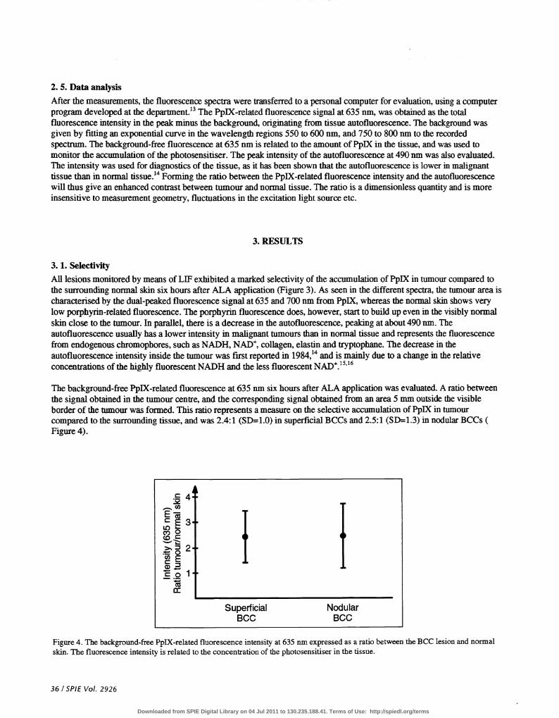
The background-free PpIX-related fluorescence at 635 nm six hours after ALA application was evaluated. A ratio betweenthe signal obtained in the tumour centre, and the corresponding signal obtained from an area 5 mm outside the visibleborder of the tumour was formed. This ratio represents a measure on the selective accumulation of PpIX in tumourcompared to the surrounding tissue, and was 2.4:1 (SD=1.O) in superficial BCCs and 2.5:1 (SD=1.3) in nodular BCCs (Figure 4).
4
E 3LC) -C) 0
olSuperficial Nodular
BCC BCC
Figure 4. The background-free PpIX-related fluorescence intensity at 635 nm expressed as a ratio between the BCC lesion and normalskin. The fluorescence intensity is related to the concentration of the photosensitiser in the tissue.
36 / SPIE Vol. 2926
Downloaded from SPIE Digital Library on 04 Jul 2011 to 130.235.188.41. Terms of Use: http://spiedl.org/terms
40
Figure 5. Thedemarcation between the tumour centre and the surrounding normal skin 5 mm outside the visible border of the tumour,when using the ratio of the background-free fluorescence intensity at 635 nmand the autofluorescence intensity at 490 urn.
If the tumour demarcation ratio is formed by dividing the PpIX-related fluorescence with the autofluorescence, a contrastenhancement of a factor of 5-7 is achieved. As seen in Figure 5, the ratio for superficial BCCs is 12:1 (SD=9) and fornodular BCCs 17:1 (SD=19). The nodular BCCs showed the highest standard deviations in both types of ratios, but thedifference was more pronounced in the lauer case.
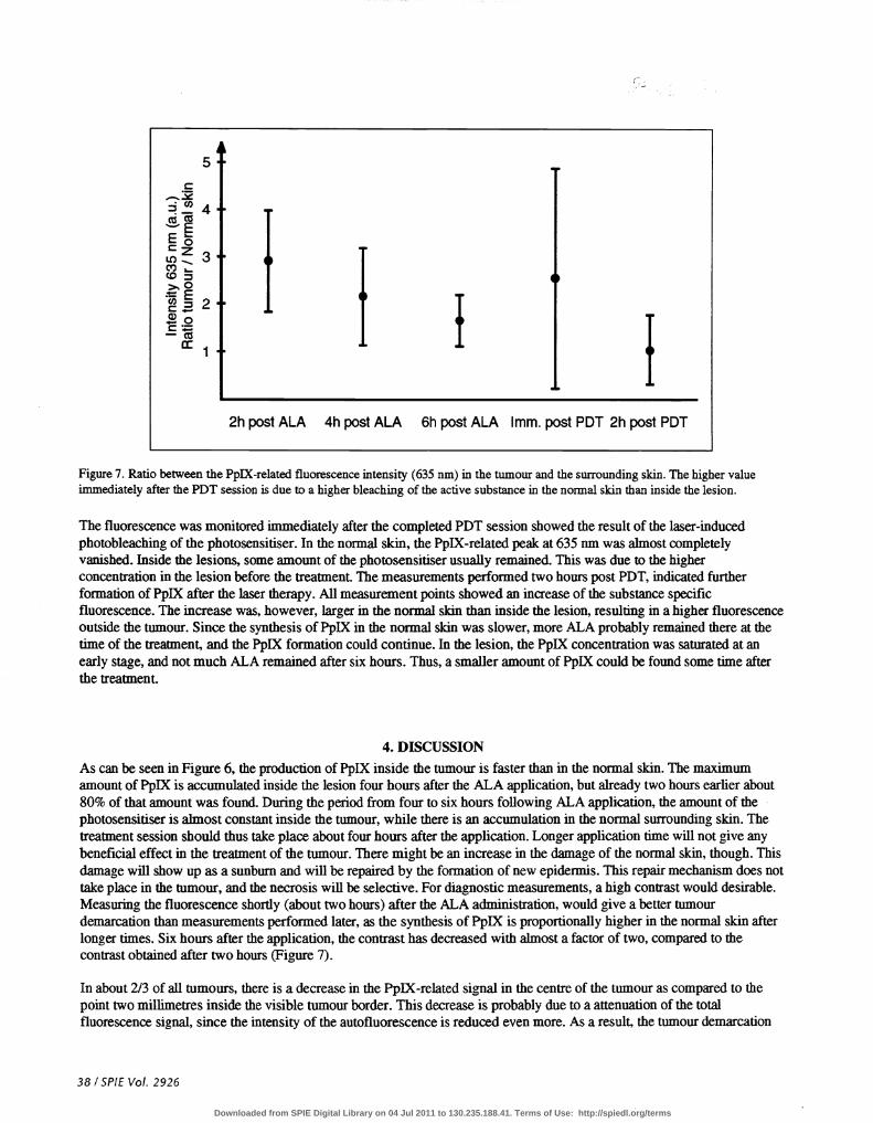
3.2. KineticsThe result of the kinetics study performed in seven lesions (five nodular and two superficial BCCs) is shown in Figure 6.Two hours after the application of ALA, most of the PpLX formation has taken place inside the tumour. During thefollowing four hours, the accumulation of the photosensitiser is proportionally greater in the normal skin. This results in alower contrast between the lesion and the surrounding tissue, as can be seen in Figure 7.
I I I I —I
Figure 6. The kinetics of the synthesis of PpIX. The fluorescence intensity at 635 nm is shown for different positions and at differenttime intervals. In the tumour, the PpIX synthesis is completed within four hours, while it continues in the normal skin. The processcontinues even after the PDT session. The average values of 7 BCC lesions are shown.
SPIEVo!. 2926/37
CCci)0—
CELC)C)10COO
SuperficialBCC
NodularBCC
a)
ECL()Cl,CO>Cl)Cci)C
2
1
2h post PDT
imm.postPDT
10mm 5mm 2mm Tumouroutside outside outside border
2 mm Tumourinside centre
Downloaded from SPIE Digital Library on 04 Jul 2011 to 130.235.188.41. Terms of Use: http://spiedl.org/terms
5C
2h post ALA 4h post ALA 6h post ALA 1mm. post PDT 2h post PDT
Figure 7. Ratio between the PpIX-related fluorescence intensity (635 nm) in the tumour and the surrounding skin. The higher valueimmediately after the PDT session is due to a higher bleaching of the active substance in the normal skin than inside the lesion.
The fluorescence wasmonitored immediately after the completed PDT session showed the result of the laser-inducedphotobleaching of the photosensitiser. In the normal skin, the PpIX-related peak at 635urnwas almost completelyvanished. Inside the lesions, some amount of the photosensitiser usually remained. This was due to the higherconcentration in the lesion before the treatment. The measurements performed two hours post PDT, indicated furtherformation of PpIX after the laser therapy. All measurement points showed an increase of the substance specificfluorescence. The increase was, however, larger in the normal skin than inside the lesion, resulting in a higher fluorescenceoutside the tumour. Since the synthesis of PpIX in the normal skin was slower, more ALA probably remained there at thetime of the treatment, and the PpLX formation could continue. In the lesion, the PpIX concentration was saturated at anearly stage, and not much ALA remained after six hours. Thus, a smaller amount of PpIX could be found some time afterthe treatment.
4_ DISCUSSION
As can be seen in Figure 6, the production of PpIX inside the tumour is faster than in the normal skin. The maximumamount of PpIX is accumulated inside the lesion four hours after the ALA application, but already two hours earlier about80% of that amount was found. During the period from four to six hours following ALA application, the amount of thephotosensitiser is almost constant inside the tumour, while there is an accumulation in the normal surrounding skin. Thetreatment session should thus take place about four hours after the application. Longer application time will not give anybeneficial effect in the treatment of the tumour. There might be an increase in the damage of the normal skin, though. Thisdamage will show up as a sunburn and will be repaired by the formation of new epidermis. This repair mechanism does nottake place in the tumour, and the necrosis will be selective. For diagnostic measurements, a high contrast would desirable.Measuring the fluorescence shortly (about two hours) after the ALA administration, would give a better tumourdemarcation than measurements performed later, as the synthesis of PpIX is proportionally higher in the normal skin afterlonger times. Six hours after the application, the contrast has decreased with almost a factor of two, compared to thecontrast obtained after two hours (Figure 7).
In about 2/3 of all tumours, there is a decrease in the PpIX-related signal in the centre of the tumour as compared to thepoint two millimetres inside the visible tumour border. This decrease is probably due to a attenuation of the totalfluorescence signal, since the intensity of the autofluorescence is reduced even more. As a result, the tumour demarcation
38 / SPIE Vol. 2926
Downloaded from SPIE Digital Library on 04 Jul 2011 to 130.235.188.41. Terms of Use: http://spiedl.org/terms
Figure 8. Ratio between the PpIX-related fluorescence and the autofluorescence six hours after topical application of 20% ALA.Superficial BCC are shown as circles, while squares indicate nodular BCC. Note the logarithmic scale on the y-axis.
ratio is higher in the tumour centre than in the border region. Thus, there might still be an increase of the PpIXconcentration in the tumour centre. In Figure 8, the average values of the logarithm of the demarcation ratio six hours afterthe ALA application is shown for different positions in the scan.
The results of the study show considerably lower values of the conirast of the ALA accumulation in tumour andsurrounding nonnal skin than reported earlier.9"7 The conirast enhancement factor, using the ratio between the Pp1X-related fluorescence and the autofluorescence was about the same in both studies. This implies that the difference lies in theaccumulation of the photosensitiser. One reason might be that in this study, the treatment was performed six hours after theALA application. In the earlier study, results were obtained when the PpIX synthesis had been active between four and sixhours. As mentioned above, the tumour demarcation ratio decreases as time passes. The position where the spectra in thesurrounding skin were recorded is also of importance. In this study the tumour centre was compared to a point 5 mmoutside the visible border of the tumour. However, there was not much change if the point 10 mm outside was used instead.
5. ACKNOWLEDGEMENTS
This work was financially supported by the Knut and Alice Wallenberg Foundation, the Kamprad Foundation, the SwedishResearch Council for Engineering Sciences, the Swedish Cancer Foundation, the Norwegian Cancer Society and Johnson& Johnson Germany, which is gratefully acknowledged.
6. REFERENCES1. T.J. Dougherty, Photoradiation therapy for cutaneous and subcutaneous malignancies, J. Invest. Dennatol. 77, 122-124
(1981).2. D. Ash and S.B. Brown, Photodynamic therapy -achievement and prospects, Br. J. Cancer 60, 15 1-152 (1989).3. S.L. Marcus, Photodynamic therapy of human cancer, IEEE 80, 869-889 (1992).4. B.W. Henderson, S.M. Waldow, T.S. Mang, W.R. Potter, P.B. Malone and T.J. Dougherty, Tumor destruction and
kinetics of tumor cell death in two experimental mouse tumors following photodynamic therapy, Cancer Res. 45, 572-576 (1985).
5. J.C. Kennedy, R.H. Pouier and D.C. Pross, Photodynamic therapy with endogenous protoporphyrin IX: Basicprinciples and present clinical experience, J. Photochem. Photobiol. B: Biol. 6, 143-148 (1990).
6. J.C. Kennedy and R.H. Pottier, Endogenous protoporphyrin IX, a clinically useful photosensitizer for photodynamictherapy, J. Photochem. Photobiol. B: Biol. 14, 275-292 (1992).
SPIE Vol. 2926 /39
010 mm 5 mm 2 mm Tumour 2 mm Tumouroutside outside outside border inside centre
Downloaded from SPIE Digital Library on 04 Jul 2011 to 130.235.188.41. Terms of Use: http://spiedl.org/terms
7. J.K. Bedwell, AJ. MacRobert, D. Phillips and S.G. Bown, fluorescence distribution and photodynamic effect of ALA-induced PP IX in the DMH rat colonic tumour model, Br. J. Cancer 65, 818-824 (1992).
8. C.S. Loh, A.J. MacRobert, J.K. Bedwell, J. Regula, N. Krasner and S.G. Bown, Oral versus intravenous administrationof5-aminolevulinic acid for photodynamic therapy, Br. J. Cancer 68, 41-51 (1993).
9. K. Svanberg, T. Andersson, D. Killander, I. Wang, U. Stenram, S. Andersson-Engels, R. Berg, J. Johansson and S.Svanberg, Photodynamic therapy of non-melanoma malignant tumours of the skin utilizing topical 6-amino levulinicacid sensitization and laser irradiation, Br. J. Dermatol. 130, 743-751 (1994).
10. N.M. Navone, C.F. Polo, A.L. Frisardi, N.E. Andrade and A.M. del C.Batlle, Heme biosynthesis in human breastcancer - mimetic "in vitro" studies and some heme enzymic activity levels, mt. J. Biochem. 22, 1407-141 1 (1990).
1 1. N.M. Navone, A.L. Frisardi, E.R. Resnik, A.M. del C.Batlle and C.F. Polo, Porphyrin biosynthesis in human breastcancer. Preliminary mimetic in vitro studies, Med. Sci. Res. 16, 61-62 (1988).
12. S. Andersson-Engels, A. Elner, J. Johansson, S.-E. Karlsson, L.G. Salford, L.-G. StrOmblad, K. Svanberg and S.Svanberg, Clinical recordings of laser-induced fluorescence spectra for evaluation of tumour demarcation feasibility inselected clinical specialities, Lasers Med. Sci. 6, 415-424 (1991).
13. R. Berg, Spectrum Evaluator- a program for evaluating fluorescence spectra, Report, Lund Institute of Technology,Lund Reports on Atomic Physics LRAP-177, (1995).
14. J. Ankerst, S. Montán, K. Svanberg and S. Svanberg, Laser-induced fluorescence studies of hematoporphyrinderivative (HpD) in normal and tumor tissue of rat, Appi. Spectr. 38, 890-896 (1984).
15. W. Lohmann and E. Paul, Native fluorescence of unstained cryosections of the skin with melanomas and naevi,Naturwissenschaften 76,424-426 (1989).
16. 5. Andersson-Engels, J. Johansson, K. Svanberg and S. Svanberg, Fluorescence imaging and point measurements oftissue: Applications to the demarcation of malignant tumors and atherosclerotic lesions from normal tissue,Photochem. Photobiol. 53, 807-814 (1991). (Invited paper).
17. I. Wang, K. Svanberg, S. Andersson-Engels, R. Berg and S. Svanberg, Photodynamic therapy of non-melanoma skinmalignancies with topical &ainino levulinic acid: diagnostic measurements, in 5th International PhotodynamicAssociation Biennial Meeting, ed. D.A. Cortese, Proc. SPIE.2371, 243-252, (1994).
40/SP1E Vol. 2926
Downloaded from SPIE Digital Library on 04 Jul 2011 to 130.235.188.41. Terms of Use: http://spiedl.org/terms