Mantica, EBSS 2011, Slide 1 Laser Spectroscopy and Nuclear Structure Study sizes and shapes of nuclei • Isotope shifts • Charge radii • Nuclear moments (m, Q) Staple at ISOL facilities for many years • CERN/ISOLDE • JAEA • Jyväskylä/IGISOL • TRIUMF/ISAC Extension to fast beam facilities • BECOLA @ NSCL/MSU Measurements made across long chains of isotopes Method applicable to nuclides over wide range of T 1/2 values http://www.physik.uni- mainz.de/Forschungsbericht/fb97/Image64.gif
Transcript
Mantica, EBSS 2011, Slide 1
Laser Spectroscopy and Nuclear Structure
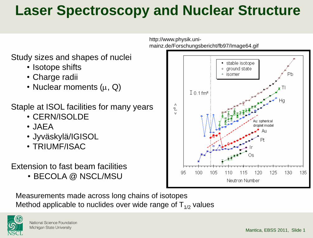
Study sizes and shapes of nuclei
• Isotope shifts
• Charge radii
• Nuclear moments (m, Q)
Staple at ISOL facilities for many years
• CERN/ISOLDE
• JAEA
• Jyväskylä/IGISOL
• TRIUMF/ISAC
Extension to fast beam facilities
• BECOLA @ NSCL/MSU
Measurements made across long chains of isotopes
Method applicable to nuclides over wide range of T1/2 values
http://www.physik.uni-
mainz.de/Forschungsbericht/fb97/Image64.gif
Mantica, EBSS 2011, Slide 2
Atomic Spectroscopy
Spectroscopy – absorption or emission of energy that induces transitions between states of a quantum mechanical system
Atomic Spectroscopy – transitions between electronic states in atom
The frequency at which energy is absorbed or emitted is related to the energy levels of the initial and final electronic states:
12 EEh
Electronic transitions typically have frequencies in the UV/Visible region of the electromagnetic spectrum
Selection rules govern the transitions that will be experimentally observed. The dipole approximation can be used to derive the selection rules for atomic spectroscopy (assuming L-S coupling applies)
0,1,0,1,0,1 SJL• ℓ is the angular momentum of the electron involved in the transition
• L, J, S refer to the vector sums for all the electrons in the atom
Mantica, EBSS 2011, Slide 3
Quantum Numbers and Terms
Hydrogen atom has good quantum numbers n, l, ml, and ms
These quantum numbers are not good for many-electron atoms or ions
no e-e repulsion e-e repulsion and
indistinguishable
spin-orbit external magnetic
field
Configuration Terms Levels States
n,l L,S J,MJ J,MJ
many-electron atoms
A useful model for atoms is to form vector sums of their orbital and spin angular momenta separately.
L, S, ML and MS can then be considered good quantum numbers for many-electron atoms
Term – group of states that has the same L and S values
Term Symbols
J
S L)12( • (2S+1) is the multiplicity
• L=0,1,2,3… are given symbols S,P,D,F…
• J is the total angular momentum
SLSLSLJ ,...,1,
Mantica, EBSS 2011, Slide 4
Atomic Fine Structure
Spin-orbit coupling breaks up a term
into levels – produces fine structure
11Na
Mantica, EBSS 2011, Slide 5
Atomic Hyperfine Structure
Interaction of atomic electron with the nucleus
Results in small shifts and splitting in the energy levels of atoms
Two main components of interaction:
Magnetic Dipole and Electric Quadrupole
electron
laser light
fluorescence
hyperfine interaction
Electric Dipole Component:
Interaction of magnetic dipole moment of the nucleus (I>0) with the magnetic field experienced by the nucleus associated with the orbital and spin angular momentum of the electrons
Electric Quadrupole Component:
Interaction of the electric quadrupole moment of the nucleus (I≥1) with the electric field gradient due to the distribution of charge within the atom
Mantica, EBSS 2011, Slide 6
Atomic Hyperfine Structure
23Na (IN=3/2)
Fine Structure Hyperfine Structure
electron+nucleus electron
~508 THz
3
3p
3s
2
1
2
1
2S1/2
2P1/2
1.77 GHz
188 MHz
110 MHz
0.5 THz 0
2P3/2 (~1/10,000,000)
(~1/1,000,000)
J=L+Se F=J+IN
NNN IJIJIJF ,...,1,
Mantica, EBSS 2011, Slide 7
Sodium Laser Hyperfine Spectra
21Na
22Na
23Na
24Na
25Na
26Na
27Na
28Na
29Na
30Na
31Na
Huber et al., PRC 18, 2342 (1978)
Na “D1” line
32S1/2 → 32P1/2
T1/2 = 0.02 s
T1/2 = 22.5 s
Mantica, EBSS 2011, Slide 8
Hyperfine Energy Shift
)12(2)12(2
)1()1(2)1(2
3
2
1
JJII
JJIIKK
BKAENN
NN
hfshfshfs
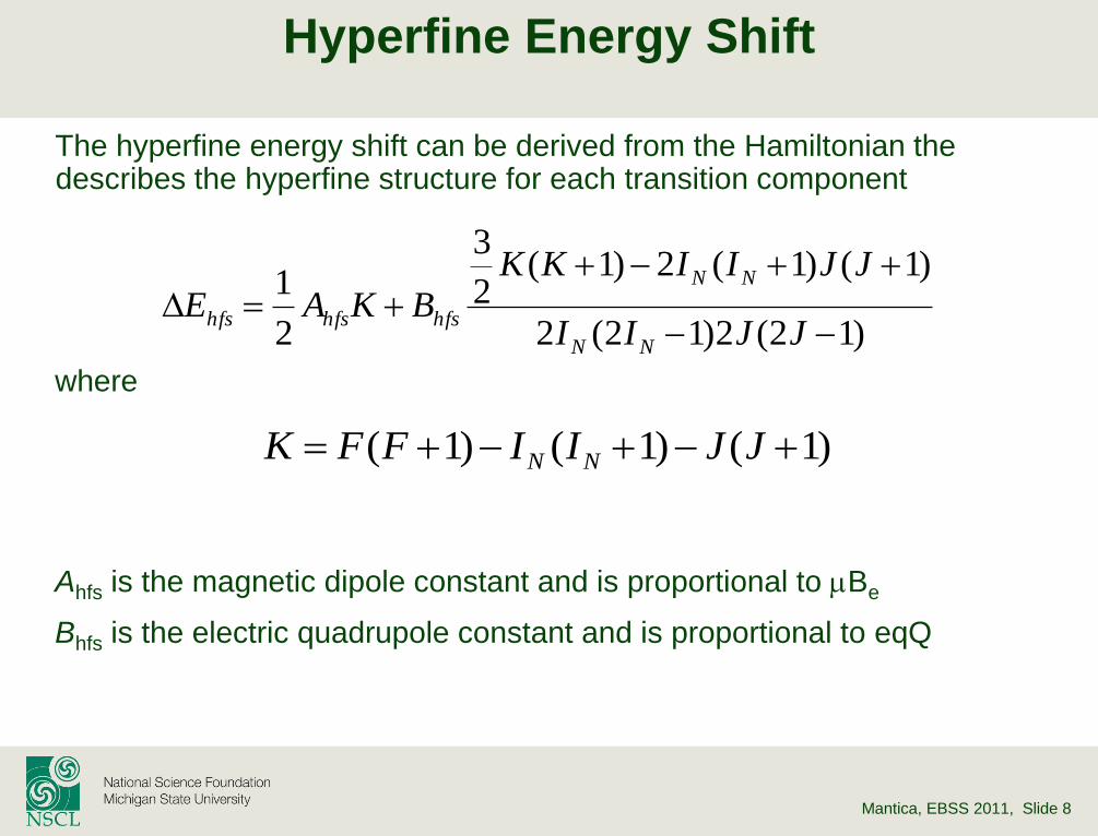
)1()1()1( JJIIFFK NN
The hyperfine energy shift can be derived from the Hamiltonian the describes the hyperfine structure for each transition component
where
Ahfs is the magnetic dipole constant and is proportional to mBe
Bhfs is the electric quadrupole constant and is proportional to eqQ
Mantica, EBSS 2011, Slide 9
Hyperfine Structure Constants: 23Na
Magnetic Dipole Constant, 32S1/2 Ahfs(32S1/2) 885.813 MHz
Magnetic Dipole Constant, 32P1/2 Ahfs (32P1/2) 94.44 MHz
Magnetic Dipole Constant, 32P3/2
Ahfs (3
2P3/2) 18.534 MHz
Electric Quadrupole Constant, 32P3/2
Bhfs (3
2P3/2) 2.724 MHz
Average magnetic field at
nucleus from atomic electrons
Average gradient of the
electric field generated by the
electrons at the nucleus
IJ
HA
eI
hfs
0m 0zzhfs VeQB
mI(23Na) = +2.217 mN Q(23Na) = +0.109 b
Mantica, EBSS 2011, Slide 10
Reference Measurements
The nuclear moments are usually deduced relative to a well-known reference measurement
• Eliminates the need to explicitly know <He(0)> and <Vzz(0)>
• High precision reference measurements are available for stable isotopes
is the hyperfine anomaly, and represents the
distribution of magnetization across the nuclear
volume (the Bohr-Weisskopf effect)
For light nuclei, it is sufficient to treat the nuclear
as a point with a pure dipole field.
In heavy nuclei, the S1/2 and P1/2 state
wavefunctions both significantly overlap the
nucleus, leading to a large value of
Bohr and Weisskopf, PR 77, 94 (1950)
IJ
HA
eI
hfs
0m
refref
refII
I
A
Amm
refref
refII
I
A
Amm 1
Mantica, EBSS 2011, Slide 11
Hyperfine Spectra of the Ga isotopes
Hyperfine spectra reveal very different structure for 73Ga compared to other odd-A isotopes
The number of peaks and relative intensities of the transitions will depend on the nuclear spin (IN)
Cheal et al., PRL 104, 252502 (2010)
42P3/2 → 52S1/2
T1/2 = 1.2 s
Mantica, EBSS 2011, Slide 12
Isotope Shifts
Isotope Shift – change in characteristic spectral line due to change in isotope
where is the center of gravity of the hyperfine components for each isotope
Mass Shift (dMS) – accounts for the difference in the relative motion between the nucleus (with mass number A) and electron
Field Shift (dFS) – proportional to the finite size of the nuclear charge distribution
Electronic Part Nuclear Part
Electron correlations (difficult)
Change in reduced mass (easy)
Change in electron density at nucleus
between spectroscopic states
FSMS
AAAA ddd ,
AA
AAMS
mm
mmk
d
SMSNMS kkk
AA
eFS rF
,
2dd 2
0
2
06
Ze
Fe
Mantica, EBSS 2011, Slide 13
Mass and Field Shifts
The Mass Shift (dMS) dominates the Isotope Shift of low-Z elements, as
the relative mass difference is large for any two isotopes.
light nuclei: dMS ~ 104 x dFS
However, it is the Field Shift dFS) that provides the desired nuclear
information
F. Schmitt et al., HI 127, 111 (2000)
AA
FS r
,
2dd
Charge radius (<r2>) – The root-mean-square (rms) charge radius is a
measure of the size of an atomic nucleus, and can be measured by both
electron scattering and atomic spectroscopy.
Optical measurements only provide the change in <r2> between two
isotopes; therefore, determination of the absolute charge radius (rc)
requires reference to a (well) known value in the isotopic chain.
Mantica, EBSS 2011, Slide 14
Charge Radius of Be isotopes
Nörtershäuser et al., PRL 102, 062503 (2009)
2.3
2.4
2.4
2.5
2.5
2.6
2.6
2.7
2.7
6 7 8 9 10 11 12
Nucle
ar
Charg
e R
adiu
s (
fm)
Mass Number (A)
Be isotopes
11Be
10Be
9Be
collinear anticollinear
2s1/2→2p1/2 for Be+
2
,9,9922
MHz/fm912.16
A
MS
A
ISc
A
c BerBerdd
measurement theory
theory
T1/2 = 13.1 s
Mantica, EBSS 2011, Slide 15
Laser Spectroscopy Methods
Collinear Laser Spectroscopy
– Fluorescence photon detection
» Na, Ga, and Be already presented
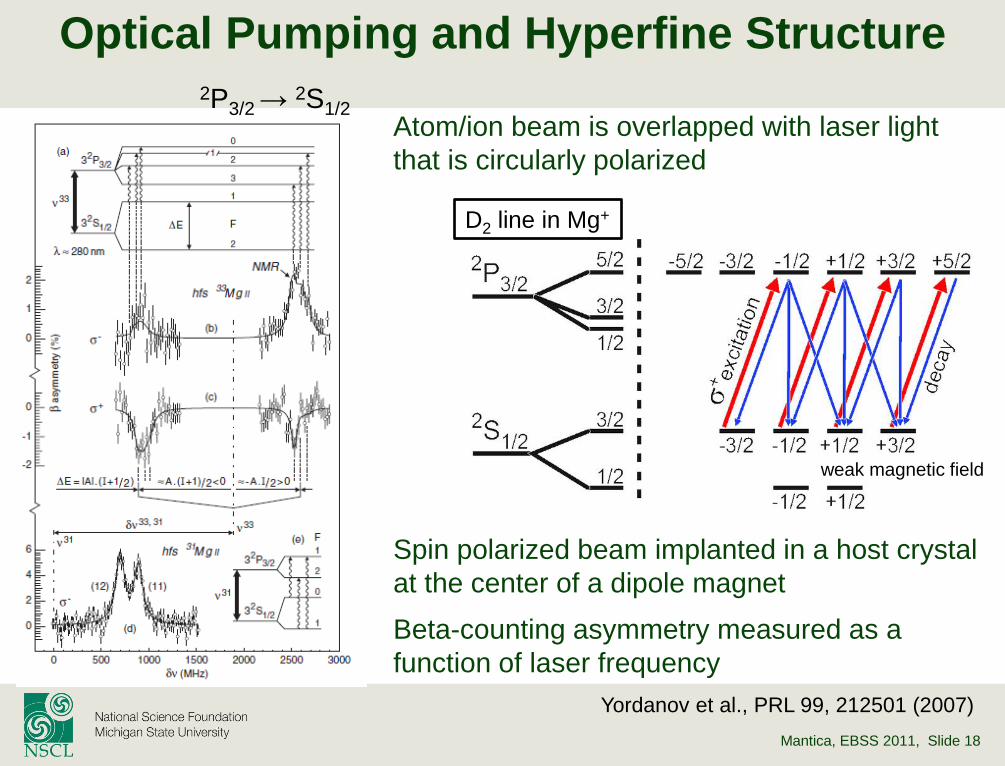
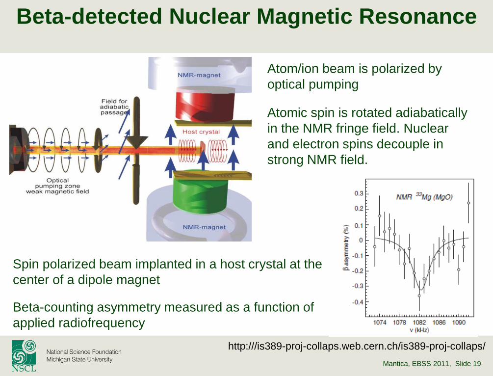
– Beta-detected nuclear magnetic resonance
» Neutron-rich 31,33Mg isotopes
» Production rate 103 ions/s for 33Mg
– Collisional ionization and ion counting
» Neutron-rich Ar isotopes
» Production rate ~106 ions/s for 43Ar
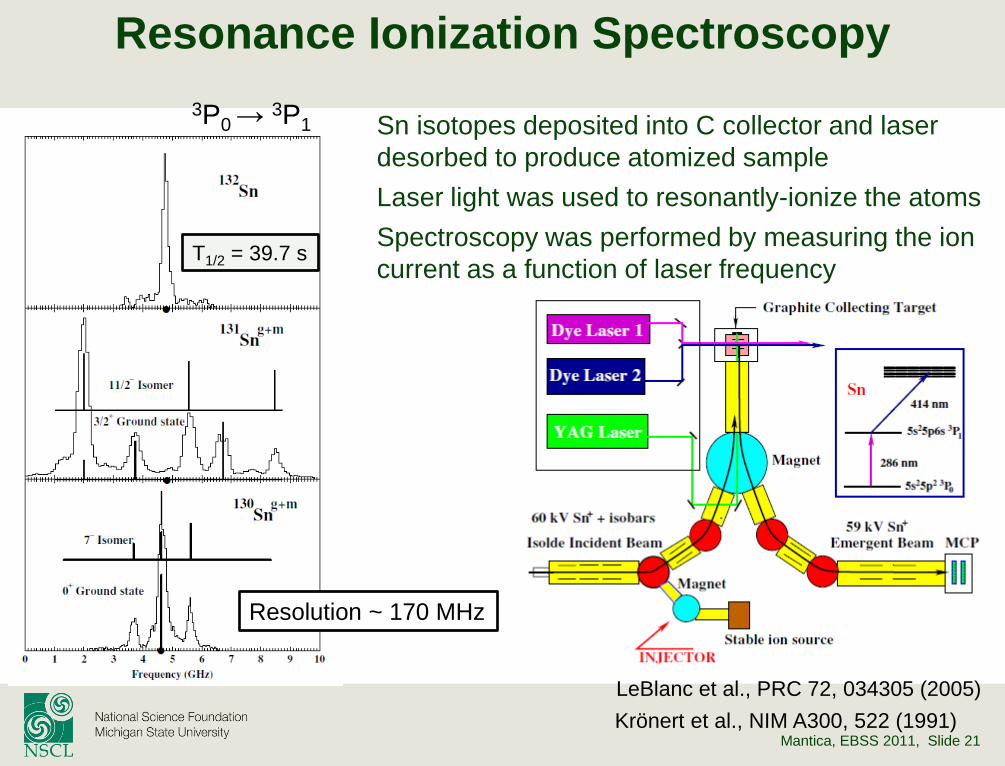
Resonance Ionization Spectroscopy
– Ion counting
» Neutron-rich Sn isotopes
» Production rate ~ 108 ions/s for 132Sn
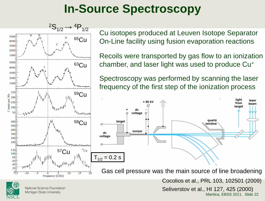
In-Source Laser Spectroscopy
– Ion Counting
» Neutron-deficient Cu isotopes
» Production rate 6 ions/s for 57Cu
Spectroscopy in Traps
– Fluorescence photon detection
» 6,8He isotopes
» Production rate 105 ions/s for 8He
Mantica, EBSS 2011, Slide 16
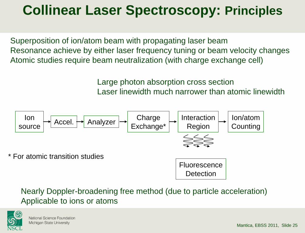
Collinear Laser Spectroscopy
COLLAPS Collaboration, CERN/ISOLDE
Photon counting
Beta-NMR
Collisional ionization Co-propagation of atom/ion beam
Laser linewidth much narrower than atomic linewidth
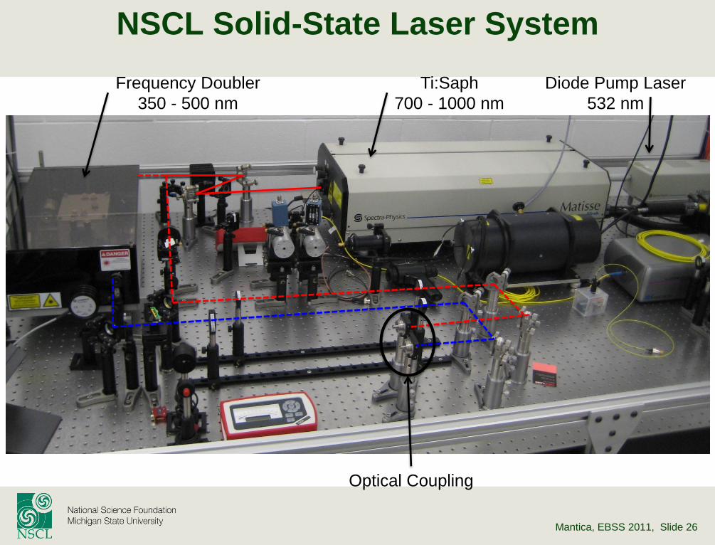
Mantica, EBSS 2011, Slide 26
Frequency Doubler
350 - 500 nm
Ti:Saph
700 - 1000 nm
Diode Pump Laser
532 nm
Optical Coupling
NSCL Solid-State Laser System
Mantica, EBSS 2011, Slide 27
Laser System Stabilization
• Short term stabilization (ms) is done with a resonator cavity that provides a feedback loop to a fast piezoelectric actuator to stabilize the system.
– Stabilization of < 1 MHz has been achieved.
• Long term stabilization (hr) is achieved via a He:Ne locking scheme.
– The He:Ne atomic transition of 633 nm is used to lock the Ti:Sapphire laser.
– Stabilization of < 8 MHz has been achieved.
– Note: The absolute frequency (10-7 precision) is not known.
Solutions to the absolute frequency
issue:
•Reference measurement
•Frequency comb
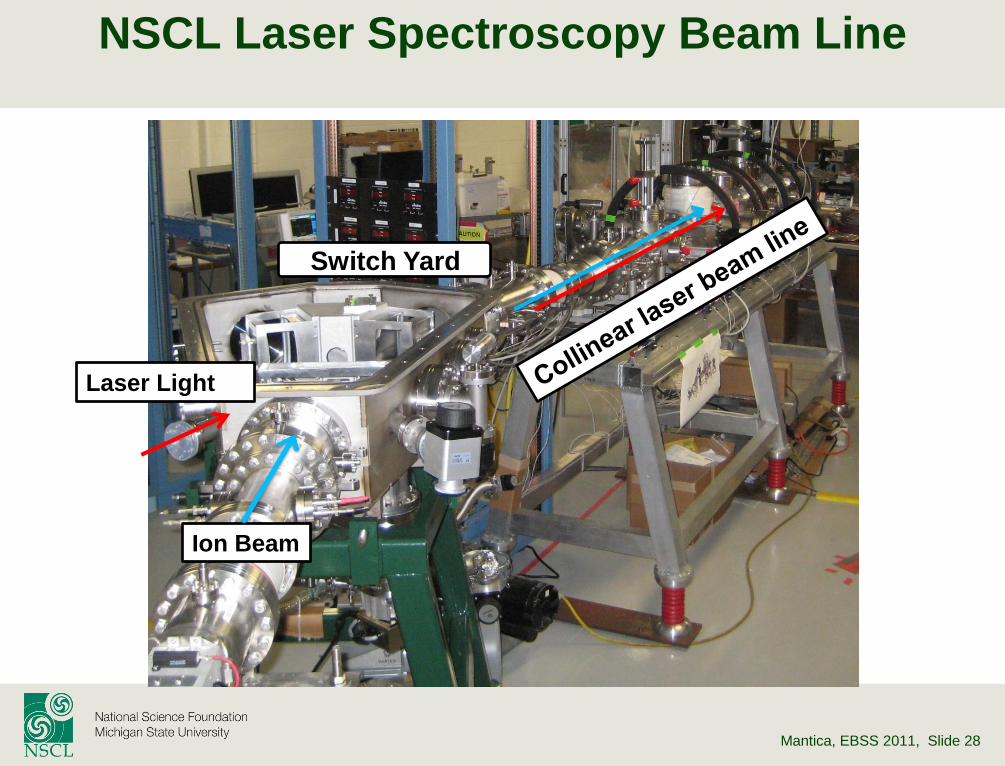
Mantica, EBSS 2011, Slide 28
NSCL Laser Spectroscopy Beam Line
Switch Yard
Ion Beam
Laser Light
Mantica, EBSS 2011, Slide 29
Charge Exchange Cells
Bacal and Reichelt, Rev. Sci. Intsrum. 45, 769 (1974)
Alkali reservoir
Heater jacket
Interaction region
HV electrodes
Liquefying region Service ports
Ion beam/laser
vertical configuration
cold end on one side
horizontal configuration
cold end on both sides
NSCL
Mainz/ISOLDE
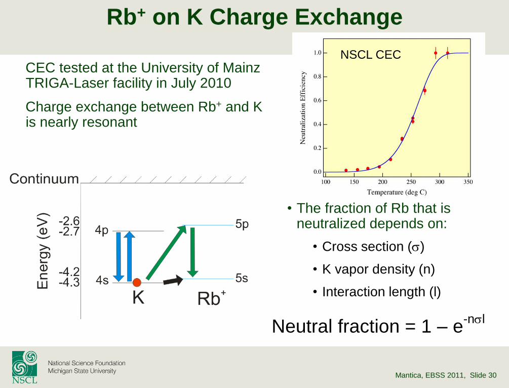
Mantica, EBSS 2011, Slide 30
Rb+ on K Charge Exchange
CEC tested at the University of Mainz TRIGA-Laser facility in July 2010
Charge exchange between Rb+ and K is nearly resonant
• The fraction of Rb that is neutralized depends on:
• Cross section (s)
• K vapor density (n)
• Interaction length (l)
Neutral fraction = 1 – e-nsl
NSCL CEC
Mantica, EBSS 2011, Slide 31
E
E
Velocity Bunching
Doppler broadening increases the linewidth of the excitation from the natural linewidth, which is problematic
By accelerating the ion beam velocity bunching occurs which allows for a decrease in Doppler broadening.
For a given E →
smaller v
at high kinetic energy (Ek) →
reduced Doppler broadening
Mantica, EBSS 2011, Slide 32
Bias Voltage to CEC
Requirements: – feasibility to scan voltage ±10 kV
» ±110 GHz for 60 keV Ca II
– stability better than 10-5
» 0.6 V for 60 kV acceleration voltage; ~7 MHz linewidth
– switching speed less than 1 ms
» fast scan around resonance to reduce systematic error from system
fluctuations
– recovery time less than a few hundreds ms
» no data taking during transition -> less efficient
– measure acceleration voltage
» voltage divider: requires 10-5 precision in dividing R
– frequent reference isotope measurement
» each measurement sandwiched by reference measurements
» feasibility to quickly go back and forth between two isotopes
» each measurement less than 1 hour and requires high counting rate