+ All Categories
Home > Documents > Lateral phage transfer in obligate intracellular bacteria (Wolbachia ...

Lateral phage transfer in obligate intracellular bacteria (Wolbachia ...

Date post: 13-Feb-2017
Category:
Upload: trinhdat
View: 220 times
Download: 2 times
Share this document with a friend
15
Lateral phage transfer in obligate intracellular bacteria (Wolbachia): Verification from natural populations Letter Meghan E. Chafee 1 , Daniel J. Funk 1 , Richard G. Harrison 2 , and Seth R. Bordenstein 1, † 1 Department of Biological Sciences, Vanderbilt University, Nashville, TN; and 2 Department of Ecology and Evolutionary Biology, Cornell University, Ithaca, NY Corresponding author: Seth R. Bordenstein, [email protected], (ph) 615.322.9087, (fax) 615.343.6707 Keywords: Lateral gene transfer, Wolbachia, bacteriophage, obligate intracellular bacteria, recombination, coinfection Running head: Lateral phage transfer Title length: 69 characters (with spaces) Abstract length: 192 words Total characters, including all legends and methods, but not Abstract: 11,132 Number of references 30
Transcript

Lateral phage transfer in obligate intracellular bacteria (Wolbachia):

Verification from natural populations

Letter

Meghan E. Chafee1, Daniel J. Funk1, Richard G. Harrison 2, and Seth R. Bordenstein1, †

1 Department of Biological Sciences, Vanderbilt University, Nashville, TN; and 2 Department of

Ecology and Evolutionary Biology, Cornell University, Ithaca, NY

† Corresponding author: Seth R. Bordenstein, [email protected], (ph) 615.322.9087,

(fax) 615.343.6707

Keywords: Lateral gene transfer, Wolbachia, bacteriophage, obligate intracellular bacteria,

recombination, coinfection

Running head: Lateral phage transfer

Title length: 69 characters (with spaces)

Abstract length: 192 words

Total characters, including all legends and methods, but not Abstract: 11,132

Number of references 30

Abstract.

Lateral transfer of mobile DNA is a hallmark of bacteria with a free-living replicative stage;

however, its significance in obligate intracellular bacteria and other heritable endosymbionts

remains controversial. Comparative sequence analyses from laboratory stocks infected with

Wolbachia pipientis provide some of the most compelling evidence that bacteriophage WO-B

transfers laterally between infections of the same insect host. Lateral transfer between

coinfections, however, has not been evaluated in natural populations nor between closely-related

Wolbachia strains. Here we analyze bacterial and phage genes from two pairs of natural

sympatric field isolates, of Gryllus pennsylvanicus field crickets and of Neochlamisus bebbianae

leaf beetles, to demonstrate WO-B transfers between supergroup B Wolbachia. N. bebbianae

revealed the highest number of phage haplotypes yet recorded, hinting that lab lines could

underestimate phage haplotype variation and lateral transfer. Finally, using the approximate age

of insect host species as the maximum available time for phage transfer between host-associated

bacteria, we very conservatively estimate phage WO-B transfer to occur at least once every 0-5.4

million years within a host species. Increasing discoveries of mobile elements, intragenic

recombination, and bacterial coinfections in host-switching obligate intracellular bacteria specify

that mobile element transfer is common in these species.

The restrictive lifestyle of obligate intracellular bacteria and inherited endosymbionts limits

exposure to novel gene pools and enhances reductive evolution, often resulting in a low mobile

DNA element composition (Moran and Plague 2004; Bordenstein and Reznikoff 2005);

however, genera prone to lateral transmission between hosts tend to establish coinfections and

integrate foreign DNA into their genomes (Bordenstein and Reznikoff 2005). For instance, in

the reproductive parasite Wolbachia, up to 21% of its genes represent mobile DNA (Wu et al.

2004; Klasson et al. 2009). Similarly, defensive bacterial symbionts of aphids that are

transmitted both maternally and horizontally have dynamic genomes exhibiting phage transfer,

recombination, and toxin-gene acquisitions (Oliver et al. 2009a).

In Wolbachia, prophage regions can comprise more than 20% of mobile DNA genes and

account for the largest fraction of absent/divergent genes between closely related strains (Ishmael

et al. 2009). Among these regions, temperate phage WO-B may inhibit Wolbachia’s capacity to

adaptively modify invertebrate reproduction (Bordenstein et al. 2006) or enhance it by encoding

ankyrin-repeat-containing genes or effectors (Iturbe-Ormaetxe et al. 2005; Sinkins et al. 2005;

Sanogo and Dobson 2006; Tanaka et al. 2009). Phage WO-B occurs in the majority of

supergroup A and B Wolbachia, can integrate as a prophage and produce active phage particles,

and rampantly transfers between Wolbachia infections (Masui et al. 2000; Bordenstein and

Wernegreen 2004; Gavotte et al. 2007). Further, multiple and divergent WO-B haplotypes

commonly co-occur in single Wolbachia infections, potentially by recurrent phage transfers

(Masui et al. 2000; Bordenstein and Wernegreen 2004; Gavotte et al. 2004; Gavotte et al. 2007).

Likewise, active transposition and frequent lateral transmission of the insertion sequence element

ISWpi1 is evident in Wolbachia (Cordaux 2008). The lateral transfer of mobile elements in

Wolbachia and other heritable symbionts has major implications for genome evolution, gene

acquisition, and the ecological interactions between selfish elements and their bacterial hosts.

However, the ecological contributions to these exchanges in natural populations remain

unexplored.

In laboratory lines of Drosophila flies, Nasonia wasps, and Ephestia moths, insect hosts

serve as incubators for phage WO-B exchange between Wolbachia coinfections (Masui et al.

2000; Bordenstein and Wernegreen 2004). For example, the rapidly evolving phage ORF7 gene

that encodes the minor capsid protein has nearly complete sequence identity between

coinfections of the divergent A and B Wolbachia in these animals. However, outstanding issues

remain. It is not known if WO-B movement between Wolbachia infections occurs in nature,

how frequent it is, or if it is possible between closely related coinfections.

Here we infer recent lateral transfer events between supergroup B Wolbachia infections

in field isolates. Specifically, Gryllus pennsylvanicus field crickets were sampled from an

Ithaca, New York population known to include individuals with coinfections of different B

Wolbachia (Mandel, Ross, and Harrison 2001). Neochlamisus bebbianae leaf beetles were

collected from Salix bebbiana (Bebb’s willow) in Caledonia County, Vermont. Crickets and

beetles were screened for single Wolbachia infections using PCR and sequences from four multi-

locus sequence typing (MLST) genes, ftsZ, gatB, hcpA, and coxA (Baldo et al. 2006).

Comparative sequence analyses of MLST genes ftsZ, gatB, and hcpA from G.

pennsylvanicus strains wG1 and wG2 showed 3.26% divergence (Fig. 1). coxA was excluded

from further consideration because we could not determine if its unusual sequence evolution

reflected gene transfer to the host genome or recombination at the coxA locus. Cloned and

sequenced PCR products of the ORF7 minor capsid gene from wG1 and wG2 revealed six and

three distinct phage WO-B haplotypes based on a cut off criteria of 1.5% divergence (Fig. 2).

Variability in phage haplotype number and diversity is typical of Wolbachia (Bordenstein and

Wernegreen 2004; Gavotte et al. 2007) and suggests that gene duplications and separate

invasions of different phage haplotypes shape the evolution of phage WO-B in Wolbachia

genomes. Notably, one of the cloned ORF7 haplotypes is shared between wG1 and wG2,

differing by 1/358 base pairs (0.28%) (Fig. 2). The evolutionarily recent transfer of a given

phage haplotype receives support if phage genetic divergence is lower than that of its two

bacterial hosts despite the more rapid rate of phage nucleotide evolution (Table 1). Indeed, a

Fisher’s exact test shows ORF7 divergence (1/358) to be significantly lower than that “expected”

from the 3.26% divergence of its Wolbachia host genes (12/358) (P = 0.001, one-tailed test).

The two N. bebbianae leaf beetle infections wNbeb1 and wNeb2, show 1.0% sequence

divergence in combined MLST gene sequences. Fig. 2 reveals seven and four ORF7 haplotypes

within wNbeb1 and wNbeb2, respectively, with the former value representing the most

haplotypes reported for any single Wolbachia infection (Gavotte et al. 2007). Three of the

cloned ORF7 haplotypes are shared between wNbeb1 and wNbeb2, suggesting these sequences

have experienced recent lateral transfer. The Fisher’s exact test yields a marginally non-

significant difference between the observed (0/358) and “expected” (4/358) divergence (P =

0.062), likely due to the lower 1% divergence in Wolbachia genes than in the crickets. Given the

two-fold higher sequence diversity in our phage gene compared to the Wolbachia ones (Table 1)

and the complete identity of the three shared haplotypes, this p-value represents a conservative

estimate of the likelihood that these three phage haplotypes transferred.

Support for lateral phage transfer between these closely related Wolbachia can also be

bolstered if the two infections arose by independent acquisitions, rather than by descent and

duplication from a common ancestor within N. bebbianae. Estimating absolute divergence times

is problematic in the absence of fossil data, but molecular clocks can provide rough estimates.

Synonymous divergence between wNbeb1 and wNbeb2 (3.56%) suggests their infections are

3.96 million years old based on a rate derived for diverse bacteria (Ochman, Elwyn, and Moran

1999). Meanwhile, N. bebbianae is 0.48 million years old based on mtDNA divergence (Brower

1994; Funk 1999). These estimates support independent acquisitions of these two Wolbachia

strains and lateral transfer of three phage haplotypes within the last 480,000 years (Table 2).

Using insect isolates with different Wolbachia infections, these experiments support

coinfection in the same insect host as a mechanism for lateral phage transfer in natural

populations of obligate intracellular bacteria. Phage transfer has been documented in all five

instances where it has been investigated, with three cases occurring between A and B Wolbachia

coinfections in lab stocks of Hymenoptera, Diptera, and Lepidoptera, and two cases (this study)

occurring between different B Wolbachia lineages in natural populations of Orthoptera and

Coleoptera (Table 2). Using estimates of insect host age as the maximal age of phage transfer

between independently acquired host-associated Wolbachia, all documented WO-B transfers

have occurred within the last 5.4 Myr. In our study, the actual time since lateral phage transfer is

expected to be considerably shorter than the maximum age estimate because the phage ORF7

genes exhibit complete or near complete nucleotide identity. N. bebbianae shows the most recent

transfers, with each of three haplotypes transferring within 480Kyr (Table 2). Our field samples

identified more phage haplotypes for G. pennsylvanicus wG1 (6) and N. bebbianae wNbeb1 (7)

than any previous reported study. Perhaps such field samples provide improved snapshots of the

extent of lateral transfer in heritable symbionts, as laboratory lines may be subject to relaxed

selection and phage loss over time (Oliver et al. 2009b). In this event, our data indicate that

bacteriophage WO-B is more abundant and capable of lateral transfer than previously thought.

Phages may thus play underappreciated roles as conduits for lateral transfer between coinfecting

bacterial strains of arthropod hosts.

Methods.

Bacteriophage and Wolbachia DNA were amplified using PCR. The phage ORF7

capsid locus (Masui et al. 2000) was amplified along with 16S rRNA, wsp and the multilocus

sequence typing (MLST) genes, ftsZ, gatB, coxA and hcpA (Baldo et al. 2006). Bidirectional

sequencing was performed directly from PCR products or from products cloned into plasmid

vectors using the GC Cloning Kit (Lucigen). Sequences were assembled, manually edited, and

aligned using MUSCLE in Geneious v. 4.6.5 (Drummond et al. 2009) and MacClade version

4.08 (Maddison and Maddison 2005). Cloned sequences showing > 1.5% nucleotide divergence

from each other were selected for analysis. K2p (Kimura 1980) genetic distances were

calculated using PHYLIP (Felsenstein 1993), and average nucleotide diversities and synonymous

divergences were determined with DnaSP (Rozas et al. 2003).

Maximum-likelihood (ML) and Bayesian methods were carried out using PAUP 4.0b10

and the Bayes plugin in Geneious (Huelsenbeck and Ronquist 2001). Model selection for the

ML analysis was estimated using the Akaike information criterion in Modeltest v3.7. The DNA

substitution model selected for the concatenated MLST loci was the GTR model in which the

gamma distribution + invariant sites were estimated from the data (GTR+I+G). Maximum

likelihood bootstrap values were generated from 100 bootstrap replicates, each using 10 random

taxon addition replications. Data sets were also analyzed with Bayesian phylogenetic methods

using GTR+I+G. Four chains were run for 1.1 million generations. Trees were sampled every

200 generations and a consensus tree was built with a burn-in of 200,000.

Figure legends.

Figure 1. Maximum Likelihood (ML) phylogeny based on a concatenated nucleotide sequence

alignment of ftsZ, gatB and hcpA Wolbachia MLST loci. The tree is midpoint rooted and

numbers denote ML bootstrap values and Bayesian posterior probabilities (in bold). Taxa are

denoted by host arthropod species and Wolbachia strain. Field specimens analyzed in this study

are shown in shaded boxes. Wolbachia supergroup A and B clades are indicated to the right of

each tree.

Figure 2. Maximum Likelihood (ML) phylogeny of the bacteriophage ORF7 nucleotide

sequences. The tree is midpoint rooted and numbers denote ML bootstrap values and Bayesian

posterior probabilities (in bold). Taxa names denote host arthropod species, Wolbachia strain

and ORF7 haplotype. Pairs of haplotypes in shaded boxes illustrate putative lateral transfer

events in field specimens collected in this study. Bold taxa represent previously published

examples of lateral transfer through laboratory segregation experiments. The three group labels

(I-III) were assigned according to the nomenclature in Bordenstein and Wernegreen (2004).

Acknowledgements

This work was supported by grants NSF IOS-0852344 and NIH R01 GM085163-01 to S.R.B.

This paper’s contents are solely the responsibility of the authors and do not necessarily represent

the official view of the NSF or NIH.

References. Baldo, L., J. C. Dunning Hotopp, K. A. Jolley, S. R. Bordenstein, S. A. Biber, R. R. Choudhury,

C. Hayashi, M. C. Maiden, H. Tettelin, and J. H. Werren. 2006. Multilocus sequence typing system for the endosymbiont Wolbachia pipientis. Appl Environ Microbiol 72:7098-7110.

Bordenstein, S. R., M. L. Marshall, A. J. Fry, U. Kim, and J. J. Wernegreen. 2006. The tripartite associations between bacteriophage, Wolbachia, and arthropods. PLoS Pathog 2:e43.

Bordenstein, S. R., and W. S. Reznikoff. 2005. Mobile DNA in obligate intracellular bacteria. Nat Rev Microbiol 3:688-699.

Bordenstein, S. R., and J. J. Wernegreen. 2004. Bacteriophage flux in endosymbionts (Wolbachia): infection frequency, lateral transfer, and recombination rates. Mol Biol Evol 21:1981-1991.

Brower, A. V. 1994. Rapid morphological radiation and convergence among races of the butterfly Heliconius erato inferred from patterns of mitochondrial DNA evolution. Proc Natl Acad Sci U S A 91:6491-6495.

Campbell, B. C., J. D. Steffen-Campbell, and J. H. Werren. 1993. Phylogeny of the Nasonia species complex (Hymenoptera: Pteromalidae) inferred from an internal transcribed spacer (ITS2) and 28S rDNA sequences. Insect Mol Biol 2:225-237.

Cordaux, R. 2008. ISWpi1 from Wolbachia pipientis defines a novel group of insertion sequences within the IS5 family. Gene 409:20-27.

Drummond, A., B. Ashton, M. Cheung, J. Heled, M. Kearse, R. Moir, S. Stones-Havas, T. Thierer, and A. Wilson. 2009. Geneious v.4.7, Available from http://www.geneious.com/.

Felsenstein, J. 1993. PHYLIP (Phylogeny Inference Package) version 3.567. Distributed by the author. Department of Genetics, University of Washington, Seattle.

Funk, D. J. 1999. Molecular systematics of cytochrome oxidase I and 16S from Neochlamisus leaf beetles and the importance of sampling. Mol Biol Evol 16:67-82.

Gavotte, L., H. Henri, R. Stouthamer, D. Charif, S. Charlat, M. Bouletreau, and F. Vavre. 2007. A survey of the bacteriophage WO in the endosymbiotic bacteria Wolbachia. Mol Biol Evol 24:427-435.

Gavotte, L., F. Vavre, H. Henri, M. Ravallec, R. Stouthamer, and M. Bouletreau. 2004. Diversity, distribution and specificity of WO phage infection in Wolbachia of four insect species. Insect Mol Biol 13:147-153.

Huelsenbeck, J. P., and F. Ronquist. 2001. MRBAYES: Bayesian inference of phylogenetic trees. Bioinformatics 17:754-755.

Ishmael, N., J. C. Hotopp, P. Ioannidis, S. Biber, J. Sakamoto, S. Siozios, V. Nene, J. Werren, K. Bourtzis, S. R. Bordenstein, and H. Tettelin. 2009. Extensive genomic diversity of closely related Wolbachia strains. Microbiology 155:2211-2222.

Iturbe-Ormaetxe, I., G. R. Burke, M. Riegler, and S. L. O'Neill. 2005. Distribution, expression, and motif variability of ankyrin domain genes in Wolbachia pipientis. J Bacteriol 187:5136-5145.

Kimura, M. 1980. A simple method for estimating evolutionary rates of base substitutions through comparative studies of nucleotide sequences. J Mol Evol 16:111-120.

Klasson, L., J. Westberg, P. Sapountzis, K. Naslund, Y. Lutnaes, A. C. Darby, Z. Veneti, L. Chen, H. R. Braig, R. Garrett, K. Bourtzis, and S. G. Andersson. 2009. The mosaic genome structure of the Wolbachia wRi strain infecting Drosophila simulans. Proc Natl Acad Sci U S A 106:5725-5730.

Maddison, D., and W. Maddison. 2005. Sinauer Associates, Sunderland, MA. Mandel, M. J., C. L. Ross, and R. G. Harrison. 2001. Do Wolbachia infections play a role in

unidirectional incompatibilities in a field cricket hybrid zone? Mol Ecol 10:703-709. Masui, S., S. Kamoda, T. Sasaki, and H. Ishikawa. 2000. Distribution and evolution of

bacteriophage WO in Wolbachia, the endosymbiont causing sexual alterations in arthropods. J Mol Evol 51:491-497.

Moran, N. A., and G. R. Plague. 2004. Genomic changes following host restriction in bacteria. Curr Opin Genet Dev 14:627-633.

Ochman, H., S. Elwyn, and N. A. Moran. 1999. Calibrating bacterial evolution. Proc Natl Acad Sci USA 96:12638-12643.

Oliver, K. M., P. H. Degnan, G. R. Burke, and N. A. Moran. 2009a. Facultative symbionts of aphids and the horizontal transfer of ecologically important traits. Annu Rev Entomol.

Oliver, K. M., P. H. Degnan, M. S. Hunter, and N. A. Moran. 2009b. Bacteriophages encode factors required for protection in a symbiotic mutualism. Science 325:992-994.

Rozas, J., J. Sanchez-DelBarrio, X. Messeguer, and R. Rozas. 2003. DnaSP, DNA polymorphism analyses by the coalescent and other methods. Bioinformatics 19:2496-2497.

Sanogo, Y. O., and S. L. Dobson. 2006. WO bacteriophage transcription in Wolbachia-infected Culex pipiens. Insect Biochem Mol Biol 36:80-85.

Sinkins, S. P., T. Walker, A. R. Lynd, A. R. Steven, B. L. Makepeace, H. C. Godfray, and J. Parkhill. 2005. Wolbachia variability and host effects on crossing type in Culex mosquitoes. Nature 436:257-260.

Tamura, K., S. Subramanian, and S. Kumar. 2004. Temporal patterns of fruit fly (Drosophila) evolution revealed by mutation clocks. Mol Biol Evol 21:36-44.

Tanaka, K., S. Furukawa, N. Nikoh, T. Sasaki, and T. Fukatsu. 2009. Complete WO phage sequences revealed their dynamic evolutionary trajectories and putative functional elements required for integration into Wolbachia genome. Appl Environ Microbiol.

Wu, M., L. V. Sun, J. Vamathevan, M. Riegler, R. Deboy, J. C. Brownlie, E. A. McGraw, W. Martin, C. Esser, N. Ahmadinejad, C. Wiegand, R. Madupu, M. J. Beanan, L. M. Brinkac, S. C. Daugherty, A. S. Durkin, J. F. Kolonay, W. C. Nelson, Y. Mohamoud, P. Lee, K. Berry, M. B. Young, T. Utterback, J. Weidman, W. C. Nierman, I. T. Paulsen, K. E. Nelson, H. Tettelin, S. L. O'Neill, and J. A. Eisen. 2004. Phylogenomics of the reproductive parasite Wolbachia pipientis wMel: a streamlined genome overrun by mobile genetic elements. PLoS Biol 2:E69.

Table 1 Average Pairwise Nucleotide Diversity Per Site (π) in Five Genes Taxa

Gene

Length of

Alignment (bp) φ

N

π

Wolbachia pipientis

ftsZ

435

38

0.058

gatB

369 38 0.066

hcpA

444 38 0.073

coxA

402 38 0.080

Bacteriophage WO-B ORF7

240 59 0.146

φ Length of the alignment is based on removal of indels

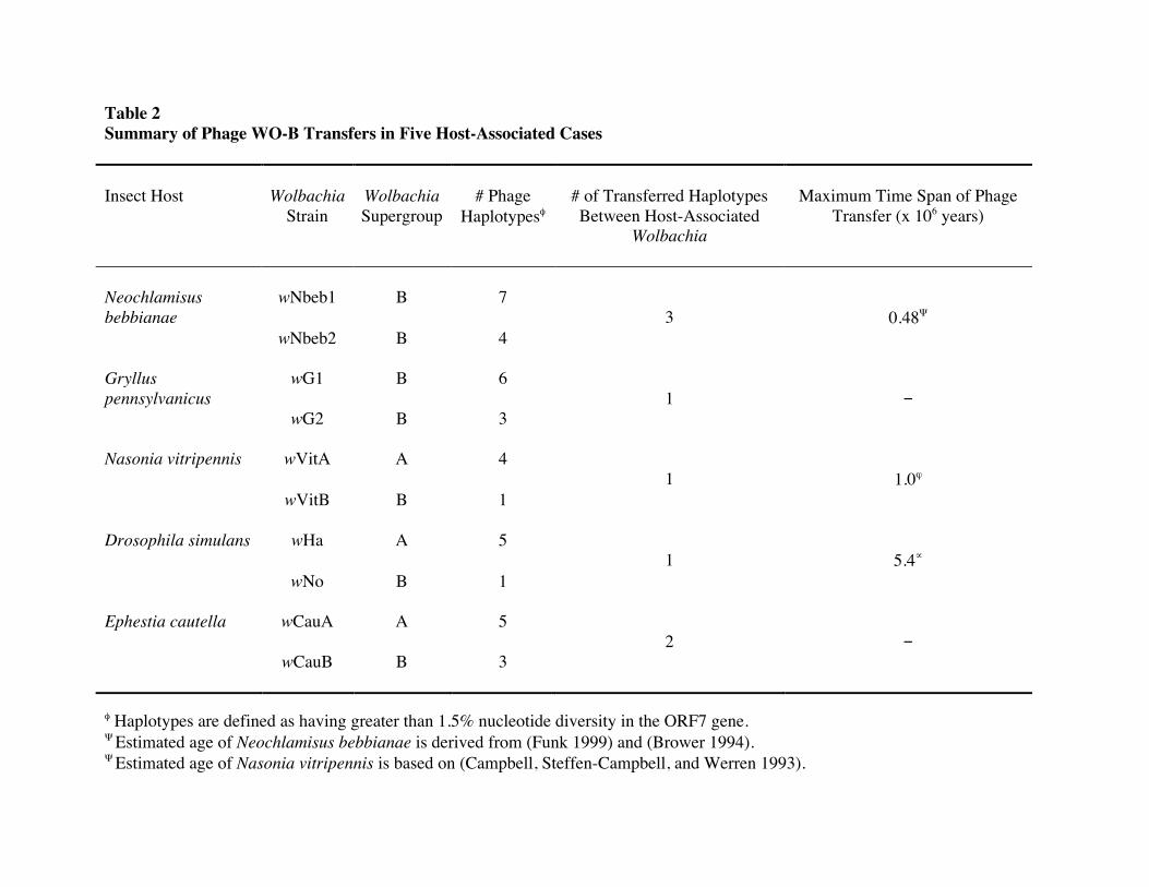
Table 2 Summary of Phage WO-B Transfers in Five Host-Associated Cases Insect Host

Wolbachia

Strain

Wolbachia Supergroup

# Phage

Haplotypesφ

# of Transferred Haplotypes Between Host-Associated

Wolbachia

Maximum Time Span of Phage

Transfer (x 106 years)

Neochlamisus bebbianae

wNbeb1

B

7

3

0.48Ψ wNbeb2

B 4

Gryllus pennsylvanicus

wG1

B

6

1

wG2

B 3

Nasonia vitripennis wVitA

A 4 1

1.0ϕ

wVitB

B 1

Drosophila simulans wHa

A 5 1

5.4∝

wNo

B 1

Ephestia cautella wCauA

A

5 2

wCauB

B 3

φ Haplotypes are defined as having greater than 1.5% nucleotide diversity in the ORF7 gene. Ψ Estimated age of Neochlamisus bebbianae is derived from (Funk 1999) and (Brower 1994). Ψ Estimated age of Nasonia vitripennis is based on (Campbell, Steffen-Campbell, and Werren 1993).

∝ Estimated age of Drosophila simulans is based on (Tamura, Subramanian, and Kumar 2004).


Recommended