16
Lead Errors Reversal of limb leads
| Date post: | 27-May-2015 |
| Category: |
Business |
| Upload: | anas-nader |
| View: | 2,379 times |
| Download: | 4 times |

Lead Errors
Reversal of limb leads
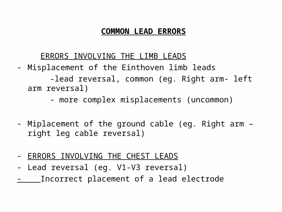
COMMON LEAD ERRORS
ERRORS INVOLVING THE LIMB LEADS- Misplacement of the Einthoven limb leads
-lead reversal, common (eg. Right arm- left arm reversal)
- more complex misplacements (uncommon)
- Miplacement of the ground cable (eg. Right arm – right leg cable reversal)
- ERRORS INVOLVING THE CHEST LEADS- Lead reversal (eg. V1-V3 reversal)
- Incorrect placement of a lead electrode
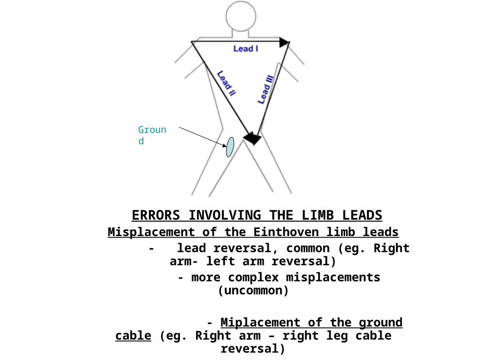
ERRORS INVOLVING THE LIMB LEADSMisplacement of the Einthoven limb leads
- lead reversal, common (eg. Right arm- left arm reversal)
- more complex misplacements (uncommon)
- Miplacement of the ground cable (eg. Right arm – right leg cable reversal)
Ground
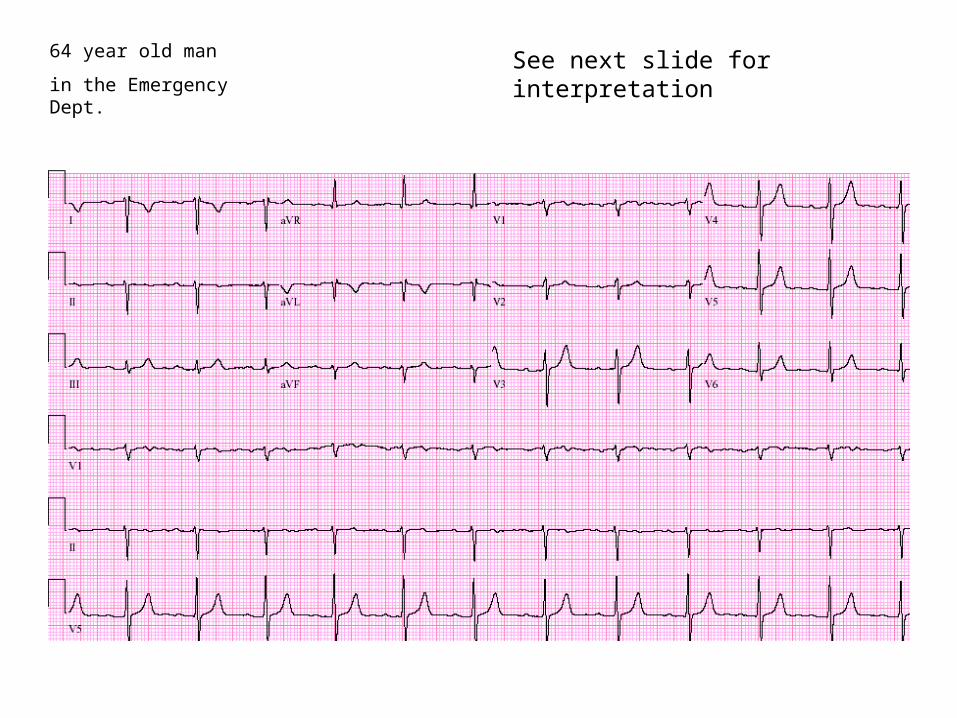
64 year old man
in the Emergency Dept.See next slide for interpretation

64 year old man
In the Emergency Dept.
P wave and QRS complex are negative in lead I and positive in aVR.
Suspect lead error: left arm – right arm lead reversal
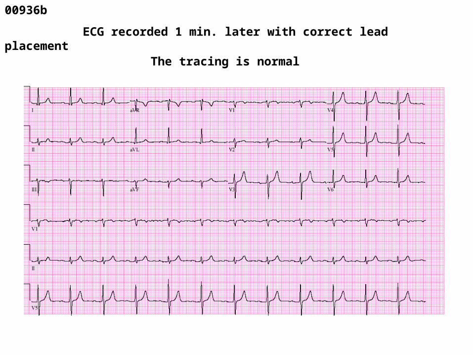
00936b
ECG recorded 1 min. later with correct lead placement
The tracing is normal
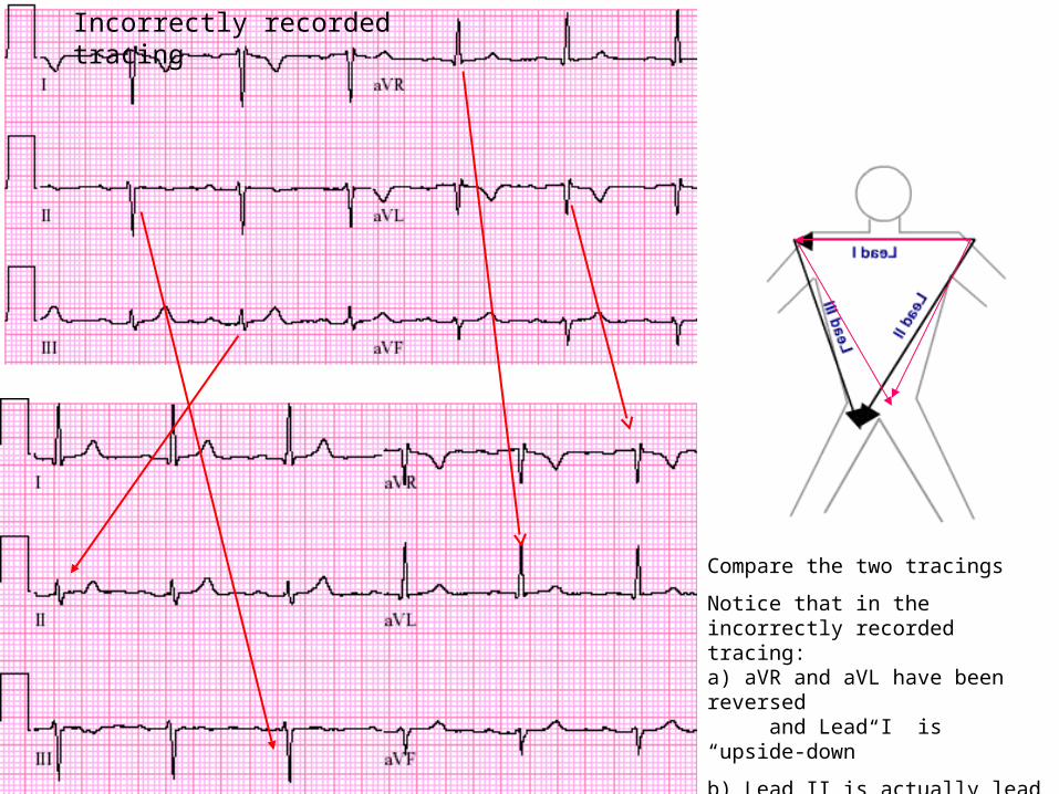
Compare the two tracings
Notice that in the incorrectly recorded tracing:a) aVR and aVL have been reversed and Lead I is “upside-down”
b) Lead II is actually lead III and lead
III is lead II
Incorrectly recorded tracing
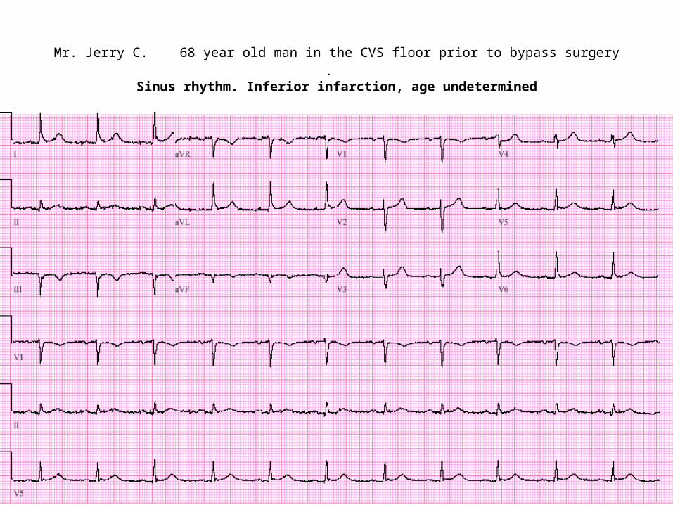
Mr. Jerry C. 68 year old man in the CVS floor prior to bypass surgery
. Sinus rhythm. Inferior infarction, age undetermined
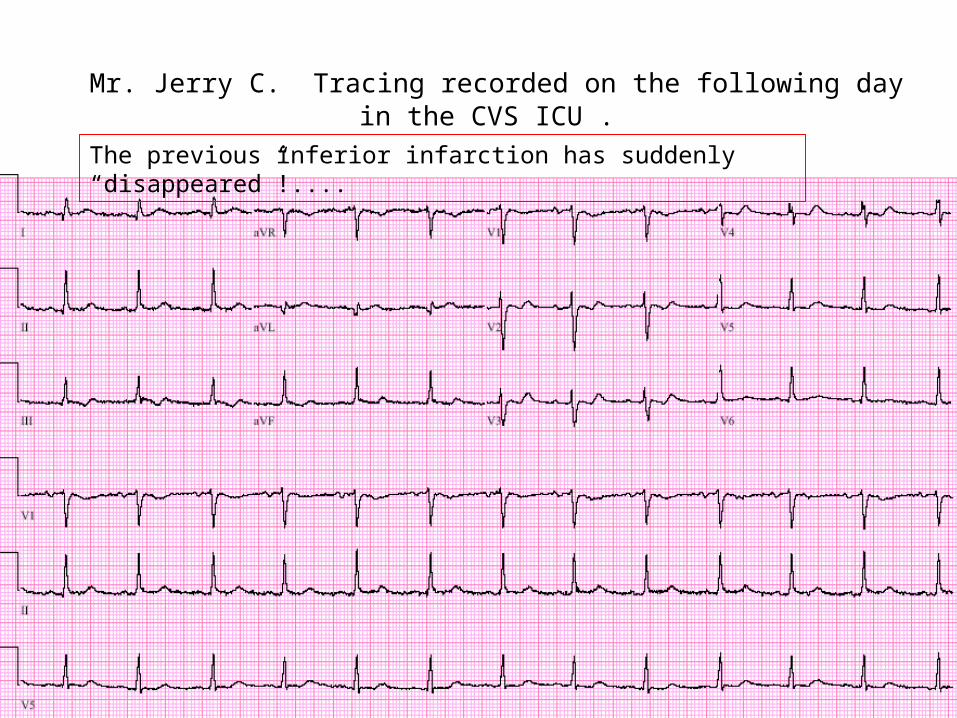
Mr. Jerry C. Tracing recorded on the following day in the CVS ICU .
The previous inferior infarction has suddenly “disappeared”!....
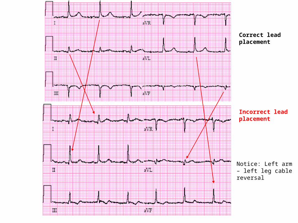
Correct lead placement
Incorrect lead placement
Notice: Left arm – left leg cable reversal
If unexpectedly an inferior infarctionappears or disappears, check whetherthe left arm and left leg electrodes might have been reversed.This lead error is frequently missed.
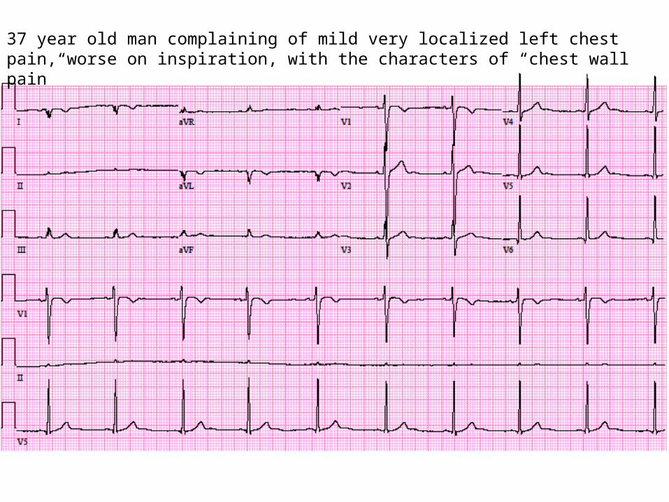
37 year old man complaining of mild very localized left chest pain, worse on inspiration, with the characters of “chest wall pain”
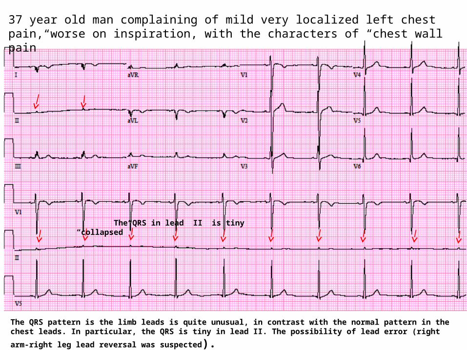
The QRS in lead II is tiny “collapsed”
The QRS pattern is the limb leads is quite unusual, in contrast with the normal pattern in the chest leads. In particular,
the QRS is tiny in lead II. The possibility of lead error (right arm-right leg lead reversal was suspected).
37 year old man complaining of mild very localized left chest pain, worse on inspiration, with the characters of “chest wall pain”
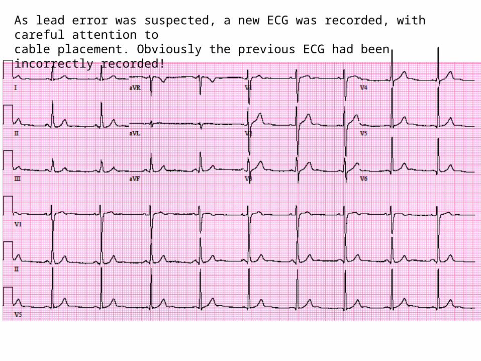
As lead error was suspected, a new ECG was recorded, with careful attention tocable placement. Obviously the previous ECG had been incorrectly recorded!
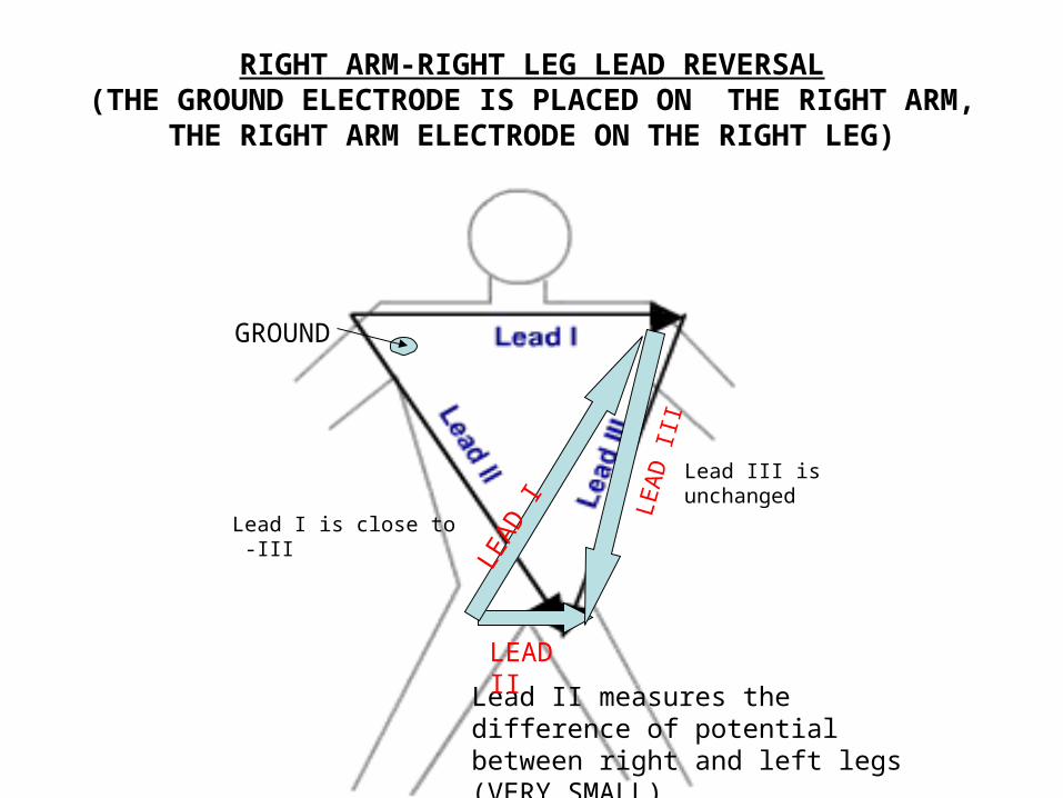
RIGHT ARM-RIGHT LEG LEAD REVERSAL(THE GROUND ELECTRODE IS PLACED ON THE RIGHT ARM,
THE RIGHT ARM ELECTRODE ON THE RIGHT LEG)
GROUND
Lead III is unchanged
Lead II measures the difference of potential between right and left legs (VERY SMALL)
LEAD IILE
AD
III
LEA
D I
Lead I is close to -III
When tracing of lead II appears “collapsed” (very small voltage),suspect that the
the placement of the right arm and right leg cables may have been reversed
Please, see the “lead errors” tutorial for an animated demonstration


