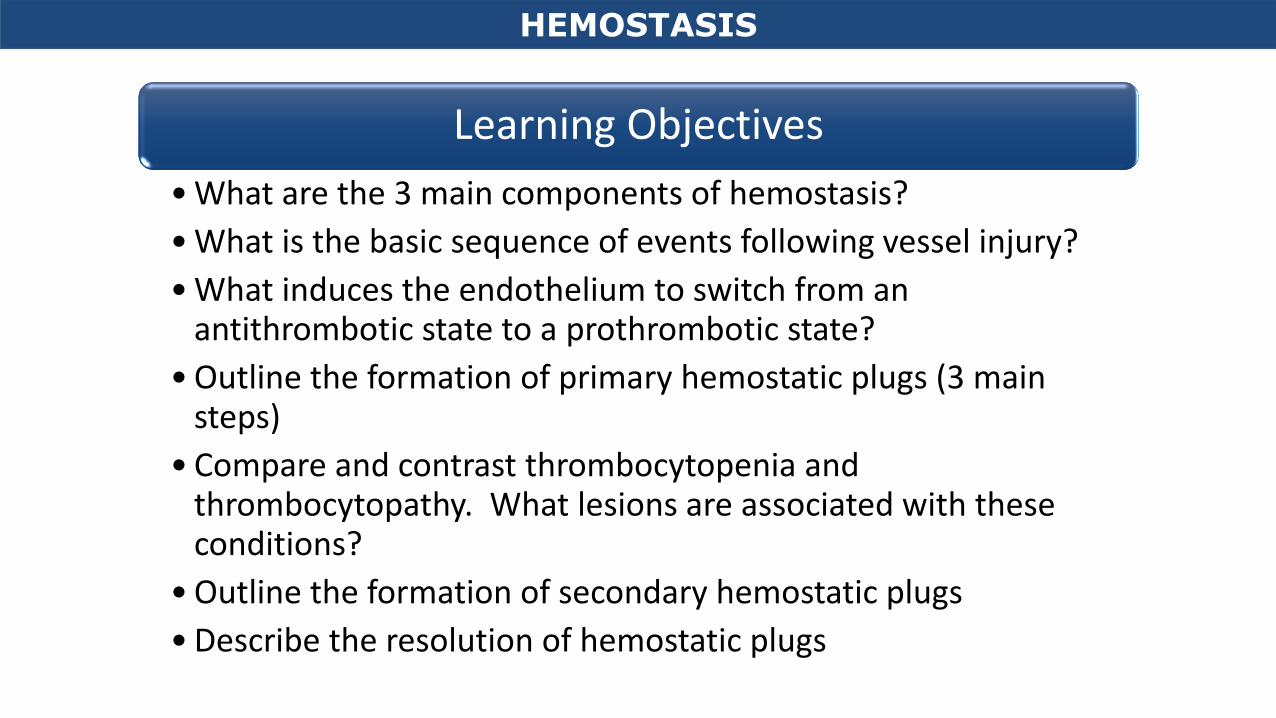
HEMOSTASIS Learning Objectives • What are the 3 main components of hemostasis? • What is the basic sequence of events following vessel injury? • What induces the endothelium to switch from an antithrombotic state to a prothrombotic state? • Outline the formation of primary hemostatic plugs (3 main steps) • Compare and contrast thrombocytopenia and thrombocytopathy. What lesions are associated with these conditions? • Outline the formation of secondary hemostatic plugs • Describe the resolution of hemostatic plugs
Transcript
HEMOSTASIS
Learning Objectives
• What are the 3 main components of hemostasis?
• What is the basic sequence of events following vessel injury?
• What induces the endothelium to switch from an antithrombotic state to a prothrombotic state?
• Outline the formation of primary hemostatic plugs (3 main steps)
• Compare and contrast thrombocytopenia and thrombocytopathy. What lesions are associated with these conditions?
• Outline the formation of secondary hemostatic plugs
• Describe the resolution of hemostatic plugs
Circulatory Disturbances 3: Hemostasis
Shannon Martinson, Feb 2016
http://people.upei.ca/smartinson/ VPM 152 General Pathology
Edema
Hyperemia and congestion
Shock
Hemorrhage
Thrombosis and embolism
Infarction
Altered hemostasis
Altered Blood flow
HEMOSTASIS
HEMOSTASIS
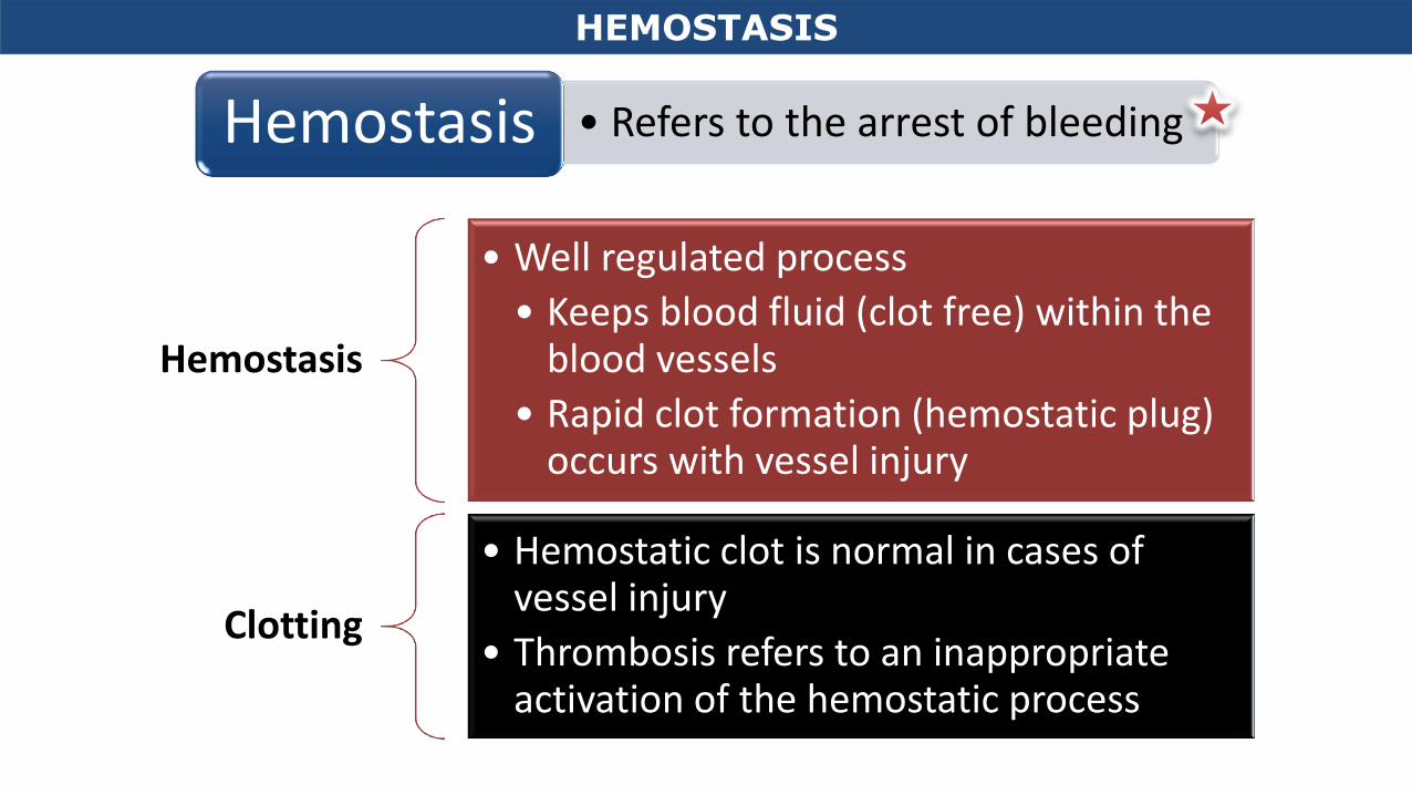
• Refers to the arrest of bleeding Hemostasis
Hemostasis
• Well regulated process
• Keeps blood fluid (clot free) within the blood vessels
• Rapid clot formation (hemostatic plug) occurs with vessel injury
Clotting
• Hemostatic clot is normal in cases of vessel injury
• Thrombosis refers to an inappropriate activation of the hemostatic process
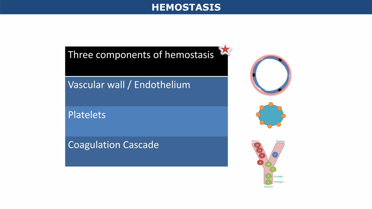
Three components of hemostasis
Vascular wall / Endothelium
Platelets
Coagulation Cascade
HEMOSTASIS
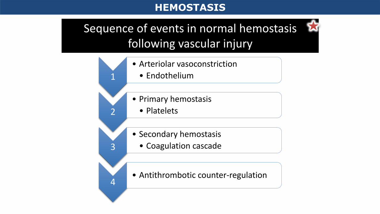
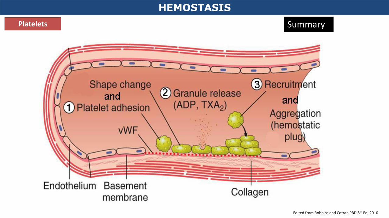
1
• Arteriolar vasoconstriction
• Endothelium
2
• Primary hemostasis
• Platelets
3
• Secondary hemostasis
• Coagulation cascade
4 • Antithrombotic counter-regulation
Sequence of events in normal hemostasis following vascular injury
HEMOSTASIS
1
• Arteriolar vasoconstriction
• Endothelium • Reflex neurogenic mechanism
• Transient
• Induced by local secretion of endothelin from endothelial cells
HEMOSTASIS
Robbins and Cotran PBD 8th Ed, 2010
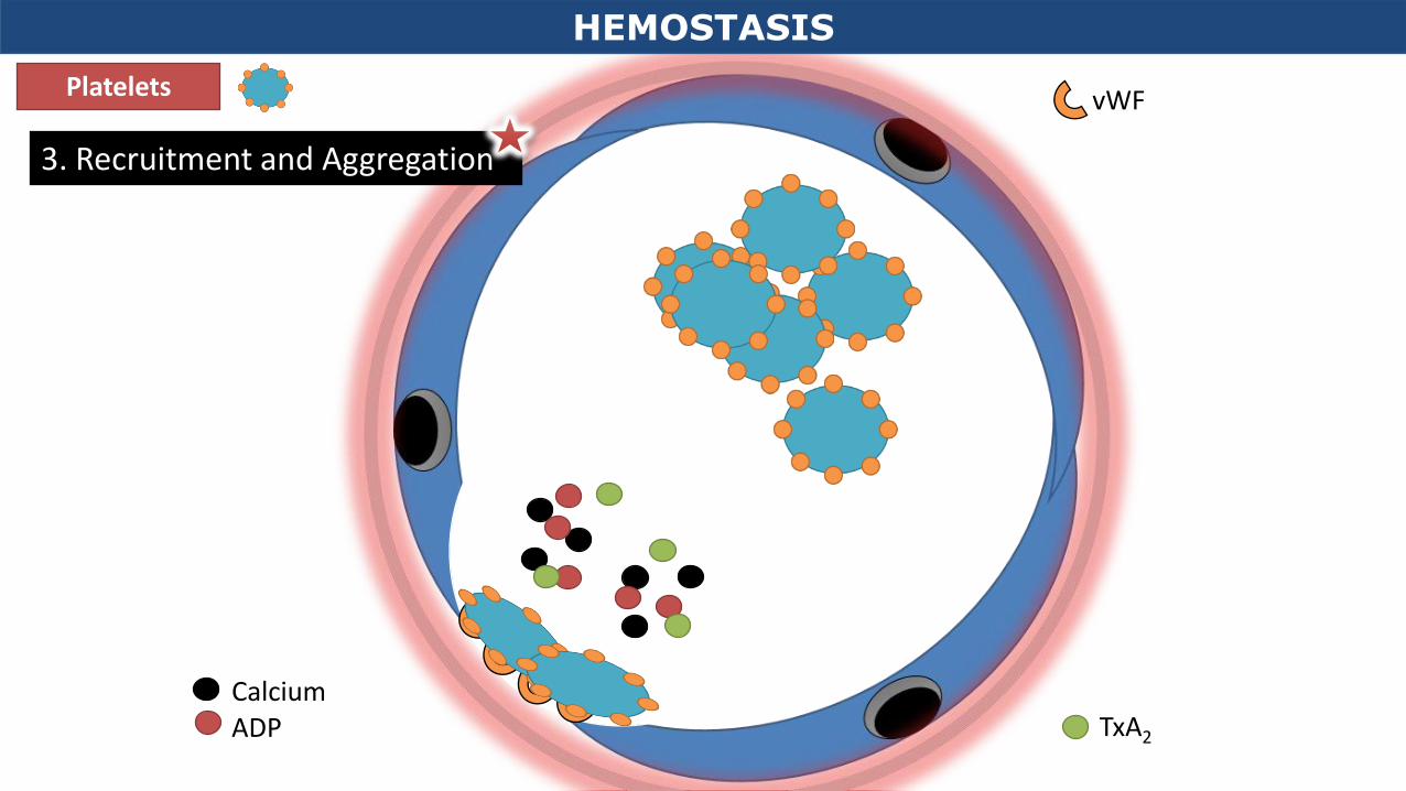
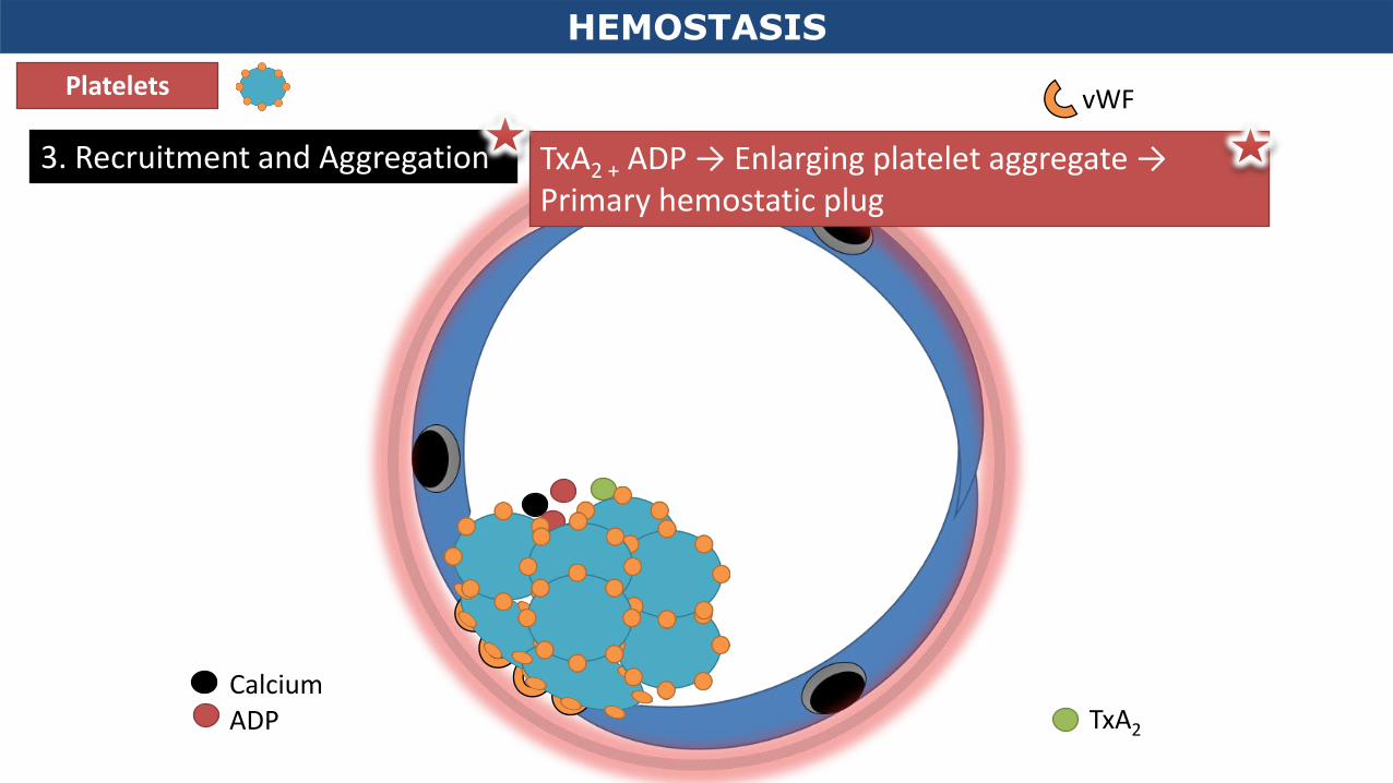
2
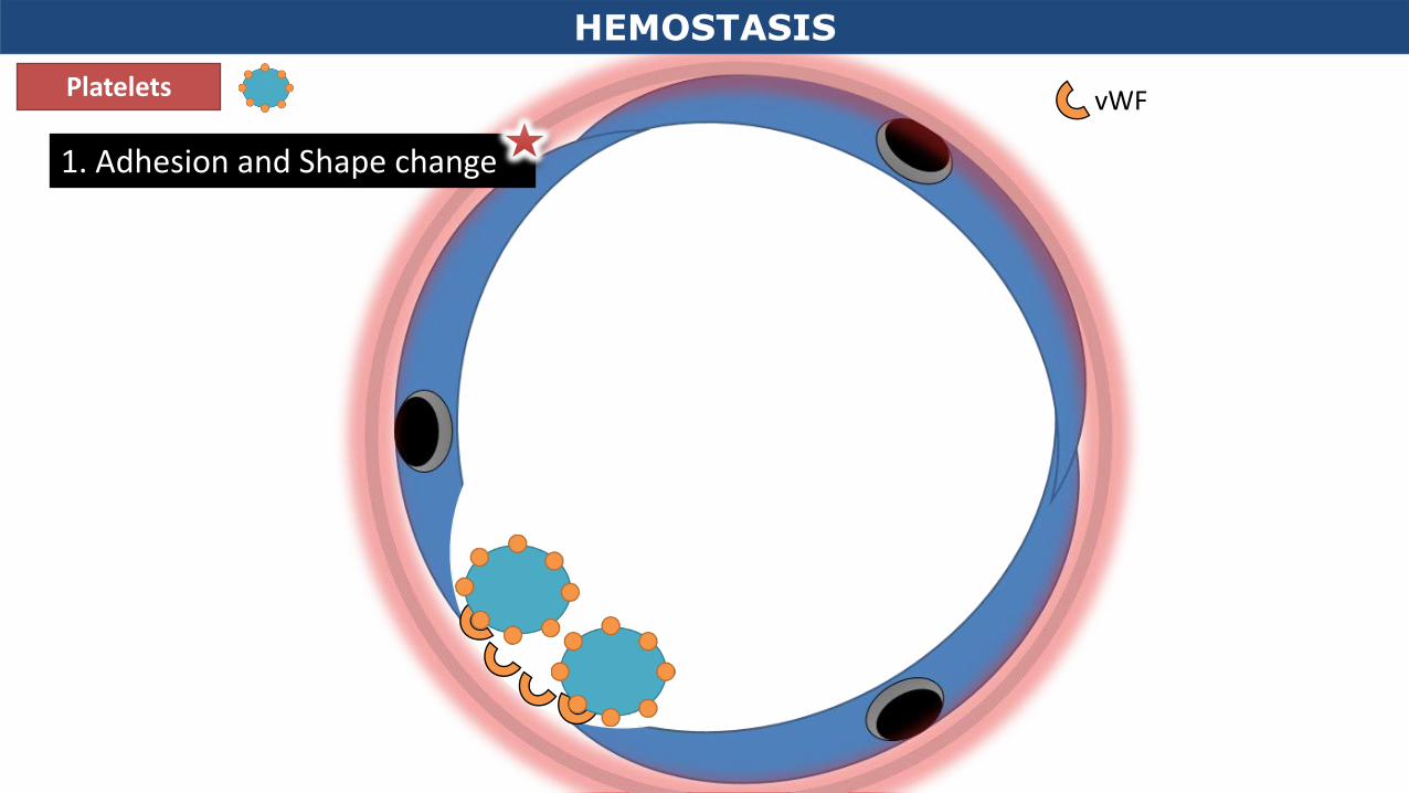
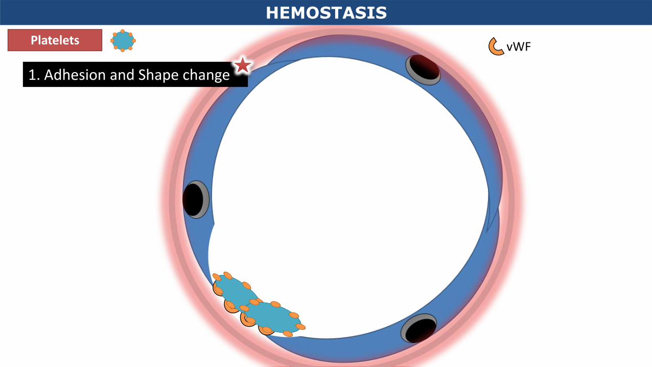
• Primary hemostasis
• Platelets
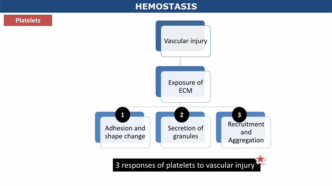
• Platelets respond to exposure of subendothelial ECM
• Result = Formation of a primary hemostatic plug
HEMOSTASIS
Robbins and Cotran PBD 8th Ed, 2010
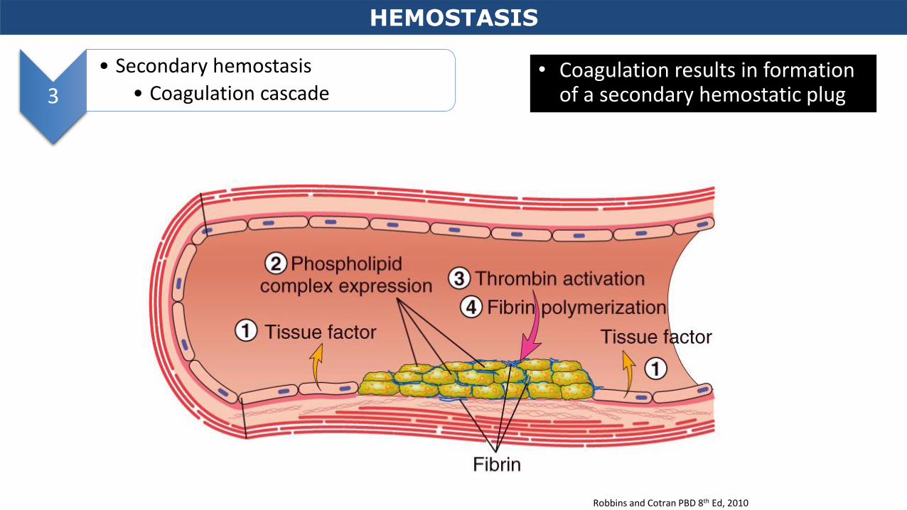
3
• Secondary hemostasis
• Coagulation cascade • Coagulation results in formation
of a secondary hemostatic plug
HEMOSTASIS
Robbins and Cotran PBD 8th Ed, 2010
4 • Antithrombotic counter-regulation
• Factors are released to limit the size of the hemostatic plug
HEMOSTASIS
Robbins and Cotran PBD 8th Ed, 2010
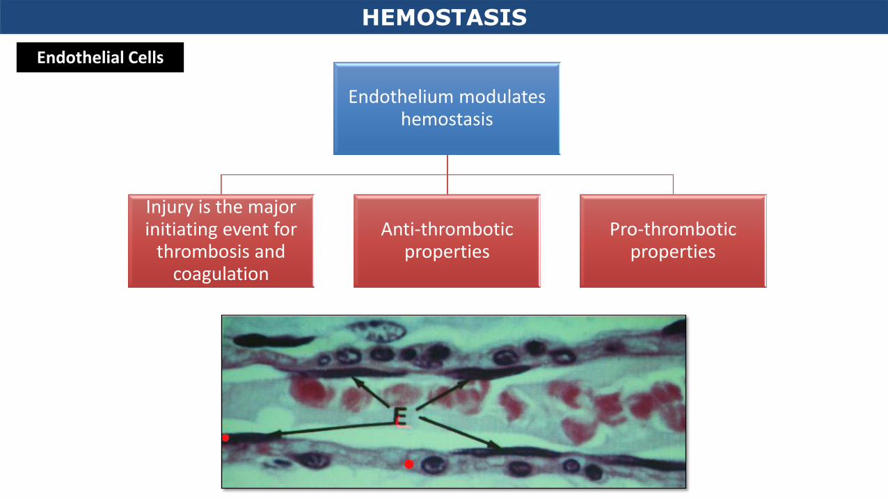
Endothelial Cells
Endothelium modulates hemostasis
Injury is the major initiating event for
thrombosis and coagulation
Anti-thrombotic properties
Pro-thrombotic properties
HEMOSTASIS
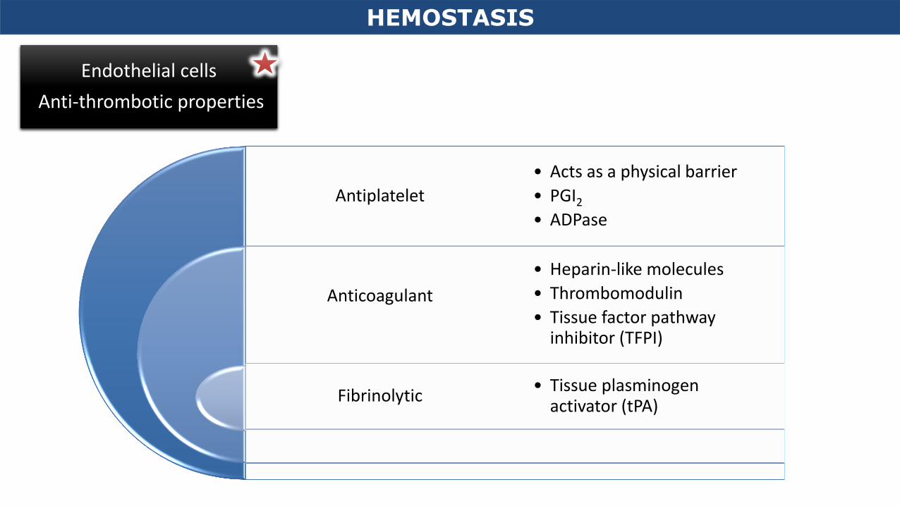
Endothelial cells
Anti-thrombotic properties
Antiplatelet
Anticoagulant
Fibrinolytic
• Acts as a physical barrier
• PGI2
• ADPase
• Heparin-like molecules
• Thrombomodulin
• Tissue factor pathway inhibitor (TFPI)
• Tissue plasminogen activator (tPA)
HEMOSTASIS
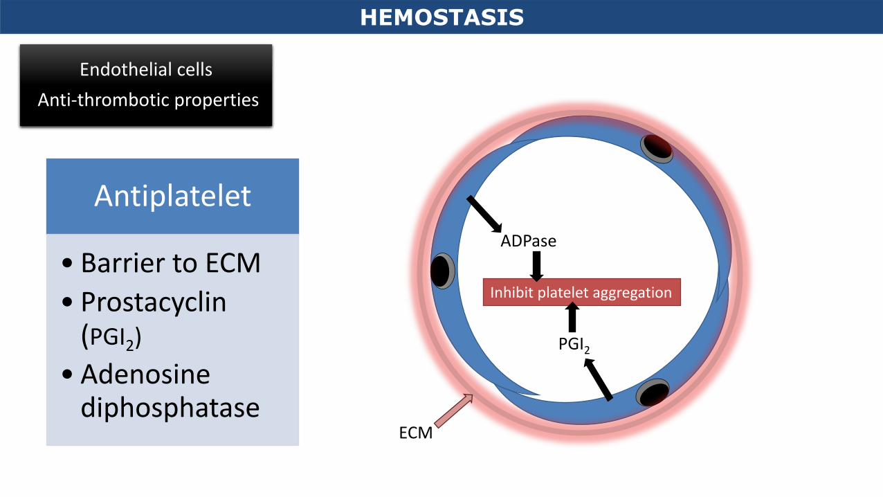
ECM
PGI2
ADPase
Inhibit platelet aggregation
Antiplatelet
• Barrier to ECM
• Prostacyclin (PGI2)
• Adenosine diphosphatase
HEMOSTASIS
Endothelial cells
Anti-thrombotic properties
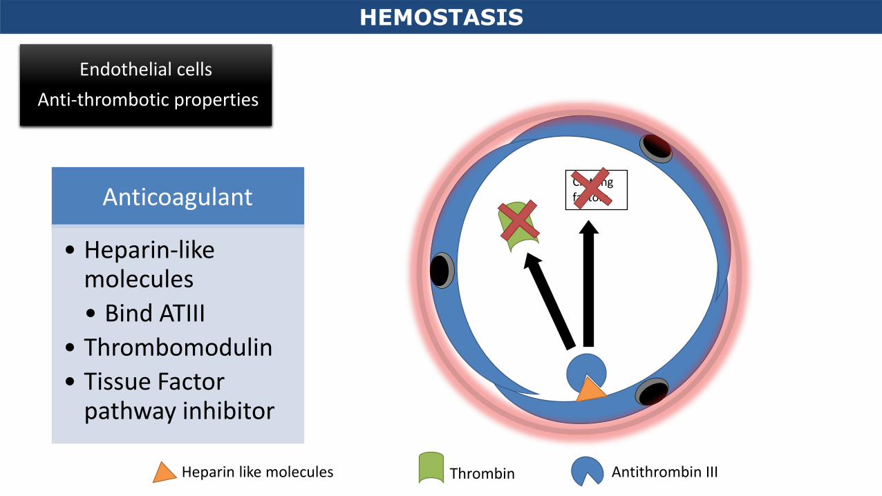
Anticoagulant
• Heparin-like molecules
• Bind ATIII
• Thrombomodulin
• Tissue Factor pathway inhibitor
Thrombin
Clotting factors
Heparin like molecules Antithrombin III
HEMOSTASIS
Endothelial cells
Anti-thrombotic properties
Thrombin
Activates protein C → cleaves clotting factors
Thrombomodulin
HEMOSTASIS
Endothelial cells
Anti-thrombotic properties
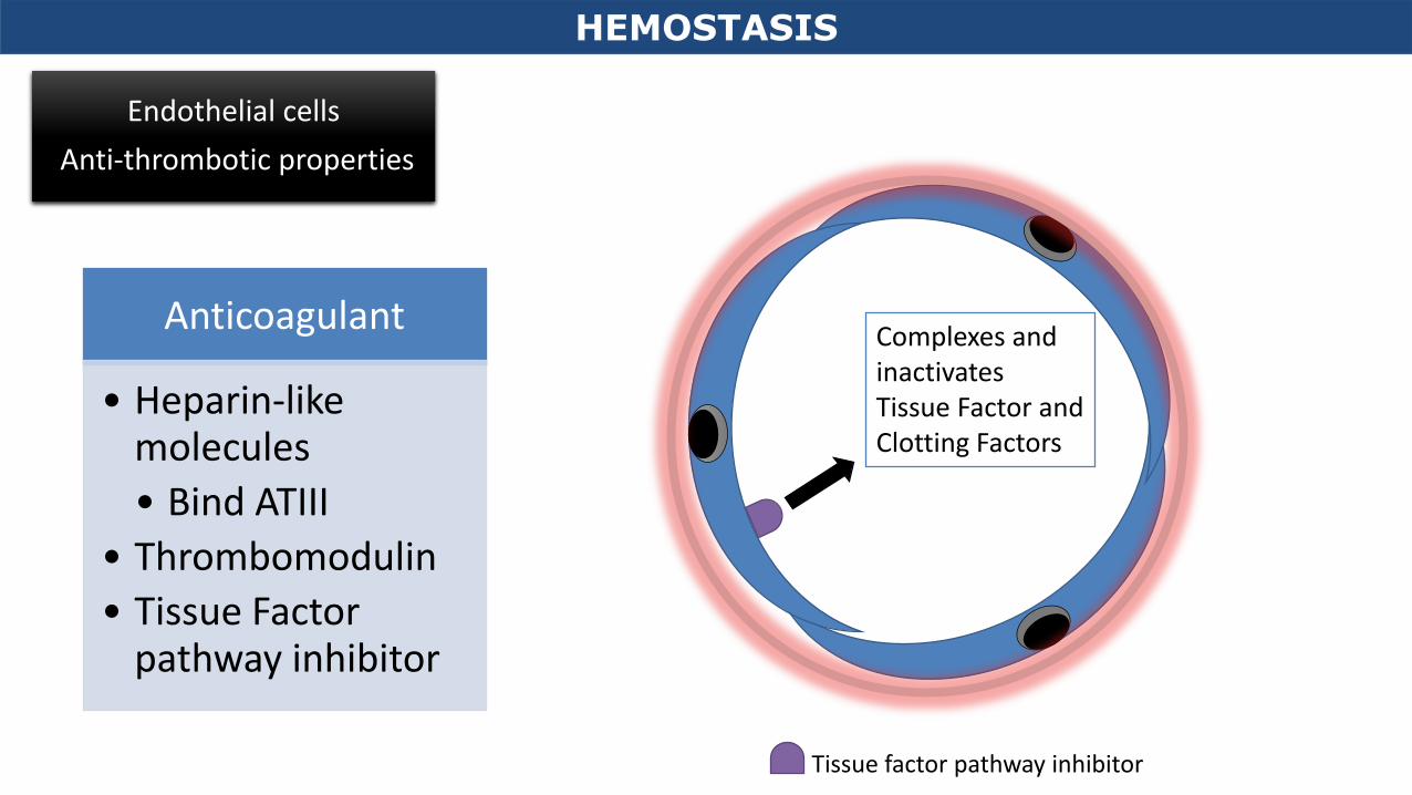
Anticoagulant
• Heparin-like molecules
• Bind ATIII
• Thrombomodulin
• Tissue Factor pathway inhibitor
Tissue factor pathway inhibitor
Complexes and inactivates Tissue Factor and Clotting Factors
HEMOSTASIS
Endothelial cells
Anti-thrombotic properties
Anticoagulant
• Heparin-like molecules
• Bind ATIII
• Thrombomodulin
• Tissue Factor pathway inhibitor
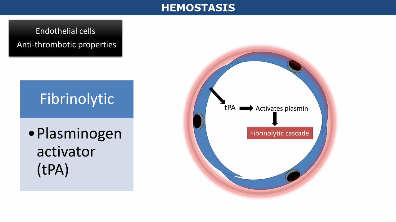
Fibrinolytic
•Plasminogen activator (tPA)
tPA
Fibrinolytic cascade
Activates plasmin
HEMOSTASIS
Endothelial cells
Anti-thrombotic properties
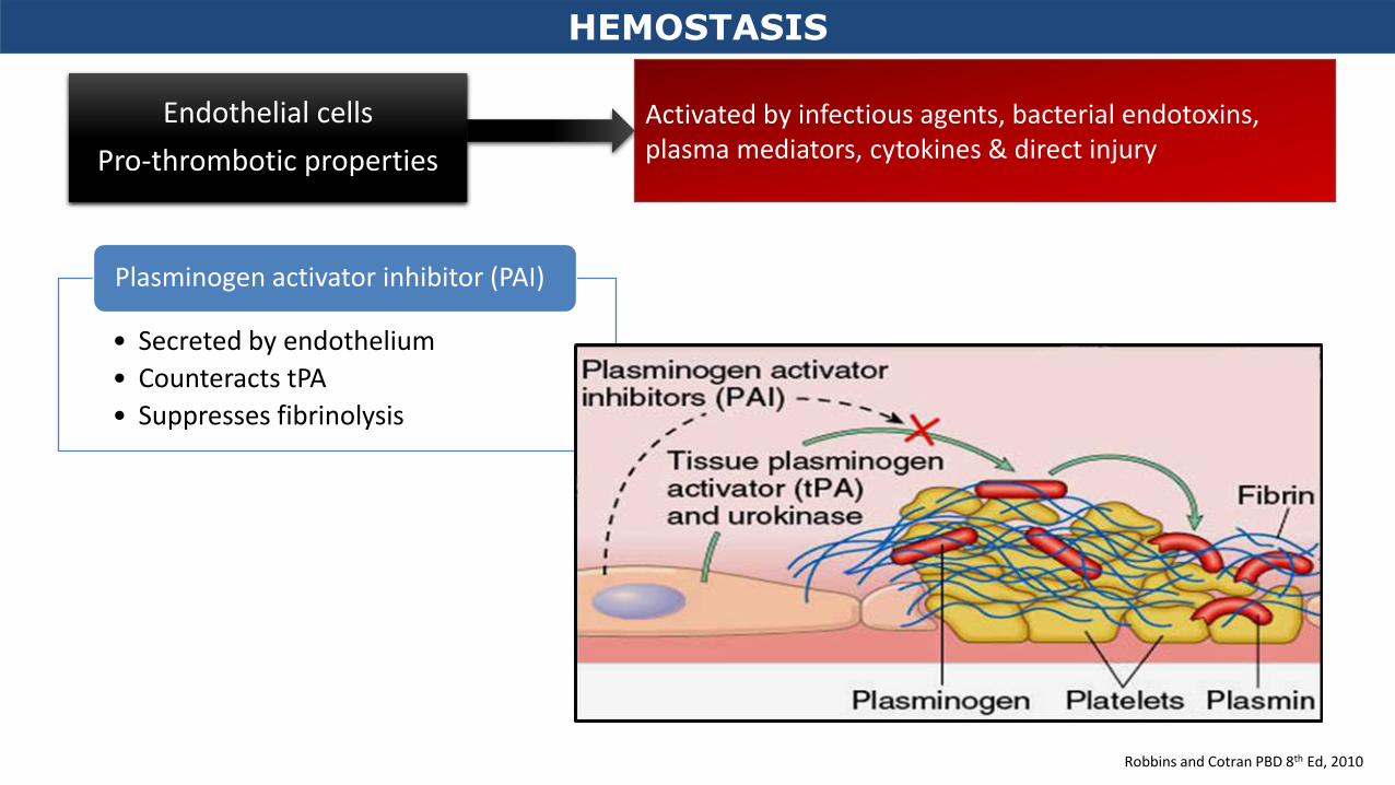
Activated by infectious agents, bacterial endotoxins, plasma mediators, cytokines & direct injury
Endothelial cells
Pro-thrombotic properties
von Willebrand Factor (vWF)
Tissue Factor (TF)
Plasminogen activator inhibitors (PAIs)
• Synthesize
• Store
• Release
• Secreted by injured endothelium
• ↓Fibrinolysis
• Counteract Plasminogen activators
HEMOSTASIS
HEMOSTASIS
Robbins and Cotran PBD 8th Ed, 2010
Activated by infectious agents, bacterial endotoxins, plasma mediators, cytokines & direct injury
Endothelial cells
Pro-thrombotic properties
• Produced by endothelium
• Binds surface of platelets
• Activates them → helps them aggregate
• Allows platelets to stick to exposed collagen
Von Willebrand factor (vWF)
• Injured endothelium secrete TF
• Activates extrinsic coagulation cascade
Tissue factor (TF)
HEMOSTASIS
Robbins and Cotran PBD 8th Ed, 2010
Activated by infectious agents, bacterial endotoxins, plasma mediators, cytokines & direct injury
Endothelial cells
Pro-thrombotic properties
• Secreted by endothelium
• Counteracts tPA
• Suppresses fibrinolysis
Plasminogen activator inhibitor (PAI)
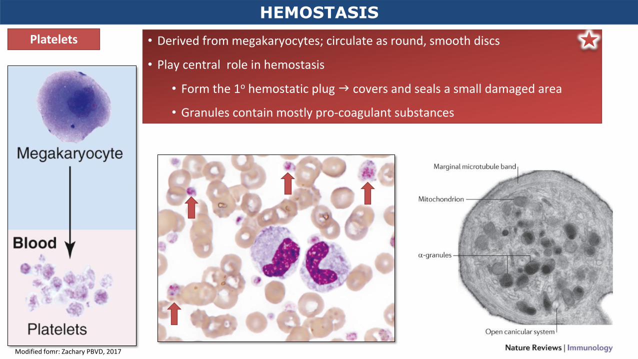
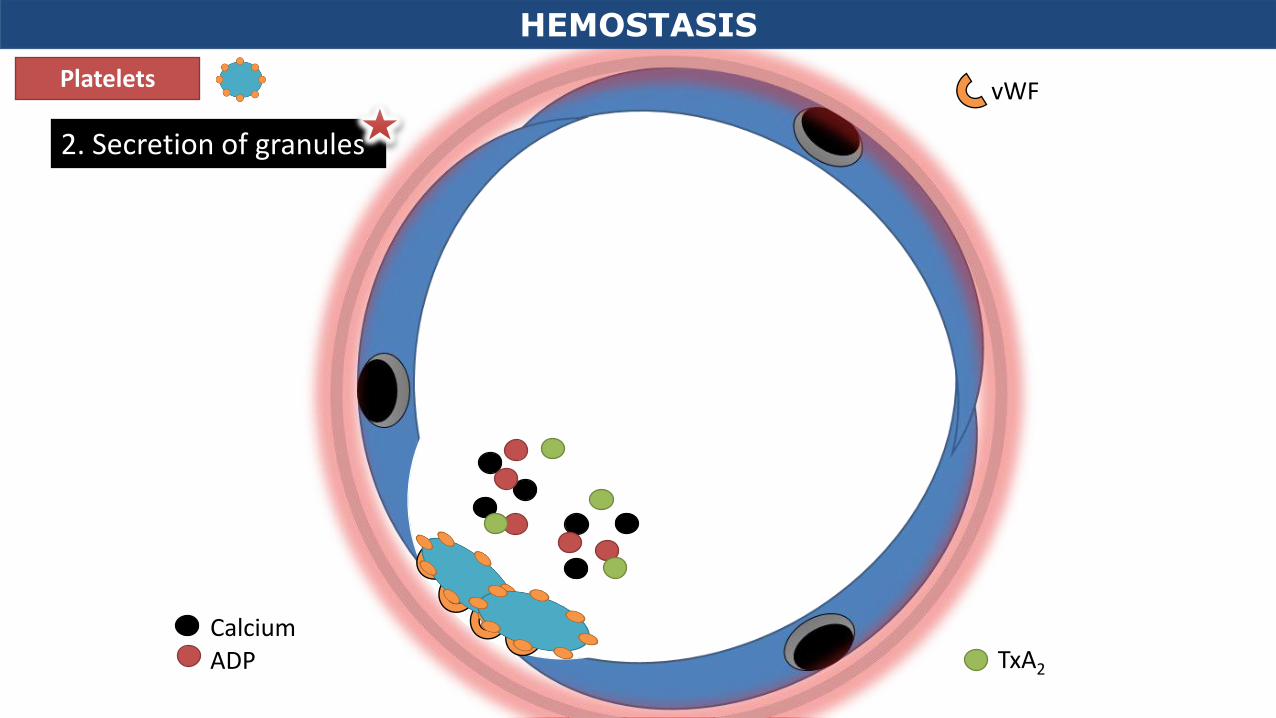
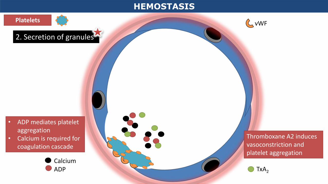
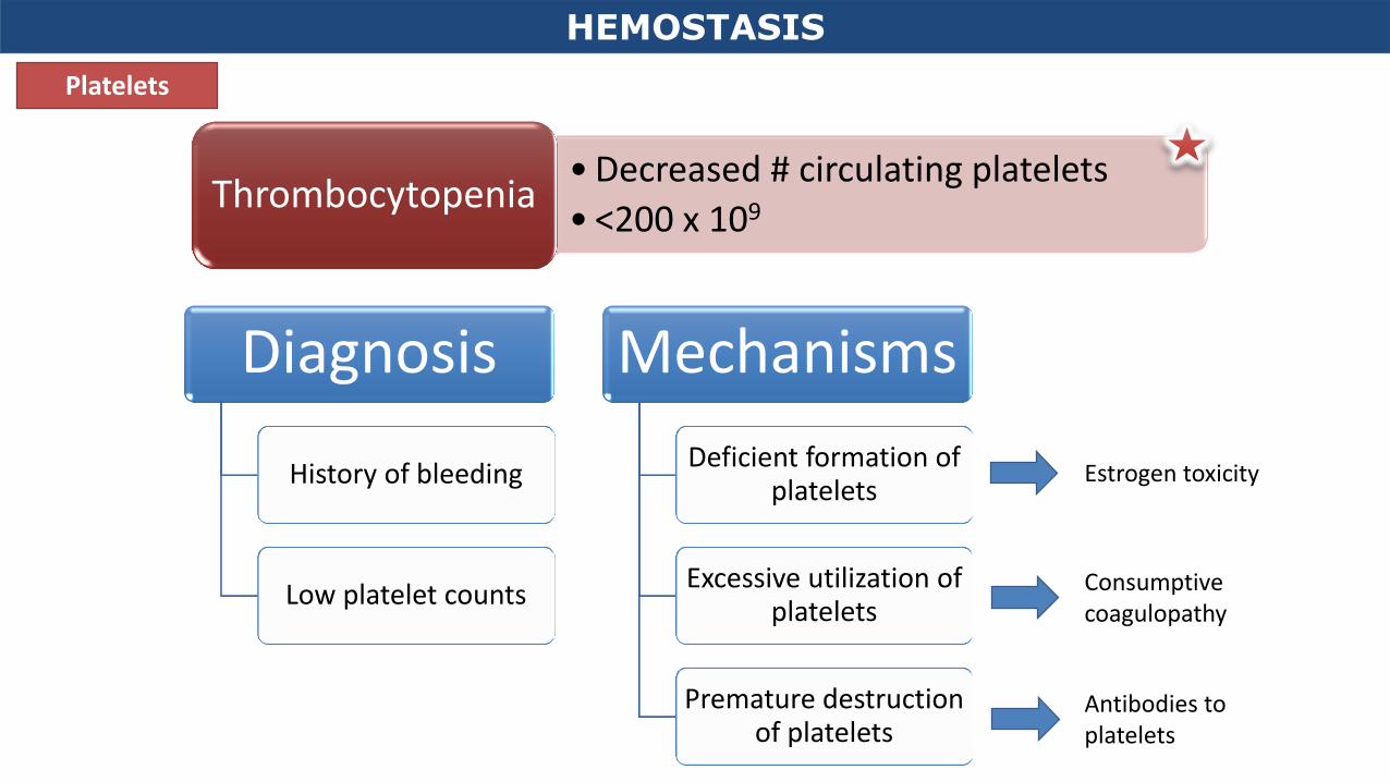
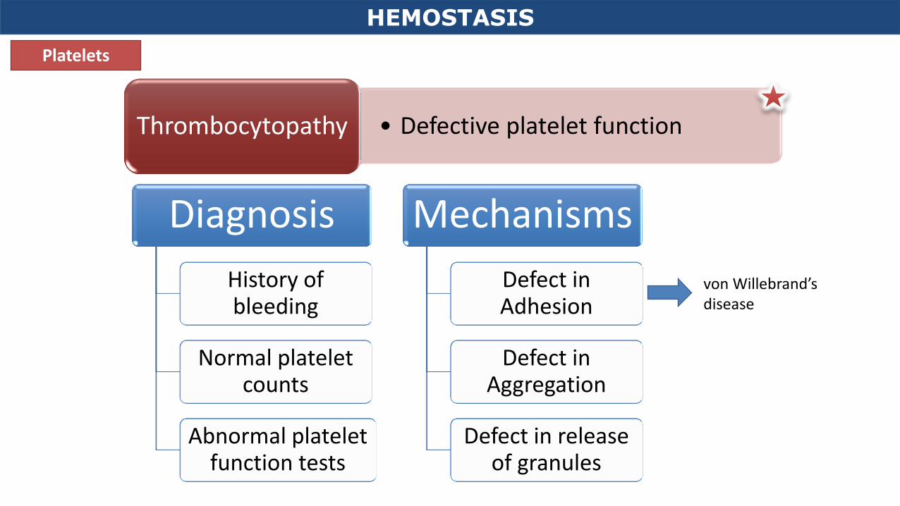
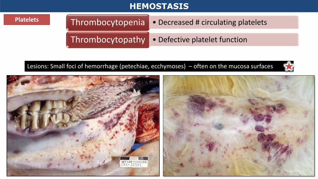
• Derived from megakaryocytes; circulate as round, smooth discs
• Play central role in hemostasis
• Form the 1o hemostatic plug covers and seals a small damaged area
Lesions: Small foci of hemorrhage (petechiae, ecchymoses) – often on the mucosa surfaces
Coagulation Cascade
Enzyme
Substrate
Activated coagulation
factor
• An enzymatic cascade • A reaction pathway that occurs on a platelet phospholipid complex and is held
together by calcium ions
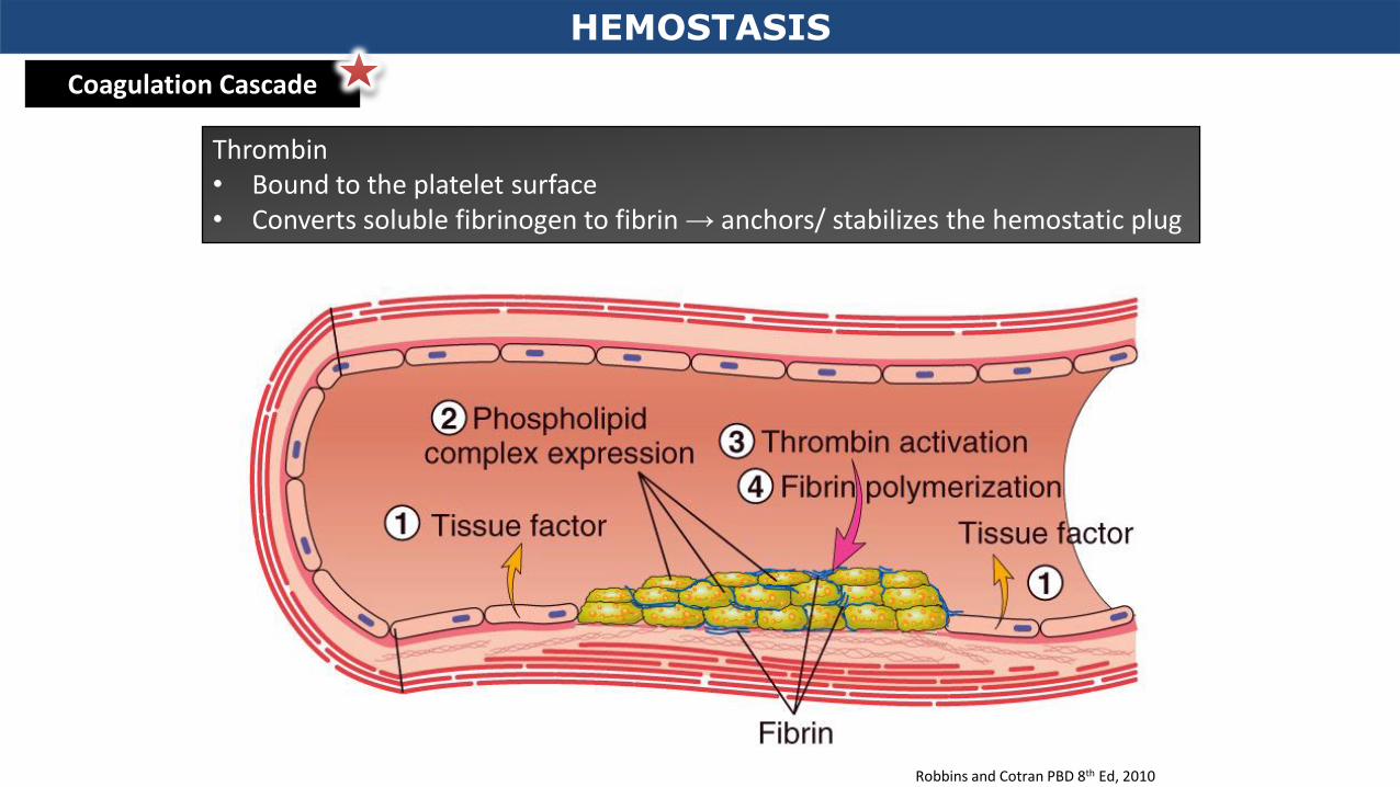
Thrombin
Coverts fibrinogen to fibrin
HEMOSTASIS
Thrombin • Bound to the platelet surface • Converts soluble fibrinogen to fibrin → anchors/ stabilizes the hemostatic plug
HEMOSTASIS
Robbins and Cotran PBD 8th Ed, 2010
Coagulation Cascade
HEMOSTASIS
Coagulation Cascade
Robbins and Cotran PBD 8th Ed, 2010
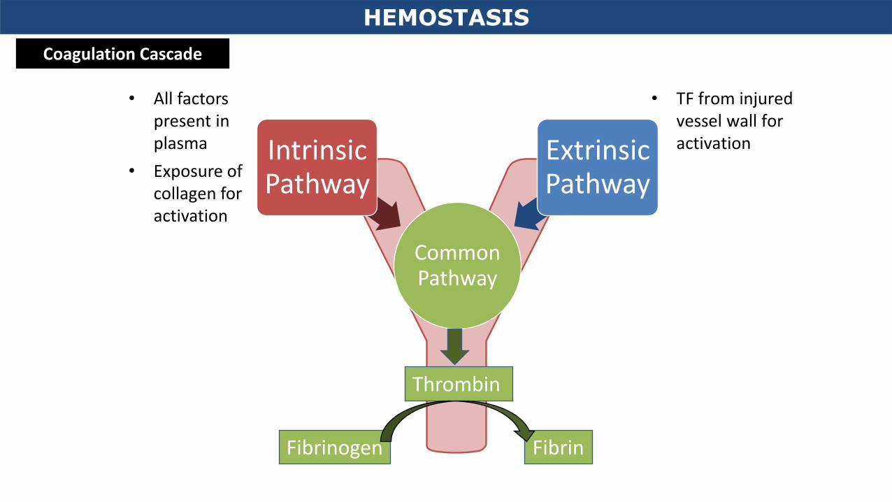
• TF from injured vessel wall for activation
• All factors present in plasma
• Exposure of collagen for activation
Common Pathway
Intrinsic Pathway
Extrinsic Pathway
Thrombin
Fibrinogen Fibrin
HEMOSTASIS
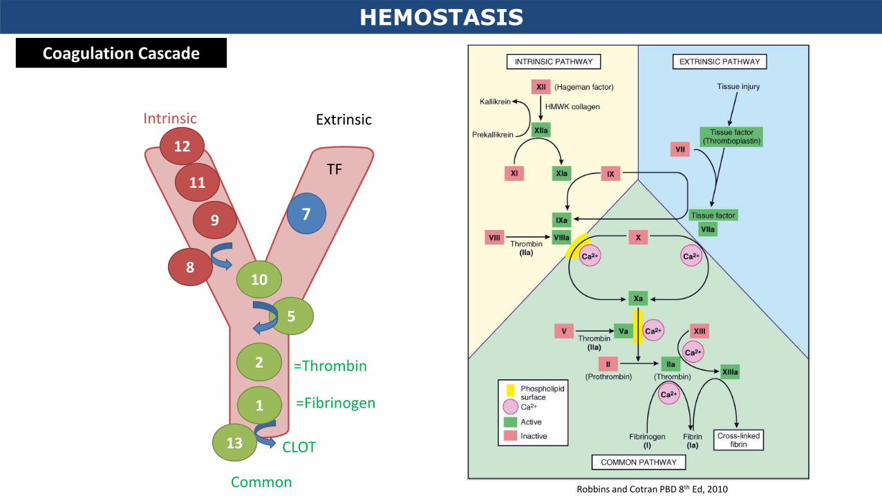
Coagulation Cascade
Common
CLOT
12
10
7
11
9
5
2
1
Intrinsic Extrinsic
=Thrombin
=Fibrinogen
8
13
TF
HEMOSTASIS
Robbins and Cotran PBD 8th Ed, 2010
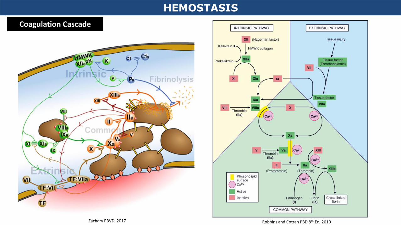
Coagulation Cascade
HEMOSTASIS
Robbins and Cotran PBD 8th Ed, 2010
Coagulation Cascade
Zachary PBVD, 2017
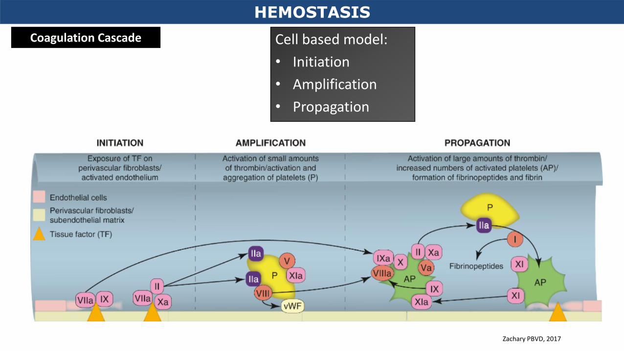
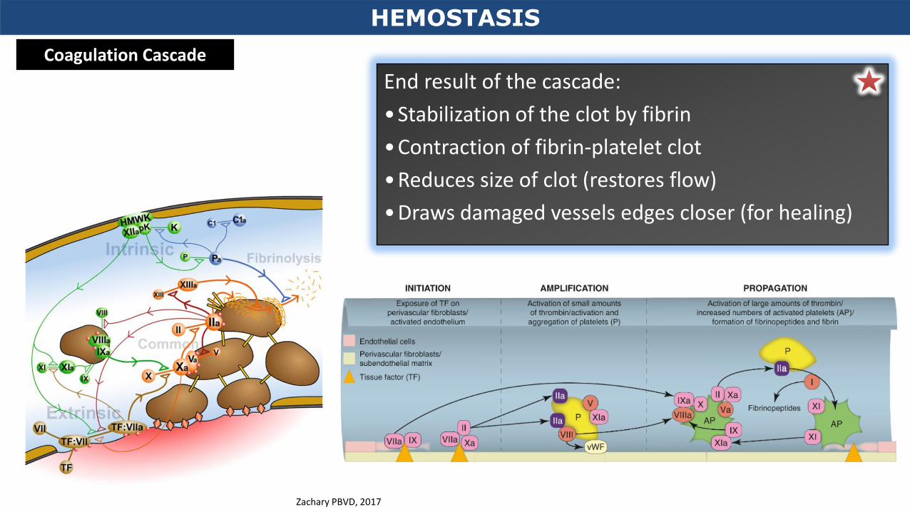
Coagulation Cascade Cell based model:
• Initiation
• Amplification
• Propagation
HEMOSTASIS
Zachary PBVD, 2017
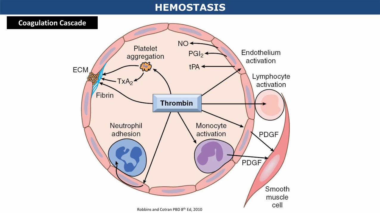
End result of the cascade:
•Stabilization of the clot by fibrin
•Contraction of fibrin-platelet clot
•Reduces size of clot (restores flow)
•Draws damaged vessels edges closer (for healing)
HEMOSTASIS
Coagulation Cascade
Zachary PBVD, 2017
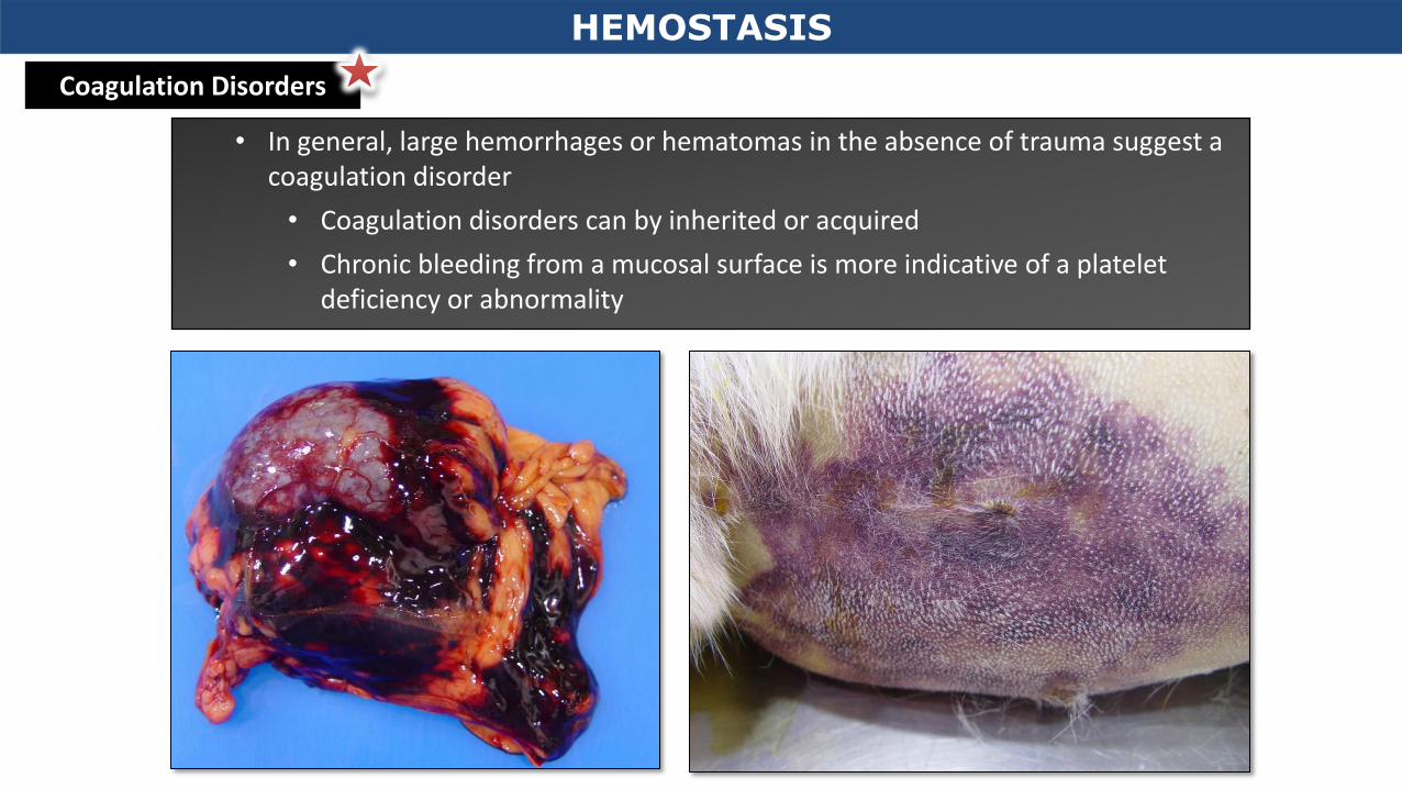
• In general, large hemorrhages or hematomas in the absence of trauma suggest a coagulation disorder
• Coagulation disorders can by inherited or acquired
• Chronic bleeding from a mucosal surface is more indicative of a platelet deficiency or abnormality
Coagulation Disorders
HEMOSTASIS
(E Box 2-1 Zachary– Pathologic Basis of Veterinary Disease, 2017)
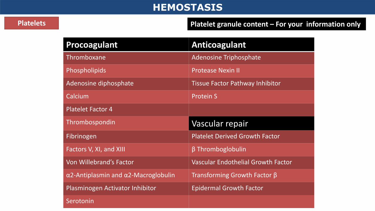
For your information only
HEMOSTASIS
Coagulation Disorders – Inherited
• Vitamin K deficiency
• Affects factors 10, 9, 7 and 2 and proteins C and S
• Anticoagulant toxicity
• Liver failure
• Site of synthesis of coagulation factors
Decreased Production of Coagulation Factors
HEMOSTASIS
Coagulation Disorders - Acquired
HEMOSTASIS
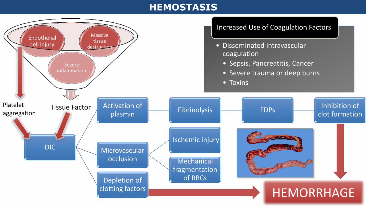
DIC
Activation of plasmin
Fibrinolysis FDPs Inhibition of
clot formation
Microvascular occlusion
Ischemic injury
Mechanical fragmentation
of RBCs Depletion of clotting factors HEMORRHAGE
Tissue Factor
Severe inflammation
Endothelial cell injury
Massive tissue
destruction
Platelet aggregation
• Disseminated intravascular coagulation
• Sepsis, Pancreatitis, Cancer
• Severe trauma or deep burns
• Toxins
Increased Use of Coagulation Factors
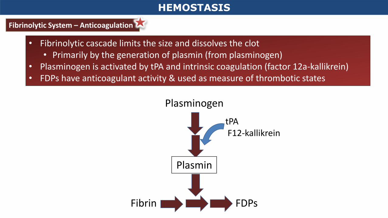
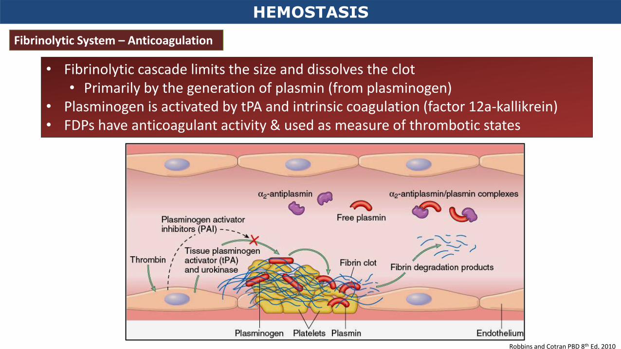
• Fibrinolytic cascade limits the size and dissolves the clot • Primarily by the generation of plasmin (from plasminogen)
• Plasminogen is activated by tPA and intrinsic coagulation (factor 12a-kallikrein) • FDPs have anticoagulant activity & used as measure of thrombotic states
Fibrinolytic System – Anticoagulation
Plasminogen
Plasmin
tPA F12-kallikrein
Fibrin FDPs
HEMOSTASIS
HEMOSTASIS
Robbins and Cotran PBD 8th Ed, 2010
Fibrinolytic System – Anticoagulation
• Fibrinolytic cascade limits the size and dissolves the clot • Primarily by the generation of plasmin (from plasminogen)
• Plasminogen is activated by tPA and intrinsic coagulation (factor 12a-kallikrein) • FDPs have anticoagulant activity & used as measure of thrombotic states