2016 / 3 / Lec(1) Virology 3 1 Introduction and discovering of viruses 1- the viruses discovered concidence during a world Adolf Meyer in 1883, research on the mosaic tobacco leaves, had identified to the present of minutes is smaller than the bacteria causing the disease. 2- followed by Russian scientist Dmitri Ivanovsky the year 1892, who managed the liquidation of sap of infected tobacco leaves using filters a not allow bacteria to pass through, and wipe the leaves is infected with filters noted hit. And is the first to called the virus name, which means in Latin poison .which is .a simple and small molecules in size. 3-The first virus infects vertebrates discovered a foot and mouth virus disease virus in 1898, all of the world Loeffler and Frosch world and the world koch. 4-Reed and the world Carol discovered the first virus infects a human virus, yellow fever virus. . Virology: the science which deals with study of viruses as causative agents of very important diseases that occurs in human, animals, plants and other living organisms (insects, bacteria,…). Viruses: They are the smallest and simplest form of life on earth, which can replicate only in living susceptible cells. Viruses consist of: 1. A nucleic acid genome either DNA or RNA. 2. A protein coat (capsid) that enclosed the genome. 3. In some cases a lipid membrane (envelope).
Transcript
2016 /3 / Lec(1) Virology 3
1
Introduction and discovering of viruses
1- the viruses discovered concidence during a world Adolf Meyer in 1883, research on
the mosaic tobacco leaves, had identified to the present of minutes is smaller than the
bacteria causing the disease.
2- followed by Russian scientist Dmitri Ivanovsky the year 1892, who managed the
liquidation of sap of infected tobacco leaves using filters a not allow bacteria to pass
through, and wipe the leaves is infected with filters noted hit. And is the first to called
the virus name, which means in Latin poison .which is .a simple and small molecules
in size.
3-The first virus infects vertebrates discovered a foot and mouth virus disease virus in
1898, all of the world Loeffler and Frosch world and the world koch.
4-Reed and the world Carol discovered the first virus infects a human virus, yellow
fever virus. .
Virology: the science which deals with study of viruses as causative agents of very
important diseases that occurs in human, animals, plants and other living
organisms (insects, bacteria,…).
Viruses: They are the smallest and simplest form of life on earth, which can replicate only
in living susceptible cells. Viruses consist of:
1. A nucleic acid genome either DNA or RNA.
2. A protein coat (capsid) that enclosed the genome.
3. In some cases a lipid membrane (envelope).
2016 /3 / Lec(1) Virology 3
1
Virion: a complete infectious virus particle that consists of an RNA
or DNA core with protein coat with external envelopes
General characters of viruses:
1. Virus particles are very small in size; they are between 20-500 nm (nanometer)
In diameter. 1 nm= 1/1000 μm, 1 μm=1/1000 mm.
2. Viruses are obligatory intra cellular microorganisms.
3. Multiply inside the cells by replicating their genomes which either DNA or
RNA, but not both.
4. The virus does not contain any organelles (ribosome's, t RNA, metabolic enzymes,
etc), but they depend on infected cells to provide all their needed organelles.
5. Virus does not affect with antibiotics.
6. Most viruses sensitive to interferon.
7. Viruses cannot grow on artificial media, but only in living cells (specific host,
9. Viruses cannot be seen by ordinary microscope, but only by Electron Microscope
. (EM).
The chemical composition of viruses
1-all viruses contain only one type of nucleic acid (NA), either DNA or RNA but not both
A protective coat of protein(capsid)-2-
3-some viruses may contain lipids & carbohydrates.(membrane)
Structure of viral nucleic acids:
2016 /3 / Lec(1) Virology 3
1
Genetic information is stored as the following:
1. Double stranded DNA cells (animal, plants, bacteria and some viruses).
2. Single-stranded DNA in other viruses (phage x 174).
3. Single stranded RNA (myxovirus).
4. Double-stranded RNA (reoviruses).
How we can differentiated between DNA and RNA?
By DNA ase or RNA ase
Between double and single stranded NA; by acridine orange stain,which is yellowish
green in double stranded and red orange in single stranded.
Chemical composition of nucleic acid:-
NA chain is composed of basic units called nucleotides each of which consist of:
1. Nitrogenous base: ring compound containing nitrogen and carbon.
2.Molecule of a 5-carbon pentose sugar which is either ribose RNA
ribose (RNA) or deoxyribose (DNA).
3. Molecule of phosphoric acid which links the base to the pentose sugar. There are
four kinds of nucleotides (bases) in DNA: Guanine, Adenine, Cytocine, and
Thymine.
In the RNA molecules the bases are guanine, adenine, cytocine and uracil.
Viral proteins:
Protein coat which encloses the viral genome called capsid which consists of
protomers that accumulated to give pentan or hexan forms producing the capsomers
which protect the viral NA and have surface characters acts to attach the virus on host cells
then penetration, also it contains antigenic determinants.
2016 /3 / Lec(1) Virology 3
1
Viral envelope:
Most viruses contain envelope or membrane surrounding the virus so they called
enveloped viruses, others have no envelope and they called naked viruses. Enveloped
viruses contain lipids like orthomyxo and paramyxo viruses, these viruses will become
sensitive to organic solvents (ether, alcohol, chloroform), these charactersused in newly
isolated virus classification. Also viral membrane contains glycolipids or glycoprotein
which appears as projections from the envelope called spikes or peplomers.
Viral shape structure
Nucleocapsid is the arrangement between the viral nucleic acid genome with the
capsid, this connection controlled by specific NA genetic information leading to different
types of symmetry. Accordingly viruses can classified in to four symmetry structures.
1. Helical symmetry.
2. Cubical symmetry.
3. Binal symmetry.
4. Complex symmetry.
1. Helical symmetry:-
This form can be seen in RNA viruses, that the capsomers surrounded the N.A in spiral
or helical manner to give helical symmetry which may be seen naked (TMV) or
enveloped in ND & Rinderpest viruses. When the tubular nucleocapsidpresented in coil
form and surrounded by lipoprotein envelope containing peplomers which is glycoprotein
projections (also called spikes) which represent haemagglutinins and neuraminidases but
in rabies virus the capsid is straight and surrounded by lipoproteinmembrane to give the
bullet shape.
2. Cubical symmetry:-
2016 /3 / Lec(1) Virology 3
1
Many RNA and DNA animal viruses the virus particles have hexagonal outlines with
20 equilateral triangular faces, i.e. icosahedrons, but when these viruses studied by
negative stain using phosphotungstic acid which can penetrate even through the finest
contours of the viral surface. Many viruses appear to be faceted and each face forms an
equilateral triangle consisting of angular arrangement of protein subunits as
capsomers. There are generally 20equilateral triangular faces.
3. Binal symmetry
This type of symmetry show both icosahedral (cubical) and helical symmetry, but
within the same virion like bacteriophage, when the head is cubical and the tail is helical.
4. Complex symmetry
Most animal viruses show either helical or cubical symmetry but pox viruses have
exceptional and their ultra structure appears to be complex. Some pox viruses are brick-
shaped, while others are ovoid and the DNA is contained in nucleoid, shaped like a
biconcave disc and surrounded by one or more membranes. Negative staining shows
that the virion contains a surface layer of hallow tube like fibrils which may give the
particles a striated appearance. In some species of pox viruses e.g. or virus, the thread
appears to be continuous and is arranged in a cross or figure-eight pattern across the
surface of the virion giving it the characteristic ball of wool appearance.
Genetics & Evolution of Viruses
Mutation: - spontaneous and random errors in the copying of viral N.A. which can
occur during the replication of viruses, leading to change in nucleic acid sequence to
produce mutant when differ somewhat than original organism. Mutant rate in RNA
viruses are higher than DNA viruses although some RNA viruses such as those of mumps
in man and Newcastle disease in poultry are remarkably stable over many years, but others
2016 /3 / Lec(1) Virology 3
1
like influenza A are labile and show tendency to variation in some their properties, leading to
producing new pandemics. Some times virus mutation may lead to loss of virulence still
immunogenic which called attenuated viruses like vaccinia which is mutant of variola, then it
can be used is vaccine against small pox in human. There are some viruses properties may
change through mutation:-
1. Loss of virulence.
2. Increase rate of reproduction.
3. Extension of natural host range.
4. Altered haemagglutination activity and changes of antigenic structure, plaque size,
morphology or resistance to heat.
Viral recombination:- the transfer of genetic material between closely related viruses infecting
the same cell, e.g. Sheep pox and Goat pox virus, then new recombinant virus will produced
with genome contain new genetic information.
The alteration of genetic information in recombinant may result from:-
1. Intramolecular recombination: usually occurs in DNA viruses and involved
dissociation and re-establishment of covalent bonds within the nucleic acid .
2. Copy-choice recombination: usually occurs between positive sense single stranded
RNA viruses. e.g. Picorna , Corona, and Toga viruses .
3. Reassortment: occurs randomly in RNA viruses with segmented genome e.g.
Orthomyxo viruses (influenza), Reo viruses and Bunya viruses. Viral cell
interactions.When an intact of infectious virus particles makes contacts with a
susceptible host cell may develop a number of reactions at the cell surface lead to
release of the enetic material at the virus within the cell. This is immediately followed
by a series of biosynthetic processes lead to formation of new virus like e.g.
1. Defective virus:- viruses that have lost ability to perform any one of the essential steps
required for successful replication.
2016 /3 / Lec(1) Virology 3
1
2. Incomplete virus:- abnormal viruses produced due to inoculation of high titer virus
solution in limited number of host susceptible cells like inoculums containing a high
rating of infective units to cells this called Von Magnus phenomena. That the produced
viruses without nucleic acid e.g. influenza virus.
Interferon Soluble substance produced by living cells of many different types in cell cultures, embryonated
eggs, in lab. Animals when infected by some animal viruses either DNA or RNA and can inhibit
multiplication of active virus e.g. influenza virus.
Characteristics of interferon molecules:-
1. It is small protein without nucleic acid.
2. Low molecular weight of about 25- 45000 Dalton.
3. Thermo stable at 4 C° and resist heating at 50 C° for I hour.
4. Interferon is active through a wide range of pH values (2-12).
5. It is relatively non-toxic, weakly antigenic and cannot neutralized by the specific antiserum.
6. Inactivated by protolytic enzymes such as trypsin.
7. Not affected with RNase & DNase.
8. Interferon specific to animal species but not to viruses species i.e.: it act against
wide variety of viruses.
Replication of Viruses
Viruses can multiply only in active host cells, the replicate cycle of viruses can be divided into
number of stages:
1. Attachment to surface receptors on the susceptible host cell.
2. Penetration and uncoating.
3-Formation of messenger RNA transcripation
2016 /3 / Lec(1) Virology 3
1
4-Formation of new genomes
5-Formation of new protein (translation)
6-Assembly.
7- Release.
8- Latency .
1-Attachment: the virus meets and then binds to a cell surface receptor.
2-Penetration and uncoating: :- the process where by the viral genome is released in
a form suitable for transcription.e.g.
a-In enveloped viruses: nucleocapsid is discharged directly into the
cytoplasm, transcription can usually proceed without complex uncoating.
b. In non- enveloped virus uncoating may results from lysosomal proteolytic
enzymes activity.
3-Formation of messenger RNA: Viruses have their own polymerase proteins
which force the cell to make a huge amount of viral mRNA and also nucleic acid.
DNA viruses make mRNA in the cell nucleus.
RNA virus make mRNA in the cytoplasm.
4- Formation of new genomes which may be aided by early proteins which are
either viral polymerases, see above, or promoting cell division to provide new
cells for the madly dividing viruses.
5- Formation of new protein is always on the host cell ribosomes. Early proteins
are viral enzymes e.g. polymerases (see above) or viral growth factors which
stimulate cell division to provide new host cells for virus.Late proteins are the
structural proteins e.g. capsids and spikes .
6-Assembly. Nucleocapsid of DNA viruses are assembled the nucleus.
Nucleocapsid of RNA viruses are assembled the nucleus. The notable exception is
the DNA poxviruses which assemble the cytoplasm. Nucleocapsid 'factories' can be
2016 /3 / Lec(1) Virology 3
1
seen as inclusion bodies by light microscopy. Glycoprotein spikes insert into the
cell-surface plasma membrane. .
7- Release. Release of many particles at once when the cell dies and then bursts. Or
each enveloped virus particle gradually buds from the cell surface.
8- Latency - certain viruses, the herpes and retro's, form ± DNA during replication
and this can remain latent in the nucleus for years but then become reactivated to
make new particles during immunosuppression.
Viral vaccines and anti viral drugs:-
A vaccine:- is a biological preparation that provides active acquired immunity to a particular disease. A vaccine typically contains an agent that resembles a disease-causing micro-organism and is often made from weakened or killed forms of the microbe, its toxins or one of its surface proteins. Vaccines can be prophylactic or therapeutic (e.g., vaccines against cancer).
advantages of veterinary vaccines:-
1- Safety:A vaccine should not cause disease, and side effects should be minimal.
2- Effectiveness: Vaccinated animals should be protected from illness due to the pathogen. Ideally, appropriate innate, cellular and humoral responses should be evoked by the vaccine, and long term immunity.
3- Practical Issues: The vaccine should be stable (for storage) .
5-Control of exotic diseases of animals and people.
6-Reduction of the need for antibiotics.
Types of Vaccines:-
1-Live, attenuated vaccines
2-Inactivated vaccines (killed vaccine)
3-Subunit vaccines
4-Toxoid vaccines
5-DNA vaccines
6-Recombinant vector vaccines.
1- Attenuated Live Vaccines: vaccines contain live, attenuated microorganisms. Many of these are active viruses that have been cultivated under conditions that disable their virulent properties, and become less dangerous organisms to produce a broad immune response. Although most attenuated vaccines are viral, some are bacterial in nature. Examples include the viral diseases yellow fever, measles, rubella, and mumps, and the bacterial disease typhoid..
1-they are effective at stimulating both the humeral and cellular immune system.
2- long-term immunity. 3- they can induce secretary immunity.
2- Killed Viral Vaccines:
vaccines contain inactivated virus, but previously virulent, micro-organisms that have been destroyed with chemicals, heat, radiation, or antibiotics without destroying the antigenicity of the virus. Examples are influenza, cholera, hepatitis A, and rabies.
killed virus vaccines are often administered along with an adjuvant, which induces an
inflammatory response and can stimulate the cellular immune system.
Advantages Killed Viral Vaccines:
stable, and usually safe since no live virus is present.
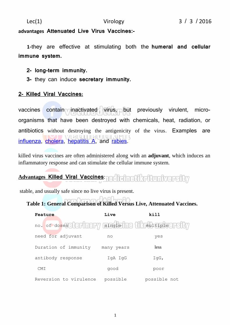
Table 1: General Comparison of Killed Versus Live, Attenuated Vaccines.
3-Subunit vaccines: viral proteins or groups of proteins are used. These proteins can be purified directly from viral particles. However this is expensive, since it is difficult to prepare virus in large enough quantities for protein purification, and potentially dangerous since there is the possibility of contaminating virulent virus. An alternative method is to use recombinant proteins produced in either procaryotic or eucaryotic (yeast, insect, animal) cells. The viral proteins that are chosen for this type of vaccine should be immunogenic (ie, good antigens) and should also induce neutralizing or protective antibodies. This method of immunization has many of the same advantages and disadvantages as the killed virus vaccine.
3-Recombinant Vaccine: In this approach, a gene encoding a major viral antigen (that is a target for neutralizing Ab) is inserted (cloned) into another, non-virulent viral vector so that the cloned gene is expressed and the protein produced during viral infection.Animals are then infected with the recombinant virus, and mount an immune response (both humoral ,cellular and induce secretory immunity) against the introduced antigen.
4-DNA- Based Vaccines: In this approach, genes (DNA) encoding specific viral proteins are injected into an animal (either in muscle or skin). The DNA is then taken up by cells, where it is transcribed into mRNA which is then translated to give rise to the viral protein. This protein is expressed on the surface of cells, either alone or in association with
2016 /3 / Lec(1) Virology 3
1
MHC molecules. It is recognized as a foreign molecule by the immune system, and elicits an immune response.
This approach has several advantages, including the following:
1- no infectious agent. 2 long term immunity. 3- strong cellular immune response.
4- Once a gene is cloned, DNA is inexpensive to make and is stable, so the vaccines should be inexpensive.
5- No adjuvant is required.
5-Toxoid Vaccines
For bacteria that secrete toxins, or harmful chemicals, a toxoid vaccine might be the answer. These vaccines are used when a bacterial toxin is the main cause of illness.
Scientists have found that they can inactivate toxins by treating them with formalin, a solution of formaldehyde and sterilized water. Such “detoxified” toxins, called toxoids, are safe for use in vaccines.
Anti-viral Drugs:
The goal of anti-viral drug development is to identify drugs that inhibit viral
replication without harming the animal host. Theoretically, anti-viral drugs could be
developed that affect/inhibit any stage of a virus life cycle
A- Penetration and Uncoating. Develop drugs that will inhibit virus binding to its
receptor, or uncoating of the virus inside the cell. An example would be using
2016 /3 / Lec(1) Virology 3
1
soluble receptor to block the cellular binding sites on the virus. Amantadine and rimantadine have been introduced to combat influenza. These agents act on penetration and uncoating.
b- Transcription/Translation (gene expression): Antisense oligonucleotides could
be used to block translation (these are oligos that are complementary to the viral
mRNAs so that they can bind to them and inhibit their translation and/or cause
their degradation). -interferon could be used to block both viral transcription and
translation, since it induces an "anti-viral state" acyclovir, lamivudine,
caliciviruses
c- Assembly and Release: Protease inhibitors can be developed to prevent the final
maturation of viral proteins in viruses that use a polyprotein expression strategy.
Rifampicin andTamiflu
Laboratory diagnosis of viral infections:-
In the diagnostic laboratory virus infections are confirmed by several methods that
include:
1-Growth of the virus in a cell culture from a specimen taken from the patient.
2-Detection of virus-specific antibodies in the blood.(IgM and IgG)
3-Detection of virus antigens(ELISA in tissues and fluids)
4-Detection of virus nucleic acids.( reverse transcriptase PCR and real time PCR)
5-Gene sequencing to characterise viral strains
6-Observation of virus particles by electron microscopy.
a phage is a virus that infects and replicates within a bacterium. Bacteriophages are composed of proteins that encapsulate a DNA or RNA genome, and may have relatively simple or elaborate structures. Their genomes may encode as few as four genes, and as many as hundreds of genes. Phages replicate within the bacterium following the injection of their genome into its cytoplasm.
Effect of physical and chemical agents on viruses
1. Heat and cold. Viral infectivity is generally destroyed by heating at 50-60 C0 for 30 mint.,
hours at 20 C0, days at 4 C0. Viruses can be preserved at -90 C0 or -196 C0
(liquid nitrogens).
2. PH
Viruses can be preserved at physiological PH (7.3).
3. Ether susceptibility :
Ether susceptibility can be used to distinguish viruses that possess
an envelope from those that do not.
4. Detergents:
Non Anionic detergents solubilize lipid constituents of viral
membranes.The viral proteins in the envelope are released. Anionic
detergents also solubilize viral envelopes; in addition, they disrupt
capsids into separated polypeptides.
5. Salts
Many viruses can be stabilized by salt in concentrations of 1 mol/L.
e.g. MgCl2, MgSO4, Na2SO4.
6. Radiation
Ultraviolet, X-ray, and high-energy particles inactivate viruses.
7. Formaldehyde: Destroys viral infectivity by reacting with nucleic acid.
8. Antibiotics: Antibacterial antibiotics have no effect on viruses.
Classification of Animal Viruses:- Animal viruses can be classified into two families according to the following characters: A- Characters of viral nucleic acid (N.A):
1. Type of N.A if it is DNA or RNA.
2. Shape of N.A strand if it is ring or straight, single or double strand, segmented or not
3. Molecular weight (size &diameter of N.A).
4. Replication site of N.A if it is in the cytoplasm or nucleus of infected cells.
5. Presence of transcriptase enzyme.
B- Characters of viral capsid:
1. Shape & size of capsid.
2. Symmetry of capsid with N.A (Helical, Cubical, Binal, Complex).
3. Site of capsid assembly inside the infected cells.
4. Lipid solvent sensitivity.
5. Number of capsomeres which consisting the capsid.
According to all above characters recently animal viruses classified into two major groups
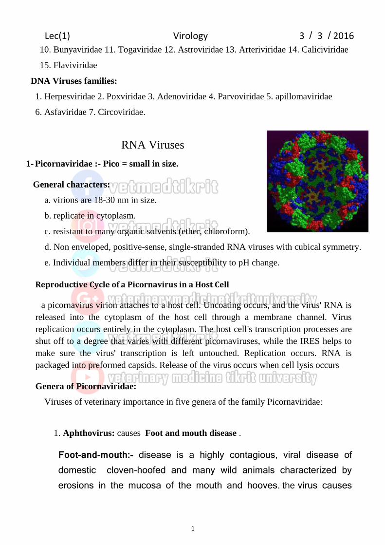
c. resistant to many organic solvents (ether, chloroform).
d. Non enveloped, positive-sense, single-stranded RNA viruses with cubical symmetry.
e. Individual members differ in their susceptibility to pH change.
Reproductive Cycle of a Picornavirus in a Host Cell
a picornavirus virion attaches to a host cell. Uncoating occurs, and the virus' RNA is
released into the cytoplasm of the host cell through a membrane channel. Virus
replication occurs entirely in the cytoplasm. The host cell's transcription processes are
shut off to a degree that varies with different picornaviruses, while the IRES helps to
make sure the virus' transcription is left untouched. Replication occurs. RNA is
packaged into preformed capsids. Release of the virus occurs when cell lysis occurs
Genera of Picornaviridae:
Viruses of veterinary importance in five genera of the family Picornaviridae:
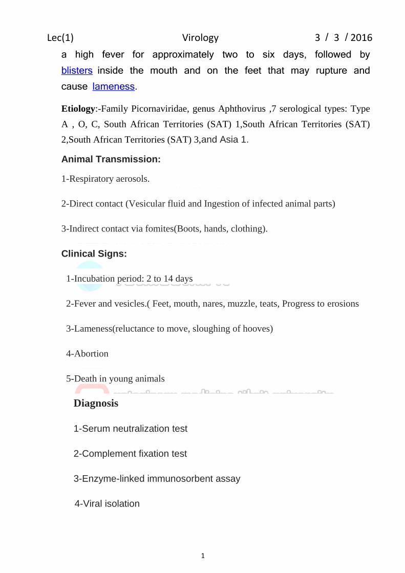
1. Aphthovirus: causes Foot and mouth disease .
Foot-and-mouth:- disease is a highly contagious, viral disease of domestic cloven-hoofed and many wild animals characterized by erosions in the mucosa of the mouth and hooves. the virus causes
2016 /3 / Lec(1) Virology 3
1
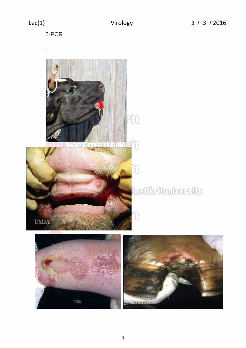
a high fever for approximately two to six days, followed by blisters inside the mouth and on the feet that may rupture and cause lameness.
Etiology:-Family Picornaviridae, genus Aphthovirus ,7 serological types: Type
A , O, C, South African Territories (SAT) 1,South African Territories (SAT)
2,South African Territories (SAT) 3,and Asia 1.
Animal Transmission:
1-Respiratory aerosols.
2-Direct contact (Vesicular fluid and Ingestion of infected animal parts)
3-Indirect contact via fomites(Boots, hands, clothing).
Clinical Signs:
1-Incubation period: 2 to 14 days
2-Fever and vesicles.( Feet, mouth, nares, muzzle, teats, Progress to erosions
3-Lameness(reluctance to move, sloughing of hooves)
Reproductive Cycle of an Orthomyxovirus in a Host Cell
It takes about 6 hours for the replication of the orthomyxovirus,
killing the host cell in the process. The virus attaches to the
permissive cells via the hemagglutinin subunit, which binds to cell
membrane glycolipids or glycoproteins containing N-
acetylneuraminic acid, the receptor for virus adsorption. The virus
is then engulfed by pinocytosis into endosomes. The acid
environemnt of the endosome causes the virus envelope to fuse
with the plasma membrane of the endosome, uncoating the
nucleocapsid and releasing it into the cytoplasm. A trans
membrane protein derived from the matrix gene (M2) forms an ion
channel for protons to enter the virion and destabilize protein
binding, allowing the nucleocapsid to be transported to the
nucleus, where the genome is transcribed by vital enzymes to yield
viral mRNA.. the virus cap sequences allow the viral mRNA to be
transported to the cytoplasm, where it is translated by host
ribosomes. The nucleocapsid is assembled in the nucleus.
Virions acquire envelope hemagglutinin is subjected to proteolytic cleavage by host enzymes during budding. This process is necessary for the released particles to be infectious
2016 /3 / Lec(1) Virology 3
1
Genera of Orthomyxoviridae:-
1. Influenza type A. This type includes influenza A viruses of human and also widespread in animals, particularly aquatic birds, chickens, ducks, pigs, horses, and seals. Influenza type A is responsible for pandemic and for most cases of epidemic influenza (antigenically highly variable). Epidemic:- is defined as an outbreak of a contagious disease that is rapid and widespread, affecting many individuals at the same time. A pandemic:- is an epidemic that becomes so widespread that it affects a region,
continent, or the world.
2. Influenza type B. This type includes influenza B viruses which are mainly found in humans. Influenza type B may exhibit antigenic changes and sometimes causes epidemics. 2. Influenza type C. This type includes influenza C viruses of human and swine. Influenza type C is antigenically stable.
Genetic variation in influenza viruses. A. Events leading to different subtypes:-
Antigenic shift: Major antigenic changes in one or both surface glycoproteins (HA and/ or NA) in influenza A viruses. This due to :
1- Genetic reassortment of surface glycoprotein genes occurs between different strains of a given type (possibly involving animal strains).
2- Reemergence of strains. Every 10-40 years when a new subtype
of influenza A appears, a pandemic results. This happened in : 1918 H1N1 (Spanish flu) 1957 H2N2 (Asian flu) 1968 H3N2 (Hong Kong flu) 1977 H1N1 (Russian flu).
B. Events leading to variation within subtypes:-
Antigenic drift: Minor antigenic changes due to accumulation of point mutations in the gene, resulting in amino acid changes in the protein. Sequence changes can alter antigenic site.
Avian influenza (AI)
Avian influenza (AI) viruses infect domestic poultry as well as pet, zoo, and wild
birds. In domestic poultry, AI viruses are typically of low pathogenicity (LP),
causing subclinical infections, respiratory disease, or drops in egg production. However, a few AI viruses cause severe systemic infections with high mortality.
This highly pathogenic (HP) form of the disease has historically been called fowl
plague.
Etiology
AI viruses are type A orthomyxoviruses characterized by antigenically homologous nucleoprotein and matrix internal proteins,
2016 /3 / Lec(1) Virology 3
1
AI viruses are further divided into 16 hemagglutinin (H1-16) and 9 neuraminidase (N1-9) subtypes based on hemagglutinin inhibition and neuraminidase inhibition tests, respectively. Most AI viruses (H1-16 subtypes) are of low pathogenicity, but some of the H5 and H7 AI viruses are highly pathogenic for chickens, turkeys, and related gallinaceous domestic poultry.
Transmission:- 1-Fecal –oral route 2- water
Clinical Findings
Clinical signs, severity of disease, and mortality rates vary depending on AI virus
strain and host species.
Low Pathogenicity Avian Influenza Viruses:
1-Sub clinical infection
2-respiratory signs such as sneezing, coughing, ocular and nasal discharge,and
swollen infraorbital sinuses in poultry.
3-decreased egg production or fertility.
High Pathogenicity Avian Influenza Viruses:
1- HP AI viruses cause severe, systemic disease with high mortality in chickens,
turkeys, and other gallinaceous poultry; mortality can be as high as 100% in a few
days. In peracute cases, clinical signs or gross lesions may be lacking before death.
However, in acute cases, lesions may include cyanosis and edema of the head,
comb, wattle, and snood (turkey); edema and red discoloration of the shanks and
feet due to subcutaneous ecchymotic hemorrhages; petechial hemorrhages on
visceral organs and in muscles; and blood-tinged oral and nasal discharges. In
severely affected birds, greenish diarrhea is common.
Swine flu (swine influenza)
2016 /3 / Lec(1) Virology 3
1
Swine influenza is an acute, highly contagious, respiratory disease that results from
infection with type A influenza virus.. Pigs are the principal hosts of classic swine
influenza virus
Etiology
Swine influenza virus (SIV) is an orthomyxovirus of the influenza A group with
hemagglutinating antigen H1 and neuraminidase antigen N1 (ie, H1N1). Recently,
new subtypes of SIV have been reported (H3N2, H1N2, and H2N3). Influenza B
and C viruses have been isolated from pigs but have not caused the classic disease.
Transmission
aerosolization and pig-to-pig contact.
Clinical Findings
A classic acute outbreak is characterized by sudden onset and rapid spread through
the entire herd, often within 1–3 days. The main signs are depression, fever (to
108°F [42°C]), anorexia, coughing, dyspnea, weakness, prostration, and a mucous
discharge from the eyes and nose. Mortality is generally 1%–4%. The overt course
of the disease is usually 3–7 days in uncomplicated infections Some increasea
bortions in late pregnancy.
Laboratory Diagnosis
1- Specimens: Nasal washings, gargles, and throat swabs. 2- Isolation:
a. Embryonated eggs b. Primary monkey kidney cells:- Cell cultures can be tested for the presence of virus by hemadsorption 3-5 days after inoculation.
3-Identification:Viral isolates can be identified by hemagglutination inhibition test(HI).
2016 /3 / Lec(1) Virology 3
1
4- Serology: Routine serodiagnostic tests in use are based on (HI) and ELISA. 5- Polymerase chain reaction (PCR). Rapid tests based on detection of influenza RNA in clinical specimens using reverse-transcription polymerase chain reaction (RT-PCR) are preferred for diagnosis of influenza. RT-PCR is rapid (<1 day), sensitive,and specific.
Prevention (vaccines): The different types of vaccines in use today for influenza included an annual vaccine available for influenza A and B typically two A strains and one B strain.
A-Inactivated-virus vaccines, are either whole virus, split or subunit (surface Ag preparations purified HA & NA) vaccines. These vaccines are administered intramuscularly. B. Live-virus vaccines A live attenuated, cold-adapted, temperature sensitive, trivalent influenza virus vaccine administered by nasal spray .
Treatment:- Amantadine and rimantadine for systemic use in treatment and prophylaxis of influenza A (blocks viral uncoating). Resistant viruses emerge during therapy. Zanamivir (Relenza) and Oseltamivir (Tamiflu) NA inhibitors.These drugs are effective against both influenza A and Bviruses.
2016 /3 / Lec(1) Virology 3
1
3- Paramyxoviridae: - Paramyxo= along - side mucous
General characters:
a. Large pleomorphic enveloped viruses (150 nmor more).
b. Negative sense single –stranded RNA, (non segmented).
c. Helical symmetrical nucleocapsid.
d. Replicate in the cytoplasm.
e. Genetically stable & not exhibit recombination but some antigenic variation may
transcription, using polymerase stuttering is the method of
transcription. Translation takes place by leaky scanning, ribosomal
shunting, and RNA termination-reinitiation. The virus assembly and
releasing from the host cell by budding.
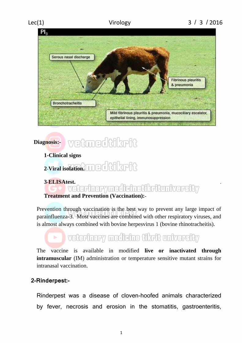
1- Bovine Parainfluenza-3 Virus(PI3)
Bovine parainfluenza-3 is a respiratory disease infected cattle population and
usually only causes mid to subclinical disease. More importantly it can predispose
an animal to secondary bacterial pneumonia.
Etiology:-
Parainfluenza-3 is classified as a RNA virus of the paramyxovirus .
Clinical Findings:- .
1-The clinical signs of parainfluenza-3 include pyrexia (fever), cough, serous
(watery) nasal and lacrimal discharge as well as increased rate of respiration
and an increase breathing sounds.
2-Bronchotracheitis.
3-Fibrinous pleuritis and pneumonia.
2016 /3 / Lec(1) Virology 3
1
Diagnosis:-
1-Clinical signs .
2-Viral isolation. .
3-ELISAtest. .
Treatment and Prevention (Vaccination):-
Prevention through vaccination is the best way to prevent any large impact of
parainfluenza-3. Most vaccines are combined with other respiratory viruses, and
is almost always combined with bovine herpesvirus 1 (bovine rhinotracheitis).
The vaccine is available in modified live or inactivated through
intramuscular (IM) administration or temperature sensitive mutant strains for
intranasal vaccination.
2-Rinderpest:-
Rinderpest was a disease of cloven-hoofed animals characterized by fever, necrosis and erosion in the stomatitis, gastroenteritis,
2016 /3 / Lec(1) Virology 3
1
tongue and infected oral mucosa, lymphoid necrosis, diarrhea with/ and blood and mucous. and high mortality.
Etiology
Rinderpest virus is a Morbillivirus .
Transmitted:-
1-direct contact.
2-drinking contamination water.
3-air
4-infected animals secreted ocular, nasal, oral and vaginal and feces secretion
Clinical Findings
After an incubation period of 3–15 days.
1-fever.
2-Necrosis and erosions in gastrointestinal tract.
3-ulcer, vesicles and erosions on the gums, buccal mucosa, and tongue. The hard and soft palates were often affected. The ocular nasal discharge became mucopurulent, and Diarrhea the final clinical sign, could be watery and bloody.
2016 /3 / Lec(1) Virology 3
1
Diagnosis:- 1-clinical signs.
2-Viral isolation.
3-agar gel immunodiffusion test.
3-ELISA.
4-RT-PCR technique.
3- Newcastle Disease in Poultry:-
Newcastle disease is an infection of domestic poultry and other bird species characterized by acute respiratory disease ,depression, nervous manifestations, or diarrhea may be the predominant clinical form. Severity depends on the virulence of the infecting virus and host susceptibility. Occurrence of the disease is reportable and may result in trade restrictions.
Etiology
NDV, is an RNA virus causes by rubella virus. The virus classification isolates into one of three virulence groups by isolated chicken embryo and chicken inoculation as virulent (velogenic), moderately virulent (mesogenic), or of low virulence (lentogenic) .Velogens and mesogens are now classified as virulent NDV
(vNDV), the cause of Newcastle disease and reportable infection, whereas
infections with lentogens, the low virulence NDV (loNDV) widely used as live
vaccines, are not reportable.
2016 /3 / Lec(1) Virology 3
1
Transmission
1-air.
2-respiratory discharges,
3-feces.
4-ingesting contaminated water or food.
5-contaminated equipment or litter
Clinical Findings
1-Onset is rapid, and signs appear throughout the flock within 2–12 days (average 5).
2-Observed signs depend on whether the infecting virus has a predilection for respiratory, digestive, or nervous systems.
3-Respiratory signs of gasping, coughing, sneezing, and rales predominate in infections with loNDV.
4-Nervous signs of tremors, paralyzed wings and legs, twisted necks, circling, clonic spasms, and complete paralysis may accompany, but usually follow, the respiratory signs in neurotropic velogenic disease. Nervous signs with diarrhea are typical in pigeons, and nervous signs are frequently seen in cormorants and exotic bird species. Respiratory signs with depression, watery greenish diarrhea, and swelling of the tissues of the head and
2016 /3 / Lec(1) Virology 3
1
neck are typical of the most virulent form of the disease, viscerotropic velogenic Newcastle disease, although nervous signs are often seen, especially in vaccinated poultry. Varying degrees of depression and inappetence are seen. Partial or complete cessation of egg production may occur.
5-Eggs may be abnormal in color, shape, or surface and have watery albumen. Mortality is variable but can be as high as 100% with vNDV infections.
Diagnosis
1-NDV can be isolated from oropharyngeal or cloacal swabs or tissues from infected birds by inoculation of the allantoic cavity of 9- to 11-day-old SPF embryonated chicken eggs.
2-hemagglutinating inhibitedtest.
3-PCR.
4-ELISA
Prevention:-
1-vaccines are used to prevent losses from sickness and death. Live lentogenic vaccines, chiefly B1 and LaSota strains, are widely used and typically administered to poultry by mass application in drinking water or by spray..
2-Oil-adjuvanted inactivated vaccines are also used after live vaccine
2016 /3 / Lec(1) Virology 3
1
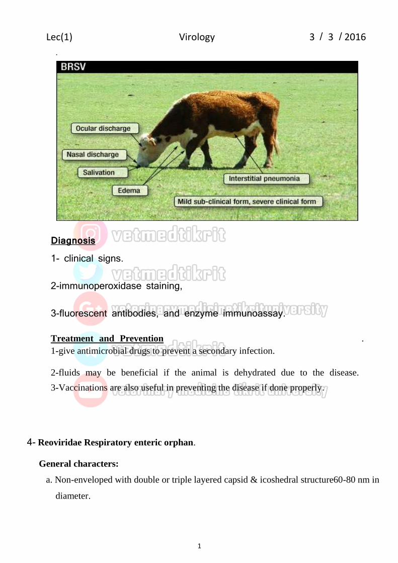
4- Bovine Respiratory Syncytial Virus
Bovine Respiratory Syncytial virus or BRSV is a common virus that affects cattle characterized by respiratory disease that can range in severity from being
fatal to the animal not even appearing ill. This virus is also important because it can predispose the animal to secondary infections.
Etiology:- It is a RNA virus causes by pneumonia virus.
Transmission 1-nose to nose contact. 2-Direct contact.
Clinical Findings . Signs of an animal having this are: Difficulty breathing, fever, reduced feed intake and production and nasal discharge.
2016 /3 / Lec(1) Virology 3
1
.
Diagnosis 1- clinical signs.
2-immunoperoxidase staining,
3-fluorescent antibodies, and enzyme immunoassay.
Treatment and Prevention .
1-give antimicrobial drugs to prevent a secondary infection.
2-fluids may be beneficial if the animal is dehydrated due to the disease.
3-Vaccinations are also useful in preventing the disease if done properly.
4- Reoviridae Respiratory enteric orphan.
General characters:
a. Non-enveloped with double or triple layered capsid & icoshedral structure60-80 nm in
diameter.
2016 /3 / Lec(1) Virology 3
1
b. Segmented (10-12 segments) double- stranded RNA.
c. Replicate in cytoplasm with intracytoplasmic inclusion bodies.
d. Genetic reassortment readily takes place.
e. Resistant to heat, organic solvents.
Genera of Reoviridae :
1.Orthoreovirus: cause arthritis & tenocynovitis in poultry.
2.Rotavirus : cause enteritis in neonatal farm animals.
3.Orbivirus:Arthropods- borne infections (Arbo), cause:African horse sickness in
. horses, Bluetongue disease in sheep, and in other domestic & wild ruminants.
Replication:-
Reovirus replication occurs in the cytoplasm. Viral attachment protein s1 binds to sialic acid and junction adhesion molecule (JAM) on host cell. Particles enter the cell via endocytosis and are partially uncoated in the endolysosome to form infectious subvirion particles or ISVPs. This process is characterized by removal of outer capsid protein s3, proteolytic cleavage of the inner capsid proteins µ1 and conformational changes in s1. Early transcription of the segmented dsRNA genome by a viral polymerase occurs inside the ISVPs. These transcripts leave the core particle and are translated in the cytoplasm. RNA is transcribed ; negative-sense strands are used as templates for positive-sense strands. These (+) sense strands serve as late mRNAs and as templates for (-) sense strand synthesis. The capsid is assembled and the particles are released
1.Orthoreovirus: cause arthritis & tenocynovitis in poultry.
2016 /3 / Lec(1) Virology 3
1
Avian orthoreovirus is a virus of the Reoviridae family and causes most importantly arthritis and tenosynovitis in poultry but can also be a cause of respiratory disease.
Tranmission
1-fecal-oral route . 2-respiratory droplets.
Clinical Signs
1-Lameness is the principle sign of disease, and may be accompanied by
. swelling, haemorrhage, skin discolouration and refusal to move.
2-In severe cases, the gastrocnemius may rupture the bird .
3-Diarrhoea, dehydration and anorexia can also be caused by the virus .
(malabsorption syndrome).
4-Neurological signs develop in severe cases..
Diagnosis
1- Clinical signs.
2- Virus can be isolated by cultured on chicken eggs and chicken embryo liver cell cultures. 3- electron microscopy and PCR. 4- Agar gel immunodiffusion and ELISA.
Treatment
Vaccination can be used in the face of an outbreak, Live attenuated and
inactivated vaccines are available. Attenuated vaccines are for use in young
birds and inactivated for breeding hens
3-Rotavirus : cause enteritis in neonatal farm animals.
Rotavirus in Calves - Rotaviral diarrhoea
2016 /3 / Lec(1) Virology 3
1
Rotaviral diarrhoea is one of the main infections causing calves scour between
five to fourteen days of age. Both the rotavirus, which is seen more often, and the
coronavirus together make up over half of diarrhoea problems in calves .
Bovine rota virus entering the small intestine, the virus attacks the villi thus
reducing the calf’s ability to absorb material into the body effectively. This
produces a concentration gradient and the change in water potential results in
water loss from the epithelial cells and therefore from the body causing
dehydration.
Transmission:-
1-oral contact with infected faeces.
2-Other sources of infection include re-infected cows which show no symptoms
at all. These cows are able to shed the virus without the knowledge of the
farmer.
Signs and Symptoms of the bovine rotavirus
1-diarrhoea which may have the appearance of water rather than normal faeces.
The diarrhoea is often yellow in color and blood and mucous may be
present.
2-The calf will appear depressed and be prevent to drink or feed.
3-As a result severe dehydration will occur.
4-Some calves may drool.
These symptoms are more commonly seen in calves between four days and
three weeks of age. It is rarely symptomatic when the cow is older than a
month old.
Diagnosis
1-clinical signs.2- ELISA.
Treatment and vaccination bovine rotavirus
1-The treatment plan usually consists of replacing lost fluids and restoring the
balance of the body’s important electrolytes.
2016 /3 / Lec(1) Virology 3
1
2-administered intravenously or orally depending on whether the oral
dehydration therapy works.
3-The infected animal must be kept in a warm and dry area, and adequate
amounts of colostrum may be given to young calves to induce recovery.
4-The use of antibiotics in a presence of a secondary bacterial infection the
antibiotics are administered.
cows may be administered the vaccine in the last one to three months of pregnancy.
3-African horse sickness:
African horse sickness (AHS) is an insect borne, viral disease of equie that is endemic Africa. It can be acute, subacute, or subclinical and is characterized by clinical signs and lesions associated with respiratory and circulatory impairment.
Etiology :-
AHS is caused by African horse sickness virus (AHSV) of the genus Orbivirus in
the family Reoviridae. There are nine immunologically distinct serotypes of HSV.
Transmission
1-Culicoides spp are the principal vectors of all nine serotypes of AHSV is seen
during warm, rainy seasons, which favor propagation of the vectors, and
disappears when cold weather stops or significantly reduces vector activity.
2-transmitted between dogs by infected mosquitoes.
Clinical Findings :-
1-The acute respiratory form is characterized by an incubation period of 3–5 days, interlobular edema, and hydropericardium. Death occurs in ~1 wk.
2016 /3 / Lec(1) Virology 3
1
2-A fever of 40°–40.5°C (104°–105°F) for 1–2 days is followed by dyspnea, spasmodic coughing, and dilated nostrils.
3-The animal stands with its legs apart and head extended.
4-The conjunctivae are congested, and the supraorbital fossae may be edematous.
5- Recovery is rare, and the animal dies of anoxia, congestive heart failure, or
both.
Diagnosis:-
1-clinical signs. 2-ELISA. 3-PCR.
Vaccination.
1- Live vaccine.2- nactivated and subunit vaccines
3- Bluetongue:-
Bluetongue disease is a non-contagious, insect-borne, viral disease of ruminants,
mainly sheep and less frequently cattle,[1]
goats, buffalo, deer, dromedaries, and
antelope
Etiology
Bluetongue virus is the type-species of the genus Orbivirus in the family Reoviridae.
There are at least 24 serotypes worldwide, although not all serotypes exist in any
one geographic area.
Transmission
The virus is transmitted by the midge Culicoides spp.
Clinical signs:-
2016 /3 / Lec(1) Virology 3
1
1-high fever.
2-excessive salivation, swelling of the face,lips and tongue and cyanosis of the
tongue. gives the tongue its typical blue appearance.
3-Some animals also develop foot lesions, beginning with coronitis, with
consequent lameness. In sheep, this can lead to knee-walking. In cattle,
constant changing of position of the feet gives bluetongue the nickname The
Dancing Disease. Torsion of the neck is observed in severely affected
Bovine leukemia virus (BLV) :- Bovine immunodeficiency virus (BIV) is a retrovirus belonging to the Lentivirus genus. It is similar to the human immunodeficiency virus (HIV) and infects cattle. The cells primarily infected are lymphocytes and monocytes/macrophages.
Infectious bursal disease (IBD) is a viral disease infected domestic chickens.
Characterized by present clinical or subclinical disease, but immune suppression
and related secondary infections are typically seen. Severity of the immune
suppression depends on the virulence of the infecting virus and age of the host.
Etiology .
IBD is caused by avibirnavirus (infectious bursal disease virus; IBDV) that is most
readily isolated from the bursa of Fabricius but may be isolated from other organs.
Two serotypes of IBDV have been identified.
1- serotype 1 viruses cause disease in chickens and, within them, antigenic variation
can exist between strains. Antigenic drift is largely responsible for this antigenic
variation, but antigenic differences can also occur through genome homologous
recombination.
2- Serotype 2 strains of the virus infect chickens and turkeys but have not caused
clinical disease or immunosuppression in these hosts.
Transmission:-
1-feces . 2-transferred from house to house by fomites.
Clinical Findings
IBD is highly contagious; results of infection depend on age and breed of chicken and
virulence of the virus.
1-Infections may be subclinical or clinical. Infections before 3 wk of age are usually
subclinical. Chickens are most susceptible to clinical disease at 3–6 wk of age when
immature B cells populate the bursa and maternal immunity has waned, but severe
infections have occurred in Leghorn chickens up to 18 wk of age.
subclinical infections They cause severe, long-lasting immunosuppression due to
destruction of immature lymphocytes in the bursa of Fabricius, thymus, and spleen.
The humoral (B cell) immune response is most severely affected;
2016 /3 / Lec(1) Virology 3
1
In clinical infections, onset of the disease occurs after an incubation of 3–4 days.
Chickens may exhibit severe prostration, incoordination, watery diarrhea, and
inflammation of the cloaca.
Diagnosis:-
1-clinical signs.
2-Viral isolation
3-PCR to identify the viral genome in bursa tissue.
4-titration of the virus and virus-neutralization assays.
Control
1-Live vaccines of chicken embryo can be administered by eye drop, drinking water,
or SC routes at 1–21 days of age
2-oil-adjuvanted, inactivated vaccine.
7- Rhabdoviridae:- Rod
General characters:
a. Have characteristic rod shapes and bullet-shaped in vertebrates .
b. enveloped RNA viruses with helical symmetry.
c. virions size (100-430 nm).
d. Stable in the pH range of 5- 10.
e. Rapidly inactivated by heating at 56 C°.
f. Sensitive to lipid solvents and UV light.
Genera of Rhabdoviridae:
1. Lyssavirus:Rabies virus .
2. Ephemero virus:Bovine ephemeral fever virus.
3. Vesiculovirus: vesicular stomatitis virus.
2016 /3 / Lec(1) Virology 3
1
1- rabies virus is a neurotropic virus that causes rabies in humans and
animals.
Transmission:-
1-Usually the bite or scratch of an infected animal, which introduces the virus
through the skin or mucous membrane. .
2-Aerosol transmission from an infected animal, usually a bat.
3-Tissue transplants (such as corneas) from infected humans.
Clinical signs:-
most animals, the virus will spread through the nerves of the bitten animal towards the brain. The virus is relatively slow moving and the average time of incubation from exposure to brain involvement is between 3 to 8 weeks in dogs, 2 to 6 weeks in cats, and 3 to 6 weeks in people. However, incubation periods as long as 6 months in dogs and 12 months in people have been reported. After the virus reaches the brain it then will move to the salivary glands where it can be spread through a bite. After the virus reaches the brain the animal will show one, two, or all of the three different phases
1-Prodromal phase
The first is the prodromal phase and usually lasts for 2-3 days in dogs.
Apprehension, nervousness, anxiety, solitude, and a fever may be noted.
Friendly animals may become shy or irritable and may snap, whereas,
aggressive animals may become affectionate and docile.
2-Furious phase
From the prodromal phase, animals may enter the furious stage; cats are
particularly prone to developing this phase. The furious stage of the disease in
dogs usually lasts for 1 to 7 days. Animals become restless and irritable and are
hyperresponsive to auditory and visual stimuli. As they become more restless,
they begin to roam and become more irritable and vicious. When caged, dogs
may bite and attack their enclosures. Animals progress to become disoriented
and then have seizures and eventually die.
3-Paralytic (dumb) phase
Animals may develop the paralytic phase either after the prodromal or furious stage. The paralytic phase usually develops within 2 to 4 days after the first signs are noted. Nerves affecting the head and throat are the first to be involved and animals may begin to salivate as a result of their inability to swallow. Deep labored breathing and a dropped jaw may result as the diaphragm and facial muscles become increasingly paralyzed. Animals may make a choking sound and many owners think that there is something lodged in the dog’s throat. The animal will get weaker and eventually go into respiratory failure and die.
Diagnosis:
1-clinical signs.
2-microscopic examination.
3-blood samples
Treatment
There is no treatment. Vaccination is the best way to prevent infection
2016 /3 / Lec(1) Virology 3
1
DNA Viruses 1- Herpesviridae: creeping
General characters:
1. Enveloped DNA viruses with icosahedral symmetry.
2. Replicate in nucleus, with intra nuclear inclusion bodies.
3. 120-200 nmin diameter.
4. Latency is a common outcome of infection with these viruses.
5.This family contains more than 100 viruses which cause different diseases in human,
birds, mammals, fish, amphibian &reptiles.
There are three subfamilies of veterinary importance:
Alpha herpesvirinae, Beta herpesvirinae, Gamma herpesvirinae which cause diseases of
the respiratory, reproductive & nervous systems, in different animal species: e.g. Herpes
infections of ruminants:
a. Bovine herpes virus cause (infectious bovine rhinotracheitis (IBR).
b. In poultry: a. infectious laryngo-tracheitis ( ILT).- Marek's disease.- Duck plaque.
c. Human Herpes simplex type 1 cause:- Fever blisters.- Chicken pox or varicella zoster
or shingles.
Replication Replication begins with the binding of the virion to the plasma membrane of the host cell and is followed by fusion of the virion
2016 /3 / Lec(1) Virology 3
1
envelope with the host cell membrane. The capsid and tegument are released into cytoplasm and are transported to into nucleus. Viral DNA is then released in the nucleus, followed by transcription, replication, and assembly of new capsids. Caspids egress from the nucleus and are released from the cell plasma membrane by exocytosis.
1-Infectious Bovine Rhinotracheitis (IBR):-
is a highly contagious, infectious disease that is caused by Bovine Herpesvirus-1 (BHV-1). In addition to causing respiratory disease, this virus can cause conjunctivitis, abortions, encephalitis, and generalized systemic infections.
Cause
IBR is caused by Bovine Herpesvirus-1 that is capable of attacking many different tissues in the body leading to a variety of clinical diseases.
Transmission:-secretions from the eye nose and reproductive organs.
Clinical signs:-clinical diseases caused by the virus can be grouped into:
5- brain infections 6- generalized infections of newborn calves.
Diagnosis:- 1-Clinical signs. 2-Elisa test.
Treatment and Control
As with other viral diseases, there is no direct treatment for the infection. Antibiotic treatment of secondary infections may be necessary. The best way to control IBR is to vaccinate before a disease outbreak occurs (replicating or non-replicating vaccines) although there are fast acting intranasal vaccines that will confer short-lived immunity.
2-Infectious laryngotracheitis (ILT):- is an acute, highly contagious,
herpesvirus infection of chickens .characterized by severe dyspnea,
coughing, ,rales, nasal and ocular discharge, tracheitis, conjunctivitis, and
mild rales.
Causes:-
The disease is caused by Gallid herpesvirus I, commonly known as
infectious laryngotracheitis virus (ILTV).
Clinical Findings
1-In the acute form, gasping, coughing, rattling, and extension of the neck
during inspiration, blood, mucus, yellow caseous exudates, or a hollow
caseous cast in the trachea are seen 5–12 days after natural exposure.
2-Reduced productivity is a varying factor in laying flocks. Affected birds
are anorectic and inactive.
2016 /3 / Lec(1) Virology 3
1
3- The mouth and beak may be bloodstained from the tracheal exudate.
Mortality varies but may reach 50% in adults and is usually due to
occlusion of the trachea by hemorrhage or exudate.
Diagnosis:-
1- Clinical signs.
2-detection of viral DNA using virus-specific PCR assays.
3-Isolation and identification of the virus is done in chicken embryos or tissue culture .
4- microscopic examination of the trachea, intranuclear inclusion lesions produced by ILTV infection .
Control
1-. Vaccination is done with live attenuated vaccines
2-viral vector recombinant vaccines.
3-Marek's disease:- Marek's disease is one of the most ubiquitous avian
infections; it is identified in chicken flocks worldwide. Every flock, except for
those maintained under strict pathogen-free conditions, is presumed to be infected.
Although clinical disease is not always apparent in infected flocks, a subclinical
decrease in growth rate and egg production may be economically important.
Etiology
Marek's disease virus is a member of the genus Mardivirus within the subfamily Alphaherpesvirinae. Within the genus Mardivirus are three closely related species previously designated as three
2016 /3 / Lec(1) Virology 3
1
serotypes of Marek's disease virus. Gallid herpesvirus 2 (MDV-1) represents all virulent Marek's disease virus strains and is further divided into pathotypes, designated as mild (m), virulent (v), very virulent (vv), and very virulent plus (vv+). Gallid herpesvirus 3 (MDV-2) and Meleagrid herpesvirus 1 (turkey herpesvirus, MDV-3) represent avirulent virus strains isolated from chickens and turkeys, respectively, and are commonly used as vaccines against Marek's disease.
Transmission The route of infection is usually respiratory and the disease is highly contagious being spread by infective feather-follicle dander, fomites, etc. Infected birds remain viraemic for life. Vertical transmission is not considered to be important.
Signs
Paralysis of legs, wings and neck. Loss of weight. Grey iris or irregular pupil. Vision impairment. Skin around feather follicles raised and roughened
Diagnosis
History, clinical signs, distribution of lesions, age affected, histopathology.
Differentiate from Lymphoid leukosis, botulism, deficiency of thiamine, deficiency
of Ca/Phosphorus/Vitamin D, especially at the start of lay.
2016 /3 / Lec(1) Virology 3
1
Treatment and vaccines
None treatment. HVT & SB-1 & RISPENS trivalent Marek’s disease vaccine contains serotype 1 (Rispens), serotype 2 (SB-1) and serotype 3 (turkey herpesvirus or HVT) Marek’s disease viruses. This combination vaccine is recommended for use in chickens as an aid in the prevention of very virulent Marek’s disease.
2- Poxviridae: General characters:
1. Largest viruses (220-450 nmx 140- 260 nm).
2. Complex symmetry, brick shaped viruses.
3. Enveloped DNA viruses replicate in cytoplasm.
4. Virions are stable at room temperature under dry conditions, but
sensitive to heat, detergents, formaldehyde and oxidizing agents.
5. Skin lesions prominent feature.
6. Genetic recombination within genera results in extensive serological cross
reaction and cross-protection.
2016 /3 / Lec(1) Virology 3
1
Genera of Poxviridae: a. Orthopoxvirus:Vaccinia, cow pox , variola virus .
b. Parapoxvirus:Orfvirus, Bovine popular stomatitis virus, pseudo
d. Avipoxvirus: Fowl pox virus, pigeon pox, Turkey pox.
e. Suipox virus: Swinepox virus.
f. Leporipox virus:Myxoma virus. (Rabbit)
g. Entomopox virus: infect insects.
1-vaccinia virus, cowpox virus , variola virus
Despite the name, the reservoir hosts of cowpox virus are rodents, from which the virus occasionally spreads to domestic cats, cows, humans, and zoo animals.
The virus produces lesions on the teats and the contiguous parts of the udder of cows and is spread through herds by the process of milking.
Sheep pox and goat pox are the most important of all pox diseases of domestic animals, causing high mortality in young animals and significant economic loss.
Eciology:- Capripox virus.
Transmission:
1-Virus particles are shed from skin lesions and in ocular and nasal discharges during the acute stages of the disease. 2-During an outbreak, the virus is probably transmitted between sheep by respiratory droplets. 3-There is also evidence that mechanical transmission by biting arthropods, such as stable flies, may be important. 4-Infection occurs through skin abrasions or by aerosol. Clinical signs:-
1- incubation period of about one week, 2-infected animals develop fever, 3-edema of the eyelids, conjunctivitis and nasal discharge. Within a few days 4-macules which rapidly develop into papules appear on the skin
and external mucous membranes. 5-Scabs form over necrotic papules.
2016 /3 / Lec(1) Virology 3
1
Lumpy skin Disease virus (within genus Capripoxvirus).
Lumpy skin disease affects cattle breeds, characterized by fever, followed shortly by the development of nodular lesions in the skin that subsequently undergo necrosis
Clinical signs
1-The incubation period is up to 14 days. 2-persistent fever accompanied by lacrimation, nasal discharge and a . drop in milk yield. 3- Superficial lymph nodes become enlarged and there is oedema of the limbs and dependent tissues. 4- skin nodules develop particularly on the head, neck, udder and perineum. 5-Nodules also develop on the mucous membranes of the mouth and nasal 6-Some skin lesions may develop into a central plug of necrotic tissue which . sloughs producing a deep ulcer. 7-Secondary bacterial infection or myiasis can exacerbate the condition. 8- Recovery may take several months. 9-pregnant cows may abortion .
3-Diseases caused by members of the genus avipoxvirus
Fowlpox and Other Avian Poxvirus Diseases
Poxviruses that are related serologically to each other and specifically infect birds have been recovered from lesions found in
2016 /3 / Lec(1) Virology 3
1
all species of poultry and many species of wild birds. Viruses recovered from various species of birds are given names related to their hosts, such as fowl pox, canary pox, turkey pox, pigeon pox. The fowl pox virus is highly infectious for chickens and turkeys, rarely so for pigeons, and not at all for ducks and canaries, but turkey pox virus is virulent for ducks.
There are two forms of fowl pox, probably associated with different routes of infection.
1-The most common, which probably results from infection by biting arthropods, is characterized by small papules on the comb, wattles, and around the beak; lesions occasionally develop on the legs and feet and around the cloaca. 2-The second form of fowl pox is probably due to droplete infection and involves infection of the mucous membranes of the mouth, pharynx, larynx, and sometimes the trachea. This is often referred to as the diphtheritic form of fowlpox because the lesions result in a necrotic pseudo-membrane, which can cause death by asphyxiation.
4-Diseases caused by members of the genus para pox virus.
1-Orf (Contagious Pustular Dermatitis):-
2016 /3 / Lec(1) Virology 3
1
Orf (contagious pustular dermatitis, scabby mouth) is an important disease in sheep and goats (especially in young lambs) caused by a para poxvirus
Transmitted through direct or indirect contact.
Clinical signs:-
The virus, which is epithelio tropic, produces proliferative wart-like lesions following entry through skin abrasions.The virus replicates in epidermal keratinocytes and infected cells release an endothelial growth factor which is implicated in epithelial cell proliferation.
1-Papular lesions progress to vesicles, pustules and eventually to scab formation. Lesion appear on the muzzle and lips gums tongue, The lesions can also affect the eyelids, feet, and the teats.
2- Infections caused by pseudo cow pox virus
Pseudocowpox, also known as milker's nodule, is caused by pseudocowpox virus, parapoxvirus with worldwide distribution. It is a common mild condition affecting the teats of lactating cows.Infection spreads slowly through milking
2016 /3 / Lec(1) Virology 3
1
herds with variation in the number of affected animals at any particular time.
Transmission
Occur by direct or indirect contact.
1-through teat cups and on milkers' hands. 2-mechanically by flies or when calves are being suckled.
3-Bovine papular stomatitis.
1-This mild viral disease of young cattle occurs worldwide. 2-Mature cattle are considered to be reservoirs of infection 3-It is caused by a parapoxvirus, bovine papular stomatitis virus, 4-is transmitted by direct or indirect contact 5-Affected calves commonly develop lesions in the Oral cavity
and on the muzzle. 6-These lesions are characterized by hyperaemic foci that develop
into papules 7-Affected animals usually recover within three weeks
diseases PoxviridaeDiagnosis of
1-Diagnosis can often be made on clinical grounds. 2-Skin biopsies or postmortem specimens may be used for
laboratory confirmation. 3-Eosinophilic intracytoplasmic inclusions may be demonstrable histologically in epidermal cells.
2016 /3 / Lec(1) Virology 3
1
4-Electron microscopy can be used for the rapid identification of poxvirus 5-lesions. can be readily distinguished from parapoxviruses. 6-Virus may be isolated in lamb testis or kidney cell monolayers, chorioallantoic membrane,…... 7-Direct ELISA has been developed for to detection of virus antigen. 8-Nucleic acid probes can be used for diagnosis 9-PCR 10-Several serological methods including virus neutralization, Western blot analysis and the indirect fluorescent antibody test and indirect ELISA.
Treatment
1-There is no specific treatment. 2-Control of secondary infected in skin, on oral mucosa or on respiratory bacterial infection is desirable.
Control
A-In endemic areas, control is based on annual vaccination by: 1-Modified live vaccines 2-inactivated vaccines 3-subunit vaccine
3. Replicate in nuclei, forming intranuclear inclusion bodies.
4. Fibers project from twelve vertices of capsid.
5. Agglutinate rat or monkey RBCs
6. Resist freezing, mild acid & lipid solvents, 56C for 10 min.
Genera of Adenoviridae:-
1. Aviadenovirus: hydropericardium hepatitis syndrome (HHS), Egg drop syndrome
in layers .
2. Mastadenovirus mammalian adenovirus which cause sever infections in dogs
(infectious canine hepatitis .pulmonary infections in equine, which fatal in
Arabian foals.
1-hydropericardium hepatitis syndrome (HHS):-
is an acute disease of young chickens associated with anemia, hemorrhagic
disorders, and hydropericardium. It is a common disease in several countries, where
broilers are severely affected, resulting in high mortality rates.
Etiology:-
The AAVs classified in the genus Aviadenovirus (formerly group I), are the etiologic agents of this condition. Although there are 12 different serotypes of AAVThese AAVs are capable of producing the disease without the immunosuppressive effects
Transmission:- 1-Vertical transmission has been described in progeny from breeder flocks infected with AAV serotypes 4 and 8.
2016 /3 / Lec(1) Virology 3
1
2- Horizontal transmission has also been demonstrated; young chicks in contact with infected chicks .
Clinical Findings:-
specific clinical signs, abrupt onset of mortality, lethargy, huddling with ruffled feathers, and yellow, mucoid droppings may be seen. The duration of the infection usually ranges from 9–14 days with morbidity of 10%–30% and a daily mortality of 3%–5%.
diagnosis
1-clinical signs.
2-Viral isolation.
3-restriction enzyme analysis, and PCR.
Treatment and Prevention
1-As with many other viral diseases, there is no treatment. Antibiotics may help
prevent secondary bacterial infections. Sulfonamides are contraindicated if evidence
of hematologic disease or immunosuppression is seen.
2-live and inactivated vaccines are used to control the syndrome.
2-Egg drop syndrome:-
is an aviadenovirus-induced disease characterized by the production of pale, soft-
shelled, and shell-less eggs by apparently healthy laying hens. The disease in laying
hens has commonly been called "egg drop syndrome," but the full name (egg drop
syndrome '76 [EDS '76]) should be used to distinguish.
Etiology:-
EDS ‘76 is caused by a double-stranded DNA virus, duck adenovirus 1 (also known as EDSV), which belongs to the genus Avai
2016 /3 / Lec(1) Virology 3
1
adenovirus. The virus commonly infects both wild and domestic ducks and geese
Clinical Findings
1-pale-shelled eggs, quickly followed by production of soft-shelled and shell-less
eggs. The thin-shelled and shell-less eggs are fragile, and the birds tend to eat
them;
Diagnosis:
1-Clinical signs.
2-viral isolation.
3-A hemagglutination-inhibition test using fowl RBCs .
4-ELISA,
5-neutralization test can be used for confirmation.
6-The double immunodiffusion test also has been used. PCR-based tests.
Control:-
There is no treatment for EDS '76. Inactivated vaccines with oil adjuvant are
available.
4- Parvoviridae:- small = picodna
General characters:
1. small (18-26 nm), non enveloped DNA viruses.
2. 2.Icosahedral symmetry, single stranded DNA.
3. Replicate in the nucleus forming intra nuclear inclusion bodies.
4. Require rapidly- dividing cells for replication.
5. Resistant to heat 56C for more than 60 minutes also resistant to lipid solvents,pH (3-9).
6. Inactivated by formalin, Propiolacton, sodium hypochlorite.
Genera of Parvoviridae:
2016 /3 / Lec(1) Virology 3
1
Parvovirus: cause of enteric & systemic disease in dogs & cats (Feline pan leukopenia
is a highly contagious, often fatal, viral disease of cats that is seen worldwide. Kittens are affected most severely. The causative parvovirus is very resistant; it can persist for 1 yr at room temperature in the environment.
Etiology:- Parvovirus
Transmission:- Cats are infected oro nasally by exposure to infected animals, their feces, secretions, or contaminated fomites(eg, shoes, clothing).
Clinical Findings
1-Acute cases show fever (104°–107°F [40°–41.7°C]), depression, and anorexia after
an incubation period of 2–7 days. Vomiting usually develops 1–2 days after the onset
of fever; it is typically bilious and unrelated to eating. Diarrhea may begin a little
later but is not always present. Extreme dehydration develops rapidly. Affected cats
Diagnosis
1-clinical signs
2-Total WBC counts <2,000 cells/μL are associated with a poorer prognosis.
3-immunochromatographic test kit intended for detection of fecal CPV antigen.
However, fecal antigen is detectable only for a short time after infection. False-
negative results are common.
2016 /3 / Lec(1) Virology 3
1
Treatment and Prevention:- 1-Successful treatment of acute cases requires vigorous fluid therapy and supportive nursing care in the isolation unit.
2-IV fluid replacement and maintenance with a balanced isotonic crystalloid solution (eg, lactated Ringer's solution with calculated potassium supplementation). B vitamins should be added to the infusion, together with 5% glucose if hypoglycemia is suspected or proved.
3- Parenteral, broad-spectrum antibiotic therapy is indicated; however, nephrotoxic drugs (eg, gentamicin, amikacin) should be avoided until dehydration.
4-Recombinant feline interferon omega (rFeIFN; 1 MU/kg/day SC for 3 days) should be considered for use in the treatment of feline panleukopenia.
5-inactivated and modified live virus vaccines
5-Papillomaviridae
General characters:
1. Non-enveloped, circular double strand DNA viruses.
2. Icosahedral symmetry, 55 nmin diameter.
3. Replications in nucleus
4. Resistant to lipid- solvents, acids, 60°C for 30 minutes.
Genera of Papillomaviridae:
Contains one genus, Papillomavirus:
a. Have not been cultured in vitro.
b. Cause papilloma & fibropapilloma in domestic animals.
2016 /3 / Lec(1) Virology 3
1
1-Bovine papillomavirus (BPV):-
is a group of DNA viruses of the family Papillomaviridae that are common in
cattle. Infection causes warts (papillomas and fibropapillomas) of the skin and
alimentary tract, and more rarely cancers of the alimentary tract and urinary
bladder. They are also thought to cause the skin tumour equine sarcoid in horses
and donkeys.
Types of papillomavirus
There are at least five strains of papillomavirus, each of which has a specific predilection
site on the cow.
1-BPV type I:- causes wart like lesion on the nose, teats or penis and affect young cattle and will usually regress over time.
2-BPV type II:- causes warts all over the skin of the head and neck of young cattle and will usually regress over time.
3-BPV type III:- causes atypical warts which are smooth and white in appearance and occur mainly on the teats and udders of older cows.
4-BPV type IV causes papillomas in gut, especially the rumen, and bladder, as well as lesions on the eye. This particular systemic form can be refered to as papillomatosis. The papillomas caused by this strain can undergo malignant transformation to alimentary carcinomas,
5-BPV type V causes tiny warts on the teat
Transmission:-
infection is spread by direct contact from cow to cow or by indirect contact from
fomites. With most strains, calves are most commonly affected.
In BPV type IV signs will be concurrent with the body system affected e.g. haematuria if in the bladder or diarrhoea and bloat if in the rumen.
On the skin, the virus will at first appear as small, smooth raised nodules in the characteristic regions, which will then enlarge. Some will become rough and cauliflower like in appearance, whilst others may become pedunculated. A characteristic feature of this disease is that the warts will regress spontaneously over a period no longer than one year.
Diagnosis
Lesions are characteristic in appearance and location along with the signalment of the animal are enough to make a presumptive diagnosis.
Treatment and Control
Often no treatment is required as spontaneous regression usually occurs. If the cow traumatises a wart then a short course of antibiotics or anti inflammatories may be used to aid its immediate relief. Any other treatment is only of cosmetic value and so is generally only performed by those wishing to show their cattle. Treatment of warts would include injection with lithium antimony thiomalate 6%, removal by cryosurgery or cold steel surgery.
Control measures are not usually necessary due to the mildness of the disease.