VI. Applications 671VII. Acknowledgments 672VIII. References 672
I. Introduction
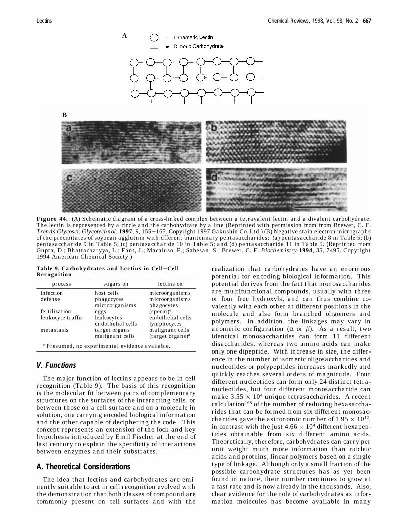
Proteins that interact with carbohydrates non-covalently occur widely in nature. Prominent ex-amples are carbohydrate-specific enzymes and anti-carbohydrate antibodies. In recent years, anotherclass of such proteins, the lectins,1-5 has come intothe forefront of biological research. Lectins bindmono- and oligosaccharides reversibly and with highspecificity, but are devoid of catalytic activity, andin contrast to antibodies, are not products of animmune response. Each lectin molecule containstypically two or more carbohydrate-combining sites,i.e., they are di- or polyvalent. Therefore, when theyreact with cells, for example erythrocytes, they willnot only combine with the sugars on their surfaces,but will also cause cross-linking of the cells and theirsubsequent precipitation, a phenomenon referred toas cell agglutination. The erythrocyte agglutinating,or hemagglutinating, activity of lectins is a majorattribute of these proteins and is used routinely fortheir detection and characterization. Lectins alsoform cross-links between polysaccharide or glycopro-tein molecules in solution and induce their precipita-tion. Both the agglutination and precipitation reac-tions of lectins are inhibited by the sugar ligands forwhich the lectins are specific.Lectins are found in most organisms, ranging from
viruses and bacteria to plants and animals. They arereadily obtainable in purified form, mostly by affinitychromatography on the immobilized ligand, and morerecently also by recombinant DNA techniques. Theyrepresent a heterogeneous group of oligomeric pro-teins that vary widely in size, structure, molecularorganization, as well as in the constitution of theircombining sites. Nonetheless, many of them belongto distinct protein families with similar sequencesand structural features. In fact, sequence similaritywith known lectins provide a novel guideline for thedetection and identification of new ones.
† Abbreviations used: CRD, carbohydrate recognition domain;ECorL, Erythrina corallodendron lectin; GNA, snowdrop ag-glutinin; Ig, immunoglobulin; LOL, Lathyrus ochrus lectin; MBP,mannose binding protein; PDP, protein data bank; PHA, kidneybean lectin; PNA, peanut agglutinin; RCA, Ricinus communisagglutinin; SAP, serum amyloid P component; SBA, soybeanagglutinin; WGA, wheat germ agglutinin. For abbreviation ofoligosaccharide names, see Table 5.* Address for correspondence: Dr. Nathan Sharon, Departmentof Membrane Research and Biophysics, The Weizmann Instituteof Science, Rehovot 76100, Israel. Telephone: 972-8-9343605.Fax: 972-8-9468256. E-mail: [email protected].
Although lectins were first described at the turnof the century, their study started to gain momentumonly in the 1960s.2,6,7 They were then shown to beinvaluable tools for the structural and functionalinvestigation of complex carbohydrates, especially
glycoproteins, and for the examination of changesthat occur on cell surfaces during physiological andpathological processes, from cell differentiation tocancer.8,9 At present, they are the focus of intenseattention because of the realization that they act asrecognition determinants in diverse biological proc-esses.10,11 These include clearance of glycoproteinsfrom the circulatory system, control of intracellulartraffic of glycoproteins, adhesion of infectious agentsto host cells, recruitment of leukocytes to inflamma-tory sites, as well as cell interactions in the immunesystem, in malignancy and metastasis. Investigationof lectins and their role in cell recognition, as wellas the application of these proteins for the study ofcarbohydrates in solution and on cell surfaces, aremaking marked contributions to the advancement ofglycobiology.12 Developments in the latter field arehaving a significant impact on lectin research, so thatthe two are now moving ahead hand in hand.During the past decade, there has been remarkable
progress in elucidating the features of lectins thatare important for carbohydrate binding. This wasmade possible by the refinement of old techniquesand development of new ones. In particular, high-resolution X-ray crystallography of lectins in complexwith their ligands allowed the identification of thechemical groups on the protein and on the carbohy-drate that interact with each other and of the typesof bond formed. Further information on the contri-bution of individual amino acids to the activity of alectin has been obtained by site-directed mutagenesisexperiments and also by molecular modeling. Ofspecial interest are the studies of lectin-oligo-saccharide complexes, since they provide a basis forthe understanding of how lectins recognize theirnatural ligands.In this article we deal mainly with the specificity
and structure of lectins, with emphasis on theircarbohydrate binding sites and the mechanism oflectin-carbohydrate interactions, and we also discussbriefly their roles and applications. For recentreviews on the subject, see refs 4 and 13-18. Bacte-rial toxins that are carbohydrate binding proteins,although sometimes considered as lectins,19 will notbe covered.
II. Carbohydrate Specificity
A. MonosaccharidesOn the basis of their specificity, lectins are classi-
fied into five groups, according to the monosaccharidefor which they exhibit the highest affinity: mannose,galactose/N-acetylgalactosamine,N-acetylglucosamine,fucose, and N-acetylneuraminic acid (sugars are ofthe D configuration except for fucose which is L)(Table 1). Relevant for the biological activities oflectins is the fact that of the numerous monosaccha-rides found in nature, only those listed above aretypical constituents of surfaces of eukaryotic cells.Only in exceptional cases does one find lectins thatexhibit affinity for other sugars. One example is thehuman serum amyloid P component (SAP) (seesection III.A.5), a lectin specific for the 4,6-cyclicpyruvate acetal of galactose;20 to date, this rare
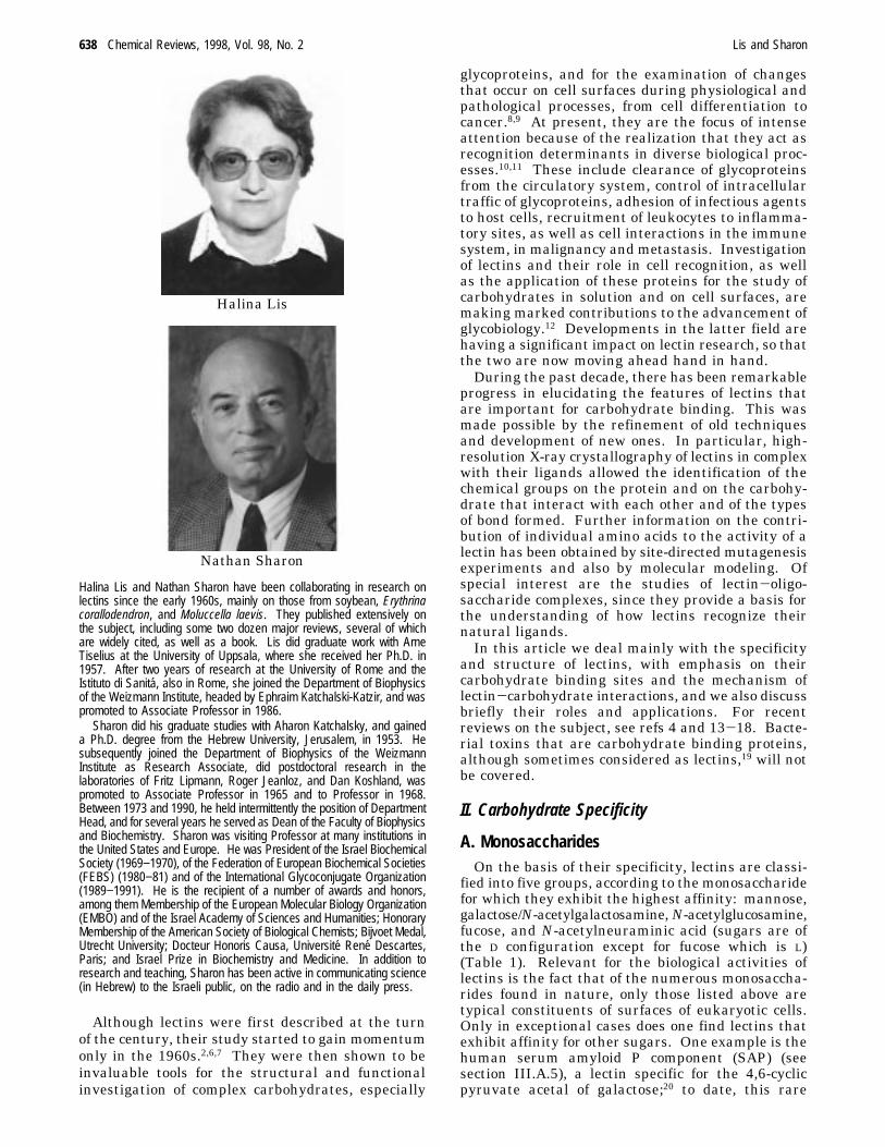
Halina Lis
Nathan Sharon
Halina Lis and Nathan Sharon have been collaborating in research onlectins since the early 1960s, mainly on those from soybean, Erythrinacorallodendron, and Moluccella laevis. They published extensively onthe subject, including some two dozen major reviews, several of whichare widely cited, as well as a book. Lis did graduate work with ArneTiselius at the University of Uppsala, where she received her Ph.D. in1957. After two years of research at the University of Rome and theIstituto di Sanita, also in Rome, she joined the Department of Biophysicsof the Weizmann Institute, headed by Ephraim Katchalski-Katzir, and waspromoted to Associate Professor in 1986.
Sharon did his graduate studies with Aharon Katchalsky, and gaineda Ph.D. degree from the Hebrew University, Jerusalem, in 1953. Hesubsequently joined the Department of Biophysics of the WeizmannInstitute as Research Associate, did postdoctoral research in thelaboratories of Fritz Lipmann, Roger Jeanloz, and Dan Koshland, waspromoted to Associate Professor in 1965 and to Professor in 1968.Between 1973 and 1990, he held intermittently the position of DepartmentHead, and for several years he served as Dean of the Faculty of Biophysicsand Biochemistry. Sharon was visiting Professor at many institutions inthe United States and Europe. He was President of the Israel BiochemicalSociety (1969−1970), of the Federation of European Biochemical Societies(FEBS) (1980−81) and of the International Glycoconjugate Organization(1989−1991). He is the recipient of a number of awards and honors,among them Membership of the European Molecular Biology Organization(EMBO) and of the Israel Academy of Sciences and Humanities; HonoraryMembership of the American Society of Biological Chemists; Bijvoet Medal,Utrecht University; Docteur Honoris Causa, Universite Rene Descartes,Paris; and Israel Prize in Biochemistry and Medicine. In addition toresearch and teaching, Sharon has been active in communicating science(in Hebrew) to the Israeli public, on the radio and in the daily press.
638 Chemical Reviews, 1998, Vol. 98, No. 2 Lis and Sharon
carbohydrate was found in certain algal polysaccha-rides, in a marine sponge, and in a yeast, but not inbacteria nor higher organisms.The affinity of the lectins for monosaccharides is
usually weak, with association constants in themillimolar range, yet it is often highly selective.2,21In particular, lectins specific for galactose do not reactwith glucose (its 4 epimer) or mannose (the 2 epimerof glucose), nor do those specific for mannose bindgalactose. Similarly, with the exception of wheatgerm agglutinin (see below), members of the N-acetylglucosamine specificity group do not combinewith N-acetylgalactosamine (and vice versa). How-ever, the selectivity of lectins for monosaccharides isnot always so high. Thus, many lectins toleratevariations at C-2 of the pyranose ring and those ofthe mannose specificity group may bind the epimericglucose as well. Most lectins that bind galactoseinteract also with N-acetylgalactosamine, in somecases preferentially, e.g., soybean agglutinin (SBA),the affinity of which for the latter monosaccharideis 25-50 times higher than that for galactose. Oth-ers bind both monosaccharides with nearly the sameaffinity, as is the case with the Erythrina corallo-dendron (coral tree) lectin (ECorL). For this reasonthey are classified as one specificity group, Gal/GalNAc, even though certain of them (e.g., peanutagglutinin, PNA) do not bind N-acetylgalactosamineat all. Occasionally, lectins combine with monosac-
charides that appear structurally unrelated, but thatpresent similar topographical features when ap-propriately viewed. For instance, wheat germ ag-glutinin binds both N-acetylglucosamine and N-acetylneuraminic acid, and in contrast to otherN-acetylglucosamine-specific lectins N-acetylgalac-tosamine as well, although more weakly. Consider-ation of the three-dimensional structures of thesemonosaccharides reveals similarity at positions C-2(acetamide group) and C-3 (hydroxyl group) of thepyranose ring of the two hexosamines with those ofC-5 and C-4 onN-acetylneuraminic acid, respectively(Figure 1); these are the positions critical for produc-tive contact with the combining site of the lectin (cf.section IV.A.2). Also, mannose-specific animal lectins(e.g., the rat mannose binding proteins, MBP’s) bindfucose too (Figure 1).Certain lectins belonging to the same specificity
group combine preferentially, or almost exclusively,either with the R- or â-glycosides of the respectivemonosaccharide, whereas others lack anomeric speci-ficity. The properties of the aglycon may markedlyinfluence the interaction of a glycoside with a lectin.In particular aromatic glycosides bind to many lectinsmuch more strongly than aliphatic ones, attesting tothe presence of a hydrophobic region close to thecarbohydrate-combining site. The hydrophobic effectis at times so strong that lectins that show a markedpreference for methyl R-glycosides over the corre-
a For references, see ref 8. b For structures of oligosaccharides not shown, see Table 3. c Relative affinity compared to that ofthe monosaccharide; usually based on hemagglutination inhibition assays. d Most lectins in this group bind also glucose, oftenwith similar affinity. e Lectin does not bind glucose. f Although termed mannose binding, this lectin binds mannose, N-acetylglucosamine and fucose with roughly equal affinities. A similar protein, designated MPB-C is found in mammalian liver.g Lectin exhibits pronounced preference for N-acetylgalactosamine. h With N-acetylgalactosamine as reference monosaccharide.i With galactose as reference monosaccharide. j Does not bind N-acetylgalactosamine.
Lectins Chemical Reviews, 1998, Vol. 98, No. 2 639
sponding â anomers exhibit an inverse anomericspecificity when tested with the corresponding p-nitrophenyl glycosides.22 The lectins within eachgroup may also differ markedly in their affinity forother derivatives. For example, concanavalin A andfavin, the lectin from fava bean, both bind glucoseequally well. However, while the affinity of the 3-Omethyl or phenyl ethers of glucose to concanavalinA is 10-20 times weaker than that of glucose, forfavin it is 3-4 times higher. Quite unusually,concanavalin A interacts also with peptides thatcontain the Tyr-Pro-Tyr motif, with an affinity closeto that of methyl R-mannoside.23,24 Such peptidesmay bind to the lectin by hydrogen bonding with thehydroxyl groups of the tyrosines (which mimic sugar
oxygens) and hydrophobic interactions with carbonson the aromatic side chain (which mimic sugarcabons). The peptide and carbohydrate ligands wereshown to bind to the lectin at the same site, thusrepresenting a case of true glycomimetics.25
B. Oligosaccharides
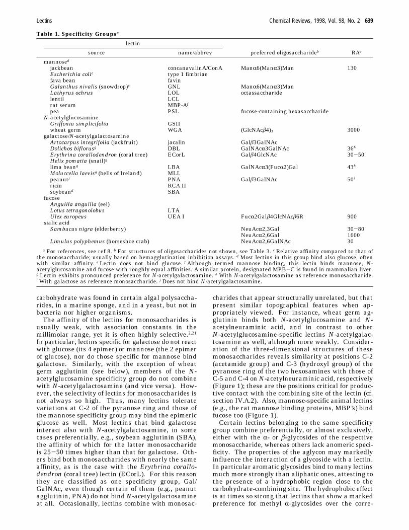
The classification of lectins according to theirmonosaccharide specificity masks the fact that theyoften exhibit an exquisite specificity for di-, tri-, andtetrasaccharides (with association constants up to1000-fold higher as compared with the monosaccha-ride) (Table 1) and that certain lectins interact onlywith oligosaccharides (Table 2). Moreover, lectins ofthe same specificity group may differ markedly intheir affinities for different oligosaccharides. Fromthe functional point of view, binding of oligosaccha-rides is of special significance since, as mentionedearlier, they are most likely the natural ligands oflectins. The affinities of lectins to oligosaccharidesmay be influenced by the shape of the latter com-pounds which are flexible molecules with consider-able freedom of rotation around the glycosidic bondsconnecting the individual monosaccharide constitu-ents. This has been demonstrated by molecularmodeling, as well as by high-resolution nuclearmagnetic resonance (NMR) studies of oligosaccha-rides in solution.26-31 For instance, in the oligosac-charide Man(R1-3)[Man(R1-6)]Man(â1-4)GlcNAc-(â1-4)GlcNAc (the pentasaccharide core, present inall asparagine-linked carbohydrate chains of glyco-proteins)12,32,33 and many of its derivatives, the R1-6-linked mannose can form two rotational isomersrelative to the C5-C6 bond of the â1-4-linkedmannose. The prevalence of either of the two isomersdepends on the type of substitution on the mannoseresidues of the core. In particular, attachment of aN-acetylglucosamine linked â1-4 to Man(â1-4) (“bi-secting” N-acetylglucosamine) fixes the orientationof the Man(R1-6)Man arm into one of the twopossible conformations and markedly decreases thebinding of the oligosaccharide to concanavalin A(Figure 2). Because of their flexibility, oligo-
Figure 1. Common structural features of mannose andfucose (A) and of N-acetylneuraminic acid and N-acetyl-glucosamine (B). Groups that occupy the same position inspace are underlined. (A) Rotation of the fucose moleculeby 180° allows superimposition of its ring oxygen, 4-OH,3-OH, and 2-OH with the ring oxygen, 2-OH, 3-OH, and4-OH of mannose, respectively. (B) Conformational similar-ity ofN-acetylglucosamine andN-acetylneuraminic acid atthe underlined positions (acetamide and hydroxyl) of thepyranose rings is observed when the sialic acid mole-cule is suitably rotated. The conformation of N-acetyl-galactosamine (not shown) at the relevant positions isidentical with that of N-acetylglucosamine. (Reprinted bypermission from ref 2. Copyright 1989 Chapman and HallLtd.)
Table 2. Lectins Specific for Oligosaccharides Only
lectin abbrev oligosaccharide
Escherichia coli type P fimbriae GalR4GalK99 fimbriae NeuGcR2,3Galâ4GlcNAc
galectins Galâ4Glc; Galâ4GlcNAcGriffonia simplicifolia IV GSIV FucR2Galâ3(FucR4)GlcNAcPhaseolus vulgaris E-PHA
640 Chemical Reviews, 1998, Vol. 98, No. 2 Lis and Sharon
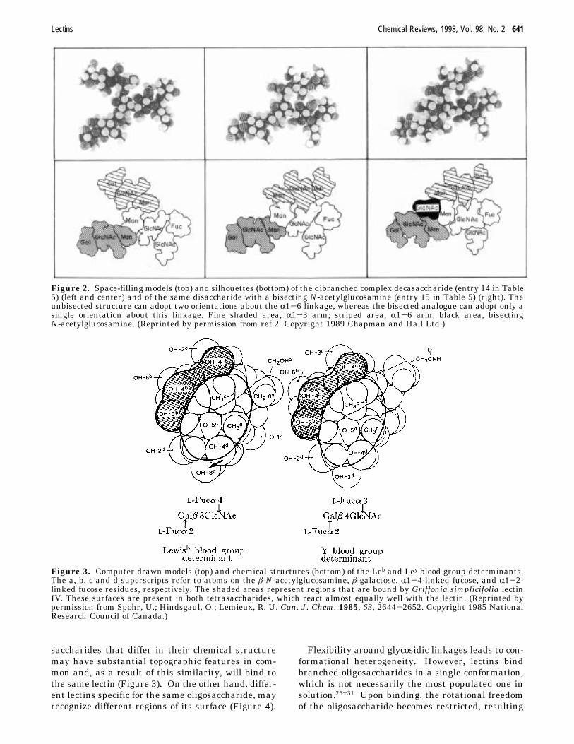
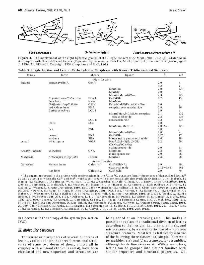
saccharides that differ in their chemical structuremay have substantial topographic features in com-mon and, as a result of this similarity, will bind tothe same lectin (Figure 3). On the other hand, differ-ent lectins specific for the same oligosaccharide, mayrecognize different regions of its surface (Figure 4).
Flexibility around glycosidic linkages leads to con-formational heterogeneity. However, lectins bindbranched oligosaccharides in a single conformation,which is not necessarily the most populated one insolution.26-31 Upon binding, the rotational freedomof the oligosaccharide becomes restricted, resulting
Figure 2. Space-filling models (top) and silhouettes (bottom) of the dibranched complex decasaccharide (entry 14 in Table5) (left and center) and of the same disaccharide with a bisecting N-acetylglucosamine (entry 15 in Table 5) (right). Theunbisected structure can adopt two orientations about the R1-6 linkage, whereas the bisected analogue can adopt only asingle orientation about this linkage. Fine shaded area, R1-3 arm; striped area, R1-6 arm; black area, bisectingN-acetylglucosamine. (Reprinted by permission from ref 2. Copyright 1989 Chapman and Hall Ltd.)
Figure 3. Computer drawn models (top) and chemical structures (bottom) of the Leb and Ley blood group determinants.The a, b, c and d superscripts refer to atoms on the â-N-acetylglucosamine, â-galactose, R1-4-linked fucose, and R1-2-linked fucose residues, respectively. The shaded areas represent regions that are bound by Griffonia simplicifolia lectinIV. These surfaces are present in both tetrasaccharides, which react almost equally well with the lectin. (Reprinted bypermission from Spohr, U.; Hindsgaul, O.; Lemieux, R. U. Can. J. Chem. 1985, 63, 2644-2652. Copyright 1985 NationalResearch Council of Canada.)
Lectins Chemical Reviews, 1998, Vol. 98, No. 2 641
in a decrease in the entropy of the system (see sectionIV.C).
III. Molecular Structure
The amino acid sequences of several hundreds oflectins, and in addition the three-dimensional struc-tures of some two dozen of them, almost all incomplex with a ligand (Tables 3 and 4), have beenelucidated and new sequences and structures are
being added at an increasing rate. This makes itpossible to replace the traditional division of lectinsaccording to their origin, i.e., plants, animals, andmicroorganisms, by a classification based on commonstructural features. Most lectins fall clearly into oneof the following three classes: (a) simple, (b) mosaic(or multidomain), and (c) macromolecular assemblies,although borderline cases exist. Within each class,lectins can be grouped into distinct families withsimilar sequences and structural properties. Oc-
Figure 4. The involvement of the eight hydroxyl groups of the H-type trisaccharide MeâFuc(R1-2)Gal(â1-4)GlcNAc inits complex with three different lectins. (Reprinted by permission from Du, M.-H.; Spohr, U.; Lemieux, R. GlycoconjugateJ. 1994, 11, 443-461. Copyright 1994 Chapman and Hall, Ltd.)
Table 3. Simple Lectins and Lectin-Carbohydrates Complexes with Known Tridimensional Structure
a The sugars are bound to the protein with conformations in the 4C1 or 1C4 pyranose form. b Structures of demetalized lectin,41as well as lectin in which the Ca2+ and Mn2+ were substituted with other metals are also available (Naismith, J. H.; Habash, J.;Harrop, S.; Helliwell, J. R.; Hunter, W. N.; Wan, T. C. M.; Weisgerber, S.; Kalb (Gilboa), A. J.; Yariv, J. Acta Crystallogr. 1993,D49, 561. Emmerich, C.; Helliwell, J. R.; Redshaw, M.; Naismith, J. H.; Harrop, S. J.; Raftery, J.; Kalb (Gilboa), A. J.; Yariv, J.;Dauter, Z.; Wilson, K. S. Acta Crystallogr. 1994, D50, 749). c Weisgerber, S.; Helliwell, J. R. J. Chem. Soc. Faraday Trans. 1993,89, 2667. d Parkin, S.; Rupp, B.; Hope, H. Acta Crystallogr. 1996, D52, 1161. e Harrop, S. J.; Naismith, J. H.; Emmerich, C.;Habash, J.; Weisgerber, S.; Kalb (Gilboa), A. J.; Yariv, J.; Helliwell, J. R. Acta Crystallogr. 1993, A49, C-94. f Reeke, G. N., Jr.;Becker, J. W. Science 1986, 234, 1108. g Delbaere, L. T. J.; Vandonselaar, M.; Prasad, L.; Wilson, K. S.; Dauter, Z. J. Mol. Biol.1993, 230, 950. h Bourne, Y.; Abergel, C.; Cambillau, C; Frey, M.; Rouge, P.; Fontecilla-Camps, J.-C. J. Mol. Biol. 1990, 214,571-584. i Loris, R.; Van Overberge, D.; Dao-Thi, M.-H.; Poortmans, F.; Maene, N.; Wyns, L. Proteins Struct. Funct. Genet. 1994,20, 330-346. j Einspahr, H.; Parks, E. H.; Suguna, K.; Subramanian, E. Suddath, F. L. J. Biol. Chem. 1986, 261, 16518. k Rini,J. M.; Hardman, K. D.; Einspahr, H.; Suddath, F. L.; Carver, J. P. J. Biol. Chem. 1993, 268, 10126.
642 Chemical Reviews, 1998, Vol. 98, No. 2 Lis and Sharon
casionally, similar families are found in phylogeneti-cally unrelated organisms, e.g., plants and animals,and are considered as belonging to one super-family.
A. Simple Lectins
Simple lectins consist of a small number of sub-units, not necessarily identical, of molecular weightusually below 40 kDa, which may contain an ad-ditional domain besides their carbohydrate bindingsite(s). This class comprises practically all knownplant lectins as well as the galectins (formerlyS-lectins), a family of galactose-specific animal lec-tins.
1. Legume
The largest and most thoroughly studied family ofthe simple lectins is that of legumes, of which closeto 100 members have been characterized, almost allisolated from seeds of the plants.8,34-36 ConcanavalinA from Jack bean, the prototype member of thisfamily, was first isolated in 1919 by James Sumner(of urease fame) and shown by him, in 1936, to bespecific for mannose and glucose. Other well-studiedlegume lectins are phytohemagglutinin (PHA) fromthe red kidney bean, SBA, PNA, and ECorL. In somecases, different lectins have been isolated from seedsof the same plant, e.g., those ofGriffonia simplicifoliaafforded lectins specific for galactose/N-acetylgalac-tosamine, N-acetylglucosamine, or a complex oligo-saccharide (Tables 1 and 2). Also, single lectins fromlegumes (or other plants) often occur as a mixture ofclosely related proteins known as isolectins. Typi-cally, legume lectins consist of two or four identical,or almost identical, subunits (or protomers) of 25-30 kDa, each with a single, small carbohydratecombining site with the same specificity. They alsocontain a tightly bound Ca2+ and a transition metalion, predominantly Mn2+, per subunit which arerequired for carbohydrate binding.37 In addition totheir carbohydrate combining site, several of thelegume lectins possess a hydrophobic site that binds
nonpolar compounds such as adenine and indole-acetic acid.The subunits of the legume lectins are commonly
made up of single polypeptide chains of about 250amino acids that may carry one or two N-linkedoligosaccharides. In some lectins (e.g., those from peaand lentil) the polypeptides are fragmented into alight (R) and heavy (â) chain. Legume lectins exhibitremarkable sequence homologies, with about 20% ofinvariant amino acids, and close to 20% of similarones. The conserved amino acids include several ofthose that participate in hydrogen-bonding or hydro-phobic interactions with the monosaccharide held inthe combining site, and almost all the residues thatcoordinate the metal ions. Concanavalin A occupiesa special position, since it exhibits an unusual homol-ogy, referred to as “circular homology”, with the otherlegume lectins. This homology is obtained by align-ing residue 119 with the amino terminal residue ofthe other lectins, proceeding to the carboxyl end ofconcanavalin A and continuing along its aminoterminal region. It is the result of an unusualrearrangement of the peptide chain that occurs in thelast step of the synthesis of the lectin38 (Figure 5).Quite surprisingly, two mannose-specific animal lec-tins (MR60/ERGIC-53 and VIP36) are homologouswith those of the legumes and also contain the twokey monosaccharide-binding residues (asparagineand aspartic acid, cf. section IV.A.1).39,40
The three-dimensional structures of 10 legumelectins, mostly in complex with carbohydrate ligands,have been elucidated by high-resolution X-ray crys-tallography (Table 3), and in one case constructed bymolecular modeling.41 The subunits are in the shapeof a dome, made up largely of two antiparallelâ-sheets, one of six strands and the other of seven(Figure 6). The structures are nearly superimpos-able, irrespective of the specificity of the lectins. Thestrands of the sheets form jellyrolls, also referred toas the lectin fold.42 The majority of residues notincluded in the â structures are in loops and â bendsthat connect the strands of the â sheets. The six-stranded sheet is almost flat, while the other is
Table 4. Multidomain (Mosaic) Lectins and Lectin-Carbohydrate Complexes with Known Structure
family lectin abbrev source ligand Å ref
viral lectinsinfluenza virus hemagglutinin HAwild type human isolates NeuAc(R2-6)Galâ4Glc 3.0 139wild type MeR4-O-Ac-NeuAc, 2.9 74wild type NeuAc derivatives 2.15-3.0 139mutant NeuAc(R2-3)Galâ4Glc 2.9 139
animal lectinsC-type mannose-binding protein MBP-A rat serumwild type Oligomannose glycopeptide 1.7 145mutant Galâ4Glc 1.9-2.0 149
mannose-binding protein MBP-C rat liver MeRMan, MeRGlcNAc, MeRFuc, 1.7-1.9 145MeâFuc, Gal
pentraxins serum amyloid protein SAP human serum Me â-4,6-O-(1-carboxyethylidene)-Gal 2.0 20
Lectins Chemical Reviews, 1998, Vol. 98, No. 2 643
concave. The combining sites of the carbohydrateand of the metal ions are located mostly in the â foldsof the seven-chain, curved sheet. The concavity ofthe face provides a shallow carbohydrate binding site,located at the top of each protomer, that is easilyaccessible not just to monosacharides, but to oligo-and polysaccharides as well. The Ca2+ and Mn2+ aresituated 4.25 Å apart and are in close proximity (9-13 Å) to the carbohydrate binding site; they help toposition the amino acids that form contacts with thecarbohydrate, but do not bind it directly. Each of thetwo metals ions is linked to four amino acid sidechains, two of which belong to aspartic acid residuesthat are shared by both metals (Figure 7). Fourwater molecules that are conserved in all legume
lectins also participate, directly or indirectly, in metalbinding.43 Large changes have been observed in thecrystallographic structure of concanavalin A upondemetalization of the lectin, which results in loss ofits carbohydrate binding ability.44 They are appar-ently initiated mainly by the removal of the calciumion, which causes the destruction of both the Ca2+
and the carbohydrate combining sites.The most common mode of subunit dimerization,
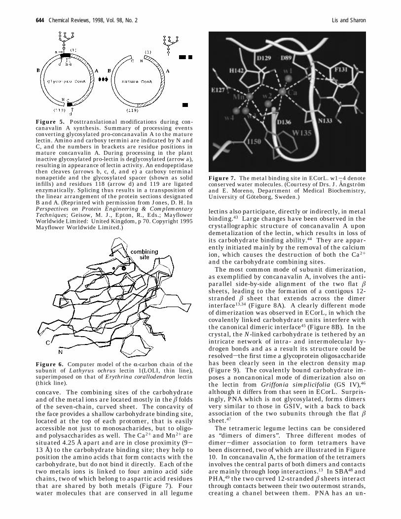
as exemplified by concanavalin A, involves the anti-parallel side-by-side alignment of the two flat âsheets, leading to the formation of a contigous 12-stranded â sheet that extends across the dimerinterface13,34 (Figure 8A). A clearly different modeof dimerization was observed in ECorL, in which thecovalently linked carbohydrate units interfere withthe canonical dimeric interface45 (Figure 8B). In thecrystal, the N-linked carbohydrate is tethered by anintricate network of intra- and intermolecular hy-drogen bonds and as a result its structure could beresolvedsthe first time a glycoprotein oligosaccharidehas been clearly seen in the electron density map(Figure 9). The covalently bound carbohydrate im-poses a noncanonical mode of dimerization also onthe lectin from Griffonia simplicifolia (GS IV),46although it differs from that seen in ECorL. Surpris-ingly, PNA which is not glycosylated, forms dimersvery similar to those in GSIV, with a back to backassociation of the two subunits through the flat âsheet.47The tetrameric legume lectins can be considered
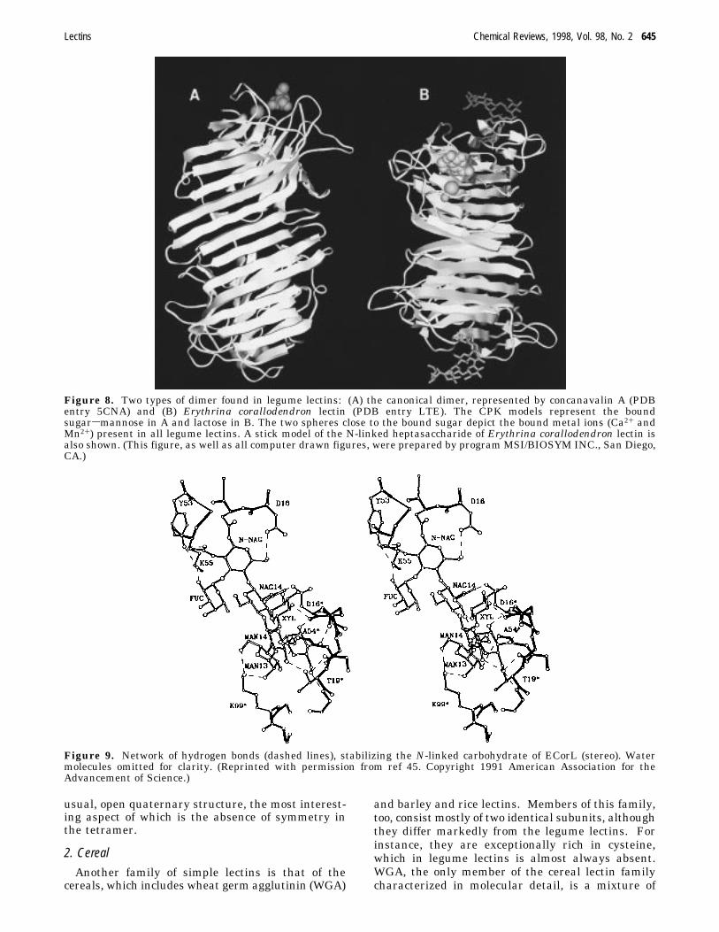
as “dimers of dimers”. Three different modes ofdimer-dimer association to form tetramers havebeen discerned, two of which are illustrated in Figure10. In concanavalin A, the formation of the tetramersinvolves the central parts of both dimers and contactsare mainly through loop interactions.13 In SBA48 andPHA,49 the two curved 12-stranded â sheets interactthrough contacts between their two outermost strands,creating a chanel between them. PNA has an un-
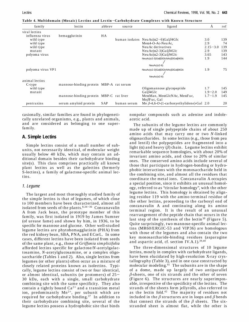
Figure 5. Posttranslational modifications during con-canavalin A synthesis. Summary of processing eventsconverting glycosylated pro-concanavalin A to the maturelectin. Amino and carboxy termini are indicated by N andC, and the numbers in brackets are residue positions inmature concanvalin A. During processing in the plantinactive glycosylated pro-lectin is deglycosylated (arrow a),resulting in appearance of lectin activity. An endopeptidasethen cleaves (arrows b, c, d, and e) a carboxy terminalnonapetide and the glycosylated spacer (shown as solidinfills) and residues 118 (arrow d) and 119 are ligatedenzymatically. Splicing thus results in a transposition ofthe linear arrangement of the protein sections designatedB and A. (Reprinted with permission from Jones, D. H. InPerspectives on Protein Engineering & ComplementaryTechniques; Geisow, M. J., Epton, R., Eds.; MayflowerWorldwide Limited: United Kingdom, p 70. Copyright 1995Mayflower Worldwide Limited.)
Figure 6. Computer model of the R-carbon chain of thesubunit of Lathyrus ochrus lectin 1(LOLI, thin line),superimposed on that of Erythrina corallodendron lectin(thick line).
Figure 7. The metal binding site in ECorL. w1-4 denoteconserved water molecules. (Courtesy of Drs. J. A° ngstromand E. Moreno, Department of Medical Biochemistry,University of Goteborg, Sweden.)
644 Chemical Reviews, 1998, Vol. 98, No. 2 Lis and Sharon
usual, open quaternary structure, the most interest-ing aspect of which is the absence of symmetry inthe tetramer.
2. Cereal
Another family of simple lectins is that of thecereals, which includes wheat germ agglutinin (WGA)
and barley and rice lectins. Members of this family,too, consist mostly of two identical subunits, althoughthey differ markedly from the legume lectins. Forinstance, they are exceptionally rich in cysteine,which in legume lectins is almost always absent.WGA, the only member of the cereal lectin familycharacterized in molecular detail, is a mixture of
Figure 8. Two types of dimer found in legume lectins: (A) the canonical dimer, represented by concanavalin A (PDBentry 5CNA) and (B) Erythrina corallodendron lectin (PDB entry LTE). The CPK models represent the boundsugarsmannose in A and lactose in B. The two spheres close to the bound sugar depict the bound metal ions (Ca2+ andMn2+) present in all legume lectins. A stick model of the N-linked heptasaccharide of Erythrina corallodendron lectin isalso shown. (This figure, as well as all computer drawn figures, were prepared by program MSI/BIOSYM INC., San Diego,CA.)
Figure 9. Network of hydrogen bonds (dashed lines), stabilizing the N-linked carbohydrate of ECorL (stereo). Watermolecules omitted for clarity. (Reprinted with permission from ref 45. Copyright 1991 American Association for theAdvancement of Science.)
Lectins Chemical Reviews, 1998, Vol. 98, No. 2 645
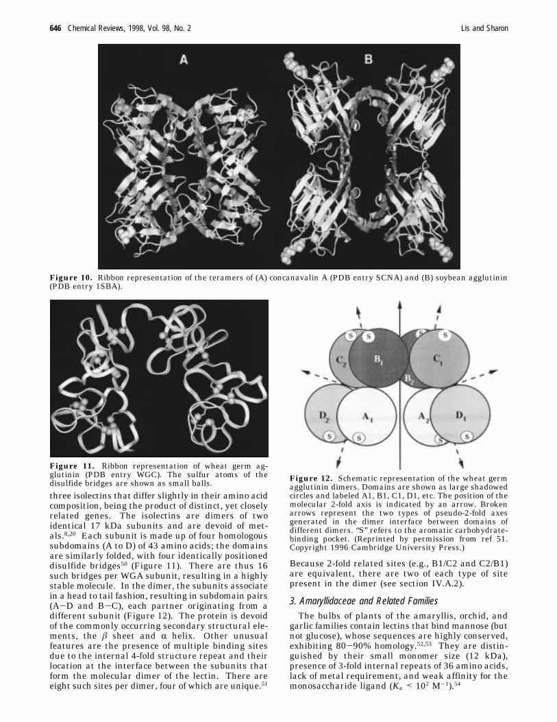
three isolectins that differ slightly in their amino acidcomposition, being the product of distinct, yet closelyrelated genes. The isolectins are dimers of twoidentical 17 kDa subunits and are devoid of met-als.8,20 Each subunit is made up of four homologoussubdomains (A to D) of 43 amino acids; the domainsare similarly folded, with four identically positioneddisulfide bridges50 (Figure 11). There are thus 16such bridges per WGA subunit, resulting in a highlystable molecule. In the dimer, the subunits associatein a head to tail fashion, resulting in subdomain pairs(A-D and B-C), each partner originating from adifferent subunit (Figure 12). The protein is devoidof the commonly occurring secondary structural ele-ments, the â sheet and R helix. Other unusualfeatures are the presence of multiple binding sitesdue to the internal 4-fold structure repeat and theirlocation at the interface between the subunits thatform the molecular dimer of the lectin. There areeight such sites per dimer, four of which are unique.51
Because 2-fold related sites (e.g., B1/C2 and C2/B1)are equivalent, there are two of each type of sitepresent in the dimer (see section IV.A.2).
3. Amaryllidaceae and Related FamiliesThe bulbs of plants of the amaryllis, orchid, and
garlic families contain lectins that bind mannose (butnot glucose), whose sequences are highly conserved,exhibiting 80-90% homology.52,53 They are distin-guished by their small monomer size (12 kDa),presence of 3-fold internal repeats of 36 amino acids,lack of metal requirement, and weak affinity for themonosaccharide ligand (Ka < 102 M-1).54
Figure 10. Ribbon representation of the teramers of (A) concanavalin A (PDB entry SCNA) and (B) soybean agglutinin(PDB entry 1SBA).
Figure 11. Ribbon representation of wheat germ ag-glutinin (PDB entry WGC). The sulfur atoms of thedisulfide bridges are shown as small balls. Figure 12. Schematic representation of the wheat germ
agglutinin dimers. Domains are shown as large shadowedcircles and labeled A1, B1, C1, D1, etc. The position of themolecular 2-fold axis is indicated by an arrow. Brokenarrows represent the two types of pseudo-2-fold axesgenerated in the dimer interface between domains ofdifferent dimers. “S” refers to the aromatic carbohydrate-binding pocket. (Reprinted by permission from ref 51.Copyright 1996 Cambridge University Press.)
646 Chemical Reviews, 1998, Vol. 98, No. 2 Lis and Sharon
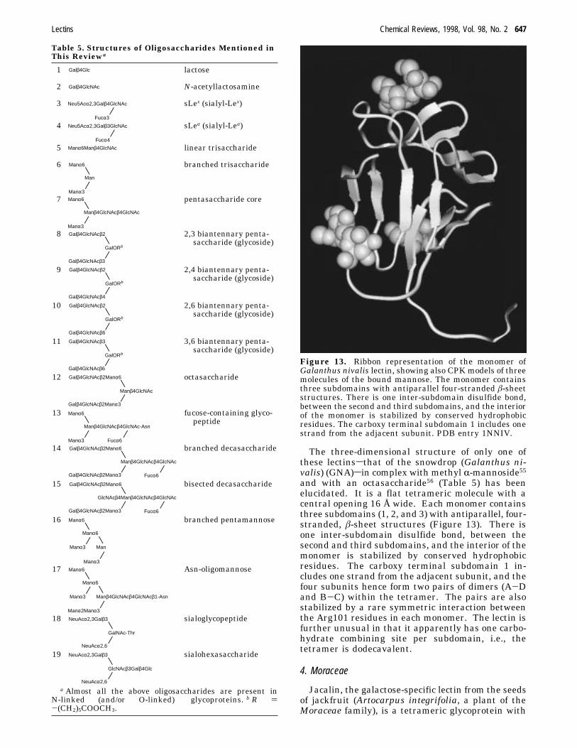
The three-dimensional structure of only one ofthese lectinssthat of the snowdrop (Galanthus ni-valis) (GNA)sin complex with methyl R-mannoside55and with an octasaccharide56 (Table 5) has beenelucidated. It is a flat tetrameric molecule with acentral opening 16 Å wide. Each monomer containsthree subdomains (1, 2, and 3) with antiparallel, four-stranded, â-sheet structures (Figure 13). There isone inter-subdomain disulfide bond, between thesecond and third subdomains, and the interior of themonomer is stabilized by conserved hydrophobicresidues. The carboxy terminal subdomain 1 in-cludes one strand from the adjacent subunit, and thefour subunits hence form two pairs of dimers (A-Dand B-C) within the tetramer. The pairs are alsostabilized by a rare symmetric interaction betweenthe Arg101 residues in each monomer. The lectin isfurther unusual in that it apparently has one carbo-hydrate combining site per subdomain, i.e., thetetramer is dodecavalent.
4. Moraceae
Jacalin, the galactose-specific lectin from the seedsof jackfruit (Artocarpus integrifolia, a plant of theMoraceae family), is a tetrameric glycoprotein with
Table 5. Structures of Oligosaccharides Mentioned inThis Reviewa
1 Galβ4Glc lactose
2 Galβ4GlcNAc N-acetyllactosamine
3 Neu5Acα2,3Galβ4GlcNAc
Fucα3
sLex (sialyl-Lex)
4 Neu5Acα2,3Galβ3GlcNAc
Fucα4
sLea (sialyl-Lea)
5 Manα6Manβ4GlcNAc linear trisaccharide
6 Manα6
Man
Manα3
branched trisaccharide
7 Manα6
Manβ4GlcNAcβ4GlcNAc
Manα3
pentasaccharide core
8 Galβ4GlcNAcβ2
GalORb
Galβ4GlcNAcβ3
2,3 biantennary penta-saccharide (glycoside)
9 Galβ4GlcNAcβ2
GalORb
Galβ4GlcNAcβ4
2,4 biantennary penta-saccharide (glycoside)
10 Galβ4GlcNAcβ2
GalORb
Galβ4GlcNAcβ6
2,6 biantennary penta-saccharide (glycoside)
11 Galβ4GlcNAcβ3
GalORb
Galβ4GlcNAcβ6
3,6 biantennary penta-saccharide (glycoside)
12 Galβ4GlcNAcβ2Manα6
Manβ4GlcNAc
Galβ4GlcNAcβ2Manα3
octasaccharide
13 Manα6
Manβ4GlcNAcβ4GlcNAc-Asn
Manα3 Fucα6
fucose-containing glyco-peptide
14 Galβ4GlcNAcβ2Manα6
Manβ4GlcNAcβ4GlcNAc
Fucα6Galβ4GlcNAcβ2Manα3
branched decasaccharide
15 Galβ4GlcNAcβ2Manα6
GlcNAcβ4Manβ4GlcNAcβ4GlcNAc
Fucα6Galβ4GlcNAcβ2Manα3
bisected decasaccharide
16 Manα6
Manα6
Manα3 Man
Manα3
branched pentamannose
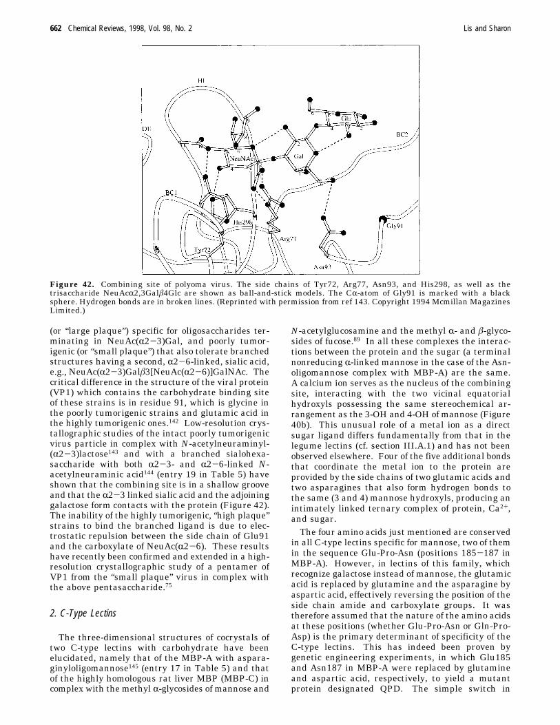
17 Manα6
Manα6
Manα3 Manβ4GlcNAcβ4GlcNAcβ1-Asn
Manα2Manα3
Asn-oligomannose
18 NeuAcα2,3Galβ3
GalNAc-Thr
NeuAcα2,6
sialoglycopeptide
19 NeuAcα2,3Galβ3
GlcNAcβ3Galβ4Glc
NeuAcα2,6
sialohexasaccharide
a Almost all the above oligosaccharides are present inN-linked (and/or O-linked) glycoproteins. b R )-(CH2)5COOCH3.
Figure 13. Ribbon representation of the monomer ofGalanthus nivalis lectin, showing also CPKmodels of threemolecules of the bound mannose. The monomer containsthree subdomains with antiparallel four-stranded â-sheetstructures. There is one inter-subdomain disulfide bond,between the second and third subdomains, and the interiorof the monomer is stabilized by conserved hydrophobicresidues. The carboxy terminal subdomain 1 includes onestrand from the adjacent subunit. PDB entry 1NNIV.
Lectins Chemical Reviews, 1998, Vol. 98, No. 2 647
a molecular weight of about 66 kDa. Each of itssubunits consists of a heavy chain (R) of 133 aminoacids and a light chain (â) of 20 residues.57 Theprimary structure of jacalin shows no significantsimilarity with any other lectin, except that fromMaclura pomifera, also a member of the Moraceaefamily. Crystallographic studies have shown that thesubunits of jacalin are made up of three four-strandedantiparallel â sheets, arranged like the faces of atriangular prism, with loops connecting strands inthe sheets58 (Figure 14). It is stabilized by hydro-phobic interactions in the core of the subunit and asmall number of hydrogen bonds involving main-chain, as well as side-chain, atoms. This recentlydiscovered arrangement, classified as the â-prismfold, has been found in a few proteins, but not in anyother lectin.
5. EuphorbiaceaeBeans of the castor tree (Ricinus communis) con-
tain two closely related lectins, ricin and Ricinuscommunis agglutinin, RCA;21 the former is one of thedeadliest poisons known: it is by weight about 10times as toxic as cobra venom and, according to someestimates, a single molecule is sufficient to kill a cell.Although classified by us as simple, ricin and RCArepresent borderline cases: their structure is morecomplex than that of the lectins discussed hitherto,but they do not fulfill the criteria that define theother two classes of lectin. Ricin is a heterodimericprotein with a MW of 60 kDa, made up of two S-Slinked chains, A and B. The latter contains twocarbohydrate binding sites specific for galactose,whereas the cytotoxic activity resides in the A chainwhich acts by enzymatically inactivating the RNA
involved in protein synthesis. The B chain is madeup of two globular domains, each of which comprisesa link domain and three homologous 40-residuesubdomains. Like the WGA subunit, the B chain isstabilized by several disulfide-linked cysteines. RCAis a dimer of two subunits, each of which is similarto ricin but it is not toxic. The three-dimensionalstructure of ricin has been determined by X-raycrystallography at 2.5-2.6 Å resolution59,60 (Figure15). The A chain is a globular protein with extensivesecondary structures, both â pleated sheet and Rhelix, and a reasonably prominent cleft, assumed tobe the active site responsible for the toxic action ofricin. The B chain folds into two topologically similardomains, each binding lactose in a shallow cleft.Preliminary crystallographic characterization of RCAhas shown that it forms an elongated molecule of 120Å × 60 Å × 40 Å with two A chains at the centerand a B chain at each end. The A chains arecovalently associated, with a disulfide bridge betweenCys157 of each of the chains. Additional contacts atresidues 114-115 stabilize the dimer interface.61
6. GalectinsThe galectins constitute a family of soluble, â-ga-
lactoside specific lectins that combine preferentiallywith lactose (entry 1 in Table 5) and N-acetyl-lactosamine62-64 (entry 2 in Table 5). They occurpredominantly in mammals, but have been found alsoin other vertebrates (e.g., frog,65) and in some inver-tebrates (for instance sponges66), but not in plants.Indeed, the first member of this family has beenisolated some 20 years ago from the electric organ ofelectric eel.67 Their structure is relatively simple andthey share a highly homologous domain (known as
Figure 14. Ribbon representation of the tetramer of jacalin, showing also the CPK model of the bound galactose. PDBentry 1JAC.
648 Chemical Reviews, 1998, Vol. 98, No. 2 Lis and Sharon
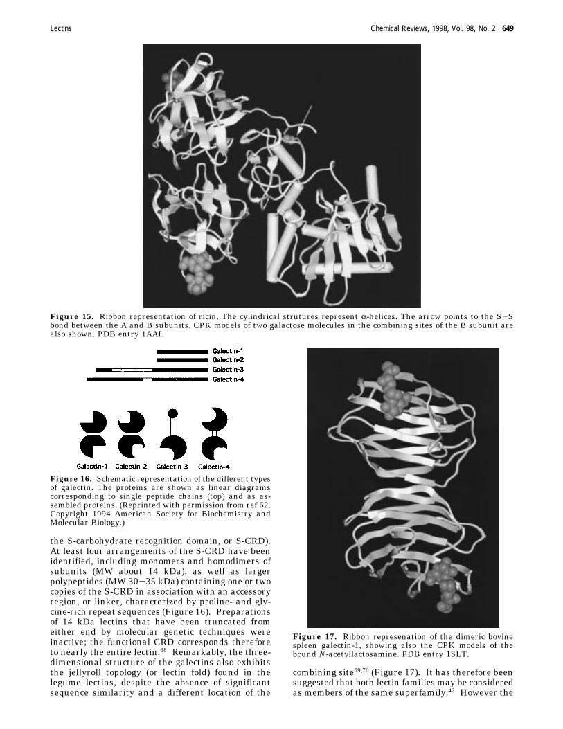
the S-carbohydrate recognition domain, or S-CRD).At least four arrangements of the S-CRD have beenidentified, including monomers and homodimers ofsubunits (MW about 14 kDa), as well as largerpolypeptides (MW 30-35 kDa) containing one or twocopies of the S-CRD in association with an accessoryregion, or linker, characterized by proline- and gly-cine-rich repeat sequences (Figure 16). Preparationsof 14 kDa lectins that have been truncated fromeither end by molecular genetic techniques wereinactive; the functional CRD corresponds thereforeto nearly the entire lectin.68 Remarkably, the three-dimensional structure of the galectins also exhibitsthe jellyroll topology (or lectin fold) found in thelegume lectins, despite the absence of significantsequence similarity and a different location of the
combining site69,70 (Figure 17). It has therefore beensuggested that both lectin families may be consideredas members of the same superfamily.42 However the
Figure 15. Ribbon representation of ricin. The cylindrical strutures represent R-helices. The arrow points to the S-Sbond between the A and B subunits. CPK models of two galactose molecules in the combining sites of the B subunit arealso shown. PDB entry 1AAI.
Figure 16. Schematic representation of the different typesof galectin. The proteins are shown as linear diagramscorresponding to single peptide chains (top) and as as-sembled proteins. (Reprinted with permission from ref 62.Copyright 1994 American Society for Biochemistry andMolecular Biology.)
Figure 17. Ribbon represenation of the dimeric bovinespleen galectin-1, showing also the CPK models of thebound N-acetyllactosamine. PDB entry 1SLT.
Lectins Chemical Reviews, 1998, Vol. 98, No. 2 649
question of the evolutionary origin, whether bydivergence from a common ancestor or by conver-gence to a similar, compact structural frameworkwith similar functional characteristics, can at presentnot be answered unambiguosly.16
7. Pentraxins
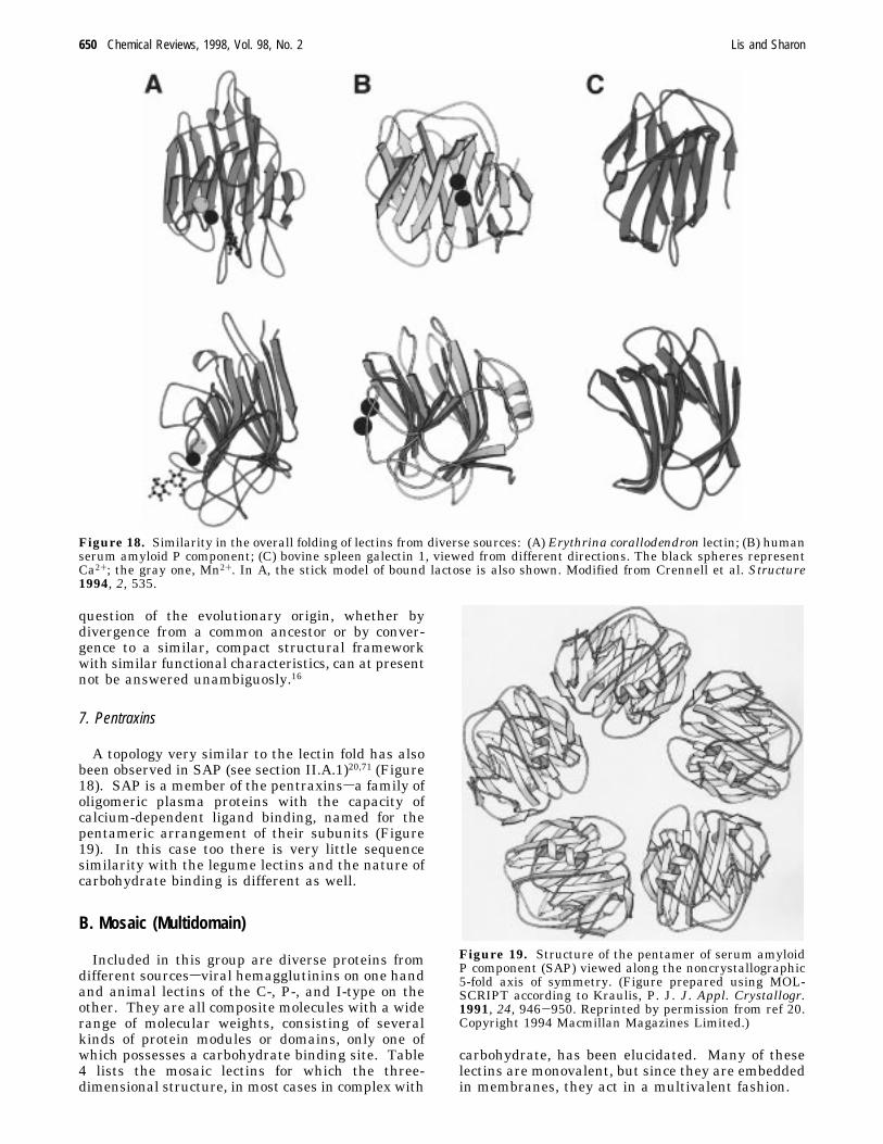
A topology very similar to the lectin fold has alsobeen observed in SAP (see section II.A.1)20,71 (Figure18). SAP is a member of the pentraxinssa family ofoligomeric plasma proteins with the capacity ofcalcium-dependent ligand binding, named for thepentameric arrangement of their subunits (Figure19). In this case too there is very little sequencesimilarity with the legume lectins and the nature ofcarbohydrate binding is different as well.
B. Mosaic (Multidomain)
Included in this group are diverse proteins fromdifferent sourcessviral hemagglutinins on one handand animal lectins of the C-, P-, and I-type on theother. They are all composite molecules with a widerange of molecular weights, consisting of severalkinds of protein modules or domains, only one ofwhich possesses a carbohydrate binding site. Table4 lists the mosaic lectins for which the three-dimensional structure, in most cases in complex with
carbohydrate, has been elucidated. Many of theselectins are monovalent, but since they are embeddedin membranes, they act in a multivalent fashion.
Figure 18. Similarity in the overall folding of lectins from diverse sources: (A) Erythrina corallodendron lectin; (B) humanserum amyloid P component; (C) bovine spleen galectin 1, viewed from different directions. The black spheres representCa2+; the gray one, Mn2+. In A, the stick model of bound lactose is also shown. Modified from Crennell et al. Structure1994, 2, 535.
Figure 19. Structure of the pentamer of serum amyloidP component (SAP) viewed along the noncrystallographic5-fold axis of symmetry. (Figure prepared using MOL-SCRIPT according to Kraulis, P. J. J. Appl. Crystallogr.1991, 24, 946-950. Reprinted by permission from ref 20.Copyright 1994 Macmillan Magazines Limited.)
650 Chemical Reviews, 1998, Vol. 98, No. 2 Lis and Sharon
1. Viral Hemagglutinins
a. Influenza Virus Hemagglutinin. The influenzavirus hemaggutinin, first described in the 1950s, isthe most thoroughly investigated of the multidomainlectins.72,73 Its subunit is composed of two polypep-tides, HA1 and HA2, with molecular masses of 36and 26 kDa, respectively, covalently linked by asingle disulfide bond. Each subunit consists of ahydrophilic, C-terminal domain located on the innerside of the membrane, a hydrophobic membranespanning region of 24-28 residues, an elongatedR-helical stem and a globular domain projecting 135Å from the membrane. The globular domain is madeup of HA1 only, and contains the carbohydratebinding site of the lectin (Figure 20). The subunitsassociate noncovalently to form trimers.b. Murine Polyoma Virus. This virus is a non-
enveloped, icosahedrically symmetrical particle, withcircular, double-stranded DNA genomes. The outershell (capsid) of the virion contains 360 copies of theviral protein VP1 (MW ∼42 kDa) arranged in pen-tamers.74 Each subunit of VP1 has two antiparallelâ sheets with a topology which resembles the lectinfold; some loops that connect the â strands areextensive and contain additional secondary structureelements. The most striking feature of the capsidstructure is the way the individual pentamers aretied together by the carboxy terminal arms of themonomers. The last 63 residues emerge from eachmonomer and “invade” a subunit of another pen-tamer, where they form a â strand that augments asheet in the target subunit.75
2. C-Type Lectins
This class of lectins has been so named becausethey require Ca2+ for activity.3,76,77 It includes over50 members, all characterized by an extracellularcarbohydrate recognition domain (C-CRD) consistingof 115-130 amino acids, of which 14 are invariantand 18 highly conserved. To the CRD is attached avariable number of domains of different kinds, whichform the bulk of the molecule. Lectins included inthis class have been grouped into three familiessendocytic lectins, collectins, and selectins, each shar-ing a common overall architecture defined by thenature of their domains (Figure 21). An exceptionis the mannose-specific macrophage surface lectinwhich has an unique structure but is included amongthe endocytic lectins because it shares with them acommon function.a. Endocytic Lectins. The prototype of this family
is the galactose/N-acetylgalactosamine specific lectinfrom rabbit hepatocytes (RHL), also known as hepaticasialoglycoprotein receptor (or hepatic binding pro-tein, HBP), the first mammalian lectin to be de-scribed in the early 1970s.78-80 Similar lectins havesubsequently been found on hepatocytes of othermammals. Examples of other endocytic lectins arethe avian hepatic lectin specific for N-acetylglu-cosamine, also present on hepatocytes,78 a galactose-specific lectin on peritoneal macrophages, and afucose-specific receptor (lectin) found on the Kupffercells of the liver.81 The endocytic lectins are type IItransmembrane proteins, consisting of a short amino
terminal cytoplasmic domain, a hydrophobic, mem-brane-spanning domain, and a neck region to whicha carboxy terminal CRD is linked. The mammalianhepatic asialoglycoprotein receptors are usually com-posed of two types of subunit: a smaller but moreabundant (MW 40-46 kDa) and a larger, less abun-dant (up to 60 kDa), that occur in varying proportionsin the different lectins. In the rat, the larger subunitexists in two forms with an identical amino acidsequence, one glycosylated and the other devoid ofcarbohydrates (Figure 22). The subunits possess
Figure 20. Ribbon presentation of the influenza virusmonomer. The broad ribbon represents subunit HA1; thenarrow one, subunit HA2. The arrow indicates the locationof the carbohydrate binding site. PDB entry HGF.
Lectins Chemical Reviews, 1998, Vol. 98, No. 2 651
similar primary structures, except for their neckregions, where considerable differences occur. Thetwo types of subunit are organized in a stericallyspecific and rigid orientation and both have to bepresent on the cell surface in order to form afunctional receptor. These mammalian lectins havea strong tendency to associate and in purified formappear to exist as hexamers, although the exactstoichiometry of the subunits in the heteropolymeris unclear. The galactose binding sites in such acomplex are tightly packed and arranged to bestaccommodate a triantennary oligosaccharide withnonreducing galactose residues that are 1.5-3.1 nmapart, which binds to the receptor with an affinity∼6 orders of magnitude higher than monovalentligands.The mannose-specific macrophage surface lectin
(MW 175 kDa) differs from the other endocytic lectinsin that it is a type I transmembrane protein (i.e., itscarboxy terminal is in the cytoplasm and the aminoterminal is outside the cell) (Figure 21). Moreover,the extracellular part of the molecule consists of threedomains: a unique cysteine-rich segment, a regionsimilar to the type II repeats of fibronectin and a
domain, closest to the membrane, containing eightCRDs.3,81-83 For a while this was the only knowncase of a C-type protein with more than one CRDwithin a single polypeptide chain. More recently,other proteins (e.g., the phospholipase A2 receptorfrom muscle) have been shown to possess the samearchitecture.84
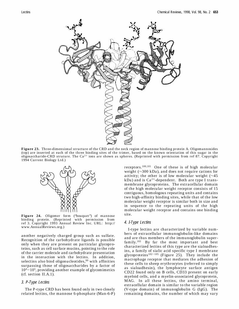
b. Collectins. The collectins are soluble proteins,composed of an amino terminal cysteine-rich domain,followed by a number of collagen-like repeats, anR-helical neck region and a carboxy terminal CRD.85,86Of the six known proteins of this group (MBP A andC, pulmonary surfactant apoproteins A and D, col-lectin CL-43 from bovine serum and bovine conglu-tinin), the MBPs have been most thoroughly inves-tigated. The structural unit of the MBP is a trimerof 32 kDa subunits, formed by a triple helix of thecollagenous portion of the subunit and is stabilizedby the association of the R helices of the neck into aparallel triple-stranded coiled coil87,88 (Figure 23).Two homologous, yet distinct forms of MBPs havebeen described, the serum type (MBP-A) and the livertype (MBP-C). The latter is characterized by aninsertion of nine amino acids in the amino terminalcysteine-rich region. MBP-A circulates in the seraof higher animals as a hexamer of trimeric units ofapparent MW of ∼650 kDa, a partial model of whichis shown in Figure 24. MBP-C is smaller andprobably consists of two associated trimers.89 Thehigh-resolution three-dimensional structure of theCRD of MBP-A, the first of a C-type lectin to beelucidated, revealed that over 50% of the CRD isformed by loops and extended structures; the remain-der comprises two short R-helices and five â-strands.90
Despite the great similarity in the architecture ofthe binding sites of the two MBP’s, and their com-parable affinity for monosaccharides, MBP-C bindsbranched mannooligosaccharides more strongly thandoes MBP-A. Binding studies, using mono- andoligosaccharides and synthetic cluster glycosides, ledto the conclusion that MBP-C has two binding sitesper subunit, one only for mannose, the other for bothmannose and N-acetylglucosamine; the former ap-pears to be extended, probably the size of a tri-mannoside. In contrast, MBP-A has only one site ofthe latter type.91,92
c. Selectins. This group consists of threememberssE-selectin (MW 115 kDa), P-selectin (140kDa), and L-selectin (90-110 kDa)sall highly asym-metric membrane-bound proteins.3,93-97 They are sonamed because they mediate selective contact be-tween cells. Each contains, in addition to the CRDlocated at the amino terminal part of the molecule,an adjoining epidermal growth factor (EGF)-likedomain, several short repeating units related tocomplement-binding protein, a membrane-spanningregion, and a cytoplasmic, carboxy terminal domain(Figure 21). The crystal structure of the CRDtogether with the EGF-like domain of E-selectinshows a very similar fold to that of the MBP-A.98 Theselectins interact specifically with sLex and its posi-tional isomer sLea (3 and 4, respectively in Table 5),with both fucose and N-acetylneuraminic acid re-quired for binding; sialic acid can be replaced by
Figure 21. Organization of membrane-bound C-typeanimal lectins: (from left to right) the mannose macro-phage receptor, a type I membrane protein; two examplesof type II endocytic receptors (the chicken hepatic lectinand the Kupffer cell receptor); and L-selectin. (Reprintedwith permission from ref 3. Copyright 1993 Annual Re-views Inc. URL: http://www.AnnualReviews.org.)
Figure 22. Models of the subunit organization of the ratasialoglycoprotein receptor: (A) heterodimer incorporatingtwo RHL1 subunits and one RHL2 or RHL3 subunit and(B) heterodimer of one RHL1 and one RHL2/3. Homotrimerof RHL1 obtained by solubilization. (Reprinted from Rice,K. G.; Lee, Y. C. Adv. Enzymol. 1993, 66, 41. Copyright1993 John Wiley & Sons Inc.)
652 Chemical Reviews, 1998, Vol. 98, No. 2 Lis and Sharon
another negatively charged group such as sulfate.Recognition of the carbohydrate ligands is possibleonly when they are present on particular glycopro-teins, such as cell surface mucins, pointing to the roleof the carrier molecule and carbohydrate presentationin the interaction with the lectins. In addition,selectins also bind oligonucleotides,99 with affinitiessurpassing those of oligosaccharides by a factor of104-105, providing another example of glycomimetics(cf. section II.A.1).
3. P-Type Lectins
The P-type CRD has been found only in two closelyrelated lectins, the mannose 6-phosphate (Man-6-P)
receptors.100,101 One of these is of high molecularweight (∼300 kDa), and does not require cations foractivity; the other is of low molecular weight (∼45kDa) and is Ca2+-dependent. Both are type I trans-membrane glycoproteins. The extracellular domainof the high molecular weight receptor consists of 15contiguous, homologous repeating units and containstwo high-affinity binding sites, while that of the lowmolecular weight receptor is similar both in size andin sequence to the repeating units of the highmolecular weight receptor and contains one bindingsite.
4. I-Type Lectins
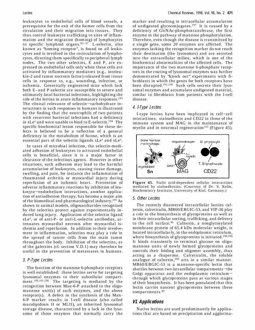
I-type lectins are characterized by variable num-bers of extracellular immunoglobulin-like domainsand are thus members of the immunoglobulin super-family.102 By far the most important and bestcharacterized lectins of this type are the sialoadhes-ins, a family of sialic acid specific type I membraneglycoproteins102-105 (Figure 25). They include themacrophage receptor that mediates the adhesion ofthese cells to sheep erythrocytes (referred to simplyas sialoadhesin), the lymphocyte surface antigenCD22 found only on B cells, CD33 present on earlymyeloid cells, and a myelin-associated glycoprotein,MAG. In all these lectins, the amino terminal,extracellular domain is similar to the variable region(V-type domain) of immunoglobulin G (IgG). Theremaining domains, the number of which may vary
Figure 23. Three-dimensional structure of the CRD and the neck region of mannose binding protein A. Oligomannosides(top) are inserted at each of the three binding sites of the trimer, based on the known orientation of this sugar in theoligosaccharide-CRD struture. The Ca2+ ions are shown as spheres. (Reprinted with permission from ref 87. Copyright1994 Current Biology Ltd.)
Figure 24. Oligomer form (“bouquet”) of mannosebinding protein. (Reprinted with permission fromref 3. Copyright 1993 Annual Review Inc. URL: http://www.AnnualReviews.org.)
Lectins Chemical Reviews, 1998, Vol. 98, No. 2 653
from 1 to 16, are similar to the C2 segment of theconstant region of IgG.With the aid of a series of mutants of I-type lectins,
from which various extracellular domains have beendeleted, it was shown that in sialoadhesin the aminoterminal, V-type domain is both necessary and suf-ficient for sialic acid dependent binding. In CD22,on the other hand, the adjacent C2-like domain isalso required, apparently for correct folding of theprotein.106 A conserved arginine was implied by site-directed mutagenesis studies to play a key role in theaffinity of the I-type lectins to glycoconjugates con-taining sialic acid.107,108 CD22 recognizes specificallyNeuAc(R2-6)Gal(â1-4)GlcNAc, known to occur invarying numbers on the N-linked oligosaccharides ofmany surface glycoproteins; the R2-6 linkage is anabsolute requirement. In contrast, all other knownI-type lectins bind structures containing N-acetyl-neuramnic acid that is R2-3 linked.
C. Macromolecular AssembliesLectins of this type are common in bacteria, usually
in the form of fimbriae (or pili). These are filamen-tous, heteropolymeric organelles present on the sur-face of the bacteria, 3-7 nm in diameter and 100 to200 nm in length, consisting of helically arrangedsubunits (pilins) of several different types, assembledin a well-defined order109-111 (Figure 26). The bulkof the fimbrial filament (shaft) is made up of poly-
mers of the major subunit, which thus plays astructural role. Only one of the subunits, usually aminor component of the fimbriae, possesses a carbo-hydrate combining site and is responsible for thebinding activity and sugar specificity of the fimbriae,e.g., for mannose (in type 1 fimbriae) or galabiose,Gal(R1-4)Gal (in P fimbriae). In type 1 fimbriae,which are made up of hundreds of subunits of fourdifferent kinds, this subunit (MW 29-31 kDa) ispresent in small numbers at intervals along thefimbrial filament and at the distal tip. However onlythe latter subunit appears to be able to mediatemannose-sensitive adhesive interactions, whereas thesubunits at the other positions are inaccessible to theligand. In other types of fimbriae (e.g., type P) thecarbohydrate-binding subunit (MW 36 kDa) is exclu-sively located at the tip. The combining sites of type1 fimbriae of E. coli and K. pneumoniae correspondto the size of a trisaccharide and are probably in theform of a depression or pocket on the surface of thelectin. Several of the carbohydrate binding subunitshave been sequenced, but in no case has the three-dimensional structure of any of them been solved.
IV. Combining SitesThe combining sites of lectins are in the form of
shallow depressions on the surface of the protein.Typically, only one or two edges or faces of thecarbohydrate ligand are bound to the protein. Thisis in contrast to carbohydrate-binding bacterial peri-plasmic receptors, specific for, e.g., glucose or galac-tose, in which the ligand is buried in the interior ofthe protein.112 In lectins, the combining sites appearto be preformed, since few conformational changesoccur upon ligand binding. In general, the siteswithin a lectin family are similar, but quite differentin different families, even if the specificity is thesame, emphasizing the fact that nature finds differ-ent solutions to the problem of the design of combin-ing sites for structurally similar ligands.6Lectins combine with carbohydrates by a network
of hydrogen bonds and hydrophobic interactions;coordination with metal ions may also play arole.13-15,113 (Table 6). The hydrogen bonds areformed between carbohydrate hydroxyl groups andNH groups, hydroxyls, and oxygen atoms of theprotein. When each of two adjacent hydroxyls of amonosaccharide interacts with a different atom of thesame amino acid (e.g., the two oxygens of the car-boxylate of glutamic or aspartic acid), they formbidentate hydrogen bonds.114 Such bonds are quitecommon in protein-carbohydrate complexes. A dif-ferent kind of hydrogen-bond characteristic for suchcomplexes is the cooperative bond, in which thehydroxyl group acts simultaneously as donor andacceptor. van der Waals forces, although ratherweak (often only a fraction of 1 kcal mol-1 for eachpair of atoms), are frequently numerous and togethermay make a significant contribution to binding.Even though carbohydrates are highly polar mol-
ecules, the steric disposition of hydroxyl groupscreates hydrophobic patches on sugar surfaces thatcan form contacts with hydrophobic regions in theprotein molecules. One common type of interaction
Figure 25. I-type lectins. (Reprinted from Kelm, S.; et al.Curr. Biol. 1994, 4, 965-972. Copyright 1994 CurrentBiology Ltd.)
Figure 26. Schematic representation of different types offimbriae of E. coli. The disks stand for the fimbrialsubunits; the black shapes symbolize the carbohydratebinding sites. In the type 1 fimbriae, the symbols withwhite dots denote carbohydrate binding sites not availableto the ligand. (Modified from ref 111.)
654 Chemical Reviews, 1998, Vol. 98, No. 2 Lis and Sharon
is the stacking of a monosaccharide on the side chainsof the aromatic amino acids such as phenylalanine,tyrosine, or tryptophan (Figure 27). In addition, themethyl group of the acetamide moiety of N-acetyl-amino sugars often interacts with aromatic residuesin lectins (e.g., WGA and influenza virus hemagglu-tinin specific for the above sugars). Since mostsaccharides are uncharged, ionic (charge-charge)interactions do not commonly participate in theformation of their complexes with proteins. Anexception is the heparin-antithrombin III complex,in which four basic amino acids of the protein forman elongated, positively charged binding site comple-mentary to a specific oligosaccharide sequence of thepolysaccharide with a unique sulfate substitutionpattern.115
Contacts between the protein and its ligands aresometimes mediated by water bridges.43,116 Wateracts as a molecular “mortar”, its small size and abilityto act as both a hydrogen donor and hydrogenacceptor conferring ideal properties for this function.Tightly bound bridging water can be thought of asstructural water, essentially an extension of theprotein surface. Comparison of a series of sugarsbound to a given lectin, or a series of related lectinsbound to a given sugar, sometimes reveal commonwater molecules, suggesting that they are importantelements in ligand recognition.
A. Simple Lectins
1. Legume
Legume lectins, irrespective of their specificity,bind ligands through the side chains of a constellationof three invariant combining site residues, an aspar-tic acid, an aparagine, and an aromatic aminoacid117,118 or leucine.41 Replacement, by site-directedmutagenesis, of the aspartic acid or asparagine byanother amino acid (e.g., alanine) in several of theselectins (e.g., ECorL,118 pea lectin,119 and GSII120)resulted in loss of sugar-binding ability. The key roleof these amino acids in ligand binding has beensimilarly demonstrated for the homologous, mannose-specific animal lectin MR60/ERGIC 53 (cf. sectionIII.A.1).121 The aspartic acid and asparagine alsoparticipate in coordinating the calcium ion presentin all members of this family, which explains themetal ion requirement for carbohydrate binding.Another characteristic of the combining site of thelegume lectins is the presence of a rare cis-peptidebond between the critical asparagine just mentionedand the preceding amino acid, which is almost alwaysalanine. This bond is necessary for the properorientation of the asparagine.The fact that the key amino acids involved in the
binding of the carbohydrate are highly conserved inall legume lectins and have an identical spatialdisposition raises the puzzling question of how dis-crimination between very similar monosaccharides,
Table 6. Amino Acids and Metal Ions in the Monosaccharide Combining Sites of Plant and Animal Lectinsa
metal
types H-bonding amino acidsb hydrophobic residues ion role
a Additional side chain interactions occur when the lectins bind oligosaccharides b Only bonds with amino acid side chains arementioned c This bond is to the N-terminal amino group
Figure 27. Stereodiagram showing the relative placement of galactose and binding site residues in different lectins:Trp65 (Galectin-1), Phe131 (ECorL), Tyr248 (ricin site 2) and Trp88 (LT, enterotoxin from E. coli). (Reprinted withpermission from ref 13. Copyright 1995 Annual Reviews Inc. URL: http://www.AnnualReviews.org.)
Lectins Chemical Reviews, 1998, Vol. 98, No. 2 655
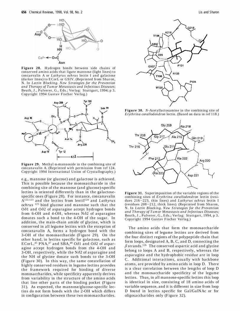
e.g., mannose (or glucose) and galactose is achieved.This is possible because the monosaccharide in thecombining site of the mannose (and glucose)-specificlectins is oriented differently than in the galactose-specific ones (Figure 28). For instance, concanavalinA122,123 and the lectins from lentil124 and Lathyrusochrus 125 bind glucose and mannose such that theOδ1 and Oδ2 of asparagine accept hydrogen bondsfrom 6-OH and 4-OH, whereas Nδ2 of asparaginedonates such a bond to the 4-OH of the sugar. Inaddition, the main-chain amide of glycine, which isconserved in all legume lectins with the exception ofconcanavalin A, forms a hydrogen bond with the3-OH of the monosaccharide (Figure 29). On theother hand, in lectins specific for galactose, such asECorL,45 PNA,47 and SBA,48 Oδ1 and Oδ2 of aspar-agine accept hydrogen bonds from the 4-OH and3-OH, respectively, while the Nδ2 of asparagine andthe NH of glycine donate such bonds to the 3-OH(Figure 30). In this way, the same constellation ofhighly conserved residues in legume lectins providesthe framework required for binding of diversemonosaccharides, while specificity apparently derivesfrom variability in the structure of the amino acidsthat line other parts of the binding pocket (Figure31). As expected, the mannose/glucose-specific lec-tins do not form bonds with the 2-OH which differsin configuration between these two monosaccharides.
The amino acids that form the monosaccharidecombining sites of legume lectins are derived fromthe four distinct regions of the polypeptide chain thatform loops, designated A, B, C, and D, connecting theâ strands.126 The conserved aspartic acid and glycinebelong to loops A and B, respectively, whereas theasparagine and the hydrophobic residue are in loopC. Additional interactions, usually with backboneatoms, are provided by amino acids in loop D. Thereis a clear correlation between the lengths of loop Dand the monosaccharide specificity of the legumelectins. Thus, in all mannose-specific lectins this loopis identical in size, consisting of 18 amino acids ofvariable sequence, and it is different in size from loopD found in lectins specific for Gal/GalNAc or foroligosaccharides only (Figure 32).
Figure 28. Hydrogen bonds between side chains ofconserved amino acids that ligate mannose (light lines) toconcanvalin A or Lathyrus ochrus lectin I and galactose(darker lines) to ECorL or GSIV. (Reprinted from Sharon,N. In Lectin Blocking. New Strategies for the Preventionand Therapy of Tumor Metastasis and Infectious Diseases;Beuth, J., Pulverer, G., Eds.; Verlag: Stuttgart, 1994; p 3.Copyright 1994 Gustav Fischer Verlag.)
Figure 29. Methyl R-mannoside in the combining site ofconcanavalin A. (Reprinted with permission from ref 124.Copyright 1994 International Union of Crystallography.)
Figure 30. N-Acetyllactosamine in the combining site ofErythrina corallodendron lectin. (Based on data in ref 118.)
Figure 31. Superimposition of the variable regions of thecombining sites of Erythrina corallodendron lectin (resi-dues 216-223, thin lines) and Lathyrus ochrus lectin I(residues 208-212, thick lines). (Reprinted from Sharon,N. In Lectin Blocking. New Strategies for the Preventionand Therapy of Tumor Metastasis and Infectious Diseases;Beuth, J., Pulverer, G., Eds.; Verlag: Stuttgart, 1994, p 3.Copyright 1994 Gustav Fischer Verlag.)
656 Chemical Reviews, 1998, Vol. 98, No. 2 Lis and Sharon
When lectins bind disaccharides, the nonreducingresidue occupies the monosaccharide combining site,i.e., where the mannose of methyl R-mannoside binds,with additional contacts to the protein provided bythe reducing residue. This is illustrated by ECorLin complex with N-acetyllactosamine (Figure 30)118or PNA in complex with Gal(â1-3)GalNAc (Figure33).127,128
In the crystal of concanavalin A with the branchedtrisaccharide Man(R1-6)[Man(R1-3)]Man,129 the R1-6-linked, nonreducing mannose of the trisaccharideoccupies the monosaccharide combining site of thelectin, and forms essentially the same contacts(Figure 34). There are bonds also to the reducingmannose. The demonstration that the trisaccharidecombines with an extended site on concanvalin A isin agreement with the results of titration micro-calorimetry.130,131 A single bridging water moleculeis seen in the trisaccharide-concanavalin A com-plex.129 In contrast, 20 water molecules are involvedin the binding of the linear trisaccharide Man(R1-3)Man(â1-4)GlcNAc to Lathyrus ochrus lectin I,(LOL I), which is also mannose-specific.132 In thiscomplex, the R1-3-linked mannose at the nonreduc-ing end of the trisaccharide occupies the monosac-charide combining site, but does not form the samecontacts as does methyl R-mannoside in the samesite125 (see below). The only direct interactionsbetween the trisaccharide and the protein are withthis mannose, whereas those of the two remainingsugars are all mediated by long-chain water bridges.An example of such a bridge is the nine watermolecules connecting the atoms of Manâ1-4 and ofN-acetylglucosamine to LOL I over a distance of 13Å.The dibranched N-acetyllactosamine-type octasac-
charide (entry 12 in Table 5) binds to LOL I with theR1-3 linked mannose occupying the monosaccharidebinding site.133 The complex is stabilized by a largenumber of hydrogen bonds, several via water, as wellas by numerous van der Waals contacts. The abovemannose and the GlcNAcâ1-2 of the R1-6-linkedbranch interact on each side of Phe123 and grip thearomatic ring as a clamp. A neighboring tyrosine is
Figure 32. Correlation between the size of binding loops and monosaccharide specificity of legume lectins. The numberof gaps present in the four loops is shown along with the specificity of the lectins on the right. The residues that are highlyconserved have been indicated with asterisks (*) and the ligand binding residues are shown in bold. (Reprinted withpermission from ref 126. Copyright 1997 Academic Press.)
Figure 33. Gal(â1-3)GalNAc in the combining site ofpeanut agglutinin. The terminal galactose of the disaccha-ride forms, in addition to the commonly occurring bondswith the side chains of asparagine (Asn127) and asparticacid (Asp83) and the main chain amide of glycine (Gly104),unique ones, namely between 6-OH and the side chain ofAsp80 and between the ring oxygen and Ser211. The 4-OHof theN-acetylgalactosamine is hydrogen-bonded to Ser211and Gly213. (Reprinted with permission from ref 128.Copyright 1997 Current Science.)
Lectins Chemical Reviews, 1998, Vol. 98, No. 2 657
stacked against the terminal galactose of the R1-6branch. As a result, the Gal(â1-4)GlcNAc of thisbranch fits in a partly hydrophobic cleft and displaysnumerous interactions with the lectin. The multi-plicity of contacts that the octasaccharide forms withthe protein explains why it exhibits an affinity to thelectin a 1000-fold higher than the monosaccharide.Although mannose specific legume lectins do not
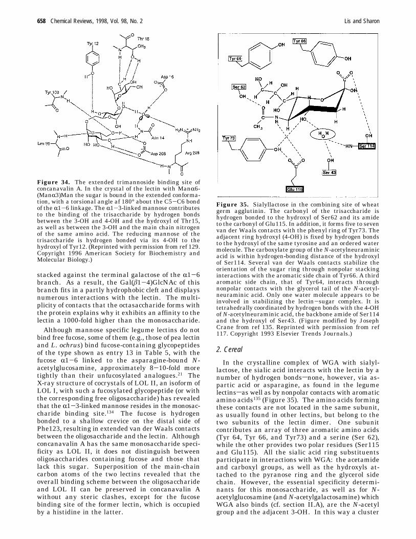
bind free fucose, some of them (e.g., those of pea lectinand L. ochrus) bind fucose-containing glycopeptidesof the type shown as entry 13 in Table 5, with thefucose R1-6 linked to the asparagine-bound N-acetylglucosamine, approximately 8-10-fold moretightly than their unfucosylated analogues.21 TheX-ray structure of cocrystals of LOL II, an isoform ofLOL I, with such a fucosylated glycopeptide (or withthe corresponding free oligosaccharide) has revealedthat the R1-3-linked mannose resides in the monosac-charide binding site.134 The fucose is hydrogenbonded to a shallow crevice on the distal side ofPhe123, resulting in extended van der Waals contactsbetween the oligosaccharide and the lectin. Althoughconcanavalin A has the same monosaccharide speci-ficity as LOL II, it does not distinguish betweenoligosaccharides containing fucose and those thatlack this sugar. Superposition of the main-chaincarbon atoms of the two lectins revealed that theoverall binding scheme between the oligosaccharideand LOL II can be preserved in concanavalin Awithout any steric clashes, except for the fucosebinding site of the former lectin, which is occupiedby a histidine in the latter.
2. Cereal
In the crystalline complex of WGA with sialyl-lactose, the sialic acid interacts with the lectin by anumber of hydrogen bondssnone, however, via as-partic acid or asparagine, as found in the legumelectinssas well as by nonpolar contacts with aromaticamino acids135 (Figure 35). The amino acids formingthese contacts are not located in the same subunit,as usually found in other lectins, but belong to thetwo subunits of the lectin dimer. One subunitcontributes an array of three aromatic amino acids(Tyr 64, Tyr 66, and Tyr73) and a serine (Ser 62),while the other provides two polar residues (Ser115and Glu115). All the sialic acid ring substituentsparticipate in interactions with WGA: the acetamideand carboxyl groups, as well as the hydroxyls at-tached to the pyranose ring and the glycerol sidechain. However, the essential specificity determi-nants for this monosaccharide, as well as for N-acetylglucosamine (andN-acetylgalactosamine) whichWGA also binds (cf. section II.A), are the N-acetylgroup and the adjacent 3-OH. In this way a cluster
Figure 34. The extended trimannoside binding site ofconcanavalin A. In the crystal of the lectin with ManR6-(ManR3)Man the sugar is bound in the extended conforma-tion, with a torsional angle af 180° about the C5-C6 bondof the R1-6 linkage. The R1-3-linked mannose contributesto the binding of the trisaccharide by hydrogen bondsbetween the 3-OH and 4-OH and the hydroxyl of Thr15,as well as between the 3-OH and the main chain nitrogenof the same amino acid. The reducing mannose of thetrisaccharide is hydrogen bonded via its 4-OH to thehydroxyl of Tyr12. (Reprinted with permission from ref 129.Copyright 1996 American Society for Biochemistry andMolecular Biology.)
Figure 35. Sialyllactose in the combining site of wheatgerm agglutinin. The carbonyl of the trisaccharide ishydrogen bonded to the hydroxyl of Ser62 and its amideto the carbonyl of Glu115. In addition, it forms five to sevenvan der Waals contacts with the phenyl ring of Tyr73. Theadjacent ring hydroxyl (4-OH) is fixed by hydrogen bondsto the hydroxyl of the same tyrosine and an ordered watermolecule. The carboxylate group of theN-acetylneuraminicacid is within hydrogen-bonding distance of the hydroxylof Ser114. Several van der Waals contacts stabilize theorientation of the sugar ring through nonpolar stackinginteractions with the aromatic side chain of Tyr66. A thirdaromatic side chain, that of Tyr64, interacts throughnonpolar contacts with the glycerol tail of the N-acetyl-neuraminic acid. Only one water molecule appears to beinvolved in stabilizing the lectin-sugar complex. It istetrahedrally coordinated by hydrogen bonds with the 4-OHof N-acetylneuraminic acid, the backbone amide of Ser114and the hydroxyl of Ser43. (Figure modified by JosephCrane from ref 135. Reprinted with permission from ref117. Copyright 1993 Elsevier Trends Journals.)
658 Chemical Reviews, 1998, Vol. 98, No. 2 Lis and Sharon
of three spatially close hydrogen bonds with theligand and a hydrophobic contact (acetamide-CH3with the aromatic ring of Tyr73) is formed in the leastexposed part of the combining site, where the con-formation of the protein is most stable.In the cocrystal of WGA with a branched sialogly-
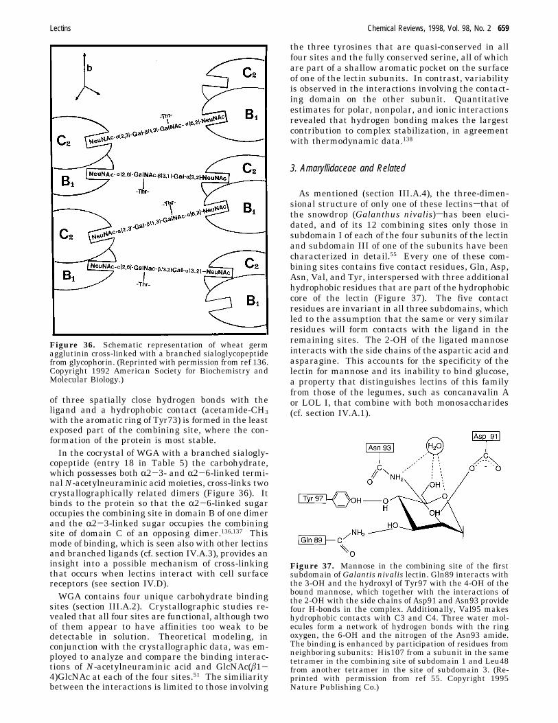
copeptide (entry 18 in Table 5) the carbohydrate,which possesses both R2-3- and R2-6-linked termi-nalN-acetylneuraminic acid moieties, cross-links twocrystallographically related dimers (Figure 36). Itbinds to the protein so that the R2-6-linked sugaroccupies the combining site in domain B of one dimerand the R2-3-linked sugar occupies the combiningsite of domain C of an opposing dimer.136,137 Thismode of binding, which is seen also with other lectinsand branched ligands (cf. section IV.A.3), provides aninsight into a possible mechanism of cross-linkingthat occurs when lectins interact with cell surfacereceptors (see section IV.D).WGA contains four unique carbohydrate binding
sites (section III.A.2). Crystallographic studies re-vealed that all four sites are functional, although twoof them appear to have affinities too weak to bedetectable in solution. Theoretical modeling, inconjunction with the crystallographic data, was em-ployed to analyze and compare the binding interac-tions of N-acetylneuraminic acid and GlcNAc(â1-4)GlcNAc at each of the four sites.51 The similiaritybetween the interactions is limited to those involving
the three tyrosines that are quasi-conserved in allfour sites and the fully conserved serine, all of whichare part of a shallow aromatic pocket on the surfaceof one of the lectin subunits. In contrast, variabilityis observed in the interactions involving the contact-ing domain on the other subunit. Quantitativeestimates for polar, nonpolar, and ionic interactionsrevealed that hydrogen bonding makes the largestcontribution to complex stabilization, in agreementwith thermodynamic data.138
3. Amaryllidaceae and Related
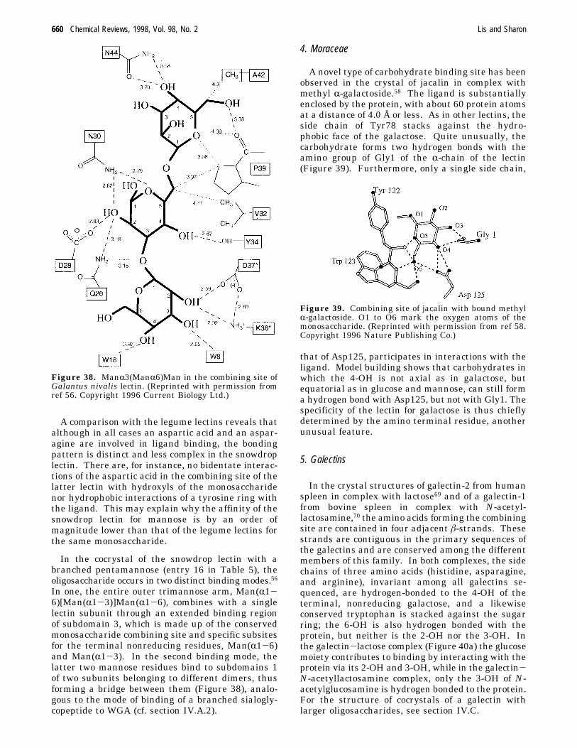
As mentioned (section III.A.4), the three-dimen-sional structure of only one of these lectinssthat ofthe snowdrop (Galanthus nivalis)shas been eluci-dated, and of its 12 combining sites only those insubdomain I of each of the four subunits of the lectinand subdomain III of one of the subunits have beencharacterized in detail.55 Every one of these com-bining sites contains five contact residues, Gln, Asp,Asn, Val, and Tyr, interspersed with three additionalhydrophobic residues that are part of the hydrophobiccore of the lectin (Figure 37). The five contactresidues are invariant in all three subdomains, whichled to the assumption that the same or very similarresidues will form contacts with the ligand in theremaining sites. The 2-OH of the ligated mannoseinteracts with the side chains of the aspartic acid andasparagine. This accounts for the specificity of thelectin for mannose and its inability to bind glucose,a property that distinguishes lectins of this familyfrom those of the legumes, such as concanavalin Aor LOL I, that combine with both monosaccharides(cf. section IV.A.1).
Figure 36. Schematic representation of wheat germagglutinin cross-linked with a branched sialoglycopeptidefrom glycophorin. (Reprinted with permission from ref 136.Copyright 1992 American Society for Biochemistry andMolecular Biology.)
Figure 37. Mannose in the combining site of the firstsubdomain of Galantis nivalis lectin. Gln89 interacts withthe 3-OH and the hydroxyl of Tyr97 with the 4-OH of thebound mannose, which together with the interactions ofthe 2-OH with the side chains of Asp91 and Asn93 providefour H-bonds in the complex. Additionally, Val95 makeshydrophobic contacts with C3 and C4. Three water mol-ecules form a network of hydrogen bonds with the ringoxygen, the 6-OH and the nitrogen of the Asn93 amide.The binding is enhanced by participation of residues fromneighboring subunits: His107 from a subunit in the sametetramer in the combining site of subdomain 1 and Leu48from another tetramer in the site of subdomain 3. (Re-printed with permission from ref 55. Copyright 1995Nature Publishing Co.)
Lectins Chemical Reviews, 1998, Vol. 98, No. 2 659
A comparison with the legume lectins reveals thatalthough in all cases an aspartic acid and an aspar-agine are involved in ligand binding, the bondingpattern is distinct and less complex in the snowdroplectin. There are, for instance, no bidentate interac-tions of the aspartic acid in the combining site of thelatter lectin with hydroxyls of the monosaccharidenor hydrophobic interactions of a tyrosine ring withthe ligand. This may explain why the affinity of thesnowdrop lectin for mannose is by an order ofmagnitude lower than that of the legume lectins forthe same monosaccharide.
In the cocrystal of the snowdrop lectin with abranched pentamannose (entry 16 in Table 5), theoligosaccharide occurs in two distinct binding modes.56In one, the entire outer trimannose arm, Man(R1-6)[Man(R1-3)]Man(R1-6), combines with a singlelectin subunit through an extended binding regionof subdomain 3, which is made up of the conservedmonosaccharide combining site and specific subsitesfor the terminal nonreducing residues, Man(R1-6)and Man(R1-3). In the second binding mode, thelatter two mannose residues bind to subdomains 1of two subunits belonging to different dimers, thusforming a bridge between them (Figure 38), analo-gous to the mode of binding of a branched sialogly-copeptide to WGA (cf. section IV.A.2).
4. Moraceae
A novel type of carbohydrate binding site has beenobserved in the crystal of jacalin in complex withmethyl R-galactoside.58 The ligand is substantiallyenclosed by the protein, with about 60 protein atomsat a distance of 4.0 Å or less. As in other lectins, theside chain of Tyr78 stacks against the hydro-phobic face of the galactose. Quite unusually, thecarbohydrate forms two hydrogen bonds with theamino group of Gly1 of the R-chain of the lectin(Figure 39). Furthermore, only a single side chain,
that of Asp125, participates in interactions with theligand. Model building shows that carbohydrates inwhich the 4-OH is not axial as in galactose, butequatorial as in glucose and mannose, can still forma hydrogen bond with Asp125, but not with Gly1. Thespecificity of the lectin for galactose is thus chieflydetermined by the amino terminal residue, anotherunusual feature.
5. Galectins
In the crystal structures of galectin-2 from humanspleen in complex with lactose69 and of a galectin-1from bovine spleen in complex with N-acetyl-lactosamine,70 the amino acids forming the combiningsite are contained in four adjacent â-strands. Thesestrands are contiguous in the primary sequences ofthe galectins and are conserved among the differentmembers of this family. In both complexes, the sidechains of three amino acids (histidine, asparagine,and arginine), invariant among all galectins se-quenced, are hydrogen-bonded to the 4-OH of theterminal, nonreducing galactose, and a likewiseconserved tryptophan is stacked against the sugarring; the 6-OH is also hydrogen bonded with theprotein, but neither is the 2-OH nor the 3-OH. Inthe galectin-lactose complex (Figure 40a) the glucosemoiety contributes to binding by interacting with theprotein via its 2-OH and 3-OH, while in the galectin-N-acetyllactosamine complex, only the 3-OH of N-acetylglucosamine is hydrogen bonded to the protein.For the structure of cocrystals of a galectin withlarger oligosaccharides, see section IV.C.
Figure 38. ManR3(ManR6)Man in the combining site ofGalantus nivalis lectin. (Reprinted with permission fromref 56. Copyright 1996 Current Biology Ltd.)
Figure 39. Combining site of jacalin with bound methylR-galactoside. O1 to O6 mark the oxygen atoms of themonosaccharide. (Reprinted with permission from ref 58.Copyright 1996 Nature Publishing Co.)
660 Chemical Reviews, 1998, Vol. 98, No. 2 Lis and Sharon
B. Mosaic Lectins
1. Viral Hemagglutinins
a. Influenza Virus. X-ray crystallographic analysisof the complex of the influenza virus hemagglutininwith N-acetylneuraminyl(R2-6)lactose placed theN-acetylneuraminic acid in the binding pocket withone side of the pyranose ring in tight contact to theprotein and the other side facing the solvent.139 Italso permitted prediction of the potential hydrogenbonds and van der Waals contacts between the sialicacid atoms and amino acids in the binding site. Thevalidity of these predictions was tested by determin-ing (a) the ability of a series of synthetic analoguesof N-acetylneuraminic acid to inhibit viral attach-ment to cells,140 and (b) the dissociation constants forthe binding of the analogues to the hemagglutininby nuclear magnetic resonance spectroscopy.73 It wasconcluded that the carboxyl of N-acetylneuraminicacid forms a bidentate bond with the hydroxyl ofSer136 and with the main chain amide of Asn137 ofthe hemagglutinin, both of which are necessary forligand binding (Figure 41). These studies also pro-vided additional proof for the critical importance ofthe hydrophobic contacts between the acetamidegroup ofN-acetylneuraminic acid with the indole ringof Trp153. On the other hand, no evidence was
obtained for the participation of the hydroxyl at C-9in ligand binding, contrary to what has been con-cluded from the crystal structure.Comparison of the primary sequences of hem-
agglutinins of the human virus with those of mutantsshowing decreased affinity for NeuAc(R2-6)Gal andincreased affinity for NeuAc(R2-3)Gal, revealed thatthey differ in a single residue, Leu226 in the parentalstrains being replaced by glutamine in the mutants.72Avian isolates and their variants showing the reversechange in specificity (from R2-3 linked to R2-6linked N-acetylneuraminic acid), again revealed asubstitution only at position 226sfrom glutamine toleucine. This illustrates that replacement of a singleamino acid can alter the sugar specificity of a lectin.Athough residue 226 is located in the carbohydratebinding site of the hemagglutinin, it is not in directcontact with the bound sugar, as shown by thecrystallographic studies of the wild-type influenzavirus hemagglutinin complexed with N-acetylneur-aminyl(R2-6)lactose and of a mutant hemagglutinincomplexed with N-acetylneuraminyl(R2-3)lactose.The suggestion was therefore made that the changein specificity is due to conformational differencesbetween the mutant and the wild-type proteins.The complexes of the hemagglutinin with N-acetyl-
neuraminic acid analogues, having naphthyl or dan-syl groups attached to the O-4 and O-6 positions,were also examined by high-resolution X-ray crystal-lography.71,141 In each, the sialic acid occupied thesame position in the combining site, while the sub-stituents interacted with adjacent hydrophobic patchesand polar residues, accounting for the high affinity(10-100-fold higher than that ofN-acetylneuraminicacid) of the derivatives to the hemagglutinin.b. Murine Polyoma Virus. Two types of strain of
murine polyoma virus are known that differ in theirability to form tumors in mice and in their specificityfor sialic acid oligosaccharides: highly tumorigenic
Figure 40. Combining sites of animal lectins: (a) bovinegalectin with bound lactose; and (b) mannose bindingprotein C with bound mannose. Hydrogen bonds and Ca2+
coordination bonds are in dashed lines. (Reprinted withpermission from ref 4. Copyright 1995 Portland Press Ltd.)
Figure 41. Complex ofN-acetylneuraminyl(R2-3)lactose(solid line) with influenza virus hemagglutinin. (Reprintedwith permission from ref 14. Copyright 1996 AnnualReviews Ltd. URL: http://www.AnnualReviews.org.)
Lectins Chemical Reviews, 1998, Vol. 98, No. 2 661
(or “large plaque”) specific for oligosaccharides ter-minating in NeuAc(R2-3)Gal, and poorly tumor-igenic (or “small plaque”) that also tolerate branchedstructures having a second, R2-6-linked, sialic acid,e.g., NeuAc(R2-3)Galâ3[NeuAc(R2-6)]GalNAc. Thecritical difference in the structure of the viral protein(VP1) which contains the carbohydrate binding siteof these strains is in residue 91, which is glycine inthe poorly tumorigenic strains and glutamic acid inthe highly tumorigenic ones.142 Low-resolution crys-tallographic studies of the intact poorly tumorigenicvirus particle in complex with N-acetylneuraminyl-(R2-3)lactose143 and with a branched sialohexa-saccharide with both R2-3- and R2-6-linked N-acetylneuraminic acid144 (entry 19 in Table 5) haveshown that the combining site is in a shallow grooveand that the R2-3 linked sialic acid and the adjoininggalactose form contacts with the protein (Figure 42).The inability of the highly tumorigenic, “high plaque”strains to bind the branched ligand is due to elec-trostatic repulsion between the side chain of Glu91and the carboxylate of NeuAc(R2-6). These resultshave recently been confirmed and extended in a high-resolution crystallographic study of a pentamer ofVP1 from the “small plaque” virus in complex withthe above pentasaccharide.75
2. C-Type Lectins
The three-dimensional structures of cocrystals oftwo C-type lectins with carbohydrate have beenelucidated, namely that of the MBP-A with aspara-ginyloligomannose145 (entry 17 in Table 5) and thatof the highly homologous rat liver MBP (MBP-C) incomplex with the methyl R-glycosides of mannose and
N-acetylglucosamine and the methyl R- and â-glyco-sides of fucose.89 In all these complexes the interac-tions between the protein and the sugar (a terminalnonreducing R-linked mannose in the case of the Asn-oligomannose complex with MBP-A) are the same.A calcium ion serves as the nucleus of the combiningsite, interacting with the two vicinal equatorialhydroxyls possessing the same stereochemical ar-rangement as the 3-OH and 4-OH of mannose (Figure40b). This unusual role of a metal ion as a directsugar ligand differs fundamentally from that in thelegume lectins (cf. section III.A.1) and has not beenobserved elsewhere. Four of the five additional bondsthat coordinate the metal ion to the protein areprovided by the side chains of two glutamic acids andtwo asparagines that also form hydrogen bonds tothe same (3 and 4) mannose hydroxyls, producing anintimately linked ternary complex of protein, Ca2+,and sugar.The four amino acids just mentioned are conserved
in all C-type lectins specific for mannose, two of themin the sequence Glu-Pro-Asn (positions 185-187 inMBP-A). However, in lectins of this family, whichrecognize galactose instead of mannose, the glutamicacid is replaced by glutamine and the asparagine byaspartic acid, effectively reversing the position of theside chain amide and carboxylate groups. It wastherefore assumed that the nature of the amino acidsat these positions (whether Glu-Pro-Asn or Gln-Pro-Asp) is the primary determinant of specificity of theC-type lectins. This has indeed been proven bygenetic engineering experiments, in which Glu185and Asn187 in MBP-A were replaced by glutamineand aspartic acid, respectively, to yield a mutantprotein designated QPD. The simple switch in
Figure 42. Combining site of polyoma virus. The side chains of Tyr72, Arg77, Asn93, and His298, as well as thetrisaccharide NeuAcR2,3Galâ4Glc are shown as ball-and-stick models. The CR-atom of Gly91 is marked with a blacksphere. Hydrogen bonds are in broken lines. (Reprinted with permission from ref 143. Copyright 1994 Mcmillan MagazinesLimited.)
662 Chemical Reviews, 1998, Vol. 98, No. 2 Lis and Sharon
position of a single amide group altered the specificityof the lectin so that galactose became its preferredligand (by a factor of 3 as compared to mannose),although with poor affinity.146
High affinity, comparable to that of rat hepaticlectin-1 (RHL-1), a paradigm of the galactose-specificC-type lectins, was obtained with a mutant of MBP-Ain which His189 was replaced by tryptophan, inaddition to the switch described above (mutantQPDW)147 (Table 7). The amino acid in this positionis variable in the mannose-specific C-type lectins, butis always either tryptophan or phenylalanine in thegalactose specific ones. However mutant QPDW stillexhibited an affinity for mannose which was only 5-8times lower than that for galactose. Insertion intothis mutant (between positions 191 and 192) of anadditional, glycine-rich, segment of five amino acidsfound in galactose specific C-lectins yielded a mutant,designated QPDWG, with both the affinity andselectivity for galactose approaching that of RHL-1148(Table 7). These experiments indicate that thedeterminants for affinity and selectivity are some-what distinct.The crystal structures of a trimeric fragment of
mutant QPDWG containing the neck and carboxyterminal CRD, both alone and in complex with themethyl â-glycosides of galactose and of N-acetyl-galactosamine revealed that, as with mannose inwild-type MBPs, the 3-OH and 4-OH groups of thesugar directly coordinate Ca2+ and form hydrogenbonds with amino acids that also serve as Ca2+
ligands.149 However the different stereochemistry ofthe 4-OH in mannose and galactose, combined witha fixed Ca2+ coordination geometry, leads to differentorientations of the bound sugar in complex with thewild-type and mutant lectins. In the latter, theapolar patch, formed by the 3, 4, 5, and 6 carbons ofgalactose or N-acetylgalactosamine, packs againstthe side chain of Trp189. In contrast, the stackingof the monosaccharide against an aromatic aminoacid, as commonly observed in complexes of galactosespecific lectins with their ligands, is not seen whenmannose is bound to the wild-type MBP. The gly-cine-rich loop in QPDWG serves as a rigid unit thatholds Trp189 in a position for optimal interactionwith galactose, but which is incompatible with man-nose binding. The results of the crystallographicstudies are consistent with earlier NMR measure-ments and mutagenesis experiments of the variousMBP mutants.147,148
Comparison of the data from NMR measurementsfor mutant QPDWG and RHL-1, both in complexwith methyl â-galactoside, revealed similar modes ofligand binding.147 Thus, carbohydrate recognition bygalactose-specific lectins the X-ray structure of whichis not known can be deduced on the basis of thecrystallographically well characterized MBP-A sys-tem. Use was made of this possibility to identify theamino acids that contribute to the preferential bind-ing of N-acetylgalactosamine over galactose by cer-tain C-type lectins. Thus, asialoglycoprotein recep-tors on both hepatocytes and macrophages recognizeterminal galactose residues of desialylated glycopro-teins. However, the hepatic receptor binds oligosac-charides with terminal N-acetylgalactosamine 60times better than those with terminal galactose,whereas the macrophage receptor binds both typesof ligand equally well. Studies with chimeric andmutant versions of the CRDs of the two receptorsproduced in bacteria have indicated that the substi-tution of only four amino acids in the macrophagereceptor with the corresponding amino acids of thehepatic receptor is sufficient to endow it with anaffinity forN-acetylgalactosamine comparable to thatof the hepatic receptor.150 Using the three-dimen-sional structure of the QPDWGmutant of MBP-A incomplex with N-acetylgalactosamine as a scaffold, amodel of the relative positions of the side chains ofthese four amino acids in the hepatic receptor hasbeen created, suggesting the presence of a bindingpocket for the acetamide of N-acetylgalactosamine.Until recently, no structural information about the
sugar binding sites in the selectins was available.Therefore, X-ray studies of unligated E-selectin,together with site-directed mutagenesis experimentsand molecular modeling based on the three-dimen-sional structures of MBP-carbohydrate complexes,have been combined to suggest possible modes ofinteraction of E-selectin and P-selectin with theirligand, sLex. The first direct data on such interac-tions were obtained from studies of a mutant of MBP-A, in which residues 211-213 (Ala-Ser-His) weresubstituted with a stretch of three positively chargedresidues corresponding to Lys111-Lys113 in theselectins, confering on MBP-A the ability to recognizesLex in addition to mannose.151 Examination, byX-ray, of complexes of this mutant with sialylatedand sulfated derivatives of Lex revealed that, aspredicted, the 2-OH and 3-OH groups of fucose formthe same set of hydrogen and cordination bonds withthe protein as in MBP-C.152 Surprisingly, the intro-duced lysines did not interact electrostatically withsialic acid. Instead, Lys211 was in direct contactwith the 4-OH of the galactose of the ligand, and viaa water molecule with its 6-OH.
3. P-Type Lectins
Although the two lectins belonging to this groupsthe cation-dependent and cation-independent man-nose 6-phosphate receptors (cf. section II.B.3)shave been extensively studied and some modifi-cation and mutagenesis data are available153 no
Table 7. Relative Affinities of Wild-Type MBP-A andIts Mutants by Competition Binding Assaya
Ka, mM
lectin MeRMan MeâGal RIAb
wild type 0.34 ( 0.11c ND 0.07 ( 0.03QPD ND 3.5 ( 0.7QPDW 0.05 ( 0.01 0.4 4.6 ( 1.1QPDWG <0.007 0.5 43 ( 8RHL-1d 1.0 110 ( 30a Calculated from data in refs 147 and 148. b RIA, ratio of
inhibitory activities of galactose vs mannose. c Ka for mannose,not MeRMan. d RHL-1, rat hepatic lectin 1, a galactose-specificC-lectin, included for comparison.
Lectins Chemical Reviews, 1998, Vol. 98, No. 2 663
protein structure exists by which to interpret theseresults.
C. Energetics of Protein−CarbohydrateInteractions
For a long time the main tool for the study of thespecificity of lectins and of their affinity for ligandswas inhibition of hemagglutination or of oligosaccha-ride precipitation, a simple and rapid method thatrequires small amounts of material. Although it isnot accurate enough for the determination of ther-modynamic parameters, it is very useful, since theinhibitory activity of a carbohydrate correlates wellwith the affinity constant, Ka, measured by differentphysicochemical methods, such as equilibrium dialy-
sis, spectrophotometry, fluorimetry, NMR, andmicrocalorimetry.45,154-156 However, a complete ther-modynamic profile of a binding interaction includeschanges in the free energy, ∆G, in the heat of binding(or enthalphy), ∆H, and in the entropy of binding,∆S, as well as heat capacity changes, ∆Cp (Table 8).These parameters, in combination with informationfrom high-resolution X-ray structures, are essentialfor the understanding of carbohydrate-based biologi-cal recognition. ∆H and ∆S for a given complexestablish the magnitude and sign of ∆G. Greater-∆H values for the binding of an oligosacchariderelative to -∆H values for those of monosaccharidesindicate the existence of an extended combining site.By monitoring changes in the thermodynamic pa-rameters of binding, resulting from the replacement
Table 8. Thermodynamic Parameters of Lectin-Carbohydrate Interactionsa
lectin carbohydrateKa,
(10-3 M-1)-∆G,
kcal/mol-∆H,
kcal/mol-T∆S,kcal/mol
∆Cp,cal mol-1 deg-1 ref
concanvalin A MeRMan 5.3 6.6 1.3 -60 b8.2 5.3 8.2 2.9 -90 130,1317.0 5.2 6.8 1.6 c
a All measurements were done by titration microcalorimetry, except for those in De Boeck et al. (ref j), where spectrophotometricmethods were used. b Williams, B. A; Chervenak, M. C.; Toone, E., Jr. J. Biol. Chem. 1992, 267, 22907. c Schwarz, F. P.; Puri, K.D.; Bhat, R. G.; Surolia, A. J. Biol. Chem. 1993, 268, 7668. d Surolia, A.; Sharon, N.; Schwarz, F. P. J. Biol. Chem. 1966, 271,17697. e Gupta, D.; Cho, M.; Cummings, R. D.; Brewer, C. F. Biochemistry 1996, 35, 15236. f From sheep spleen. g Ramkumar,R.; Surolia, A.; Podder, S. K. Biochem. J. 1995, 308, 237. h Recombinant lectin from Chinese hamster ovary cells. i Synthesticligand with two mannose residues with an intersugar distance of 25 Å. j DeBoeck, H.; Lis, H.; van Tilbeurgh, H.; Sharon, N.;Loontiens, F. G. J. Biol. Chem. 1984, 259, 7067. k Schwarz, F. P.; Puri, K.; Surolia, A. J. Biol. Chem. 1991, 266, 24344.
664 Chemical Reviews, 1998, Vol. 98, No. 2 Lis and Sharon
of a particular hydroxyl in the ligand with hydrogenor fluor (as in monodeoxy or fluorodeoxy derivatives),the contribution of that hydroxyl can be assessed.131,156There is however no way to evaluate the contributionof individual hydrogen bonds. This is clearly il-lustrated by a recent study, in which thermodynamicparameters of binding of the trimannoside Man(R1-6)[Man(R-3)]Man to concanavalin A, as measured bytitration microcalorimetry, were compared with thoseof the interaction of the lectin with a series of mono-,di-, and trideoxy analogues of the ligand.131 Theresults were in agreement with the X-ray crystalstructure of the concanavalin A-trisaccharide com-plex with respect to the hydroxy groups involved inbinding (cf. section IV.A.1). The free energy andenthalpy contribution of the individual groups washowever not linear, indicating that the differencesmeasured are due not only to the loss of hydrogenbonds but also to differential contributions of otherfactors, such as protein and solvent effects.Both the protein and the ligand in aqueous solu-
tions are normally hydrogen-bonded to water mol-ecules. In the process of complexation, these bondsare replaced by protein-ligand bonds and the re-leased water returns to bulk solvent. The net bindingenergy represents the differences between the hy-drogen bond energies of the protein and of thecarbohydrate with water (solute-solvent interac-tions) and those of the protein and carbohydrate witheach other (solute-solute interactions).The calorimetric data reveal that protein-carbo-
hydrate interactions are enthalpy driven and inalmost all cases the enthalpy of binding is morenegative than, or equal to, the free energy of binding(Table 8). A significant fraction (25-100%) of theenthalpy of complexation arises from solvent reor-ganization.157 The data also show strong linearenthalpy-entropy compensation. Thus, the unfavor-able loss in entropy resulting from changes in rota-tional degrees of freedom as torsion angles aroundthe glycosidic bonds are frozen upon binding iscompensated by an advantageous change in enthalpydue to the removal of ligated water. ∆Cp for lectin-carbohydrate binding, a term generally thought toreflect changes in solvent structure during binding,are small (<100 cal mol-1 deg-1) and negative. Amore detailed discussion of the energetics of protein-carbohydrate interactions can be found in ref 155.
D. Multivalent Binding
The most striking features of the monosaccharide-lectin interactions are that they are relatively weak(in the millimolar range), and may show relaxedspecificity, when compared with the strict specificityof enzyme-substrate interactions, for example. Thus,as mentioned, a lectin may bind different sugars thathave little in common except the orientation of a fewhydroxyls, for instance in mannose and fucose, or ahydroxyl and an acetamide in N-acetylglucosamineand N-acetylneuraminic acid (see Figure 2; sectionII.A). Nevertheless, lectins exhibit both high affinityand exquisite specificity for oligosaccharides of cellsurface glycoproteins and glycolipids, a prerequisite
for their function as recognition molecules in biologi-cal processes. It has therefore been suggested thatmultiple protein-carbohydrate interactions cooper-ate in each recognition event to give the necessaryfunctional affinity (or avidity) and specificity.158-160
There are several possible ways, either alone ortogether, in which this is achieved (a) ligand multi-valency; (b) an extended binding region capable ofinteraction with more than just a single monosac-charide residue of an oligosaccharide, as seen in e.g.concanvalin A and LOL I (cf. section IV.A.1); and (c)clustering of several identical binding sites by forma-tion of protein oligomers. Such an oligomer can bindsimultaneously to different, appropriately spaced,arms of a branched oligosaccharide, or to separatecarbohydrate chains of the same glycoprotein.A synthetic polymer carrying multiple mannose
residues exhibited a 105-fold higher affinity for con-canavalin A than methyl R-mannoside.161 About thesame increase in affinity (from ca. 2 × 10⁻⁵ to 0.5-2.6 × 10⁻⁹) was observed with the human macro-phage mannose receptor and a series of lysine-basedcluster mannosides when the number of mannoseresidues per molecule increased from two to six.162On average, expansion of the cluster with the addi-tion of an R-mannose group resulted in a 10-foldincrease in affinity. Similarly, the affinity of the rathepatic asialoglycoprotein receptor for aminotris-(hydroxymethyl)methane to which three lactose resi-dues have been attached was 100-fold higher thanfor the derivative with a single lactose subsitiuent.163By varying the structure of the carrier and introduc-ing flexible linkers of different length between thescaffold and the carbohydrate, trivalent lactose de-rivatives that were 1000-fold better ligands thanlactose were generated.159 The above studies em-phasize the importance not only of the number ofcarbohydrate residues in the ligand but that of theirspacing and orientation as well.Ligand multivalency also affects the specificity of
lectin-carbohydrate interactions. Thus, whereasconcanavalin A binds methyl R-mannose with a 4-foldhigher affinity than methyl R-glucose, it discrimi-nates between polyvalent analogues of the corre-sponding monosaccharides with an up to 160-folddifference in affinity.161 Recently, the effect of ligandclustering on the specificity of lectins was demon-strated with a solid-phase carbohydrate library ofapproximately 1300 related di- and trisaccharidesattached to beads so that each bead containedclusters of a single carbohydrate species.164 Out ofthis pool, Bauhinia purpurea lectin (that binds N-acetyllactosamine) was able to pick out the beadscarrying two specific disaccharides, namely the N-p-nitrobenzoyl and isovaleroyl derivatives of lac-tosamine. In solution, these derivatives showed nohigher affinity to the lectin thanN-acetyllactosamine.Therefore, the amplified affinity and specificity of thelectin to the beads containing the two derivativesappear to result from their polyvalent presentation.Since carbohydrates on the beads are analogous toclusters on cell surfaces, the above results can be seenas illustrating the selectivity of lectins in biologicalsystems.
Lectins Chemical Reviews, 1998, Vol. 98, No. 2 665
The disposition of the individual binding sites of apolymeric lectin plays an important role in definingwith which multivalent saccharide it can interactstrongly. In the trimeric MBP-A, for example, thesites project in one direction and are widely spaced(cf. Figure 23), thus making branched oligo-saccharides that can span the required distancebetween the binding sites optimal ligands for theselectins. In the rat MBP the distance between thecombining sites in the trimer is 53 Å (ref 87), and inthe human analogue it is 45 Å (ref 88). Suchdistances are far too great to allow contact withmultiple terminal sugar residues of mannose-con-taining oligosaccharides of the type found in mam-malian glycoproteins, a situation of great relevancefor the biological activity of this class of lectin (seesection V.D.2.b). The ability of different members ofthe plant legume lectin family to form tetramersthrough different types of dimer-dimer contacts,thereby projecting pairs of binding sites with differ-ent orientations, may represent another means ofdiversifying their preference for particular ligands.The multivalency of both lectins and oligosaccha-
rides provides them with a potential to form linear,as well as more complex arrays. The former areobtained, for example, with divalent oligosaccharidesand dimeric lectins, in which equivalent binding sitesare located at the two sites of the dimer. Suchstructures have been visualized by high-resolutionX-ray studies of three crystal forms of galectin-1, abovine heart muscle lectin, in complex with anoctasaccharide and an asparaginyl-nonasaccharide,both dibranched (or biantennary), containing N-acetyllactosamine at the nonreducing end of thebranches.165,166 With each of these ligands, threecrystal structures are formedshexagonal, trigonal,and triclinicsarising from the selection of differentlow-energy oligosaccharide conformations present insolution; electron densities in each of the forms areidentical for both the octasaccharide and the aspar-aginyl-nonasaccharide. The structures reveal infi-nite chains of lectin dimers cross-linked through theN-acetyllactosamine units of the oligosaccharides(Figure 43). In the hexagonal crystal form theensemble of cross-linked molecules consists of roughlyparallel motifs, in the triclinic form the lectin-saccharide motifs are bent with a large bendingradius, and in the monoclinic crystal the chains arehelicoidal, each turn of the helix containing betweenfour and five galectin-oligosaccharide complexes.One or more of the binding modes observed may berepresentative of those that are formed when thegalectins combine with sugars on cell surfaces.However, available techniques do not permit visual-ization of lectin-mediated cross-links on cells.Lectins with more than two binding sites, as found
on, e.g., tetrameric soybean agglutinin, allow for theformation with divalent oligosaccharides of cross-linked, three-dimensional lattices and precipitationof the lectin-oligosaccharide complex.167 Homoge-neous precipitates, with distinct lattice patterns, areformed even from solutions containing a single suchlectin in the presence of a mixture of two oligosac-charides or, for that matter, a single oligosaccharide
and two lectins that have the same specificity (Figure44). It has been suggested that these findings pointto the presence of long-range order and well-definedgeometry in the cross-linked complexes and thuspossibly a new source of specificity for lectins, namelythe ability to selectively cross-link and aggregateglycoproteins in mixed systems. This could haveimportant implications for the interaction of lectinswith cells, where they are confronted with large,nearly planar arrays of oligosaccharides, and mayalso account for the biological activity of lectins, forwhich cross-linking and clustering of receptors isrequired.
Figure 43. Ribbon model of bovine heart galectin-1 cross-linked by a biantennary octasaccharide (shown as stickmodel) as found in the hexagonal crystal form. (Reprintedwith permission from Lobsanov, Y. D.; Rini, J. M. TrendsGlycosci. Glycotechnol. 1997, 9, 145-154. Copyright 1997Gakushin Co. Ltd.)
666 Chemical Reviews, 1998, Vol. 98, No. 2 Lis and Sharon
V. Functions
The major function of lectins appears to be in cellrecognition (Table 9). The basis of this recognitionis the molecular fit between pairs of complementarystructures on the surfaces of the interacting cells, orbetween those on a cell surface and on a molecule insolution, one carrying encoded biological informationand the other capable of deciphering the code. Thisconcept represents an extension of the lock-and-keyhypothesis introduced by Emil Fischer at the end oflast century to explain the specificity of interactionsbetween enzymes and their substrates.
A. Theoretical Considerations
The idea that lectins and carbohydrates are emi-nently suitable to act in cell recognition evolved withthe demonstration that both classes of compound arecommonly present on cell surfaces and with the
realization that carbohydrates have an enormouspotential for encoding biological information. Thispotential derives from the fact that monosaccharidesare multifunctional compounds, usually with threeor four free hydroxyls, and can thus combine co-valently with each other at different positions in themolecule and also form branched oligomers andpolymers. In addition, the linkages may vary inanomeric configuration (R or â). As a result, twoidentical monosaccharides can form 11 differentdisaccharides, whereas two amino acids can makeonly one dipeptide. With increase in size, the differ-ence in the number of isomeric oligosaccharides andnucleotides or polypeptides increases markedly andquickly reaches several orders of magnitude. Fourdifferent nucleotides can form only 24 distinct tetra-nucleotides, but four different monosaccharide canmake 3.55 × 10⁴ unique tetrasaccharides. A recentcalculation168 of the number of reducing hexasaccha-rides that can be formed from six different monosac-charides gave the astronomic number of 1.95 × 10¹²,in contrast with the just 4.66 × 10⁴ different hexapep-tides obtainable from six different amino acids.Theoretically, therefore, carbohydrates can carry perunit weight much more information than nucleicacids and proteins, linear polymers based on a singletype of linkage. Although only a small fraction of thepossible carbohydrate structures has as yet beenfound in nature, their number continues to grow ata fast rate and is now already in the thousands. Also,clear evidence for the role of carbohydrates as infor-mation molecules has become available in many
A
B
Figure 44. (A) Schematic diagram of a cross-linked complex between a tetravalent lectin and a divalent carbohydrate.The lectin is represented by a circle and the carbohydrate by a line (Reprinted with permission from from Brewer, C. F.Trends Glycosci. Glycotechnol. 1997, 9, 155-165. Copyright 1997 Gakushin Co. Ltd.) (B) Negative stain electron micrographsof the precipitates of soybean agglutnin with dfferent biantennary pentasaccharides: (a) pentasaccharide 8 in Table 5; (b)pentasaccharide 9 in Table 5; (c) pentasaccharide 10 in Table 5; and (d) pentasaccharide 11 in Table 5. (Reprinted fromGupta, D.; Bhattacharyya, L.; Fant, J.; Macaluso, F.; Sabesan, S.; Brewer, C. F. Biochemistry 1994, 33, 7495. Copyright1994 American Chemical Society.)
Table 9. Carbohydrates and Lectins in Cell-CellRecognition
Lectins Chemical Reviews, 1998, Vol. 98, No. 2 667
systems.12,32 Since lectins are geared to distinguishbetween different oligosaccharides, they are admi-rably suited as decoders of the carbohydrate-encodedinformation.
B. Microbial LectinsViral and microbial surface lectins mediate the
adhesion of the organisms to host cells, a prerequisitefor infection to occur.109,110,169 This was first demon-strated in the 1940s for the influenza virus hem-agglutinin, a lectin specific for N-acetylneuraminicacid, a sialic acid. Removal of this sugar from thecell membranes by sialidase abolishes viral bindingand prevents infection, while enzymatic reattach-ment ofN-acetylneuraminic acid or insertion of sialicacid-containing oligosaccharides (for example, in theform of glycolipids) into the membranes of sialidase-treated cells restores the ability of the cells to bindthe virus and to be infected.Lectin-carbohydrate interactions also function in
bacterial infections. An interesting illustration forthe role of bacterial lectins (occasionally referred toas sugar-specific adhesins) in recognition of host cellsby the bacteria is provided by E. coli K99. Thisorganism binds to glycolipids containing N-glycolyl-neuraminic acid but not to those containing N-acetylneuraminic acid, two sugars that differ in asingle hydroxyl group, present in the acyl substituenton the 4-OH group of the former compound andabsent in the latter. N-Glycolylneuraminic acid isfound on intestinal cells of newborn piglets, but itdisappears when the animals develop and grow. Itis also not formed normally by humans. This ex-plains why E. coli K99 can cause diarrhea (oftenlethal) in piglets, but not in adult pigs nor in humans.Compelling evidence for the role of lectins in
bacterial infection derives from experiments in whichblocking the lectins by suitable sugars providedprotection against such infection170-172 (Table 10).Thus, mannose and methyl R-mannoside inhibitedspecifically infection of the urinary tract of mice andrats by different strains of type 1 fimbriated E. coliand Klebsiella pneumoniae, respectively, while N-glycolylneuraminic acid containing glycopeptides,administered orally, protected colostrum-deprived,newborn calves against lethal doses of enterotoxi-genic E. coliK99. Similarly,N-acetylneuraminic acidconsiderably reduced colonization of lung, liver, andkidney by Pseudomonas aeruginosa injected intra-venously to the animal. Further, introduction of
galactose into the trachea of rabbits infected withBordetella pertussis prevented colonization of therespiratory tract by the bacteria and blocked pulmo-nary edema. In a clinical trial in humans, patientswith otitis externa (a painful swelling with secretionfrom the external auditory canal caused by P. aerugi-nosa) were treated locally with a solution containinga mixture of galactose, mannose, and N-acetyl-neuraminic acid. The results were fully comparableto those obtained with local antibiotic treatment.Very recently, it was shown that oral adminstrationof NeuAc(R2-3)lactose to patients infected withHelicobacter pylori, the etiologic agent in the devel-opment of gastroduodenal ulcers and gastric cancers,significantly decreased the gastric bacterial load.173These findings illustrate the great potential of car-bohydrates in the prevention of infections caused bybacteria that express surface lectins and provide abasis for the development of antiadhesion therapy ofmicrobial infections. New classes of drugs such asantiadhesives are in great need, because of theincreased occurrence of pathogenic organisms thatare resistant to conventional antibiotics.Attachment of a pathogen to a body site does not,
in and of itself, initiate a disease. It must be coupledto a specific response that leads to infection. In viralinfections, attachment of the virus by its hemagglu-tinin to N-acetylneuraminic acid residues on thesurface of the target cells is followed by fusion of theviral and cellular membranes, allowing release of theviral genome into the cytoplasm and subsequentreplication. Studies with P-fimbriated E. coli (as wellas purified P-type fimbriae) and uroepithelial cellssuggested that such adherence induces a two-wayflow of biological cross-talk via the lectin bridge,affecting both partners: the target cell is activated,with resultant production of cytokines that engenderacute inflammation and symptoms of disease, whilein the bacterium the interaction leads to up-regula-tion of two signal transduction systems that allowresponses to the changing environment.174,175
Bacteria, e.g., Klebsiella pneumoniae, can attachby their surface lectins also to mast cells and suchattachment results in activation of the target cellsand production of high levels of certain cytokines, inparticular TNF-R. Activation can also be induced bythe purified type 1 fimbriae, as well as by the isolatedcarbohydrate binding subunit of the fimbriae.176-178
The cytokines released by the activated mast cellscause rapid recruitment of neutrophils into the site
Table 10. Inhibitors of Sugar-Specific Adhesion Prevent Infection in Vivoa
organismanimal,siteb inhibitor
Escherichia coli type 1 mice, UT MeRManmice, GIT Mannosemice, UT Anti-Man antibody
Klebsiella pneumoniae type 1 rats, UT MeRManShigella flexnerii type 1 guinea pigs, eye MannoseEscherichia coli type P mice Globotetraose
a Based on refs 170-172. b UT, urinary tract; GIT, gastrointestinal tract.
668 Chemical Reviews, 1998, Vol. 98, No. 2 Lis and Sharon
of infection, resulting in early clearance of thebacteria. This is supported by the finding that mastcell deficient mice were significantly less efficient inclearing enterobacteria in either an intranasal orintraperitoneal infection with pathogenic K. pneu-moniae.Earlier it has been shown that some bacterial
surface lectins may allow the specific attachment ofthe bacteria to human polymorphonuclear cells andhuman and mouse macrophages, an interactionwhich usually is mediated by serum constituentstermed opsonins (mainly IgG antibodies and frag-ments of the C3 component of complement) as partof the immune reponse of the body to the invasionby bacteria. In both cases attachment leads toactivation of the phagocytes and ingestion and killingof the bacteria. Phagocytosis resulting from lectin-mediated binding has been named lectinophagocy-tosis, in analogy to opsonophagocytosis, in whichrecognition is mediated by the opsonins.179 It refersalso to phagocytosis resulting from the interactionbetween bacteria or other infectious agents andmacrophages, which is mediated by a lectin on the,latter cells that binds to carbohydrates on the surfaceof the infectious agent.Lectinophagocytosis may function as a defense
mechanism against microbial infections in vivo atsites low in serum, such as the renal medulla andperitoneal cavity, especially during dialysis, or insituations where opsonic activity is poor, e.g., inpatients infected by microorganisms before the de-velopment of an immune response. Evidence for thepossible occurrence of lectinophagocytosis in vivocomes from animal experiments. Injection of type 1fimbriated E. coli into the peritoneal cavity of miceled to the activation of the peritoneal macrophages;no activation was observed in the presence of methylR-mannoside or when the nonfimbriated bacteriawere used.
C. Plant LectinsAlthough known longer that lectins from other
organisms, and also more extensively studied, therole of plant lectins is still not well understood. Ofthe many theories proposed, only two are currentlyunder serious consideration. The first assumes thatlectins function in the establishment of symbiosisbetween nitrogen-fixing bacteria, mainly rhizobia,and leguminous plants, a process of cardinal impor-tance in both the nitrogen cycle of terrestrial life andin agriculture. However, it can account only for therole of lectins from one plant family. The othertheory is more general and proposes that plant lectinsare defense agents against different kinds of preda-tory invertebrates and higher animals, as well asagainst phytopathogenic fungi.10,180The ability of legumes to associate specifically and
form symbiosis with soil bacteria of the rhizobiafamily, thus making them independent from soilnitrogen supplies, has long been an intriguing phe-nomenon. When rhizobia encounter root hairs in thesoil, several profound developmental events takeplace in the infected roots. The mechanism wherebymost of these events occur is still obscure, but it is
believed that invasion into the root cells requires ahighly specific association between the bacteria andthe root hair surface. Thus, rhizobia that infect andnodulate soybeans cannot nodulate garden peas orwhite clover, and vice versa. The idea that lectinsare responsible for this association was advancedover 20 years ago. It was based on the finding thata lectin from a particular legume binds in a carbo-hydrate-specific manner to the corresponding rhizo-bial species and not to bacteria that are symbiontsof other legumes. The suggestion was therefore madethat rhizobial attachment to plant roots occurs byinteraction between the rhizobial surface carbohy-drates and lectins present in the roots of the legumeplants. This became known as the “lectin recognitionhypothesis”. From the start, however, this hypoth-esis became the subject of controversy, mainly be-cause of the lack of unequivocal evidence and manyinconsistencies.10 For most host-symbiont systems,there is no proof for the presence of lectins and theirreceptors on plant roots and bacteria, respectively,at precisely the right time and location. Further-more, the correlation between the carbohydratespecificity of the host lectin and its ability to recog-nize nodulating bacteria specific for that host is notvery strict. For instance, heterologous rhizobia ad-here to pea roots equally well as does its symbiont,R. leguminosum, and sugars for which the pea lectinis specific do not inhibit the adhesion of this symbiontto pea root hairs. Also, several mutants of soybeanswere found that lack the seed lectin, but they all arenodulated normally by the corresponding rhizobialsymbiont.Application of the techniques of molecular genetics
gave results that bolstered the lectin recognitionhypothesis, but did not fully settle the contro-versy.180,181 Transformation of clover plants with thegene of the pea lectin conferred upon their roots theability to be nodulated by the pea specific rhizobia.In the roots of the transformed clover, pea lectinappeared to be present at sites similar to those onpea roots. Furthermore, transfection with a pealectin gene in which a key combining site residue(Asn125) was mutated so that the lectin lost itsability to combine with sugars, did not render theclover roots susceptible to infection by the pea rhizo-bia.182 In an extension of these studies, Lotus andalfalfa plants were transfected with the soybeanagglutinin gene.181 However, while the transgenicLotus plants responded to Bradyrhizobium japoni-cum, the symbiont for soybean, the transgenic alfalfadid not.The toxicity of various plant lectins for animals and
their growth inhibitory effect on fungi are the basisfor the assumption that they function in the defenseof plants against phytopathogenic fungi and preda-tory animals. This assumption has received consid-erable attention during the last two decades, but itstill lacks definite proof.180-183 Much of the informa-tion about the toxic effects of plant lectins on animalscomes from feeding experiments with PHA andaccidental poisoning of humans by raw or insuf-ficiently cooked beans.184-186 Ingested PHA binds tothe brush border cells of the intestine, where it is
Lectins Chemical Reviews, 1998, Vol. 98, No. 2 669
rapidly endocytosed. Upon entering the cells, thelectin enhances their metabolic activity which even-tually leads to hyperplasia and hyperthrophy of thesmall intestine. Moreover, ingestion of PHA or rawbeans causes acute nausea followed by vomiting anddiarrhea. The discomfort is so severe that experi-mental animals are very reluctant to consume a dietcontaining PHA, and in some instances they ratherstarve. The bark lectins of black locust (Robiniapseudoacacia) and elderberry (Sambucus nigra),among others, provoke similar toxic effects. Theseexamples illustrate the potential of lectins in protec-tion against predators. Indeed, since both barklectins mentioned are abundant, elderberry and blacklocust are never attacked by rodents, deer, or otherwildlife, whereas the bark of lectin-free plants, e.g.,poplar, willow, and wild apple, is a favorite food forthe same animals.
D. Animal Lectins
1. Galectins
The galectins are believed to function in celladhesion. These lectins are found both inside thecytoplasm and the nucleus of different cells andoccasionally also on the cell surface and outside thecell. Their expression is developmentally regulated,i.e., their synthesis in a given tissue takes place onlyduring particular developmental or physiologicalstages.187 They are therefore thought to be essentialfor the normal development and differentiation of allmulticellular animals. For instance, galectin-1 isprominently expressed at early stages of embryonaldevelopment. However, “knock out” mice, i.e., ge-netically engineered animals in which the gene forgalectin-1 has been inactivated, showed no apparentabnormality.188 These results suggest that animalsmay maintain some compensation system in case oneimportant gene is not functional. The elevated levelsof galectin-3 present on the surface of metastaticmurine and human cancer cells may be responsiblefor the adhesion of the cells to target organs, a stepnecessary for metastasis.189,190 For instance, a goodcorrelation was found between the amount of thelectin expressed on melanoma cells and the formationof pulmonory metastases after injection of the cellsinto syngeneic mice. Exposing highly metastatic cellsto lactose or its derivatives before injecting them intothe mice reduced the metastatic spread almost byhalf. Therefore, antiadhesive drugs may turn out tobe antimetastatic.171
2. C-Type Lectins
a. Endocytic Lectins. The endocytic lectins, a classof C-type lectins, are membrane-bound receptors withdifferent specificities (Table 11). In experimentalanimals, the mammalian hepatic asialoglycoproteinreceptor, specific for galactose and N-acetylgalac-tosamine, was shown to facilitate the clearance fromthe circulation of glycoproteins with complex oligo-saccharide units (e.g., ceruloplasmin and R1-acidglycoprotein) from which the terminal sialic acid hasbeen removed, exposing subterminal galactose. Thesefindings were traditionally interpreted as represent-
ing a physiological mechanism for regulating theturnover of serum glycoproteins (and cells).78 Thisis however far from certain.191 Genetically engi-neered mice that lack the ability to synthesize thereceptor do not exhibit increased levels of desialylatedforms of predominant circulating glycoproteins.192 Itis possible that the natural ligands for the hepaticlectin are minor species of desialylated serum glyco-proteins or perhaps glycoproteins that bear terminalN-acetylgalactosamine, for which it has a 60-foldhigher affinity that for galactose.The mannose-specific receptor present on the sur-
face of macrophages has been implicated in anti-microbial defense. It binds infectious organisms thatexpose mannose-containing glycans on their surface,leading to their ingestion and killing by lectino-phagocytosis3,83,179 (cf. section V.B). This type ofdefense, which does not depend on antibodies to theinfectious agents, is known as innate immunity.b. Collectins. A similar function, albeit by a
different mechanism, is performed by the MBP’s ofmammalian serum and liver.3,85,86 These lectins bindto oligomannosides of infectious microorganisms,causing activation of complement without participa-tion of antibody, and subsequent lysis of the patho-gens, thus acting in innate immunity. As mentioned(cf. section IV.B), the elucidation of the spatialarrangement of the subunits in the lectin trimerprovides a structural basis for understanding the factthat the mannose-binding protein can target theinnate immune response to pathogens such as bac-teria and yeast, while avoiding the endogenoussugars of the cell surfaces and soluble mannose-containing glycoproteins of the host. It is likely thatthe oligo- and polymannosides found in the outerwalls of yeasts, as well as the N-acetylglucosamine-containing polymers in many bacterial walls, serveas targets for mannose-binding proteins because theypresent monosaccharides at regularly repeating in-tervals, suitable for interacting with widely spacedbinding sites in the lectin.The soluble MBPs also enhance phagocytosis of the
invading organisms by acting as opsonins, thusbypassing the need for an antibody-binding step.Clinical evidence for their importance has come fromthe identification of a MBP deficiency syndrome. Itis caused by a mutation of a single amino acid in thecollagen-like domain of the lectin and results inrecurrent, severe bacterial infections in infants.3c. Selectins. The selectins, another family of
C-type lectins, provide the best paradigm for the roleof sugar-lectin interactions in biological recognition.The selectins mediate the adhesion of circulating
Table 11. Clearance and Targeting of Glycoproteins
670 Chemical Reviews, 1998, Vol. 98, No. 2 Lis and Sharon
leukocytes to endothelial cells of blood vessels, aprerequisite for the exit of the former cells from thecirculation and their migration into tissues. Theythus control leukocyte trafficking to sites of inflam-mation and the migration (homing) of lymphocytesto specific lymphoid organs.93-97 L-selectin, alsoknown as “homing receptor”, is found on all leuko-cytes and is involved in the recirculation of lympho-cytes, directing them specifically to peripheral lymphnodes. The two other selectins, E and P, are ex-pressed on endothelial cells only when these cells areactivated by inflammatory mediators (e.g., inteleu-kin-2 and tumor necrosis factor) released from tissuecells in response to, e.g., wounding, infection, orischemia. Genetically engineered mice which lackboth E- and P-selectin are susceptible to severe andultimately fatal bacterial infections, highlighting therole of the lectins in acute inflammatory responses.193The clinical relevance of selectin-carbohydrate in-teractions in such responses in humans is illustratedby the finding that the neutrophils of two patientswith recurrent bacterial infections had a deficiencyin sLex and were unable to bind to E-selectin.194 Thespecific biochemical lesion responsible for these de-fects is believed to be a reflection of a generaldeficiency in the metabolism of fucose, which is anessential part of the selectin ligands sLex and sLea.In cases of microbial infection, the selectin-medi-
ated adhesion of leukocytes to activated endothelialcells is beneficial, since it is a major factor inclearance of the infectious agents. However in othersituations, such adhesion may lead to the harmfulaccumulation of leukocytes, causing tissue damage,swelling, and pain, for instance the inflammation ofrheumatoid arthritis or myocardial injury duringreperfusion of an ischemic heart. Prevention ofadverse inflammatory reactions by inhibition of leu-kocyte-endothelium interactions, another applica-tion of antiadhesion therapy, has become a major aimof the biomedical and pharmacological industry.195 Asshown in animal models, oligosaccharides recognizedby the selectins protect against experimentally in-duced lung injury. Application of the selectin ligandsLex, or of anti-P- or anti-L-selectin antibodies, at-tenuates myocardial necrosis after myocardial is-chemia and reperfusion. In addition to their involve-ment in inflammation, selectins may play a role inthe spread of cancer cells from the main tumorthroughout the body. Inhibition of the selectins, asof the galectins (cf. section V.D.1) may therefore beuseful in the prevention of metastases in humans.
3. P-Type Lectins
The function of the mannose 6-phosphate receptorsis well established: these lectins serve for targetinglysosomal enzymes to their subcellular compart-ment.100,101,196 The targeting is mediated by therecognition between Man-6-P attached to the oligo-mannose unit(s) of such enzymes, and the abovereceptor(s). A defect in the synthesis of the Man-6-P marker results in I-cell disease (also calledmucolipidosis II or MLII), an inherited lysosomalstorage disease, characterized by a lack in the lyso-some of those enzymes that normally carry the
marker and resulting in intracellular accumulationof undigested glycoconjugates.197 It is caused by adeficiency of GlcNAc-phosphotransferase, the firstenzyme in the pathway of mannose phosphorylation.Therefore, even though the disease is transmitted bya single gene, some 20 enzymes are affected. Theenzymes lacking the recognition marker do not reachtheir destination (the lysosomes) and are secretedinto the extracellular milieu, which is one of thebiochemical abnormalities of the affected cells. Theimportance of the two mannose 6-phosphate recep-tors in the routing of lysosomal enzymes was furtherdemonstrated by “knock out” experiments with fi-broblasts in which the genes for both receptors havebeen disrupted.101,196 Such cells secrete their lyso-somal enzymes and accumulate undigested material,similar to fibroblasts from patients with the I-celldisease.
4. I-Type LectinsI-type lectins have been implicated in cell-cell
interactions: sialoadhesin and CD22 in those of theimmune system and MAG in the maintenance ofmyelin and in neuronal regeneration105 (Figure 45).
5. Other LectinsThe recently discovered intracellular lectins cal-
nexin, calreticulin, MR60/ERGIC-53, and VIP-36 playa role in the biosynthesis of glycoproteins as well asin their intracellular sorting, trafficking, and deliveryto the cell surface.40 Calnexin, a nonglycosylatedmembrane protein of 65.4 kDa molecular weight, islocated intracellularly, in the endoplasmic reticulum,where biosynthesis of glycoproteins is initiated.198,199It binds transiently to terminal glucose on oligo-mannose units of newly formed glycoproteins andcontrols their folding and oligomer assembly, thusacting as a chaperone. Calreticulin, the solubleanalogue of calnexin,200 acts in a similar manner.MR60/ERGIC-53 is a mannose-specific lectin thatshuttles between two intracellular compartmentsstheGolgi apparatus and the endoplasmic reticulumsthrough which glycoproteins pass at various stagesof their biosynthesis. It has been postulated that thislectin carries nascent glycoproteins between thesecompartments.40
VI. ApplicationsNative lectins are used predominantly for applica-
tions that are based on precipitation and agglutina-
Figure 45. Sialic acid-dependent cellular interactionsmediated by sialoadhesins. (Courtesy of Dr. S. Kelm,Biochemistry Institute, University of Kiel, Germany.)
Lectins Chemical Reviews, 1998, Vol. 98, No. 2 671
tion reactions or for mitogenic stimulation of lym-phocytes (see below). For numerous purposes,however, lectin derivatives are required. Thus, lec-tins derivatized with fluorescent dyes, gold particles,or enzymes are employed as histochemical and cy-tochemical reagents for detection of glycoconjugatesin tissue sections, on cells and subcellular organelles,and in investigations of intracellular pathways ofprotein glycosylation.201 Lectin binding has beenused to demonstrate that membrane receptors forhormones, growth factors, neurotransmitters andtoxins are glycoconjugates. Immobilized lectins, suchas those that are covalently bound to Sepharose, areindispensable for the purification and isolation byaffinity chromatography of glycoproteins, glycopep-tides, and oligosaccharides.202-205
Mouse and human cortical (immature) thymocytesdiffer markedly from the medullary (mature) ones intheir surface carbohydrates, as evidenced by the factthat the former are bound and agglutinated bypeanut agglutinin (PNA+ cells), whereas the latterare not (PNA-).206 Separation with peanut ag-glutinin provides facile access to the individualthymocyte subpopulations and makes it possible toexamine in vitro their developmental and functionalrelationship.Selective agglutination by SBA permits separation
of B and T mouse splenocytes. The main applicationof this lectin is for purging human bone marrow fortransplantation.207 It is employed routinely for trans-plantations into children born with severe combinedimmune deficiency (“bubble children”, since they arehighly susceptible to microbial infections and haveto be kept all the time in a plastic bubble) with closeto 70% success. SBA purging is also used experi-mentally in bone marrow transplantation of leukemicpatients, as an alternative to other accepted tech-niques for T cell depletion, such as monoclonalantibodies.Another clinical application of lectins is in blood
typing.208 Thus, the lectins from Lotus tetragonolo-bus and Ulex europaeus, both specific for fucose, areemployed to identify blood type O cells, and for theidentification of secretors of blood group substances.The lectin from Dolichos biflorus is used to distin-guish between A1 and A2 subgroups and that fromVicia graminea, specific for blood type N, to dif-ferentiate between M and N cells. In addition PNA,specific for Gal(â1-3)GalNAc (see Table 1), is em-ployed in the detection of “polyagglutination” (or“polyagglutinability”), a condition accompanying cer-tain bacterial and viral infections, in which humanerythrocytes become agglutinable by antibodies nor-mally present in the sera of nearly all adults. If notdiagnosed in time, it may lead to serious complica-tions and death.Certain lectins are potent mitogens, activating
lymphocytes and inducing them to divide; PHA andconcanavalin A, for example, stimulate T lympho-cytes, while pokeweed mitogen (PWM) stimulatesboth T and B cells.209,210 The mitogenic lectins arepolyclonal activators, in that they activate lympho-cytes irrespective of their antigenic specificity. Priorto the advent of monoclonal antibodies to cell surface
antigens, lectins were the major tool for studies ofthe mechanism of cell activation. Mitogenic stimula-tion by lectins provides an easy and simple meansto assess the immunocompetence of patients sufferingfrom a diversity of diseases, including AIDS, and tomonitor the effects of various immunosuppressiveand immunotherapeutic manipulations. It has beenused to examine the effect of stress, both physical andpsychological, on the immune system, e.g., sport,weightlessness in space, bereavement. It is alsoemployed for the preparation of chromosome mapsfor different purposes, such as karyotyping, sexdetermination, and detection of chromosome defects,since chromosomes are easily visualized in the stimu-lated cells.
VII. AcknowledgmentsOur sincere thanks to Dr. Miriam Eisenstein from
the Department of Structural Biology for the prepa-ration of the computer drawings.
VIII. References(1) (1) Liener, I. E., Sharon, N., Goldstein, I. J., Eds.; The Lectins:
Properties, Functions and Applications in Biology and Medicine;Academic Press, Inc.: Orlando, 1986; 600 p.
(2) Sharon, N.; Lis, H. Lectins; Chapman and Hall: London, 1989;127 pp.
(3) Drickamer, K.; Taylor, M. E. Annu. Rev. Cell Biol. 1993, 9, 237.(4) Sharon, N.; Lis, H. Essays Biochem. 1995, 30, 59.(5) Kilpatrick, D. C., van Driessche, E., Bøg-Hansen T. C., Eds.
Lectin Reviews; Sigma Chemical Co.: St. Louis, MO., 1991; Vol.1, 218 pp.
(6) Sharon, N.; Lis, H. Trends Biochem. Sci. 1987, 12, 488-491.(7) Kocourek, J. In The Lectins: Properties, Functions and Applica-
tions in Biology and Medicine; Liener, I. E., Sharon, N.,Goldstein, I. J., Eds.; Academic Press, Inc.: Orlando, 1986; p 1.
(8) Goldstein, I. J.; Winter, H. C.; Poretz, R. D. In Glycoproteins II;Vliegenthart, J. F. G., Montreuil, J., Schachter, H., Eds.; ElsevierScience B.V.: Amsterdam, 1997; p 403.
(9) Lis, H.; Sharon, N. In The Lectins: Properties, Functions andApplications in Biology and Medicine; Liener, I. E., Sharon, N.,Goldstein, I. J., Eds.; Academic Press, Inc.: Orlando, 1986; p293.
(10) Sharon, N.; Lis, H. Science 1989, 246, 227.(11) Sharon, N.; Lis, H. Sci. Am. 1993, 268 (1), 82.(12) Dwek, R. A. Chem. Rev. 1996, 96, 683.(13) Rini, J. M. Annu. Rev. Biophys. Biomol. Struct. 1995, 24, 551.(14) Weis W.; Drickamer K. Annu. Rev. Biochem. 1996, 65, 441.(15) Cambillau, C. In Glycoproteins I; Montreuil, J., Schachter, H.,
Vliegenthart, J. F. G., Eds.; Elsevier Science B.V.: Amsterdam,1995; p 29.
(16) Gabius, H.-J. Eur. J. Biol. 1997, 243, 543.(17) Sharon, N.; Lis, H. In Glycoproteins II; Vliegenthart, J. F. G.,
(18) Loris, R.; Bouckaert, J.; Hamelryck, T.; Wyns, L. Biochim.Biophys. Acta 1998, in press
(19) Burnette, W. N. Structure 1994, 2, 151.(20) Emsley, J.; White, H. E.; O’Hara, B. P.; Oliva, G.; Srinivasan,
N.; Tickle, I. J.; Blundell, T.; Pepys, M. B.; Wood, S. P. Nature1994, 367, 338.
(21) Goldstein, I. J.; Poretz, R. D. In The Lectins: Properties,Functions and Applications in Biology and Medicine; Liener, I.E., Sharon, N., Goldstein, I. J., Eds.; Academic Press, Inc.:Orlando, 1986; p 35.
(22) Lis, H.; Sharon, N. In Biology of Carbohydrates; Ginsburg, V.,Robbins, P. W., Eds.; John Wiley & Sons: New York, 1984; Vol.2. p 1.
(23) Oldenberg, K. R.; Loganathan, D.; Goldstein, I. J.; Schultz, P.G.; Gallop, M. A. Proc. Natl. Acad. Sci. U.S.A. 1992, 89, 5393.
(24) Scott, J. K.; Loganathan, D.; Easley, R. B.; Gong, X.; Goldstein,I. J. Proc. Natl. Acad. Sci. U.S.A. 1992, 89, 5398.
(25) Kaur, K. J.; Khurana, S.; Salunke, D. M. J. Biol. Chem. 1997,272, 5539.
(26) Carver, J. P. Pure Appl. Chem. 1993, 65, 763.(27) Bush, A. C. Curr. Opin. Struct. Biol. 1992, 2, 655.(28) Homans, S. W. In Molecular Glycobiology; Fukuda, M., Hinds-
gaul, O., Eds.; Oxford University Press: Oxford, 1994; p 230.
672 Chemical Reviews, 1998, Vol. 98, No. 2 Lis and Sharon
(29) Homans, S. W.; Rutheford, T. Biochem. Soc. Trans. 1993, 21,449.
(34) Sharon, N.; Lis, H. FASEB J. 1990, 4, 3198.(35) Konami, Y.; Yamamoto, K.; Osawa, T.; Irimura, T. Glycoconju-
gate J. 1995, 12, 128.(36) Pusztai, A. Plant Lectins; University Press: Cambridge, 1991;
263 pp.(37) Emmerich, C.; Helliwell, J. R.; Redshaw, M.; Naismith, J. H.;
Harrop, S. J.; Raftery, J.; Kalb (Gilboa), J. A.; Yariv, J.; Dauter,Z.; Wilson, K. S. Acta Crystallogr. 1994, D50, 749.
(38) Min, W.; Jones, D. H. Nature Struct. Biol. 1994, 1, 502.(39) Fiedler, K.; Simons, K. Cell 1994, 77, 625.(40) Roche, A.-C.; Monsigny, M.Chemtracts Biochem. Mol. Biol. 1996,
6, 188.(41) Imberty, A.; Casset, F.; Gegg, C. V.; Etzler, M. E.; Perez, S.
Glycoconjugate J. 1994, 11, 400.(42) Srinivasan, N.; Rufino, S. D.; Pepys, M. B.; Wood, S.; Blundell,
T. L. Chemtracts Biochem. Mol. Biol. 1996, 6, 149.(43) Loris, R.; Stas, P. P. G.; Wyns, L. J. Biol Chem. 1994, 269, 26722.(44) Bouckaert, J.; Loris, R.; Poortmans, F.; Wyns, L. Proteins 1995,
23, 510.(45) Shaanan, B.; Lis, H.; Sharon, N. Science 1991, 254, 862.(46) Delbaere, L. T. J.; Vandonselaar, M.; Prasad, L.; Quail, J. W.;
Nikrad, P. V.; Pearlstone, J. R.; Carpenter, M. R.; Smillie, L.B.; Spohr, U.; Lemieux, U. Can. J. Chem. 1990, 68, 1116.
(47) Banerjee, R.; Das, K.; Ravishankar, R.; Suguna, K.; Surolia, A.;Vijayan, M. J. Mol. Biol. 1996, 259, 281.
(48) Dessen, A.; Gupta, D.; Sabesan, S.; Brewer, C. F.; Sacchettini,J. C. Biochemistry 1995, 34, 4933.
(49) Hamelryck, T. W.; Dao-Thi, M.-H.; Poortmans, F.; Chrispeels,M. J.; Wyns, L.; Loris, R. J. Biol. Chem. 1996, 271, 20479.
(50) Wright, C. S. J. Mol. Biol. 1987, 194, 501.(51) Wright, C. S.; Kellog, G. E. Protein Sci. 1996, 5, 1466.(52) Van Damme, E. J. M.; Goldstein, I. J.; Peumans, W. J. Phyto-
chemistry 1991, 30, 509.(53) Van Damme, E. J. M.; Balzarini, J.; Smeets, K.; van Leuven,
F.; Peumans, W. J. Glycoconjugate J. 1994, 11, 321.(54) Chervenak, M. C.; Toone, E. J. Biochemistry 1995, 34, 5685.(55) Hester, G.; Kaku, H.; Goldstein, I. J.; Wright, C. S.Nature Struct.
Biol. 1995, 2, 472.(56) Wright, C. S.; Hester, G. Structure 1996, 4, 1339.(57) Kabir, S.; Daar, A. S. Immunol. Invest. 1994, 23, 167.(58) Sankaranarayanan, R.; Sekar, K.; Banerjee, R.; Sharma, V.;
Surolia, A.; Vijayan, M. Nature Struct. Biol. 1996, 3, 596.(59) Rutenber, E.; Katzin, B. J.; Collins, E. J.; Mlsna, D.; Ernst, S.
E.; Ready, M. P.; Robertus, J. D. Proteins 1991, 10, 240.(60) Rutenber, E.; Robertus, J. D. Proteins 1991, 10, 260.(61) Sweeney, E. C.; Palmer, R. A.; Tonevitsky. A. G.; Temiakov, D.
E. Proteins 1997, 28, 586.(62) Barondes, S. H.; Cooper, D. N. W.; Gitt, M. A.; Leffler, H. J.
Biol. Chem. 1994, 269, 20807.(63) Kasai, K.; Hirabayashi, J. J. Biochem. 1996, 119, 1.(64) Leffler, H. Trends Glycosci. Glycotech. 1997, 9, 9.(65) Ahmed, H.; Pohl, J.; Fink, N. E.; Strobel, F.; Vasta, G. R. J. Biol.
bach, B.; Steffen, R.; Pancer, Z.; Muller, E. G. Glycobiology 1996,6, 785.
(67) Teichberg, V. I.; Silman, I.; Beitsch, D. D.; Resheff, G. Proc. Natl.Accad. Sci. U.S.A. 1975, 72, 1383.
(68) Abbott, W. M.; Feizi, T. J. Biol. Chem. 1991, 266, 5552.(69) Lobsanov, Y. D.; Gitt, M. A.; Leffler, H.; Barondes, S. H.; Rini,
J. M. J. Biol. Chem. 1993, 268, 27034.(70) Liao, D.-I.; Kapadia, G.; Ahmed, H.; Vasta, G. R.; Herzberg, O.
Proc. Natl. Acad. Sci. U.S.A. 1994, 91, 1428.(71) Srinivasan, N.; White, H.; Emsley, J.; Wood, S. P.; Pepys, M.
B.; Blundell, T. L. Structure 1994, 2, 1017.(72) Wiley, D. C.; Skehel, J. J. Annu. Rev. Biochem. 1987, 56, 365.(73) Sauter, N. K.; Hanson, J. E.; Glick, G. D.; Brown, J. H.;
Crowther, R. L.; Park, S.-J.; Skehel, J. J.; Wiley, D. C. Biochem-istry 1992, 31, 9609.
(74) Rayment, I.; Baker, T. S.; Caspar, D. L. D.; Murkami, W. T.Nature 1982, 295, 110.
(75) Stehle, T.; Harrison, S. C. EMBO J. 1997, 16, 5139.(76) Drickamer, K. J. Biol. Chem. 1988, 263, 9557.(77) Drickamer, K. In Molecular Glycobiology; Fukuda, M., Hinds-
gaul, O., Eds.; Oxford University Press: Oxford, 1994; p 53.(78) Ashwell, G., Harford, J. Annu. Rev. Biochem. 1982, 51, 531.(79) Spiess, M. Biochemistry 1990, 29, 10008.(80) Stockert, R. J. Physiol. Rev. 1995, 75, 591.(81) Stahl, P. D. Curr. Opin. Immunol. 1992, 4, 49.
(82) Taylor, M. E.; Conary, J. T.; Lennartz, M. R.; Stahl, P. D.;Drickamer, K. J. Biol. Chem. 1990, 265, 12156.
(83) Ezekowitz, R. A. B.; Sastry, K.; Bailly, P.; Warner, A. J. Exp.Med. 1990, 172, 1785.
(84) Drickamer, K. Curr. Opin. Struct. Biol. 1995, 5, 612.(85) Epstein, J.; Eichbaum, Q.; Sheriff, S.; Ezekowitz, R. A. B. Curr.
Opin. Immunol. 1996, 8, 29.(86) Holmskov, U.; Malhotra, R.; Sim, R. B.; Jensenius, J. C.
Immunol. Today 1994, 15, 67.(87) Weis, W. I.; Drickamer, K. Structure 1994, 2, 1227.(88) Sheriff, S.; Chang, C. Y.; Ezekowitz, R. A. B. Nature Struct. Biol.
1994, 1, 789.(89) Ng, K. K.-S.; Drickamer, K.; Weis, W. I. J. Biol. Chem. 1996,
W. A. Science 1991, 254, 1608.(91) Quesenberry, M. S.; Lee, R. T.; Lee Y. C. Biochemistry 1997,
36, 2724.(92) Lee, R. T.; Lee, Y. C. Glycoconjugate J. 1997, 14, 357.(93) Rosen, D. D.; Bertozzi, C. R. Curr. Opin. Cell Biol. 1994, 6, 663.(94) Lasky, L. A. Annu. Rev. Biochem. 1995, 64, 113.(95) Springer, T. A. Annu. Rev. Physiol. 1995, 57, 827.(96) McEver, R. P.; Moore, K. L.; Cummings, R. D. J. Biol. Chem.
1995, 270, 11025.(97) Nelson, R. M.; Venot, A.; Bevilacqua, M. P.; Linhardt, R. J.
Stamenkovic, I. Annu. Rev. Cell Dev. Biol. 1995, 11, 601.(98) Graves, B. J.; Crowdther, R. L.; Chandran, C. Rumberger, J.
M.; Li, S.; Huang, K.-S.; Presky, D. H.; Familletti, P. C.;Wolitzky, B. A.; Burns, D. K. Nature 1994, 367, 532.
(99) O’Connell, D.; Koenig, A.; Jennings, S.; Hicke, B.; Han, H. L.;Fitzwater, T.; Chang, Y. F.; Varki, N.; Parma, D.; Varki, A. Proc.Natl. Acad. Sci. U.S.A. 1996, 93, 5883.
(100) Kornfeld, S. Annu. Rev. Biochem. 1992, 61, 307.(101) Ludwig, T.; Le Borgne, R.; Hoflack, B. Trends Cell Biol. 1995,
5, 202.(102) Powell, L. D.; Varki, A. J. Biol. Chem. 1995, 270, 14243.(103) Varki, A. Curr. Opin. Cell Biol. 1992, 4, 257.(104) Kelm, S.; Schauer, R.; Crocker, P. R. Glycoconjugate J. 1996,
Vinson, M.; Mucklow, S. Biochem. Soc. Trans. 1996, 24, 150.(106) Nath, D.; Van der Merwe, P. A.; Kelm, S.; Bradfield, P.; Crocker,
P. R. J. Biol. Chem. 1995, 270, 26184.(107) Vinson, M.; van der Merwe, P. A.; Kelm, S.; May, A.; Jones, E.
Y. Crocker, P. R. J. Biol. Chem. 1996, 271, 9267.(108) Van der Merwe, P. A.; Crocker, P. R.; Vinson, M.; Barclay, A.
N.; Schauer, R.; Kelm, S. J. Biol. Chem. 1996, 271, 9273.(109) Ofek, I.; Sharon, N. Curr. Top. Microbiol. Immunol. 1990, 152,
91.(110) Ofek, I.; Doyle, R. J. Bacterial Adhesion to Cells and Tissues;
Chapman and Hall: London 1994; 578 pp.(111) Gaastra, W.; Svennerholm, A.-M. Trends Microbiol. 1996, 4, 444.(112) Quiocho, F. A. Pure Appl. Chem. 1989, 61, 1293.(113) Sharon, N.; Lis, H. Chem. Brit. 1990, 26, 679.(114) Quiocho, F. A. Biochem. Soc. Trans. 1993, 21, 442.(115) Grootenhuis, P. D. J.; van Boeckel, C. A. A. J. Am. Chem. Soc.
1991, 113, 2743.(116) Lemieux, R. U. Acc. Chem. Res. 1996, 29, 373.(117) Sharon, N. Trends Biochem. Sci. 1993, 18, 221.(118) Adar, R.; Sharon, N. Eur. J. Biochem. 1996, 239, 668.(119) van Eijsden, R. R.; De Pater, B. S.; Kijne, J. W. Glycoconjugate
J. 1994, 11, 375.(120) Zhu, K.; Bressan, R. A.; Hasegawa, P. M.; Murdock, L. L. FEBS
Lett. 1996, 390, 271(121) Itin, C.; Roche, A.-C.; Monsigny, M.; Hauri, H.-P.Mol. Biol. Cell
1996, 7, 483.(122) Derevenda, Z.; Yariv, J.; Helliwell, J. R.; Kalb, A. J.; Dodson, E.
J.; Papiz, M. Z.; Wan, T.; Campbell, J. EMBO J. 1989, 8, 2189.(123) Naismith, J. H.; Emmerich, C.; Habash, J.; Harop, S. J.;
Helliwell, J. R.; Hunter, W. N.; Raftery, J.; Kalb (Gilboa), A. J.;Yariv, J. Acta Crystallogr. 1994, D50, 847.
M. Curr. Science 1997, 72, 855.(129) Naismith, J. H.; Field, R. A. J. Biol. Chem. 1996, 271, 972.(130) Mandal, D. K.; Kishore, N. Brewer, C. F. Biochemistry 1994,
33, 3, 1149.(131) Gupta, D.; Dam, T. K.; Oscarson, S.; Brewer, C. F. J. Biol. Chem.
1997, 272, 6388.(132) Bourne, Y.; Rouge, P. Cambillau, C. J. Biol. Chem. 1990, 265,
18161.
Lectins Chemical Reviews, 1998, Vol. 98, No. 2 673
(133) Bourne, Y.; Rouge, P.; Cambillau, C. J. Biol. Chem. 1992, 267,197.
(135) Wright, C. S. J. Mol. Biol. 1990, 215, 635.(136) Wright, C. S. J. Biol. Chem. 1992, 267, 14345.(137) Wright, C. S.; Jaeger, J. J. Mol. Biol. 1993, 232, 620.(138) Bains, G.; Lee, R. T.; Lee, Y. C.; Freire, E. Biochemistry 1992,
31, 12624.(139) Weis, W. I.; Brown, J. H.; Cusack, S.; Paulson, J. C.; Skehel, J.
J.; Wiley, D. C. Nature 1988, 333, 426.(140) Kelm, S.; Paulson, J. C.; Rose, U.; Brossmer, R.; Schmid, W.;
Bandgar, B. P.; Schreiner, E.; Hartmann, M.; Zbiral, E. Eur. J.Biochem. 1992, 205, 147.
(141) Watowich, S. J.; Skehel, J. J.; Wiley, D. C. Structure 1994, 2,719.
(142) Freund, R.; Garcea, R. L.; Sahli, R.; Benjamin, T. L. J. Virol.1991, 65, 350.
(143) Stehle, T.; Yan, Y.; Benjamin, T. L.; Harrison, S. C.Nature 1994,369, 160.
(144) Stehle, T.; Harrison, S. C. Structure 1996, 4, 183.(145) Weis, W. I.; Drickamer, K.; Hendrickson, W. A. Nature 1992,
360, 127.(146) Drickamer, K. Nature 1992, 360, 183.(147) Iobst, S. T.; Wormald, M. R.; Weis, W. I.; Dwek, R. A.; Drickamer,
K. J. Biol. Chem. 1994, 269, 15505.(148) Iobst, S. T.; Drickamer, K. J. Biol. Chem. 1994, 269, 15512.(149) Kolatkar, A.; Weis, W. I. J. Biol. Chem. 1996, 271, 6679.(150) Iobst, S. T.; Drickamer, K. J. Biol. Chem. 1996, 271, 6686.(151) Blanck, O.; Iobst, S. T.; Gabel, C.; Drickamer, K. J. Biol. Chem.
1996, 271, 7289.(152) Ng, K. K.-S.; Weis, W. I. Biochemistry 1997, 36, 979.(153) Dahms, N. M. Biochem. Soc. Trans. 1996, 24, 136.(154) Wiseman, T.; Williston, S.; Brandts, J. F.; Lin, L.-N. Anal.
Biochem. 1989, 179, 131.(155) Toone, E. J. Curr. Opin. Struct. Biol. 1994, 4, 719.(156) Schwarz, F. P.; Misquith, S.; Surolia, A. Biochem. J. 1996, 316,
123.(157) Chervenak, M. C.; Toone, E. J. J. Am. Chem. Soc. 1994, 116,
10533.(158) Drickamer, K. Struct. Biol. 1995, 2, 437.(159) Lee, Y. C.; Lee, R. T. Acc. Chem. Res. 1995, 28, 321.(160) Kiessling, L. L.; Pohl, N. L. Chem. Biol. 1996, 3, 71.(161) Mortell, K. H.; Weatherman, R. V.; Kiessling, L. J. Am. Chem.
Soc. 1996, 118, 2297.(162) Biessen, E. A. L.; Noorman, F.; van Teijlingen, M. E.; Kuiper,
J.; Barrett-Bergshoeff, M.; Bijsterbosch, M. K.; Rijken, D. C.;van Berkel, T. J. C. J. Biol. Chem. 1996, 271, 28024.
(163) Lee, R. T.; Lin, P.; Lee, Y. C. Biochemistry 1984, 23, 4255.(164) Liang, R.; Yan, L.; Loebach, J.; Ge, M.; Uozumi, Y.; Sekanina,
K.; Horan, N.; Gildersleeve, J.; Thompson, C.; Smith, A.; Biswas,K.; Still, W. C.; Kahne, D. Science 1966, 274, 1520.
(166) Sharon, N. Nature Struct. Biol. 1994, 1, 843.(167) Brewer, C. F. Chemtracts Biochem. Mol. Biol. 1996, 6, 165.(168) Laine, R. A. Glycobiology 1994, 4, 759.(169) McCoy, J. J.; Mann, B. J.; Petri, W. A. Jr. Infect. Immun. 1994,
62, 3045.(170) Kahane, I., Ofek, I., Eds. Towards Anti-Adhesion Therapy for
Microbial Diseases; Plenum Press; New York, 1996; 288 pp (Adv.Exp. Med., 408).
(171) Beuth, J., Pulverer, G., Eds. Lectin Blocking. New Strategies forthe Prevention and Therapy of Tumor Metastasis and InfectiousDiseases; Gustav Fischer Verlag; Stuttgart, 1994; 171 pp.
(172) Zopf, D.; Roth, S. Lancet 1996, 347, 1017.(173) Simon, P. M.; Goode, P. L.; Mobasseri, A.; Zopf, D. Infect. Immun.
1997, 65, 750.(174) Svensson, M.; Lindstedt, R.; Radin, N. S.; Svanborg, C. Infect.
Immun. 1994, 62, 4404.
(175) Normark, S. Abstracts of Microbial Pathogenesis and ImmuneResponse II, October 25, 1995; New York Academy of Sciences:New York, 1995; abstract S9.
(176) Echtenacher, B.; Mannel, D. N.; Hultner, L. Nature 1996, 381,75.
(177) Malaviya, R.; Ikeda, T.; Ross, E.: Abraham, S. N. Nature 1996,381, 77.
(178) Abraham, S. N.; Malaviya, R. Infect. Immun. 1997, 65, 3501.(179) Ofek, I.; Goldhar, J.; Keisari, Y.; Sharon, N. Annu. Rev. Micro-
biol. 1995, 49, 239.(180) Kijne, J. W. Chemtracts Biochem. Mol. Biol. 1996, 6, 180.(181) Hirsch, A. M.; Brill, L. M.; Lim, P. O.; Scambray, J.; van Rihn,
P. Symbiosis 1995, 19, 155.(182) van Eijsden, R. R.; Diaz, C. D.; de Patter, B. S.; Kijne J. W. Plant
Mol. Biol. 1995, 29, 431.(183) Etzler, M. E. In Glycoconjugates; Allen, H. J., Kisailus, E. C.,
Eds.; Marcel Dekker: New York, 1992; p 521.(184) Peumans, W. J.; van Damme E. J. M. Plant Physiol. 1995, 109,
347.(185) Pusztai, A., Bardocz S. Eds. Lectins: Biomedical Perspectives.
Taylor & Francis: London, 1995; 331 pp.(186) Pusztai, A.; Bardocz, S. Trends Glycosci. Glycotechnol. 1996, 8,
(191) Ashwell, G. In Lectin Blocking. New Strategies for the Preventionand Therapy of Tumor Metastasis and Infectious Diseases;Beuth, J., Pulverer, G., Eds.; Gustav Fischer: Stuttgart, 1994;p 26.
(192) Ishibashi, S.; Hammer, R. E.; Herz, J. J. Biol. Chem. 1994, 269,27803.
(193) Frenette, P. S.; Mayadas, T. N.; Rayburn, H.; Hynes, R. O.;Wagner, D. D. Cell 1996, 84, 563.
(194) Etzioni, A.; Phillips, L. M.; Paulson, J. C.; et al. In Cell adhesionand human disease; Marsh, J., Goode, J. A., Eds.; Ciba Founda-tion Symposium 189; Wiley: Chichester, 1995; p 51.
(195) Parekh, R. B.; Edge, C. J. Trends Biotechnol. 1994, 12, 339.(196) Munier-Lehmann, H.; Mauxion, F.; Hoflack, B. Biochem. Soc.
Trans. 1996, 24, 133.(197) Neufeld, E. F. Annu. Rev. Biochem. 1991, 60, 257.(198) Hammond, C.; Helenius, A. Curr. Opin. Cell Biol. 1995, 7, 523.(199) Williams, D. B. Glycoconjugate J. 1995, 12, iii-iv.(200) Spiro, R. G.; Zhu, Q.; Bhoyroo, V.; Soeling, H. D. J. Biol. Chem.
1996, 271, 11588.(201) Rhodes, J. M., Milton, J. D., Eds.; Lectin Methods and Protocols;
Humana Press Inc.: Totowa, NJ, 1998; 616 pp.(202) Carlson, S. R. In Glycobiology, a Practical Approach; Fukuda,
M., Kobata, A., Eds.; IRL Press: Oxford, 1994; p 1.(203) Debray, H.; Montreuil, J. Adv. Lectin Res. 1991, 4, 51.(204) Hasselbeck, A.; Hosel, W. In Glycoprotein Analysis in Biomedi-
cine. Hounsell, E. L., Ed. Methods Mol. Biol. 1993, 14, 161.(205) Yamamoto, K.; Tsuji, T.; Osawa, T. In Glycoprotein Analysis in
Biomedicine. Hounsell, E. L., Ed. Methods Mol. Biol. 1993, 14,17.
(206) Sharon, N. Adv. Immunol. 1983, 34, 213.(207) Aversa, F.; Tabillo, A.; Terenzi, A.; Velardi, A.; et al. Blood 1994,
84, 3948.(208) Kilpatrick, D. C.; Green, C. Adv. Lectin Res. 1992, 5, 51.(209) Borrebaeck, C. A. K.; Carlsson, R. Adv. Lectin Res. 1989, 2, 10.(210) Di Sabato, G.; Hall, J. M.; Thompson, L.Methods Enzymol. 1987,
150, 3.
CR940413G
674 Chemical Reviews, 1998, Vol. 98, No. 2 Lis and Sharon