1 Lecture. 10 Prof .Dr. Adel F. Ibraheem ShAdE MATCHING (SELECTION) Matching two objects that reflect similar color (wave lengths of electromagnetic spectrum) Shade Selection in fixed prosthodontics: Process of replicating of the color of the adjacent teeth in an artificial prosthesis. The success of dental treatments as perceived by our patients is often evaluated on appearance, rather than long-term health, function and comfort. Everyone, it seems, is primarily interested in color, Color is light, modified by an object as perceived by an eye”. Color that is perceived is the result of a light source, the object that absorbs, transmits, reflects or scatters the light from the source, and the interpretation of the result by the human visual system. Without Light Color Does Not Exist Color & Light The color of an object is determined by the light that enters the human eye from that object What is commonly called "the color of a tooth" is actually the color of the reflected light. So, what is light????? Light is a Form of visible energy that is part of the radiant energy spectrum. Radiant energy possesses specific wavelengths, which may be used to identify the type of energy. The eye is only sensitive to the visible portion of the spectrum (380 – 750nm) Different wavelengths constitute the different colors we perceive
Transcript
1
Lecture. 10 Prof .Dr. Adel F. Ibraheem ShAdE MATCHING (SELECTION) Matching two objects that reflect similar color (wave lengths of electromagnetic spectrum)
Shade Selection in fixed prosthodontics: Process of replicating of the color of the adjacent teeth in an artificial prosthesis. The success of dental treatments as perceived by our patients is often evaluated on appearance, rather than long-term health, function and comfort. Everyone, it seems, is primarily interested in color, Color is light, modified by an object as perceived by an eye”. Color that is perceived is the result of a light source, the object that absorbs, transmits, reflects or scatters the light from the source, and the interpretation of the result by the human visual system. Without Light Color Does Not Exist Color & Light The color of an object is determined by the light that enters the human eye from that object What is commonly called "the color of a tooth" is actually the color of the reflected light.
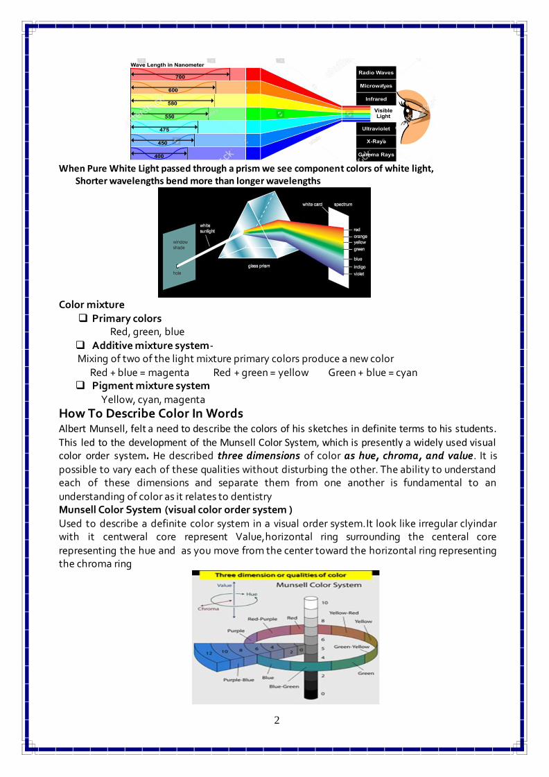
So, what is light????? Light is a Form of visible energy that is part of the radiant energy spectrum. Radiant energy possesses specific wavelengths, which may be used to identify the type of energy. The eye is only sensitive to the visible portion of the spectrum (380 – 750nm) Different wavelengths constitute the different colors we perceive
2
When Pure White Light passed through a prism we see component colors of white light,
Shorter wavelengths bend more than longer wavelengths
Color mixture
Primary colors Red, green, blue
Additive mixture system- Mixing of two of the light mixture primary colors produce a new color
Red + blue = magenta Red + green = yellow Green + blue = cyan Pigment mixture system
Yellow, cyan, magenta
How To Describe Color In Words Albert Munsell, felt a need to describe the colors of his sketches in definite terms to his students. This led to the development of the Munsell Color System, which is presently a widely used visual color order system. He described three dimensions of color as hue, chroma, and value. It is
possible to vary each of these qualities without disturbing the other. The ability to understand each of these dimensions and separate them from one another is fundamental to an
understanding of color as it relates to dentistry Munsell Color System (visual color order system )
Used to describe a definite color system in a visual order system.It look like irregular clyindar with it centweral core represent Value,horizontal ring surrounding the centeral core
representing the hue and as you move from the center toward the horizontal ring representing the chroma ring
3
Munsell define three dimension or qualities for color
1. Hue Quality by which we distinguish one color family from another ( variety of color ). We have ten hue color families; 1.R-red 2.YR-yellowgreen 3.Y-yellow 4.GY-greenyellow 5.G-green 6.BG-bluegreen 7.B-blue 8.PB-purpleblue 9.P-purple 10.RP-redpurple. Each of these ten hues is further subdivided into ten numbered segments. The middle red would thus be 5R
2. Chroma Quality of color by which we distinguish a strong color from a weak one (the intensity or saturation of hue). The degree of departure of a color sensation from that of white or gray ; the intensity of a distinctive hue, color intensity _Range= 0 – 12
3. Value Quality by which we distinguish a light color from a dark one or the relative brightness of object (lightness or darkness), range from zero to ten, black is zero(0) and white is ten (10)
The value of a color is determined by which one of the grays it matches on the scale. Colors with low value numbers are termed dark colors, and one with high value numbers are called light colors. A black-and-white television tube emits only a range of values 9 Value is generally considered to be the most important of the three dimensions of color • One reason is that lightness and darkness differences are readily detected by individuals untrained in color perception • Another reason is that value differences are more easily detected at a variety of viewing distances (both close-up and at a distance), whereas differences in hue and chroma become more difficult to quantify as the viewing distance increases Color of Human Teeth Dr. E. B. Clark was the first to accurately describe the color of teeth. In 1931, he reported his color data from a visual analysis of 6000 teeth from 1000 patients over an 8-year period (Hue range from 6 YR to 9.3 Y) ( Value range of 4 to 8) ( Chroma range from 0 to 7) Factors influence the apparent color of an object (teeth):
1) Nature of light We have three light source Incandescent Light, Fluorescent Light and Natural Daylight. Most dental offices are outfitted with incandescent and fluorescent lights. Incandescent Light Emits high concentration of yellow waves matching. while, Fluorescent Light Emits high concentration of blue waves Both of two Not suitable for shade matching.Chair light not recommended for colour matching as it is over powering and interferes with fine discrimination of three dimensions of colour Natural Daylight considered the best Closest to emitting the full spectrum of white light Used as the standard by which to judge other light sources. At Morning and evening light spectrum rich in yellow/orange, lacks blue/green because Shorter wavelengths scatter before penetrating atmosphere, While. At Mid-day time (Hours around noon) where Full spectrum of colors visible consider ideal time for color matching 2) Physical properties of object When light strikes an object, and according to the physical properties some wavelengths are absorbed by the object, while other transfer throw it, the remaining are reflected ,color of an object – light that is actually reflected by the object. True color characteristic and appearance of depth transluscency in a natural tooth cannot be correctly perceived unless the tooth is free of plaque and surface stains. With increasing opacity of teeth the grey scale value decreases and the brightness ( value) increases , The
4
Higher the brightness (values ) the lower the transparency becomes. The more transparent a tooth the more grey it appears. Opal Effect: Fine particles in enamel (hydroxyapatite crystals) responsible for opal effect Fine particles reflect short wave lengths and allow longer wave lengths to pass through. Hence areas within a tooth or a restoration with higher translucency will have a lower value because light transilluminates through and away from the viewer. When evaluating enamel translucency, the observer will often focus on the opalescent blue areas that is why Translucent areas of the teeth appear grey while opaque incisal edge appears white. -hence tooth shows •Bluish areas in reflected light •Orange red areas in transmitted areas
Tooth must be kept moist during shade selection. The color environment surrounding an object
influences our color perception of the tooth significantly (gum, lip color and color behind the object). The Gumy gingival mask (color contractor) was developed to neutralize the influence of the color environment on our color perception during visual shade selection.
Metemerism phenomenon occuring when the color of the two objects appear to match under one lighting source but not under a different source, that is why , shade selection must be evaluate under multiple light sources (different light sources) 3) Subjective assessment of observer The light first penetrates a layer of nerve fibers, then passes through several layers of cells, and finally reaches the rods and cones, which are embedded underneath • The rods and cones of the retina form the chief component of the retinal receptor complex. The rods detect only lightness and darkness (value). The cones perceive the chromatic aspects of an object (hue and chroma) .In Color - Deficient person which is Defect in color vision attack 8% males 0.5% females ,Several variations exist. Achromatism – complete lack of hue sensitivity Dichromatism – sensitivity to two primary hues Anomalous Trichromatism – sensitivity to all three hues, with abnormality in retinal cones affecting one of primary pigments. Dentists should have their color vision evaluated. If any deficiency is detected, a dentist should seek assistance when selecting tooth shades
Shade Selection: Traditional shade taking involves matching one or more selected colors from a range of shade tabs to the teeth adjacent or contralateral to the teeth to be restored. This serves as a guide to the lab technician fabricating the crown or the bridge. i.e it is Process of replicating of the color of the adjacent teeth in an artificial prosthesis, we have different methods for shade selection;
5
1) Visual shade matching. Visual shade selection by comparison of a patient’s tooth with a color standard (i.e. commercially available shade guide) .A Dental Shade Guide is a set of simulated teeth used to select prosthetic teeth by color. Shade guide are Examples of various color combinations available from manufacturers of denture teeth, restorative resins and porcelains. These samples are compared with the natural teeth and the closest color match is determined, most commonly use shade guide in fixed prosthodontics;
a) Vita Classic Shade Guide b) Vita-3D –Master shade guide c) VITA Linearguide 3D-MASTER
Principles of Shade Selection 1. Teeth to be matched must be clean & moist 2. Remove bright colors from field of view
3. View patient at eye level 4. Evaluate shade under multiple light sources 5. Make shade comparisons at beginning of appointment 6. Shade comparisons should be made quickly to avoid eye fatigue. 7. selection distance- a selection made at 3-6 feet from the oral cavity is often more useful, since
it is representative of the conditions under which the patient teeth will most often be observed. 8. Use of color Contrastors ; The color environment play important role in our color perception of
the grey tooth significantly 9. Shade tap position; Shade tap should be held above the mand tooth or Below the max tooth to
be match and aligned as close as possible to the plane of orientation of the facial surface Of the tooth being matched. Holding the shade tab over the a tooth can give a false impression of the color as the background of the tab is the tooth color rather than the oral
a) Vita Classic Shade Guide Very popular shade guide, Tabs of similar hue are clustered into letter groups;
A (red-yellow) , B (yellow) , C (grey) , D (red-yellow-gray)
How to use Vita Classic Shade Guide Manufacturer recommended sequence for shade matching as follow
1. Hue Selection 2.Chroma Selection 3. Value Selection: Because value level is not involve in this shade guide, Use of second, value ordered shade guide is recommended (Value oriented shade guide) Value oriented shade guide B1, A1, B2, D2, A2, C1, C2, D4, A3, D3, B3, A3.5, B4, C3, A4, C4, (bright >>>>>>>>>>>>decrease>>>>>>>>>>>>>>>>>>> dark)
4. Final Check / Revision b) Vita-3D –Master shade guide
More precise shade guide, tooth color divided into 5 level of value, for each value group deviation from medium hue towards yellow or red. In the medium (M) hue there are three levels of color samples for the chroma , deviation toward more yellowish hues (L) or more reddish hue (R) exist in 2 chroma
6
How to use Vita-3D –Master shade guide Step 1 Determine the lightness level ( value).Hold shade guide to patient’s mouth and start with darkest group moving to left . Select Value group 1, 2, 3, 4, or 5
Step 2 Select the chroma from From your selected Value group, remove the middle tab (M) and spread the samples out like a fan Select one of the three shade samples to determine chroma.
Step 3 Determine the hue and Check whether the natural tooth is more yellowish or more reddish than the shade sample selected
c) VITA Linearguide 3D-MASTER SHADE GUIDE
The all-new VITA 3D-Master Linearguide enables the quick determination of precise tooth shades and uses the same scientific principles and 29 shades found in the popular VITA 3D-Master shade guide. The Linearguide features a sleeker, linear design that makes the process of precise shade determination even faster and easier. In two simple steps the final shade is achieved, first by selecting from five value tabs, then by choosing the proper mix of chroma and hue within the selected value range.
7
With the VITA Linearguide 3D-MASTER you can determine the correct tooth shade swiftly and accurately. The modern design and systematic structure of the VITA Linearguide enable the appropriate 3D-MASTER shade to be found quickly.
Shade taking in two steps: Step 1:Remove the VITA Valueguide 3D-MASTER from the top of the Linearguide. The correct degree of lightness can now be determined by removing the Valueguide . Make an initial choice using the Valueguide. In doing so, you determine the correct degree of lightness 0 to 5. Step 2: Make a detailed choice within the determined degree of lightness from step 1 using the corresponding VITA Chroma/Hueguide.
Photography & shade matching
Consider as effective technique for shade matching Photography greatly simplifies the shade taking process, particularly for treatment in the aesthetic
zone; providing the ceramist with a “palate” of shades rather than trying to match a single shade. (technicians need more information than just a single shade tab
A shade tab with the shade that is closest to the shade of the tooth is placed next to the tooth in question and is photographed with the tooth.
If needed, several photographs can be taken with different shade tabs. 2) Instrumental color analysis (Digital shade-scanning devices) Digital devices are available that can be used to select the shade • Tooth should be clean & free of debris • Need to hold probe perpendicular to tooth • There is variation in the color depending on where the probe is located • Tip centered (1 – 2 mm from gingiva and incisal edge) or do 3 zones (gingival, middle, and incisal)
The advantages of a digital shade-matching system include objective readings and accuracy. There are two types of digital shade-matching devices commonly used in dentistry: the spectrophotometer and the colorimeter.
The spectrophotometer consistently and accurately measures natural tooth coloration in reference to any known specific color or can be based on any shading system. It measures the color characteristics of the natural tooth precisely and scientifically, indicating the deviations and gradations of value, chroma, and hue from a standard and provides all the information that is necessary to create an accurate restoration, or to modify an existing one such that it will accurately match the tooth. The spectrophotometer develops an accurate interpretation of the tooth shade on a given color system , which can then be related to an existing shade tab within dentistry or to a color that is interpolated between the shade tabs. In either case a lab technician is given all the color clues to recreate a shade that is very natural in appearance and very close to the target coloration.
The colorimeter analyzes the tooth coloration based on preloaded data that is related to a shade system. It determines the shade tab that is closest to the actual color of the tooth. The colorimeter is typically less accurate than the spectrophotometer but may suffice in most dental situations.
Because both spectrophotometers and colorimeters tend to eliminate ambient light by standardizing the immediate environs of the target tooth, the shade can be taken in any operatory with
8
any kind of lighting streaming in through the window. Digital shade taking therefore is far easier, far more practical, and far more accurate than shade taking using color tabs and the naked eye in a variable environment.
The current best approach to shade taking is the spectrophotometer. It provides the most accurate method for matching the coloration of the tooth. Some systems provide readings of translucence and reflectivity as well. Spectrophotometers provide consistent shade measurement regardless of the environment, lighting conditions, or other operatory variables including the dental team member who is conducting the shade-taking process. With some systems, a further comparative analysis can be undertaken on shade scans taken before and after treatment to provide the color difference between the two measurements. This is particularly useful for tooth-whitening procedures An example of digital shade scanning devices
a) VITA EASYSHADE b) VITA EASYSHADE COMPACT c) MHT SPECTROSHADE SYSTEM
CYNOVAD SHADESCAN The system is user friendly and is integrated with computed-aided design and manufacturing (CAD/CAM) technologies. The shade is measured by a handheld optical device from a single image of the whole tooth at the click of a button. The dentist can instantly obtain a shade map of the whole tooth with various established and popular shade systems.
Fixed Partial Denture (Bridge)
Components of Bridge
Bridge Retainer: That component of an FPD which unites the abutment to the remainder of restoration takes
support from the abutment tooth and provides stabilization & retention to the prosthesis. It takes support from the abutment tooth and provides retention to the prosthesis. It could be
seat over (on or in) the abutment tooth, connecting the pontic to the abutment. Bridge retainer could be (divided into);
1. Major retainer: Which are all these used in fixed-fixed, spring, and cantilever bridge.
Fixed-mobile Bridge has one major retainer at one end of the pontic. Major retainer
preparation must be retentive & with conventional bridge must cover the whole occluding surface of the tooth. (rigid connec.to pontic)
2. Minor retainer: Represent the lesser retainer of fixed-mobile bridge into which a
movable connector from a pontic seated or attach. It doesn't need full occlusal coverage. (flexible connec. to pontic )
9
Types of retainers: Major or Minor Retainer Designs;
1. Based on preparation deign 1) Extra coronal retainer (complete crown, partial crown) 2) Intra coronal (Inlay, onlay).
3) Intra radicular (Post & cor).
2. Based on material used 1) All metal retainers. 2) Metal ceramic retainers. 3) All ceramic retainer.
4) Zirconium retainers. 5) Acrylic retainers.
All metal retainers are most conservative, the simplest, & the least expensive to produce. Most of the time they used in posterior region when the esthetic is not critical or patient does not mind about appearance. Metal ceramic, All Ceramic and Zirconium are used for replacement of anterior teeth where esthetic is critical. Acrylic retainer used with temporary bridge Criteria for choosing suitable retainer (assessment factors):
When abutment teeth are more or less parallel to each other, either complete or partial crown retainer can be used .If abutments are not parallel, complete crown retainer with
common path of insertion can not obtain without a destructive reduction of the abutment.
2. Appearance (Esthetic): The esthetic of retainer must be acceptable to the patient. If esthetic is critical, P.F.M, All Ceramic, Zirconium or ¾ crown may be used also some patient may worry about metal display on occlusal surface of posterior teeth , this need to have porcelain extend to cover occlusal
surface of metal. This lead to destruction preparation which is not always possible or desirable. 3. The condition of the abutment: Partial crown can not be used because of presence of a caries or large restoration involving
the buccal surface or because of loss of buccal surface due to fracture, in such cases a complete crown is used. Pulp condition play a vital role in the selection of retainer design, incisal relationship has great effect especially when clinical length of the crown is short for both anterior & posterior teeth (worn teeth). 4. conservation of tooth structure: Inlays, Onlays are more conservative than Partial coverage which is more conservative than Complete coverage, Full Metal more conservative than P.F.M. which more conservative
than All Ceramic crown. So when we have sound buccal enamel and dentine,Partial coverage is indicated to more conservative preparation. This will not only reduce the affect
10
of cutting procedure on the pulp & periodontium, also will not destroy the natural appearance. However if there is sound indication for complete crown, this should be done. 5. Cost: Partial Crown & Complete Crown (Retainer) may be less expensive than P.F.M., which is less expensive than All Ceramic or Zirconium Crown (retainer) . Metal Retainer are least expensive. When there is no other factor affecting choice, this is obviously of considerable importance. 6. Size and position of the abutment: Partial crown need sufficient large and long tooth. Position may affect esthetic. 7. Caries susceptibility: Patient with poor oral hygiene is indicated for complete crown.
8. Retention required: The retention of a bridge retainer should be at least as great as for similar restoration made
as single unit. Retention of crown vary according to preparation feature, Crown material properties and type of luting cement
Factors affecting the amount of required retention: 1) Length of span & rigidity: The longer the span the greater the stresses on the retainer & the more will become un cemented. Further more the casting will be more liable to flex, so you must a certain they are sufficiently rigid (The longer, the span, the stronger must be all the component bridge).
2) Type of bridge: Certain types of bridges induce greater stresses on the cementing media of bridge retainer than other.
Thus strong retainers are required fixed-fixed than fixed-mobile bridge. In deed, little retention is needed for the minor retainer of fixed-mobile bridge design. Thus when it is
desirable to preserve tooth tissue, the fixed-mobile design is normally indicated as for lighter retainer can be used. For example ; replacement of upper 4 by fixed-mobile bridge using 3/4 crown or fully coverage on upper 5 as major retainer & a class III inlay on the distal of canine as the minor retainer. Such design will be: 1. Conservative 2. Esthetic
3. Incisal edge not included. 3) Strength of the bite: The strength of the bite determines to large extent the degree of retention required to resist it, this will vary with age, sex, & muscular development of the patient concerned. The heavier
the bite the stronger & thicker the retainer material needed to prevent failure of the retainer or pontic.
4) Tooth or teeth to be replaced: The size & position of the pontic have direct effect on the type of retainer required (stress
amount). Thus the replacement of a molar cause greater stress to the abutment than lower incisor. Also forces acting on canine are more likely than that acting on an incisor.
5) Occlusal coverage: There are several reasons for full. occlusal coverage, It is always (nearly) indicated because :
a) It gives abutment complete protection during mastication. b) There is no fear of cusp fracture (M O D inlay, or endo. Treated teeth).
Full occlusal coverage always indicated in fixed-fixed bridge. Occlusal reduction must be sufficient to provide enough thickness for material to be rigid.
11
6) Habits of patient: Various habits might induce stress on the bridge retainer such as pipe smoking, clenching; most important is grinding in (Bruxism). So if large number of patient natural teeth is severely warm, then any metal occluding surface that use in retainer construction will similarly worm unless the habit can be corrected. There for retainer must be thicker & stronger than normal (very hard alloy lead to wear off does not proceed at the same level as natural teeth do). Requirements of ideal retainer:
1) Provide maximum retention. 2) Give maximum esthetic. 3) Preserve vitality of preparation tooth. 4) Need conservative preparation(less amount of traumatic reduction).
5) Biological accepted to the surrounding tissue. 6) With stand masticatory forces.
7) Easily constructed. Specific Retainer design;
Major retainer Minor retainer Full Crown. Full Crown.
Post Crown & 3/4 Crown 3/4 Crown MOD inlay with
full occlusal coverage. Class III incisal
withdrawal inlay. pinlay & pinledge.
7/8 partial crown. All the restorations above that used as major retainer can be used as minor retainer but the
reverse is not possible. Major or Minor Retainer Designs;
1) Extra coronal retainer (complete crown, partial crown) 2) Intra coronal (Inlay, onlay).
3) Intra radicular (Post & care). Specific Retainer design;
Standard Full Metal Crown , 3/4 Crown , 7/8 Crown , Post Crown, PFM , full Zirconium,Combination
Non Standard Implant, Inlay, Onlay , All Ceramic, Resin Bonded (“Maryland Bridge”) Requirements of ideal retainer:
1) Provide maximum retention.
2) Give maximum esthetic. 3) Preserve vitality of preparation tooth.
4) Need conservative preparation(less amount of traumatic reduction). 5) Biological accepted to the surrounding tissue.
6) With stand masticatory forces. 7) Easily constructed.
12
General factors in tooth preparation: In order to obtain these ideal retainer requirements as far as related to tooth preparation, if the case permit, the design of the preparation of abutment tooth for a metal or porcelain crown restorations are limited by five principles:-
1. Preservation of tooth structure. 2. Retention and resistance from. 3. Structural durability of the restoration. 4. Preservation of periodontium. 5. Marginal integrity.
Factors affecting retention and resistance of crown restorations; 1) Taper of the preparation.
The more nearly parallel the opposing walls of preparation the greater will be the retention.(leaving undercut&seating problems)
*(5-6) degree convergence angle is mostly used to provide the needed retention. 2) Surface area of the preparation, 3) Length and height of the preparation. 4) Diameter of the tooth (tooth width). 5) Texture of the preparation. 6) Accessary mean.
Structural Durability:
The preparation must be designed so that it provide S.D. to the restoration i.e. the crown rest.
Must be rigid enough to not flex, perforate or even fracture. For rest. To be rigid it need bulk....to provide enough bulk to the crown restoration Sufficient T.S. must be removed from the prep.
Tooth to create enough space. By doing so the restoration allowed to withstand the forces of occlusion, prevent wearing holes in the gold and allow proper contouring and carving of
anatomy in the restoration.
13
Preservation of periodontal tissue;
1-Whenever possible the margin of the preparation should be supragingivally. 2-The casting should have proper contact, Embrasure form, Occlusion and a healthy
occluso-gingival contour.
Margin(F.L.) placement: Finishing line can be placed either:
1. Supragingival: Placing the margin above the gingival tissue for these reasons:-
a- can easily prepared and finished. b- To provide good vision for the dentist during preparation.
c- impression can be easily made. d- the patient can keep the area clean easily.
e-most of the time such position is situated on hard enamel. f-Less destructive The factors that influence such position of finish line are :-
a- When the esthetic is a factor. b- When we need extra retention.
c- When we have carries or filling at the area of finish line. 2. Subgingival: Placing the margin below the gingival tissue. 3. Placing the margin with in the level of the gingiva. Marginal Integrity:-
The restoration can survive in the biological environment of the oral cavity only if the margins are
closely adapted to the CSL OF the prep. The configuration Of the F.L. determine the shape and the bulk of the rest. Margin that affect both marginal adaptation and the degree of seating of the restoration. The restoration margins should:
a-They must fit as closely as possible against the finishing line of preparation. b-They must have sufficient strength. c-Whenever possible they should be placed in an area where the dentist can finish
and clean them properly.
Finishing line of the preparation (f.l): Requirement of F.L. :-
1. It must be clear ,smooth and well defined. 2. It must be continuous from one surface to the other.
3. It must lie on sound tooth structure
Factors affecting selection of F.L. Design; 1) Type of the restoration
2) Materials used in construction 3) The amount of occlussal force (stress) the restoration will bear
Types (design or configuration) of finish line: The following designs for finish line(margin of preparation ) could be used: depending on the type of the crown restoration: 1. Knife edge 2. Shoulder 3. Shoulder with bevel 4. Radial shoulder 5. Chamfer 6. Heavy chamfer
14
Complete Cast Crown ( FMC )
It is one of the most commonly used retainer for the posterior teeth. Because it made of metal, it should be used when the patient doesn’t mind the appearance of metal or when esthetic not a factor. This type of retainer provides better retention and resistance because all the axial surfaces of the teeth are including in the preparation.
Indications 1) It is indicated when the bridge located posteriorly . 2) The abutment teeth with extensive tooth destruction as a result of caries or trauma.
3) As a retainer on teeth receive clasp. 4) Existing previous restoration that precludes the use of a more conservative restoration
5) Need for superior retention and strength 6) Endodontically treated tooth 7) Patient with high caries index.
Contra-Indications 1) When the abutment teeth located in the appearance zone.
2) Patient with low caries index. 3) Whenever a more conservative retainer is feasible
Advantages; 1) Provide greater retention and resistance 2) high resistance to deformation (Strong)
3) Less tooth structure is removed and easy to prepared (conservative). 4) Strong even in thin sections
Disadvantages 1) Poor esthetic.
2) Difficulty to test the vitality of the abutment tooth especially by electrical pulp tester
3) Interfere with taste
4) Tarnish and corrosion so it needs prophylactic measures.
15
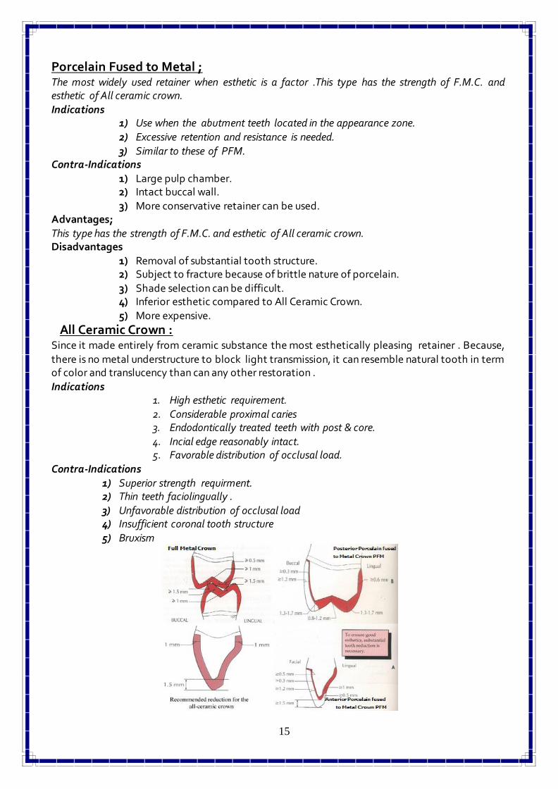
Porcelain Fused to Metal ; The most widely used retainer when esthetic is a factor .This type has the strength of F.M.C. and esthetic of All ceramic crown. Indications
1) Use when the abutment teeth located in the appearance zone. 2) Excessive retention and resistance is needed.
3) Similar to these of PFM. Contra-Indications
1) Large pulp chamber. 2) Intact buccal wall. 3) More conservative retainer can be used.
Advantages; This type has the strength of F.M.C. and esthetic of All ceramic crown. Disadvantages
1) Removal of substantial tooth structure. 2) Subject to fracture because of brittle nature of porcelain. 3) Shade selection can be difficult. 4) Inferior esthetic compared to All Ceramic Crown. 5) More expensive.
All Ceramic Crown : Since it made entirely from ceramic substance the most esthetically pleasing retainer . Because,
there is no metal understructure to block light transmission, it can resemble natural tooth in term of color and translucency than can any other restoration .
Indications 1. High esthetic requirement. 2. Considerable proximal caries 3. Endodontically treated teeth with post & core. 4. Incial edge reasonably intact. 5. Favorable distribution of occlusal load.
Contra-Indications
1) Superior strength requirment. 2) Thin teeth faciolingually .
3) Unfavorable distribution of occlusal load 4) Insufficient coronal tooth structure
5) Bruxism
16
Partial veneer crown (three quarter crown): It’s a cast restoration which cover 3/4 of the clinical crown (occlusal, incisal, lingua and proximal surfaces) leaving the buccal or labial surface untouched. It has less retention and resistance to
displacement compared to full metal, full veneer with facing. Indication:
1) Short span bridge. 2) On teeth with clinical crown of good (average) length and thickness labio-lingually. 3) Patient with good oral hygiene and low caries index. 4) When the abutment tooth in good axial relationship to facilitate the path of insertion.
1) Possibility of recurrent caries along to cavosurface line angle. 2) Possibility of showing gold especially in the lower anterior and posterior teeth. 3) Difficulty in preparation compared to other types of crowns (limited adjustment can be
Post crown: It is a fixed artificial restoration which replaced the coronal portion of the natural tooth completely .It retained itself by a mean of post (dowel) that extended and cemented to the root
canal space of endodontically treated tooth The post crown will reinforce the remaining tooth structure against forces by distributing the
forces to the supporting tissue. Indication:
1) It commonly indicated for endodontically abutment tooth. 2) Abutment tooth with short clinical crown. 3) Re-alignment of malposed abutment. When the preparation of full metal and full
veneer will cause exposure of the pulp. The retention of the post crown depends on:
1) Taper of the root canal. parallel sided prep. Is more retentive than tapered (diverage occluasly)
2) Post length. Longer length more retention (2/3 length of root, Equal to length of clinical crown, 4-5 mm
from apex, 8 mm deep from CEJ ) 3) Post diameter.
one third the root diameter at C.E.J .and should be at least 2mm less than root diameter at mid root area)
4) Post surface texture. For multi-rooted posterior teeth, the post should be placed in the largest canal.
For the maxillary molar use the palatal canal and for the mandibular molar use the distal canal. For the maxillary premolar, the post should be placed in the buccal canal.
Factors affect the selection of a tooth for post crown retainer: 1- The root of the abutment should be of sufficient length, width and without sharp angulations in the middle third. 2- The root should be without internal or external resorption. 3- Quality of the root-filling: the canal should be filled with well-condensed gutta-
Esthetically appealing Economical , conservative , functional & do not irritate soft or hard tissues
Indication 1) As retainers of FPD for abutment with sufficient enamel to etch 2) Splinting of periodontally compromised teeth
3) Stabilizing dentition after orthodontic treatment. Contraindication
1) In patients with sensitivity to base metal alloys 2) When facial esthetic of abutment require improvement
3) Inadequate enamel surface to bond eg;caries,existing restoration,attrition 4) Incisor with extremely thin facio-lingual dimension
18
Pinledge A partial veneer retainer preparation incorporating pins holes to provide retention. Indications
1) High esthetic requirement 2) Undamaged anterior teeth 3) When proximal grooves are impossible to prepare
4) To alter lingual contour of maxillary anterior teeth
INLAY
A fixed intracoronal restoration; a dental restoration made outside of a tooth to correspond to the form of the prepared cavity, which is then luted into the tooth. Inlay may be used as
a single tooth restorations for proximo-occlusal or gingival lesions with minimal to moderate extensions.
Minor retainer for fixed partial denture They may be made up of gold alloy or ceramic material.
Contraindications 1) high caries index
2) Poor plaque control 3) MODs
4) Poor dentinal support require wide preparation
ONLAY A restoration that restores one or more cusps and adjoining occlusal surfaces or the entire
occlusal surface and is retained by mechanical or adhesive mean. It is used for restoring more extensively damaged posterior teeth needing wide mesio-occluso-distal restorations.
It can be used as a retainer for fixed partial denture Contraindications