Lecture 22: Single-molecule Dynamics: rotation, diffusion, energy transfer, and powerful application in biological systems Why and how to study molecule dynamics: rotation and diffusion. 2 research cases. Single-molecule fluorescence resonance energy transfer (FRET) and application in protein dynamic structural imaging. Imaging of long-range molecule diffusion: monitoring virus attacking of a living cell, and implication for drug delivery.
Transcript
Lecture 22: Single-molecule Dynamics: rotation, diffusion, energy transfer, and powerful application in biological systems
Why and how to study molecule dynamics: rotation and diffusion. 2 research cases.
Single-molecule fluorescence resonance energy transfer (FRET) and application in protein dynamic structural imaging.
Imaging of long-range molecule diffusion: monitoring virus attacking of a living cell, and implication for drug delivery.
Part 1: Imaging Molecule dynamics
Help reveal the basic properties beyond the ensemble average.
Molecule properties measured at single-molecule level • Optical properties: excited states (formation and decay), fluorescence wavelength and
quantum yield and dependence on environment (sensor); energy transfer (dependent on distance --- for protein conformation detection).
• Electronic properties: electron transfer (electronic devices) and separation (photovoltaic), binding recognition (changing fluorescence properties via redox processes).
• Molecule dynamics: rotation and diffusion. 1. Critical for understanding and justifying the application in probing of surface
properties and protein dynamic structures --- for any probing, the behavior of the probe itself should be characterized and controlled first.
2. Compared to particles (like pollen grains), diffusion of molecules are quite difficult to be measured at single-molecule level (mainly due to the small signal with respect to the fast motion), although Brownian motion has been known for long time. Fortunately, high sensitivity of fluorescence helps to image such a motion with confocal microscopy.
3. However, molecular rotation cannot be simply detected by fluorescence measurement. It should employ polarization technique --- since only polarized fluorescence depends on molecule dipole, which in turn depends on rotation. Such orientation dependence can only revealed at single-molecule imaging level. Why?
Experimental techniques for measuring molecule rotation
1. NMR: for both solution and solid. Advantages: high spatial resolution (atomic). Disadvantages: low sensitivity for C13, and challenging for selectivity for H1.
2. Polarized fluorescence at single-molecule level: offering in situ measurement of fast rotation of molecules, through milliseconds modulation of excitation polarization.
Experimental techniques for measuring molecule diffusion
1. Diffusion NMR: for high spatial resolution measurement, particularly powerful for bio-species. Potential challenge: diffusion range and time scale.
2. Fluorescence correlation spectroscopy (FCS): suitable for fast processes (micro-seconds) and large area diffusion. Draw a scheme.
3. Absorption: based on change of absorption coefficient upon mixing of different components (diffusion). Advantages: simple and cheap. Disadvantages: good for phase diffusion, like toluene diffusion in hexane, but not sensitive enough for molecules diffusion, particularly at low concentration.
4. Single-molecule fluorescence based confocal microscopy: increased sensitivity and resolution.
Case 1: imaging molecular rotation
Polarized emission • Fixed polarization: the angle of polarizer is fixed, so the excitation polarization is
constant. Only the molecules with dipole parallel to the polarization can be excited, whereas the molecule with dipole perpendicular to the polarization cannot be excited. If a molecule is immobilized, the emission intensity will be somewhere between zero and the maximum (parallel). By measuring large number of molecules, one can reveal the distribution of molecule orientation. This cannot be done by the ensemble measurement, which gives only averaged result that does not depend on the polarization angle. Draw a scheme.
• Modulated polarization: the polarization angle changes with time (here milliseconds).
1. If the molecule is immobilized, the intensity will be modulated with the polarization following, I = a cos2(x-b) + c, where a is the maximum intensity, x is the polarization angle, b is the in plane dipole angle, is the background correction.
2. If the molecule is mobile --- rotating: the intensity will show a irregular behavior.
Fig 1
• From the fit, the in-plane dipole orientation can be extracted with an accuracy of 0.2 o.
• In this case, the molecule used is DNA (20 base pairs)---C6 linker---fluorophore (Texas red or TMR). Out of 200 molecules investigated, 97% showed stationary dipoles.
Fig 2
• A dark state was observed, indicating either a rotation or transition into a non-emitting state.
• From the fit, the in-plane dipole orientation before (-42.8 o ± 0.8 o) and after ((-41.3 o ± 0.8 o) the dark state are the same --- the dark state is not due to rotation.
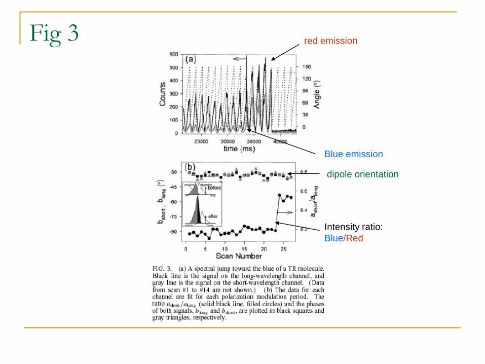
Fig 3
Blue emission
red emission
dipole orientation
Intensity ratio: Blue/Red
Fig 3
• A sudden increase intensity: due to spectral change (shift of absorption upon continuous irradiation) or molecule rotation?
• Spectral change: the dipole angle does not change.
• The constant dipole from the fitting suggests a spectral change.
• This is consistent with the increase in the intensity at short wavelengths.
Fig 4
Fig 4
• 5 out of more than 200 molecules observed showed molecule rotation.
• Only phase changes, the maximum intensity does not change due to the full angle modulation of the polarization.
Adachi, K. et al. Proc. Natl. Acad. Sci. USA 97, 7243–7247 (2000).
Single-molecule rotation imaged as movie clips in various time intervals
Case 2: the first report on single-molecule diffusion
• Fluorescence labeled lipid embedded in fluid lipid membranes.
• Detector: LN cooled CCD.
• Objective: Zeiss, 100X, NA 1.3.
• S/N was obtained at 28, and the positional accuracy for molecule is
achieved at 30 nm, far below the diffraction limit of optical microscopes.
Fig 1 • A mean value of (172±15) counts per dye molecule was found for six dye
densities covering the range from 10 to 103 dyes/µm2 --- indicating the well isolated distribution of fluorescent molecule within the lipid membranes.
• Images at this density were characterized by large fluctuations of the intensity as shown in Fig. 1 A.
• The sequence was obtained from consecutive illuminations of the same membrane area lasting 5 ms at 35-ms intervals.
• The two molecules undergo lateral motion (Brownian motion), with position uncertainty of 30 nm, 7 times smaller than the diffraction limit, here ~ 300 nm.
• This position accuracy allows for monitoring of the molecule movement within the time interval of 30 ms.
Fig 2 • Statistics based on repeated measurements over one molecules: averaged
intensity is 154 counts, as shown in the figure.
• Statistics based on measurements over large number of different molecules: averaged intensity is 172 counts.
• The consistence indicates that the observed emission spots (peaks) is due to isolated molecules.
•
<r2> = 6 D t + C D: diffusion coefficient t: time C: constant
Fig 3
• Analysis of trajectories of 531 individual molecules showed that the direction of diffusion steps of simultaneously observed molecules were uncorrelated.
• The mean-square displacement (MSD), and the time lag (tlag) yielded the expected linear relationship as shown in Fig. 3 (Inset). The lateral diffusion constant Dlat was estimated as 1.42 ± 0.13 x 10⁻⁸ cm2/s.
Part 2: Imaging Protein dynamics via fluorescence resonance energy transfer (FRET)
where physics and biology meet, DNA and RNA dance, enzymes wiggle and flutter, and dye
molecules FRET and ... die!
Two advantages of single-molecule imaging for biological systems
T. Ha @ UIUC
Single molecule imaging/spectroscopy of biological systems
Heterogeneous: most (if not all) biological systems are heterogeneous.
Single molecule imaging/spectroscopy reveals distribution of molecular properties (like the colors of the balls), by looking at one molecule at a time and tabulate the result (similar to taking a census).
Dynamic: most (if not all) biological systems are dynamic, keeping change in shape, conformation, composition, or position.
Single molecule imaging/spectroscopy measures the entire time trajectories, which include multiple pathways and intermediates involved. See the last slide.
Though elegant biochemical and structural methods are available, they don’t provide with the real time measurements of conformational changes.
Typical fluorescence properties used for single molecule probing and imaging
Fluorescence depends on local polarity --- for both wavelength and quantum yield (intensity). This is normally used for single fluorophore labeling. Fluorescence intensity fluctuation reflects conformational changes of the host system (like a protein or membrane). Draw scheme (sigmoidal plot for intensity and spectral shift). Also see a slide next.
Fluorescence polarization --- revealing rotation and diffusion of molecules (see last lecture), which in turn reflecting the conformation changes of the host systems.
Fluorescence resonance energy transfer (FRET) --- revealing dynamic conformational changes involving two domains, where the fluorophores are labeled. This is unique for probing the coincident structural change within proteins or other large biological systems. a typical example is the signal transducing membrane proteins functional in neuron cells, where upon a ligand binding the membrane protein experience great structure change; some domains may transform across the membrane. Such a large change can be revealed by labeling two dyes at the active domains. Draw a scheme for a later slide.
SMS measurement based on intensity fluctuation and wavelength shift
(top panel) Fluorescence fluctuation monitored with photon counting (left) and spectra recording (right). (bottom panel) A scheme showing the basic method to get the statistic mean value of ON and OFF times.
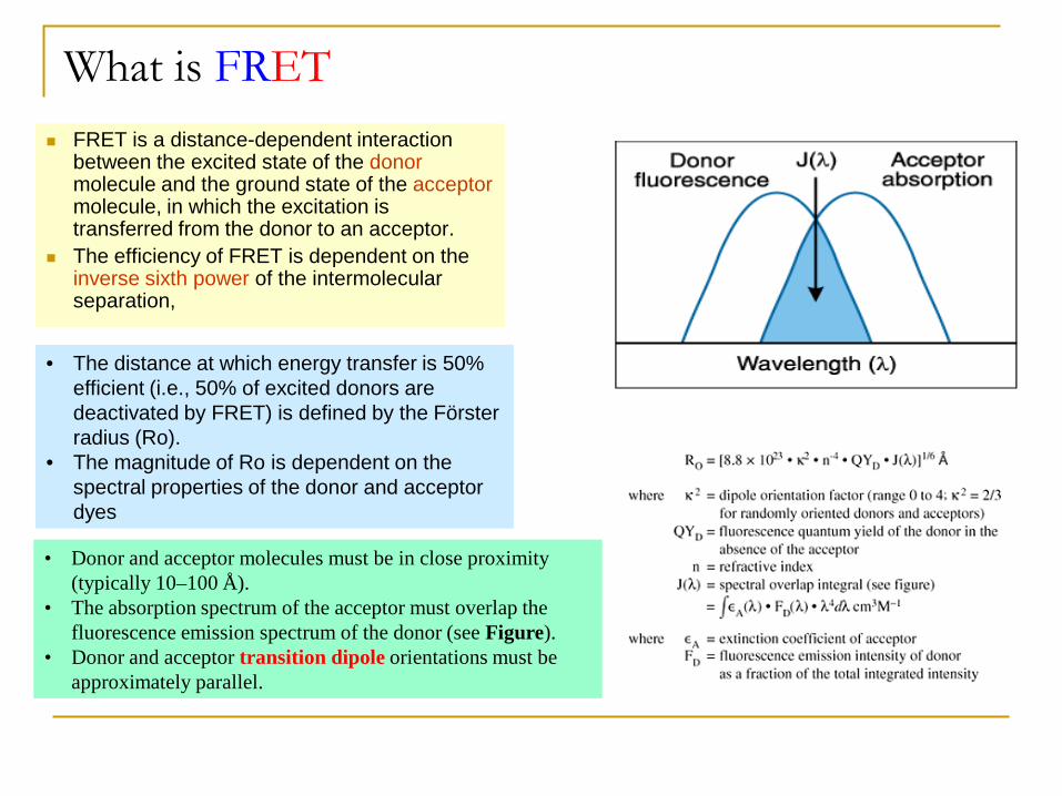
What is FRET FRET is a distance-dependent interaction
between the excited state of the donor molecule and the ground state of the acceptor molecule, in which the excitation is transferred from the donor to an acceptor.
The efficiency of FRET is dependent on the inverse sixth power of the intermolecular separation,
• The distance at which energy transfer is 50% efficient (i.e., 50% of excited donors are deactivated by FRET) is defined by the Förster radius (Ro).
• The magnitude of Ro is dependent on the spectral properties of the donor and acceptor dyes
• Donor and acceptor molecules must be in close proximity (typically 10–100 Å).
• The absorption spectrum of the acceptor must overlap the fluorescence emission spectrum of the donor (see Figure).
• Donor and acceptor transition dipole orientations must be approximately parallel.
Determines changes in distance (conformation) rather than absolute distances as ‘E’ depends on orientation of the dyes. FRET is particularly useful for biological systems. most of single-molecule-FRET studies have been performed on bio-molecules.
T. Ha @ UIUC
What is FRET
Selected Applications of FRET bio-research Structure and conformation of proteins Spatial distribution and assembly of protein complexes Receptor/ligand interactions Immunoassays Probing interactions of single molecules Structure and conformation of nucleic acids Real-time PCR assays and SNP detection Detection of nucleic acid hybridization Primer-extension assays for detecting mutations Automated DNA sequencing Distribution and transport of lipids Membrane fusion assays Membrane potential sensing Fluorogenic protease substrates Indicators for cyclic AMP and zinc
Ha et al - First FRET in single molecules between single donor and acceptor under non-physiologic conditions (dry glass surface).
Ha, T. et al. Proc. Natl. Acad. Sci. USA 93, 6264–6268 (1996).
Deniz et al – Single molecule FRET and predicted distance.
Deniz, A.A. et al. Proc. Natl. Acad. Sci. USA. 97, 5179–5184 (2000).
Brasselet et al – GFP’s in single-molecule-FRET for studying calmodulin. Brasselet, S., Peterman, E.J.G., Miyawaki, A. & Moerner, W.E. J. Phys. Chem. B 104,
3676–3682 (2000).
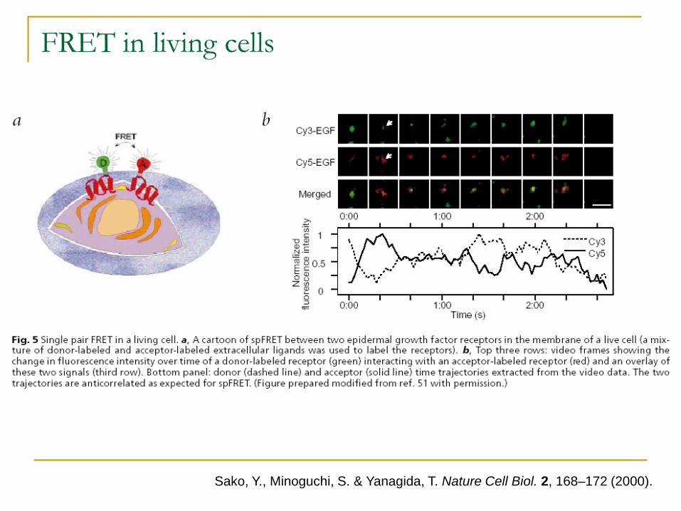
Sako et al – extended it to invivo system and used it for studying epi’dermal growth factor dimerisation.
Sako, Y., Minoguchi, S. & Yanagida, T. Nature Cell Biol. 2, 168–172 (2000).
Dyes Used Green fluorescent protein (GFP) and siblings (RFP, CFP, YFP,
BFP) – unique and amenable for mutation within the host proteins without disturb the protein structure or functions. But suffers from limited sensitivity, low stability against photobleaching, and time consuming for sample assembly.
Cysteine reactive dyes --- selectively reactive to cysteine moiety, which can be selectively mutated inside the proteins.
Amine reactive dyes --- reactive to amine groups at some amino acids, but the selectivity is not as good as the Cysteine reactive dyes, since there several amino acids have amine groups.
Membrane permeant fluorescein and rhodamine Cyanine Dyes (Cy3, Cy5, Cy7) – well separated emission maxima
Cy3-Cy5 pair widely used for cellular imaging. Semiconductor nanocrystals (Q-dots) --- CdSe, CdTe or the core-
shell nanocomposites. Good photostability, narrow emission band, tunable emission wavelength. But suffering from surface modification to get water soluble, and the potential poisonous.
The Nobel Prize in Chemistry 2008 "for the discovery and development of the green fluorescent protein, GFP"
Photo: U. Montan Photo: U. Montan Photo: U. Montan
Osamu Shimomura Martin Chalfie Roger Y. Tsien
1/3 of the prize 1/3 of the prize 1/3 of the prize
FRET efficiency correlates with the Protein folding states
A dynamic gauge for protein folding processes.
FRET efficiency correlates with the Protein folding states
• Many methodologies have been developed and applied to protein folding, but most conducted on an ensemble of proteins.
• Since it is very difficult to synchronize the folding reactions over an ensemble of molecules, these measurements usually fail to resolve detailed dynamic information.
• SMS, and in particular single-molecule-FRET, can provide distance information between a pair of points on a polypeptide chain as folding progresses — providing a reaction coordinate that affords a global view of the conformational distributions, intermediates and dynamics as the protein tumbles down the free energy folding funnel.
Single-molecule-FRET histograms of wild type CI2 at 3, 4 and 6 M denaturant concentrations
Shimon Weiss lab
Immobilization of Bio-Systems
Why immobilization? – Observing conformational changes can be impeded by molecule diffusion in solution (see last lecture). To quantitatively measure the changes in the conformation, immobilization is required.
Nonspecific immobilization: DNA or proteins attached to charged surface like aminopropylsilane coated
surface via electrostatic interaction. This is a easy and universal technique. Trapping molecules in pores of agarose gels. Agarose is a widely used support
material in molecular biology. Agarose gels have large "pore" size, which would be just appropriate for hosting and immobilizing proteins (MW 50 – 150 kDa), and on the other hand, there is still enough free room for the proteins to keep the dynamic activity.
Specific immobilization Using a biotin – streptavidin tether to the surface. Histidine tags for recombinant proteins immobilized on Ni – NTA coated
surface.
Since the diffusion is quite limited due to the binding to the host, most control is for molecular rotation, which determines the fluorescence polarization. Also the FRET efficiency depends on the relative orientation between donor and acceptor --- parallel gives the highest efficiency.
Check for anisotropy in solution at ensemble levels. If the anisotropy is low, it means that the dye molecule interacts weakly with the host molecule (large flexibility for rotation) --- this assumes the host motion (rotation) is slower than the lifetime of fluorescence.
In case the molecular rotation is significant, another polarization control should be done at single molecule level with immobilization of proteins on surface. This measures the rotation rate --- if the rotation is far slower or faster than the FRET process, the signal can be resolved for FRET. But if the two rates are close, it would be a problem for FRET measurement.
Control of molecular rotation for FRET measurement
FRET in living cells
Sako, Y., Minoguchi, S. & Yanagida, T. Nature Cell Biol. 2, 168–172 (2000).
Shimon Weiss, SCIENCE, VOL 283, page 1686.
FRET for nuclease-DNA interaction
Part 3: Imaging virus diffusion and attacking to living cell
Learn from the bad thing; Turn it into something good.
Basics of virus Definition: A virus is a small particle that infects cells in biological organisms. Viruses are
obligate intracellular parasites; they can only reproduce by invading and taking over other cells as they lack the cellular machinery for self reproduction.
Structure: virus carry a small amount of nucleic acid (either DNA or RNA) surrounded by some form of protective coat consisting of proteins, lipids, and glycoproteins. Importantly viral genomes code not only for the proteins needed to package its genetic material, but for proteins needed by the virus during lysogenic and lytic cycles, the reproductive cycles.
Non-living or alive: A virus makes use of existing enzymes and other molecules of a host cell to create more virus particles. Viruses are neither unicellular nor multicellular organisms; they are somewhere between being living and non-living. Viruses have genes and show inheritance, but are reliant on host cells to produce new generations of viruses.
A supra-molecule? Many viruses have similarities to complex molecules. Like DNA, viruses undergo molecular replication and they can often be crystallized.
Size: The viral capsid may be either spherical or helical and is composed of proteins encoded by the viral genome. Helical virus: 1 µm to a few µm; spherical virus: 20 – 400 nm. Hard to see by optical microscope, but observable by confocal microscope based on fluorescence labeling.
Rabies virions are bullet-shaped with 10-nm spike-like glycoprotein peplomers covering the surface. The ribonucleoprotein is composed of RNA encased in nucleoprotein, phosphorylated or phosphoprotein, and polymerase.
Spherical virus
Schematic representation of the structure of HIV:
Three types of virus
Three types of viruses: a bacterial virus, otherwise called a bacteriophage (left center); an animal virus (top right); and a retrovirus (bottom right). Viruses depend on the host cell that they infect to reproduce. When found outside of a host cell, viruses consist of genomic nucleic acid, either DNA or RNA (depicted as blue), surrounded by a protein coat, or capsid, with or without a glycoprotein envelope. Retroviruses contain RNA and reverse transciptase. it relates to some tumor formation.
Viruses as probes for exploring basic cellular processes
Viruses are important to the study of molecular and cellular biology because they provide simple systems that can be used to manipulate and investigate the functions of cell types.
Viral replication depends on the metabolism of the host ---- the study of viruses can provide fundamental information about aspects of cell biology and metabolism. With high sensitivity of fluorescence labeling, the metabolic dynamics can be revealed.
Because of the complexity of an animal cell genome, viruses have been even more important in studies of animal cells than in studies of bacteria.
Viruses as tools for genetic engineering
Geneticists regularly use viruses as vectors to introduce DNA into cells that they are studying. Attempts to treat human diseases through genetic engineering have also made use of viruses in similar ways. Deaths have occurred through virus infections caused by virus vectors used in gene therapy, so their application to human subjects is still nascent --- how about artificial vector?
The steps of gene delivery and replication: draw scheme Attachment, sometimes called absorption: The virus attaches to
receptors on the host cell wall. Injection: The nucleic acid of the virus moves through the plasma
membrane and into the cytoplasm of the host cell. For bacterial virus, the capsid remains on the outside. For animal virus, they enter the host cell intact.
Replication: The viral genome contains all the information necessary to produce new viruses. Once inside the host cell, the virus induces the host cell to synthesize the necessary components for its replication.
Assembly: The newly synthesized viral components are assembled into new viruses --- self-assembly?
Lysis: Assembled viruses are released from the cell and can now infect other cells, and the process begins again.
The influenza A virus
• A RNA virus: 8 single strand RNA. • a globular particle (about 100 nm in diameter). • Each year, 10-20% US residents get infected by flu, and ~36,000 people die of flu-related
comlications. • 1918 Spanish flu, 21 M people died over the world --- but still a mystery why it was so deadly
in terms of genomes. • Studying the cellular entry and the replication dynamics helps to understand the mystery of the
special class of supra-molecules, and to avoid another time of pandemic attack of flu.
Electron microscope image of stained viurs
Visualizing individual influenza virus particles in living cells
• Cellular replication of Influenza viruses: receptor-mediated endocytosis endocytic trafficking of influenza to acidic endosomes fusion of the viral membrane with endosomes deliver the viral genome (ribonucleoprotein, vRNPs) into the cell the vRNPs are then imported to the nucleus to initiate viral gene expression and replication.
• Despite intensive studies of influenza infection, many important aspects of cellular entry process of influenza still remain elusive.
• Labeling the lipid membrane of virus with fluorophores.
• Using fluorescence microscope equipped with oil immersion objective, NA1.4.
• 3 stages of virus transportation to be imaged (see above).
Three Stages of action: 1. moving at cell periphery, 2. moving from cell periphery to nuclear periphery, 3. moving at nuclear periphery.
Stacked, time-lapsed images of two viruses in living cells. The sudden color change from blue/pink to white indicates a dramatic fluorescence dequenching, signaling the fusion of the virus with an endosome. The viruses are labeled with membrane dyes.
Zhuang, PNAS, 2003, vol. 100, 9280–9285
Three Stages of action: 1. moving at cell periphery, 2. moving from cell periphery to nuclear periphery, 3. moving at nuclear periphery.
Endocytic trafficking and fusion of individual viruses. (a) The trajectory of a DiD-labeled virus inside a cell. The color of the trajectory codes time with the colored bar indicating a uniform time axis from 0 s (black) to 500 s (yellow). The red star indicates the fusion site. (b) Time trajectories of the velocity and the DiD fluorescence intensity of the virus. The labels I, II, and III indicate stage I, II, and III movement, respectively. The fluorescence dequenching signal of the lipophilic dye, DiD, near 400 s indicates viral fusion. Movie shown on the last slide. (c) The trajectory of a Cy3/CypHer5-labeled virus inside a cell. The green star indicates the initial acidification site (to pH 6). (d) Time trajectories of the velocity and the fluorescent emission ratio of CypHer5 (a pH-dependent dye) and Cy3 (a pH-independent dye) of the virus. Scale bars: 10 µ m.
Zhuang, PNAS, 2003, vol. 100, 9280–9285
Entry mechanisms of influenza viruses: via CCP internalization
While many viruses are known to infect cells via receptor-mediated endocytosis, the exact endocytic mechanisms have remained unclear for most of them.
Snapshots of a virus (labled with DiD, red) internalized by a clathrin-coated pit (CCP, labeled with EYFP, green). Overlay of green and red signals appears yellow. T = 0 s: the virus (red) binds to the cell. t = 115 s: a CCP labeled with EYFP (green) begins to form at the virus site. T = 175 s: the clathrin coat rapidly disassembles. T = 181 s, 202 s, and 235 s: transport of the virus on microtubules. Clathrin-coated pits are 150 nm invaginated structures on the plasma membrane that occupy about 2% of the plasma membrane surface
Entry mechanisms of influenza viruses: without CCP
Time-trajectories of viruses fused with endosomes. ( a ) A virus internalized via a CCP. ( b ) A virus internalized without association with a CCP. Black symbols are the velocity time-trajectories of the viruses. Red symbols are the integrated DiD fluorescence intensities of the viruses. Viral fusion can be identified as a dramatic increase of the DiD signal. Green symbols are the integrated fluorescence intensities of EYFP-clathrin associated with the viruses.
Nuclear import of influenza genome
Zhuang, Biophys. J. 87, 2749-2758 (2004)
Regulated nuclear import of influenza vRNPs. ( a ) A DIC image of an injected cell. ( b ) A fluorescence image of the same cell, taken 30 minutes after the microinjection of dye-labeled vRNPs, showing nuclear import of the labeled vRNPs. (c) A fluorescence image of a cell taken 2 minutes after injection of vRNP. The ring at the nuclear envelope indicates association of vRNPs to the nuclear envelope. (d) A fluorescence image of a cell that was co-injected with labeled vRNP and anti-NPC antibodies. (e) A fluorescence image of a pre-infected cell taken 10 min after injection with vRNPs. The cell was infected with influenza viruses 6 hours prior to injection. No fluorescent ring is observed at the nuclear envelope. DIC: differential interference contrast; NPC: nuclear pore complexes
2 min after injection of vRNP
30 min after injection of vRNP
differential interference contrast
Co-injection of vRNP and anti-NPC
injection of vRNP to a pre-infected cell.
Cellular entry and trafficking of polymeric gene-delivery vectors
Zhuang, at Harvard
• Gene delivery: emerging as a promising therapeutic method for a variety of diseases including cystic fibrosis, Parkinson's disease, and certain cancers.
• Polymer-mediated gene delivery replaces the conventional viral vector (potential infection) with a cationic polymer, which binds to the anionic DNA or siRNA, causing it to condense into a small complex with a net positive charge --- similar to the virus structure.
• At present, the best polymeric gene-delivery vectors is still a few orders of magnitude less efficient than viral vectors.
• A detailed understanding of the entry and trafficking mechanisms of polymer-DNA complexes is critical for the rational design of polymer vectors with much improved efficiency.


![Nanostructuredhigh-entropymaterials · H–P effect include dislocation- or diffusion-induced grain-boundary shearing and sliding, grain rotation, and two-phase– based models [16].](https://static.documents.pub/doc/80x56/606ecc16cb0351003666a522/nanostructuredhigh-entropymaterials-hap-effect-include-dislocation-or-diffusion-induced.jpg)


