Page 1
Lecture 4 Calcium & Phosphate Disorders Loh
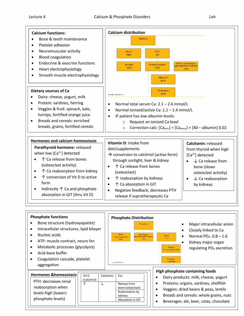
Calcium functions:
Bone & teeth maintenance
Platelet adhesion
Neuromuscular activity
Blood coagulation
Endocrine & exocrine functions
Heart electrophysiology
Smooth muscle electrophysiology
Calcium distribution
Normal total serum Ca: 2.1 – 2.6 mmol/L
Normal ionized/active Ca: 1.1 – 1.4 mmol/L
IF patient has low albumin levels:
o Request an ionized Ca level
o Correction calc: [Cacorr] = [Cameas] + [40 – albumin] 0.02
Dietary sources of Ca
Dairy: cheese, yogurt, milk
Protein: sardines, herring
Veggies & fruit: spinach, kale,
turnips, fortified orange juice
Breads and cereals: enriched
breads, grains, fortified cereals
Hormones and calcium homeostasis
Parathyroid hormone: released
when low [Ca2+] detected
↑ Ca release from bones
(osteoclast activity)
↑ Ca reabsorption from kidney
↑ conversion of Vit D to active
form
Indirectly ↑ Ca and phosphate
absorption in GIT (thru Vit D)
Vitamin D: intake from
diet/supplements
conversion to calcitriol (active form)
through sunlight, liver & kidney
↑ Ca release from bones
(osteoclast)
↑ reabsorption by kidneys
↑ Ca absorption in GIT
Negative feedback, decreases PTH
release if supratherapeutic Ca
Calcitonin: released
from thyroid when high
[Ca2+] detected
↓ Ca release from
bone (slows
osteoclast activity)
↓ Ca reabsorption
by kidneys
Phosphate functions
Bone structure (hydroxyapatite)
Intracellular structures, lipid bilayer
Nucleic acids
ATP: muscle contract, neuro fxn
Metabolic processes (glycolysis)
Acid-base buffer
Coagulation cascade, platelet
aggregation
Phosphate Distribution
Major intracellular anion
Closely linked to Ca
Normal PO4: 0.8 – 1.6
Kidney major organ
regulating PO4 excretion
Hormones &homeostasis
PTH: decreases renal
reabsorption when
levels high (lowers
phosphate levels)
Vit D (Calcitriol)
Calcitonin Fxn
↑ ↓ Release from bone (osteoclast)
Reabsorption by kidneys
Absorption in GIT
High phosphate containing foods
Dairy products: milk, cheese, yogurt
Proteins: organs, sardines, shellfish
Veggies: dried beans & peas, lentils
Breads and cereals: whole grains, nuts
Beverages: ale, beer, colas, chocolate
Page 2
Lecture 4a Calcium Disorders Loh
Goals of therapy
Improve & resolve S/S
Correct underlying
causes
Normalize serum Ca
Prevent further
complications (renal
failure)
Hypercalcemia S/S: “bones, stones, abdominal groans & psychic moans”
CNS: fatigue, confusion, weakness, lethargy, seizures
CV: bradycardia, changes on cardiac ECG (shortened QTc), arrhythmias
MSK: weakness, tenderness, bone pain
GI: anorexia, nausea, vomiting, constipation
GU: kidney stones, polyuria acute renal insufficiency
Disease causes
Malignancy: production of PTH-like peptides, ectopic PTH, calcitriol;
metastatic cancers to bone
Hyperparathyroidism: primary (adenoma), secondary (renal disease)
Granulomatous diseases: sarcoidosis, tuberculosis
Other: adrenal insufficiency, thyrotoxicosis, immobility
Drug causes
Thiazide diuretics: reduces renal excretion of Ca
Lithium carbonate: increases Ca threshold for suppression of PTH
Tamoxifen: increased bone resorption
Increased Ca absorption via intake/use (milk-alkali syndrome): calcium
supplements, calcitriol/calcipotriol/Vit D, vitamin A, antacid overdose
General approach
Correct intravascular
volume depletion
Block body’s
reabsorption of Ca
Block osteoclast bone
resorption
Treatment algorithm
Mild T: 2.6 – 3 Ion: 1.4 – 2
Address underlying issues (ex/ drug-related causes)
Volume repletion if needed
Mod T: 3 – 3.5 Ion: 2 – 2.5
Repletion with normal saline
Bisphosphonate therapy
Severe T: > 3.5 Ion: > 2.5
Repletion with normal saline
Bisphosphonate therapy
Salmon calcitonin
Dialysis (if indicated)
Normal Ca:
Total: 2.1 – 2.6
Ion: 1.1 – 1.4
Monitoring
Serum Ca levels: return to normal range in 12-48h
Sx: decrease/absence
Volume status: euvolemia, JVP, edema on exam
Renal fxn: SCr normalizes or returns to baseline,
good urine output
Hypercalcemia: hyperparathyroidism – Cinacalet
Binds Ca receptor on parathyroid gland, sensitizes it
to serum Ca levels & reduces PTH
Used in CRF pts; symptomatic hypercalcemia in pts
prior to or who cannot undergo parathyroidectomy
Treatment options
Isotonic saline: restores intravascular
volume, promotes Ca excretion
1 L NS bolus, then 200-300 mL/h
maintenance
Monitoring: urine output, JVP,
overall volume status, SCr
Bisphosphonates: disrupts osteoclasts by
binding to bone surface, toxic to
osteoclasts
Use IV pamidronate or zoledronic acid
Onset 48-96 h and duration 10-35 days
Monitor: flu-like sx, PO4 and Mg levels,
renal fxn, jaw osteonecrosis w/
prolonged/repeated used
Furosemide IV:
promotes Ca
excretion &
removal
Only if
hypervolemic
as well
Monitor:
volume
status, SCr, K
levels
Dialysis in
severe cases
Salmon calcitonin: reduces calcium re-absorption, inhibits maturation of osteoclasts
Onset 6 h BUT tachyphylaxis occurs quickly (2 days)
Use as temporary bridge therapy for symptomatic pts til bisphosphonates work
Monitor: avoid if fish allergy (anaphylaxis), watch phosphate level (can lower)
Page 3
Lecture 4a Calcium Disorders Loh
Hypocalcemia S/S
CNS: irritability, confusion, fatigue, syncope, seizures
CV: changes on cardiac ECG (prolonged QTc),
arrhythmias, acute heart failure
HEENT: difficulty swallowing
MSK: cramps, weakness, spasms of hands and feet
CHRONIC: coarse hair, dry skin, brittle nails, dental issues
Classic signs
Trousseau sign: carpopedal spasm when inflating BP
cuff above SBP for 3 minutes
Chvostek sign: facial muscle twitches when tapping
facial nerve anterior to the ear
Disease causes
PTH deficiency: parathyroidectomy (hungry
bone); genetic; autoimmune diseases; neck
irradiation
Hypoalbuminemia (malabsorption,
malnutrition, chronic alcoholism): binding
of Ca2+ to proteins
Hyperphosphatemia: binding of Ca2+ to
phosphate
Pancreatitis
Drug causes
Bisphosphonates Reduces osteoclast activity Denosumab
Sodium citrate (blood transfusion)
Chelates with calcium (physical binding) EDTA
Cisplatin Causes hypomagnesemia affecting Ca levels
Aminoglycoside abx
Cinacalcet Inhibits PTH release
Anticonvulsants (phenytoin, phenobarbital)
Increase Vitamin D catabolism
Goals of therapy
Improve and resolve S/S
Correct underlying causes
Normalize serum calcium
Prevent further
complications (ex// tetany)
General approach
Address underlying
cause
Replete with IV or PO
calcium
Resupplement other
electrolytes, vit D
Drug-drug interactions with Ca
Space Ca apart from oral alendronate, iron supplements,
quinolones, oral phosphate, certain antacids,
levothyroxine, bile acid sequestrants
Watch for calcium’s effects on digoxin
Treatment algorithm (hypocalcemia T: < 2.1 Ion: <1.1)
Mild-mod hypocalcemia
PO Elemental Ca 1-2 g/day in 2-4 divided doses
IV Ca gluconate 1 g over 30 min PRN
Vitamin D supplement PRN
Acute hypocalcemia or Sx present
IV Ca gluconate 1-3 g in 50-100 mL fluid over 15-30 minutes
May follow
3-5 g of Ca gluconate in 500-1000 mL IV solution continuous infusion over 3-12 h
Vitamin D supplement PRN
Monitoring
Ca2+: normal w/in 6-10 h (parenteral); 24-48 h (oral)
Sx: decrease or absence; negative Chvostek/Trosseau
Mg2+: maintained or returns to normal (0.6 – 1.2)
Renal fxn: SCr normalizes or returns to baseline
Calcium options Salt Form Strength Elemental Ca
Ca acetate Tab 667 mg 169 mg
Ca carbonate
Tab 500 mg 200 mg
1250 mg 500 mg
1500 mg 600 mg
Ca chloride Inject 100 mg/mL 27.3 mg/mL
Ca compound
Effervescent Tab
500 mg 500 mg
1000 mg 1000 mg
Liquid 100 mg/5mL
20 mg/mL
Ca gluconate
Tab 650 mg 60 mg
Inject 100 mg/mL 9 mg/mL
Vitamin D options Vitamin D3 (cholcalciferol)
Tab 400, 1000, 10000 units
Vitamin D2 (ergocalciferol)
Capsule 50,000 units
Liquid 207 mcg/mL
D-Vi-Sol Solution (50 mL) 400 units/mL
Alfacalcidol Capsule 0.25, 1 mcg
Solution 2 mcg/mL
Calcitriol Capsule 0.25, 0.5 mcg
Injection 1 or 2 mcg/mL
Page 4
Lecture 4b Phosphate Disorders Loh
Hyperphosphatemia
Normal: 0.8 – 1.6
Mild: > 1.6
Moderate: > 2.1
Severe: >2.26
Hyperphosphatemia S/S
CNS: decreased levels of consciousness, seizures
HEENT: band keratopathy, cornea calcification
CVS: cardiac deposition of Ca/PO4 precipitate; arrhythmias; prolonged QTc on
cardiac ECG
MSK: soft tissue calcification; weakness, cramps, tetanus
GI/GU: precipitation in gastric mucosa & kidey; NVD; chronic renal failure
Drug causes
Increase serum phosphate
Phosphate-containing laxative and meds
Vitamin D
Calcitriol
Causes nephrotoxicity can indirectly lead to hyperphosphatemia
ACE inhibitors/ARBs
NSAIDs
Aminoglycosides
IV contrast dye
Amphotericin B
Disease causes
Renal disease: often in acute kidney insufficiency;
almost always in chronic kidney disease
Dietary intake: problem if impaired renal function
Intracellular release: tumor lysis syndrome,
rhabdomyolysis, hemolysis
Hypoparathyrodisim: increase in phosphate
reabsorption by kidneys
Transcellular shifts: acidosis (lactic, diabetic)
Goals of therapy
Improve and resolve
S/S
Correct underlying
causes
Normalize PO4 levels
Prevent further
complications
(ex// Ca/PO4 deposits)
General approach
Intervene only if
impaired renal
function
Dietary control
and/or phosphate
binders
Enhanced elimination
of phosphate
Monitoring
Serum PO4 levels: return to normal w/in
24-48h (phosphate binders), 12h (dialysis)
Sx: decrease/absence; signs of Ca deposits
Ca levels: no S/S; w/in normal range
Renal fxn: normalizes if patient has acute
kidney injury
Treatment options
Oral phosphate binders
Binds phosphate in GIT
Insoluble compound not absorbed
IV normal saline and/or furosemide
Dilutes serum phosphate and enhances renal elimination
Dialysis Management in pts with CKD
Phosphate binders
Ca acetate Ca carbonate
Tab 667 mg
500 mg
1250 mg
1500 mg
Aluminum hydroxide
Tab 600 mg
Sevelamer HCl Tab 400 mg, 800 mg
Sevelamer carbonate
Tab 800 mg
Lanthum carbonate
Tab 500 mg, 750 mg
Page 5
Lecture 4b Phosphate Disorders Loh
Hypophosphatemia
Normal: 0.80 – 1.6
Mild: 0.65 – 0.8
Moderate: 0.32 – 0.65
Severe: < 0.32
Hypophosphatemia S/S
CNS: weakness, paresthesia, confusion, seizures
RESP: resp. failure, hypoventilation
CVS: decreased contractility (acute heart failure), reversible cardiomyopathy
MSK: weakness, myalgias
GI/GU: nausea, vomiting, acute kidney insufficiency
HEME: hemolysis, thrombocytopenia
Disease causes
Internal redistribution (cellular shifts)
Respiratory alkalosis
Re-feeding syndrome (malnutrition)
Sepsis
Post-parathyroidectomy (hungry bone)
Increased urinary excretion
Hyperparathyroidism
Renal tubular diseases
Chronic alcoholism
Decreased GI absorption
Malnutrition (decreased intake)
Vomiting diarrhea
Chronic alcoholism
Drug causes
Certain drug overdoses (insulin, ASA)
Drug overdoses w/ acid-base effects may cause PO4, redistribution from extra to intra-cellular compartments
Furosemide Increases urinary phosphate excretion
Oral phosphate binders
Decreases GI absorption of phosphate
Vitamin D deficiency
Goals of therapy
Improve and resolve S/S
Correct underlying
causes
Normalize phosphate
Prevent further
complications
(ex// acute resp/heart
failure)
General approach
Phosphate
replacement over
several days
Oral replacement
limited by diarrhea
IV replacement
often used
Monitoring
Serum phosphate: normal w/in 24h
(parenteral), 48 h (oral)
Sx: decrease or absence; no signs of resp.
or cardiac failure
Serum Mg, K: follow if re-feeding syndrome
Serum Ca: remains normal after repletion
Treatment algorithm
Mild and symptomatic
Oral Phosphate solution 500 mg (4 mL) bid-qid
Mild-mod & asymptomatic
IV Na phosphate 15 mmol IV via peripheral or central line
K phosphate 15 mmol IV (higher doses require central line)
Severe and/or symptomatic
IV Sodium phosphate 15-45 mmol IV