1 1 Lecture 5 (Ch6) - Viruses • Topics – Characteristics – Structure/Classification – Multiplication – Cultivation and replication – Non-viral infectious agents – Treatment 2 Virus Characteristics • obligate intracellular parasites • not cells • tiny! - 20nm -450nm (no light scope) • do not independently fulfill characteristics of life • active only inside the cell • surface molecules confer high specificity • use hosts genetic material • lack enzymes or machinery for synthesis Viral Host Range 3 Most infect only specific host (attachment) Can be so specific only infect specific type of cell in specific host Some generalists – infect many kinds of cells in many different hosts
Transcript
1
1
Lecture 5 (Ch6) - Viruses
• Topics
– Characteristics
– Structure/Classification
– Multiplication
– Cultivation and replication
– Non-viral infectious agents
– Treatment
2
Virus Characteristics
• obligate intracellular parasites
• not cells
• tiny! - 20nm -450nm (no light scope)
• do not independently fulfill characteristics of life
• active only inside the cell
• surface molecules confer high specificity
• use hosts genetic material
• lack enzymes or machinery for synthesis
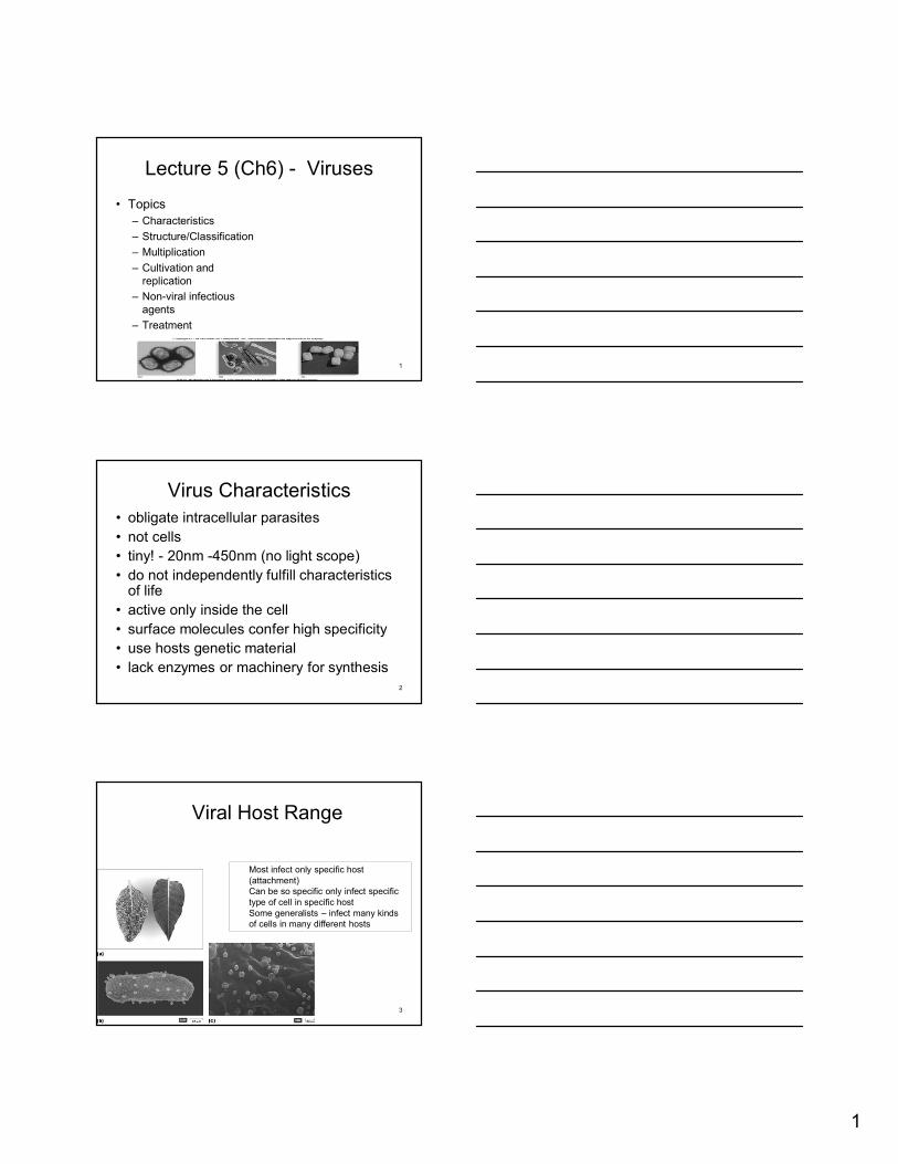
Viral Host Range
3
Most infect only specific host (attachment)Can be so specific only infect specific type of cell in specific hostSome generalists – infect many kinds of cells in many different hosts
2
4
Structure
• Size and morphology
• Capsid
• Envelope
• Complex
• Nucleic acid
Virus- inside & out• Extracellular
– Called virion
– Protein coat (capsid) surrounding nucleic acid
– Nucleic acid and capsid also called nucleocapsid
– Some have phospholipid envelope
– Outermost layer provides protection and recognition sites for host cells
• Intracellular
– Capsid removed
– Virus exists as nucleic acid
5
6
Size Comparison
Size comparison of viruses
3
More Size…
7
8
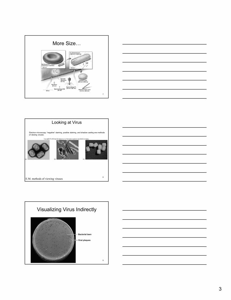
Looking at Virus
E.M. methods of viewing viruses
Electron microscopy, “negative” staining, positive staining, and shadow casting are methods of viewing viruses.
Visualizing Virus Indirectly
9
4
10
The two major structure types for viruses:
Generalized viral structures
naked nucleocapsid virus enveloped virus
11
Capsid
• Protective outer shell that surrounds viral nucleic acid
• Capsid spikes
• Composed of capsomer subunits
• Two types of capsids (based on shape):
– Helical
– Icosahedral
12
Helical capsid
• Naked helical virus
– Nucleocapsid is rigid and tightly wound into a cylinder-shaped package
– Example: Tobacco mosaic virus
• Enveloped helical virus
– Nucleocapsid is more flexible than naked virus
– Examples: Influenza, measles, rabies
5
13
Helical capsids:rod-shaped capsomers form hollow discs, like a bracelet.
Helical nucleocapsid assembly
14
Comparison: naked helical plant virus and an enveloped helical human virus.
Typical variation of viruses with helical Nucleocapsids.
15
Icosahedron capsid
• Three-dimensional, 20-sided with 12 evenly spaced corners
• Variation in capsomer number
– Polio virus 32 capsomers
– Adenovirus 240 capsomers
6
16
The structure and formation of an adenovirus
17
Icosahedral viruses –can be naked or enveloped.
Two types of icosahedral viruses
18
Viral Envelope
• Lipid and proteins
• Envelope spikes
• During release of animal viruses, a part of the host membrane is taken
• Enable pleomorphic shape of the virus
– Spherical
– Filamentous
• Recognition & Attachment
7
19
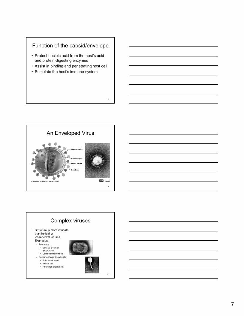
Function of the capsid/envelope
• Protect nucleic acid from the host’s acid-and protein-digesting enzymes
• Assist in binding and penetrating host cell
• Stimulate the host’s immune system
An Enveloped Virus
20
21
Complex viruses
• Structure is more intricate than helical or icosahedral viruses. Examples:– Pox virus
• Several layers of lipoproteins
• Course surface fibrils
– Bacteriophage (next slide)
• Polyhedral head
• Helical tail
• Fibers for attachment
8
22
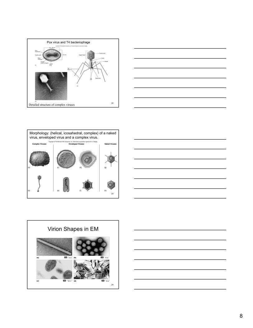
Pox virus and T4 bacteriophage
Detailed structure of complex viruses
23
Morphology: (helical, icosahedral, complex) of a naked virus, enveloped virus and a complex virus.
Virion Shapes in EM
24
9
25
Viral nucleic acid
• Viruses contain either DNA or RNA
• Possess only the genes to invade and regulate the metabolic activity of host cells– Examples:
• Protein particle with no nucleic acid, no envelope, no capsid
• Diseases
– Creutzfeldt-Jakob
– “mad cow disease”
Prion Diseases
– Fatal neurological degeneration, fibril deposits in brain, and loss of brain matter
– Large vacuoles form in brain
• Characteristic spongy appearance
– Spongiform encephalopathies – BSE, vCJD, kuru
– Prions only destroyed by incineration or autoclaving in 1 N NaOH
62
Prions (cont…)
– Cellular PrP protein• Made by all mammals
• Normal structure with -helices called cellular PrP
– Prion PrP• Disease-causing form with -sheets called prion
PrP
– Prion PrP converts cellular PrP into prion PrP by inducing conformational change
63
22
Prion Protein Folding
– Normally, nearby proteins and polysaccharides force PrP into cellular shape
– Excess PrP or PrP mutations result in formation of prion PrP
• Cause newly synthesized cellular PrP to refold into prion PrP
64
Stable Prion Protein (PrP) Forms
65
The Prion Diseases
66
23
67
Satellite viruses
• Dependent on other viruses for replication
• Ex. Delta agent, which is only expressed in the presence of hepatitis B virus, depend on it for replication- the only viroid like infectious agent of animals.