HISTOLOGY OF LARYNX & TRACHEA LEARNING OBJECTIVES At the end of the lecture the student shouldbe able to know : • Different layers of larynx • Histological characteristics of each layer of larynx • Histological classification of laryngeal cartilage • Structure of trachea and its layer • Different layers of trachea and their histological characteristics
Transcript
HISTOLOGY OF LARYNX & TRACHEA
LEARNING OBJECTIVES
At the end of the lecture the student shouldbe able to know :
• Different layers of larynx
• Histological characteristics of each layer of larynx
• Histological classification of laryngeal cartilage
• Structure of trachea and its layer
• Different layers of trachea and their histological characteristics
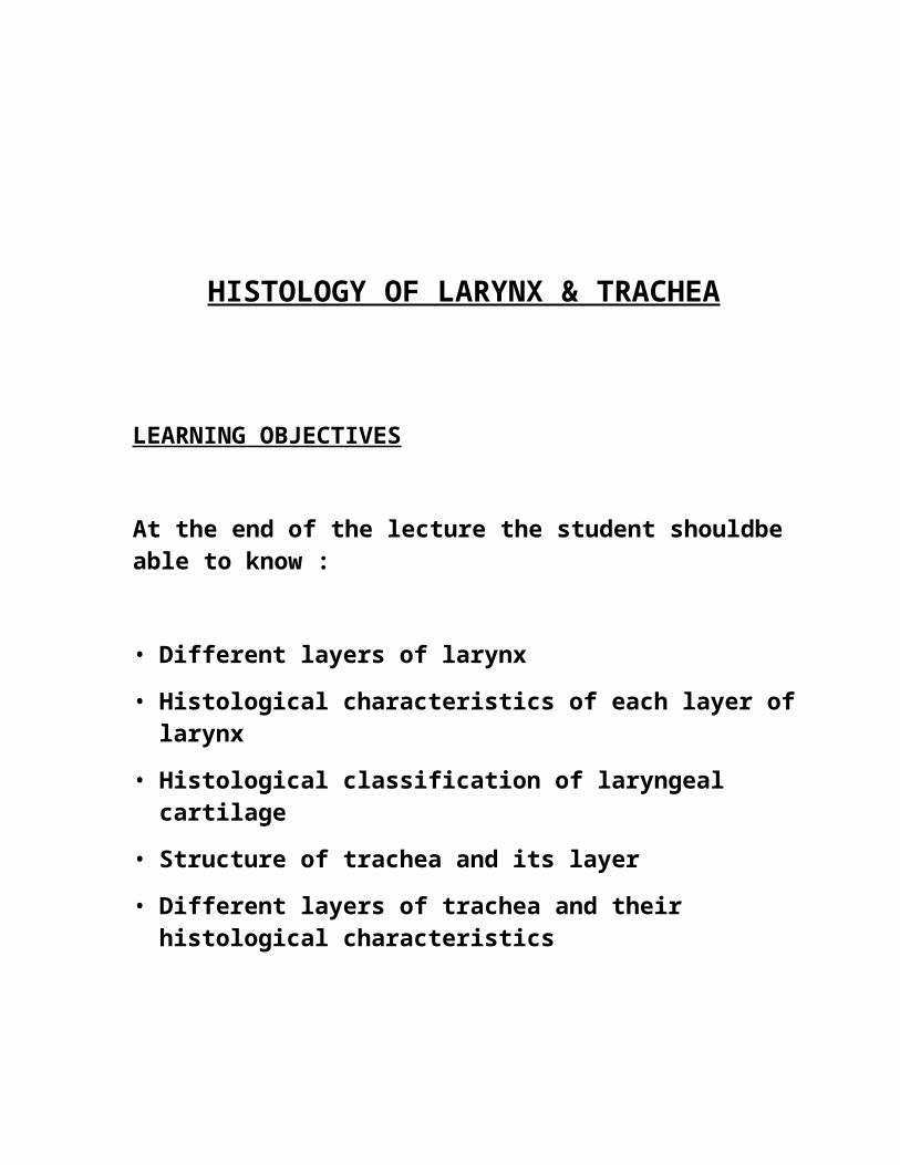
LARYNX
An irregular tube that connects pharynx to the trachea.
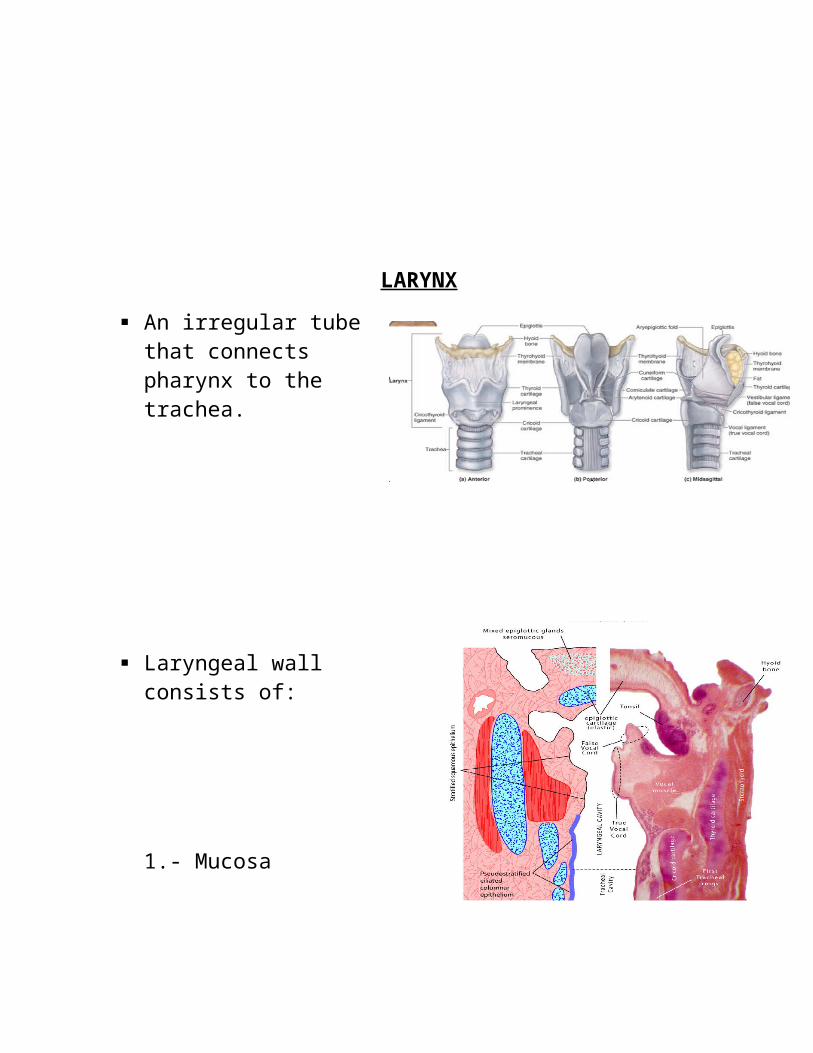
Laryngeal wall consists of:
1.- Mucosa
2.- Cartilages
3.- Intrinsic muscles
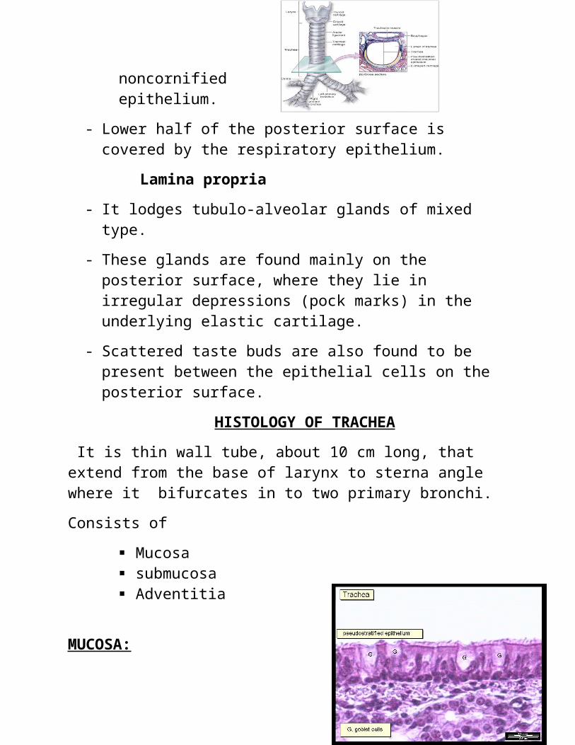
MUCOSA :
comprises of:
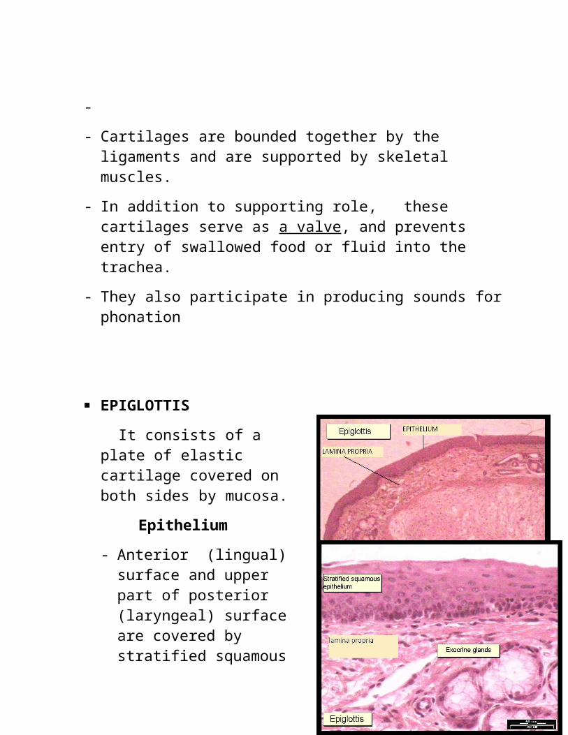
Epithelium
Lamina propria
- Mucosa forms 2 pairs of folds that extend into the lumen of the larynx.