+ All Categories
Home > Documents > Lecture 9 Introduction to Proteases and X-ray Crystallography

Lecture 9 Introduction to Proteases and X-ray Crystallography

Date post: 28-Feb-2022
Category:
Upload: others
View: 5 times
Download: 0 times
Share this document with a friend
22
Biological Chemistry Laboratory Biology 3515/Chemistry 3515 Spring 2022 Lecture 9 Introduction to Proteases and X-ray Crystallography 8 February 2022 ©David P. Goldenberg University of Utah [email protected]
Transcript

Biological Chemistry LaboratoryBiology 3515/Chemistry 3515

Spring 2022

Lecture 9

Introduction to Proteases and X-ray Crystallography

8 February 2022©David P. Goldenberg

University of [email protected]

Computer Labs

Computer Labs this week.• Start at 2:00 PM!• Room 150 Biology Building

This week: Molecular modeling with PyMOL.

We will use the computers in the lab, not personal laptops.

But, you should still install SciDAVis and PyMOL on your own computer.Use the versions available on Canvas.

First Quiz: Thursday, 10 February

In class, during second half.

Study materials:• Quizzes from previous years (Canvas).• Problems in lab manual.• Answers will not be posted, but the TAs and instructors are available for

discussion.

Review session with TAs:• 5:30 PM, Wednesday, 9 February• Room 210, Aline Skaggs Biology Building

The General Protease Reaction

H

N

O

O

N

H

CH 3

O

NH 3

+

+ + H

3 N

CH 3

O

NH 3

+

O

O

H

N

O –

H2OH

N

H

N

residue residue residue residue

About 2% of genes in most organisms encode proteases.(Hedstrom, L. 2002, Chem. Rev. 102, 4429)

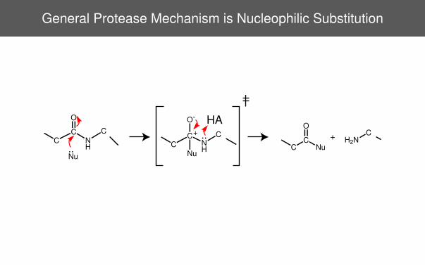
General Protease Mechanism is Nucleophilic Substitution

H2NC

C

C

O

NuC

C

O

NH

C

Nu

+C

C

O-

NH

C

Nu

HA

+

Water Can Act as the Nucleophile,but Must be Activated by a Base

H2NC

C

C

O

OHC

C

O

NH

C +

C

C

O-

NH

C

HA

H

O

H

H

O-

H

O

B

BH+

Why is this reaction so slow in the absence of an enzyme?

How do enzymes enhance the rate?

Carboxyl Groups Activate H2O in Aspartyl Proteases

H

O

H

C

O

NH

CαC

O O-

C

O

OH

Asp Asp

C

O-

NH

H

O

C

O

C

HO O

Asp Asp

OH

Examples include pepsin and HIV protease

Serine Proteases Employ a Two-Step Mechanism

Enzyme-Substrate Complex Acyl Intermediate Enzyme + Products

O

O

O H

H

OH

O

N H

+ H 3 N

OH

O

O –

+ H 3 N

In step 1, a serine hydroxyl is the nucleophile.

In step 2, a water molecule is the nucleophile.

Both steps require activation by a base.

Examples include trypsin, chymotrypsin, blood clotting factors and manyothers.

Clicker Question #1

What bond does a protease cleave?

H

N

O

O

N

H

CH 3

O

NH 3

+

A B

C

How Do We Know What We Know About Serine Proteases?

Information

Observations

Experiments

Data

Knowledge

Organized information

Theories

Predictions

Chemical and biochemical data:• Enzyme kinetics• Studies with inhibitors• Chemical analysis, e.g., active site labeling

Structural analysis, mostly X-ray crystallography

Clicker Question #2: How Big is an Enzyme?

A) 10−10m

B) 10−9m

C) 10−8m

D) 10−7m

E) 10−6m

?

No wrong answers (for now)!

Image Formation with a Lens

Duck Duck Image

Lens

Optical Magnification

Duck

Magnified Duck Image

Lens

A B

As the object is brought closer to the lens:

Image moves further from the lens and becomes larger.

Magnification = B=A

Magnification, in principle, is not limited, but resolution is.

Imaging With a Lens - a Wave Interpretation

Image is formed at points where waves are brought back in phase.

Points in the object must be separated by at least ∼ 1=2 wavelength togive rise to separate points in the image.

The Electromagnetic Spectrum

Illustration From: McMurry, J. & Fay, R. (2004). Chemistry . Prentice-Hall, 4th edition.

Why Not an X-Ray Microscope?

Scattering from individual atoms is very weak, especially from elementswith low atomic numbers.

Very difficult to make lenses for X-rays.

In crystallography:• Use crystals to increase the total scattering intensity.

• Use a mathematical technique, the Fourier transform, to do the job of a lens.

Diffraction from a Duck

Film

A Real Diffraction Pattern From a Pretend Duck

Taylor, C. & Lipson, H. (1964). Optical Transforms: Their preparation and application to X-ray diffractionproblems. Cornell Univ. Press, Ithaca, NY.

Steps in Protein Crystallography

1. Grow Crystals

Entirely empirical andidiosyncratic.

Protein crystals are about 50%water and are kept suspended in asalt solution; close to physiologicalconditions.

Resolution of final structure ishighly dependent on how wellordered the crystals are.

Crystal pictures from: http://biophysics.uoguelph.ca/central/facilities.htm

Steps in Protein Crystallography

2. Collect Diffraction Data

Pictures from:http://www.nsrrc.org.tw/english/research8_1_circle_Diffractometer.aspx

http://www.bnl.gov/nufo/facilities.asp

Steps in Protein Crystallography

3. Determine PhasesDiffraction data contain intensities of scattered X-ray waves, but not theirphases. Both are needed to reconstruct structure.

One way: Comparing diffraction intensities of crystals containingdifferent heavy-atom derivatives.

4. Calculate electron density map

Molecular model is built into the electron density map.

Figure from Berg, Tymoczko and Stryer Biochemistry, 5thed. (2002) W.H. Freeman, New York.

Atomic Coordinates are Depositedin the Protein Data Bank (PDB)

http://www.rcsb.org


Recommended