+ All Categories
Home > Documents > Lecture on Biology and genetics principles specialty 226 ...

Lecture on Biology and genetics principles specialty 226 ...

Date post: 19-Mar-2022
Category:
Upload: others
View: 1 times
Download: 0 times
Share this document with a friend
76
NATIONAL PHARMACEUTICAL UNIVERSITY DEPARTMENT OF VETERINARY MEDICINE AND PHARMACY Lecture on Biology and genetics principles specialty 226 Pharmacy Lecturer: associate professor Department of Veterinary medicine and pharmacy Ph.D. Dotsenko Roman Valeryevich INTRODUCTION TO BIOLOGY
Transcript

NATIONAL PHARMACEUTICAL UNIVERSITY

DEPARTMENT OF VETERINARY MEDICINE AND PHARMACY

Lecture on Biology and genetics principlesspecialty 226 Pharmacy

Lecturer: associate professor Departmentof Veterinary medicine and pharmacyPh.D. Dotsenko Roman Valeryevich

INTRODUCTION

TO BIOLOGY

LECTURE PLAN

Questions for self-examination:

Classification of microorganisms.

Microscopy. Types of Microscopes.

Basic form of bacterial cells

Introduction to biology. History of biology.

The characteristics of life. Levels of life organization.

The scientific method in biology.

Experimental methods in biology.

Microscopy as one of the basic methods in biology.

Preparing specimens for microscopes.

The elements of life and their role in biological systems.

Biological properties of water. pH and its biological significance.

Carbohydrates and their biological importance.

Lipids and their role in biological systems.

Proteins: structure, functions. Denaturation of proteins.

Nucleic acids and their role in the living organisms.

ATP as energetic molecule of the cell.

Recommended literatureMaloshtan L.M., Filiptsova O.V. 2011. Biology and genetics principles, Publisher house of NUPh, Kharkiv, pp. 4-18,56-68.

https://www.britannica.com/list/6-cell-organelles

Craig, Nancy (2014). Molecular Biology, Principles of Genome Function. ISBN 978-0-19-965857-2.

Darwin, Charles (1859). On the Origin of Species, John Murray.

"biology, n". Oxford English Dictionary online version. Oxford University Press. September 2011. Retrieved 1 November 2011.

Microbiology: A Guide to Laboratory Lessons. Study a manual for students of higher educational institutions / IL Wild, I.I. Sidorchuk, I.Yu. Kholupiak, N.E. Sheveleva, MM Great, N.A. Volkova, L.F. Silayeva, O.P. Strilec, O.G. Heyderich, V.E. Litarov - Kh.: Publishing house of NfaU; Golden Pages, 2002. 444 p.

BIOLOGY

Zoology

biological

chemistry

(KROK 1)

Physiology

Pharmacology

(KROK 1)

Microbiology

(KROK 1)

Botany

(KROK 1)

Ecology

Pathologic

physiology

(KROK 1)

Organic

chemistry

(KROK 1)

History of biology

Hippocratesundated bust.

© Photos.com/ThinkstockEbers papyrus prescription for asthma treatment.U.S.

National Library of Medicine/National

Institutes of Health

1665

Robert Hooke

Observed cork and saw little boxes that

he called “cells”

1675

Anton von

Leeuwenhoek

Observed pond water and saw

“animalcules”

Watson and Crick discovers DNA

Feb 28th 1953

Atoms

Molecules

Cells

Tissues

Organs

Organ systems

Prions

Viruses

Virology (the study of viruses) – is such a

significant part of microbiology

A Virus is a non-cellular particle made up

of genetic material and protein that can

reproduction only in the living cells

A complete virus particle as it exists

outside the cell called virion (or viral

particle)

Viruses can infect all types of cells: bacteria,

protozoa, plants, animals, human.

1. small size: cannot be viewed with a light microscope, range of size = 30-400 nm

2. characteristic shapes - spherical (complex), helical, rodor polyhedral, sometimes with tails or envelopes. Mostcommon polyhedron is the icosahedron which as 20 triangular faces.

3. obligate intracellular parasites: Viruses do not containwithin their coats the machinery for replication. For thisthey depend upon a host cell and this accounts for theirexistence as obligate intracellular parasites. Each viruscan only infect certain species of cells. This refers to thevirus host range.

4. no built-in metabolic machinery: Viruses have nometabolic enzymes and cannot generate their ownenergy.

General characteristics of Viruses

5. no ribosomes: Viruses cannot synthesize their own

proteins. For this they utilize host cell ribosomes

during replication.

6. only one type of nucleic acid: Viruses contain either

DNA or RNA (never both) as their genetic material.

The nucleic acid can be single-stranded or double

stranded.

7. do not grow in size: Unlike cells, viruses do not grow

in size and mass leading to a division process. Rather

viruses grow by separate synthesis and assembly of

their components resulting in production of a "crop" of

mature viruses.

General characteristics of Viruses

Classification of Viruses

Viruses are classified on the basis of host range,

morphology (size, shape), type of nucleic acid

(DNA, RNA, single-stranded, double-stranded, linear,

circular, segmented, etc.) and occurrence of auxilliary

structures such as tails or envelopes.

International Committee on Taxonomy of Viruses names

them based on three characteristics:

1. Type of nucleic acid;

2. Is the nucleic acid double or single stranded:

3. Presence or absence of nuclear envelope.

Comparative size and shape of various groups of viruses

Viruses have fundamentally three morphologies:

1. icosahedron (E, F, G, H, L, N);

2. helical (D, I, J, K, M; B is controversial); 3. complex (A).

Virus structure

Viruses contain with nucleic acid and protective protein

coat. There are 2 large groups of viruses, which differ

one from another morphologically.

The first group is naked viruses or simple viruses,

The secod – envelope viruses or complex viruses.

1 2

Scheme of the structure of viruses

Envelope viruses Nonenvelope viruses

Double-stranded DNA

Single-stranded DNA

Double-stranded RNA

Single-stranded RNA

Double-stranded DNA

Single-stranded RNA

Inner Core - contains genetic information in the

form of RNA or DNA (never both)

Capsid (Protein Coat) – serves to protect genetic

information inside viral particle and permit attachment

to appropriate host.

Outer Envelope - complex viruses also have a

quasi-membrane or envelope (proteins and

phospholipids) around the Capsid to enhance

adhesion to an appropriate host

Capsid Construction - made of identical protein

subunits termed capsomeres

The protein spikes on viruses surface that allow it to

bind and fuse with host cells.

Study of the structure of the variola virus

with an electron microscope

Photos of viruses

Virus structure

25

Virus structure (HIV)

26

Viral Replication

Viruses require living cells for reproduction. The cell

that may be infected named host cell or target cell.

Viruses are obligate intracellular parasites that are

dependent on cellular energy production and cellular

machinery for synthesis of their components.

Viruses have unique replication strategies –

disjunctive mode of reproduction. This mode

consists of separable synthesis of viral components

in a host cell and spontaneous macromolecular

interaction for their maturation.

There are permissive and nonpermissive cells in

human organism. The permissive cell has a

suitable cell receptors and intracellular

requirements are sufficient for supporting the

replication of a particular virus. A productive viral

infection proceeds in permissive cells with form

new virion.

Virus replication is not supported in the

nonpermissive cell because it does not have

specific receptors. The virus cannot replicate itself

in nonpermissive cell.

Viral life cycles in cells is terms viral repduction

(replication). Our knowledge of viral replication

is now very detailed and is expanding rapidly.

Every viral family has a different strategy of

replication. Process of viral replication may be

short – 4-12 hours or very long – for all the

organism life.

An understanding of viral replication provides a

basis for understanding pathogenesis, immunity,

chemotherapy, and role of viruses in cancer.

There are two main ways that viruses reproduce

Viral Replication - Lytic Cycle

Lytic - the virus may be "lytic" (assembling new viral particles from the host's biomolecules) and eventually lysing the host to release the newly-assembled viral population. Lytic stages include:

Adsorption - virus attaches to host's cellular membrane

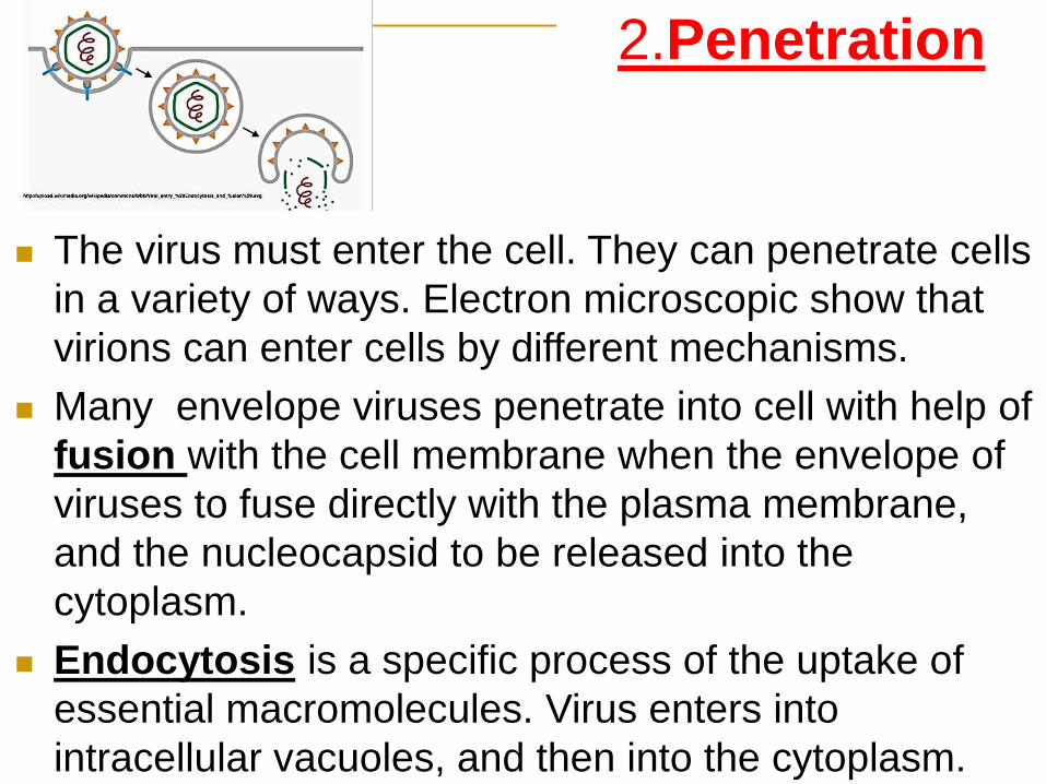
Penetration - virus injects genetic material through membrane

Uncoating - viral coat digested releasing viral genetic material

Synthesis - virus commences cellular take-over and assembly

Release - newly-made viral particles released, cell lysed.

Specific molecules on the surface of the virions

attach specific receptors on the plasma membrane

of cell. Specific-binding proteins of the virus

interact with specific receptors on the target cell

surface. Many kinds of virus receptors are now

known. Adsorption occurs to specific cellular

receptors. Some receptors are glycoproteins,

others are phospholipids or glycolipids.

1.Attachment (Adsorption) This is

the first step in viral replication.

Some viruses have a very narrow host range,

meaning that they can only infect one or a small

number of cell types. For example, HIV infects only

human T cells, because the surface protein interacts

with CD4 receptors on the surface of the T cell.

Some complex viruses have broad host ranges,

meaning that they can infect a large number of

different cell types, for example, Influenzavirus and

Herpesviruses.

The expression of receptors on the surface of cells

largely determines the TROPISM of viruses. Tropism

is the type of cell in which viruses are able to replicate.

1.Attachment (Adsorption)

Adsorption of

HIV on the

surface of

Т-helper

Virion

Т-helper

The virus must enter the cell. They can penetrate cells

in a variety of ways. Electron microscopic show that

virions can enter cells by different mechanisms.

Many envelope viruses penetrate into cell with help of

fusion with the cell membrane when the envelope of

viruses to fuse directly with the plasma membrane,

and the nucleocapsid to be released into the

cytoplasm.

Endocytosis is a specific process of the uptake of

essential macromolecules. Virus enters into

intracellular vacuoles, and then into the cytoplasm.

2.Penetration

Uncoating is the step in which viral enzymes degrade the

virus capsid; that then exposes the viral genome to permit

multiplication of the virus.

4.Strategies of Viral Replication, Gene expression

(biosynthesis):

synthesis of viral mRNA (transcription), synthesis of viral

proteins (translation), and genome replication.

The viruses must use host cellular machinery to replicate

and make functional and structural proteins. Strategies for

genomic expression for different taxonomic group of

viruses are different.

All virulent viruses shut off cellular protein synthesis and

disaggregate cellular polyribosomes, favoring a shift to

viral synthesis.

3.Uncoating

The process assembly of the new components into

virions.

The process of virion assembly involves bringing

together newly formed viral nucleic acid and the

structural proteins to form the nucleocapsid of the

virus. There are basically two strategies that viruses

employ.

For example, for many nonenveloped viruses, the viral

capsid is partially assembled, the newly synthesized

capsid proteins associate together into a capsid-like

structure. The viral genome is then inserted into the

capsid to form a nucleocapsid.

5.Virion assembly (maturation)

of new infectious virus. There are basically two

mechanisms for the release of mature virions from the

infected cell: cytopathogenic or noncytopathogenic.

Most nonenveloped viruses accumulate within the

cytoplasm or nucleus. Release occurs only when the

cell lysis or until the cell slowly degenerates and dies.

Most enveloped viruses are released by budding from

the plasma membrane. It’s a process which can occur

over a prolonged period without much damage to the

cell.

Viruses can replicate tens of thousands of times within

only a few hours.

6.Release

Release of envelope viruses

42

Structure of Bacteriophages1. Size: Most phages range in size from 24-

200 nm in length.

2. Head or Capsid: Some are icosahedral

others are filamentous. The head is

composed of proteins. Inside the head is

found the nucleic acid. The head acts as the

protective covering for the nucleic acid.

3. Tail: Many but not all phages have tails

attached to the phage head. The tail is a

hollow tube through which the nucleic acid

passes during infection. In the more

complex phages the tail is surrounded by a

contractile sheath which contacts during

infection of the bacterium. At the end of the

tail the more complex phages have a base

plate and one or more tail fibers attached to

it. The base plate and tail fibers are involved

in the binding of the phage to the bacterial

cell.

All bacteriophages have a lytic or infectious cycle,

in which the virus, incapable of replicating itself, injects

its genetic material into a bacterium. By pirating its

host’s enzymes and protein-building capacities, the

virus can reproduce and repackage, making about

100 new copies before it bursts from and destroys the

bacteria.

Some bacteriophages, however, behave differently

when they infect a bacterium. The injected genetic

material instead integrates itself into its host DNA,

passively replicating with it to be inherited by bacterial

daughter cells. In about 1 in 100,000 of these

lysogenic cells, the viral DNA spontaneously activates

and starts a new lytic cycle.

CULTIVATION OF VIRUSES:

1. Laboratory animals

2. Embryonated eggs

3. Cell culture

Primary cell cultures

Diploid cell strains

Continuous cell lines

Laboratory animals are

used for primery isolation of certain

viruses and for studies of the

pathogenesis of viral diseases.

They are inoculated by intracerebral or

intranasal routes. Specimens may also be

inoculated by intraperitoneal and

subcutaneous routes.

Growth of virus

on embryonated eggs

This method is used for

the cultivation of more than

30 species of viruses.

The material containing

the virus is inoculated into

the amnion, allantois, yolk

sac and others.

After inoculation of the

chick embryo, it is

incubated and examined

daily for virus growth.

The virus is inoculated into the allantois of the embryonated egg

The cell culture

method is

employed nowadays

for identification and

cultivation of viruses.

Cell suspension is

distributed in tubes,

bottles or Petri dishes.

Cytopathic effect (CPE)

The development of the cytopathic effect is an indicator

of the replication of the virus in cell cultures.

CPE are of the following types:

total destruction of the monolayer sheet (cell necrosis and

lysis), e.g. enteroviruses;

syncytium (multinucleated giant cells) formation, e.g. measles

virus;

cellular clumping, e.g. adenovirus;

inclusion bodies;

discrete focal degeneration, e.g. herpes virus;

transformation, e.g. oncogenic virus.

Hemadsorbtion

Inclusion body Cell necrosis

Cytopathic effect (CPE)

Syncytium

Conclusions:

The main differences between viruses:

Small dimensions

Lack of cellular structure

Lack of own protein synthesis systems.

There are only one of two nucleic acids in viruses

Genetic parasitism.

Viruses do not grow

Reproduction through disjunctive reproduction

Ability to integrate into the cellular genome and

synchronous replication

Cells types

PROCARYOTIC

CELL

STRUCTURE

Procaryotic cells are

morphologically

much simpler than

eucaryotic cells

Differences between procaryotic

and eucaryotic cellsProcaryotic cell

Nucleus ABSENT:

Nuclear membrane

Nucleolus

Mitotic division

CHROMOSOME:

One

DNA:

Circular

Eucaryotic cell

Nucleus PRESENT:

Nuclear membrane

Nucleolus

Mitotic division

CHROMOSOME:

More

DNA:

Linear

Differences between procaryotic

and eucaryotic cellsProcaryotic cell

Cytoplasm ABSENT:

Cytoplasmic streaming

Mitochondria

Lysosomes

Golgi apparatus

Endoplasmic reticulum

CYTOPLASMIC

RIBOSOME:

70s

Eucaryotic cell

Cytoplasm PRESENT:

Cytoplasmic streaming

Mitochondria

Lysosomes

Golgi apparatus

Endoplasmic reticulum

CYTOPLASMIC

RIBOSOME:

80s

Differences between procaryotic

and eucaryotic cellsProcaryotic cell

Miscellaneous

DIAMETER:

1 μm

OXIDATIVE

PHOSPHORYLATION

SITE:

periplasmic space of

cell membrane

CILIA:

Absent

PILI:

Present

Eucaryotic cell

Miscellaneous

DIAMETER:

10 μm

OXIDATIVE

PHOSPHORYLATION SITE:

Mitochondria Chloroplast

CILIA:

Present

PILI:

Absent

Procaryotic Cell MembranesThe plasma membrane encompasses the cytoplasm of

cell. Membranes contain both proteins and lipids. Lipids are

with polar (hydrophilic) and nonpolar (hydrophobic) ends

and are called amphipathic. This property of lipids enables

them to form a bilayer in membranes. Cell membranes are

very thin structures, about 5 to 10 nm thick.

Roles of the Plasma Membrane

Selectively permeable barrier

Mechanical boundary of cell

Nutrient and waste transport

Location of many metabolic processes

(respiration, photosynthesis)

Detection of environmental cues for

chemotaxis.

Internal Membrane Systems

Mesosomes - are invaginations of the plasma membrane

in the shape of vesicles, tubules, or lamellae.

Mesosomes may be involved in cell wall formation during

division or play a role in chromosome replication and

distribution to daughter cells.

Mesosomes also may be involved in secretory processes

and respiration processes.

The Cytoplasmic Matrix

Inclusion Bodies:

Organic –

Glycogen, PHB (Poly-β-hydroxybutyrate) – are carbon storage reservoirs providing material for energy and biosynthesis.

Carboxysomes - serve as a reserve of this enzyme and may be a site of CO2 fixation.

Gas vacuole - give bacteria buoyancy.

Inorganic –

Volutin – storage reservoirs for phosphate.

Sulfur granules - storage reservoirs for sulfur.

Metachromatic granules - energy reserve.

Ribosomes

The cytoplasmic matrix often is packed with

ribosomes; they also are loosely attached to

the plasma membrane. Ribosomes are actually

very complex objects made of both protein and

ribonucleic acid (RNA). They are the sites of

protein synthesis.

Nucleoid

The procaryotic chromosome, almost always a

single circle of double-stranded

deoxyribonucleic acid (DNA), is located in an

irregularly shaped region called the nucleoid

(other names are also used: the nuclear body,

chromatin body, nuclear region).

Plasmids

Many bacteria possess plasmids in addition to their

chromosome. These are circular, double-stranded

DNA molecules that can exist and replicate

independently of the chromosome or may be

integrated with it. Plasmid genes can render bacteria

drug-resistant, give them new metabolic abilities, make

them pathogenic, or endow them with a number of

other properties.

Procaryotic Cell Wall

Most bacteria have strong walls that give them

shape and protect them from osmotic lysis.

The cell walls of many pathogens have

components that contribute to their

pathogenicity. The wall can protect a cell from

toxic substances and is the site of action of

several antibiotics. After Christian Gram developed the

Gram stain in 1884, it soon became evident that

bacteria could be divided into two major groups based

on their response to the Gram-stain procedure.

Gram-positive cell wall

The gram-positive cell

wall consists of a single

20 to 80 nm thick

homogeneous

peptidoglycan or

murein layer lying

outside the plasma

membrane.

Gram-negative cell wallThe gram-negative cell wall is quite complex. It has a 1-to 3nm

peptidoglycan layer next to the plasma membrane. The outer

membrane lies outside the thin peptidoglycan layer. A space

between the plasma membrane and the outer membrane is

called the periplasmic space.It contains many proteins,

hydrolytic enzymes

the Cell Wall External Components

Some bacteria have a layer of material lying

outside the cell wall. When the layer is well

organized and not easily washed off, it is called

a capsule. Capsule help bacteria resist

phagocytosis by host phagocytic cells.

Capsules contain a great deal of water and can

protect bacteria against desiccation. They

exclude bacterial viruses and most hydrophobic

toxic materials such as detergents.

Pili and Fimbriae

Many gram-negative bacteria have short, fine, hairlike

appendages that are thinner than flagella and not involved

in motility. These are usually called fimbriae (s., fimbria).

Some types of fimbriae attach bacteria to solid surfaces

such as rocks in streams and host tissues.

Sex pili (s., pilus) are similar appendages,

Pili often are larger than fimbriae. They are genetically

determined by sex factors or conjugative plasmids and

are required for bacterial mating.

Flagella

Most motile bacteria move by use of flagella. Bacterial

species often differ distinctively in their patterns of flagella

distribution.

Monotrichous bacteria (trichous means hair) have one flagellum.

Amphitrichous bacteria (amphi means "on both sides") have a

single flagellum at each pole.

Lophotrichous bacteria (lopho means tuft) have a cluster of

flagella at one or both ends.

Flagella are spread fairly evenly over the whole surface of

peritrichous (peri means "around") bacteria.

Flagella

A - Monotrichous

B –Lophotrichous

C - Amphitrichous

D - Peritrichous

The Bacterial Endospore

Spore Formation,

sporogenesis or sporulation It is complex process and may be divided into seven

stages. An axial filament of nuclear material forms (stage I),

followed by an inward folding of the cell membrane to

enclose part of the DNA and produce the forespore septum

(stage II). The membrane continues to grow and engulfs

the immature spore in a second membrane (stage III).

Next, cortex is laid down in the space between the two

membranes, and both calcium and dipicolinic acid are

accumulated (stage IV). Protein coats then are formed

around the cortex (stage V), and maturation of the spore

occurs (stage VI). Finally, lytic enzymes destroy the

sporangium releasing the spore (stage VII). Sporulation

requires only about 10 hours.

A number of gram-positive bacteria can form a special

resistant, dormant structure called an endospore.

These structures are resistant to environmental

stresses such as heat, ultraviolet radiation, chemical

disinfectants, and desiccation.

Spore position in cell differs among species, making it

of considerable value in identification.

Spores may be centrally located, close to one end

(subterminal), or definitely terminal. Sometimes a

spore is so large that it swells the sporangium.

Conclusions: Procaryotic cells are morphologically much simpler than eucaryotic cells

The cell envelope of gram-positive cells is relatively simple, consisting of

two to three layers: the cytoplasmic membrane, a thick peptidoglycan layer,

and in some bacteria an outer layer called the capsule. The structure and

function of these layers are described below.

This is a highly complex, multilayered structure. The cytoplasmic membrane

(called the inner membrane in gram-negative bacteria) is surrounded by a

single planar sheet of peptidoglycan to which is anchored a complex layer

called the outer membrane. An outermost capsule may also be present. The

space between the inner and outer membrane is called the periplasmic

space.

Thank you for attention


Recommended