Craniometry and Functional Craniometry and Functional Craniology Craniology Part I: Part I: Anthropometry, Craniometry and Cephalometry Anthropometry, Craniometry and Cephalometry Michael S. Yuan, DDS, MA, PhD Michael S. Yuan, DDS, MA, PhD Assistant Professor of Clinical Dentistry Assistant Professor of Clinical Dentistry Division of Orthodontics Division of Orthodontics School of Dental and Oral Surgery School of Dental and Oral Surgery Columbia University Columbia University November 25, 2003 November 25, 2003 Lecture outline Lecture outline 1. Introduction: the scope and history 2. Definition and objectives 3. Identification of anatomical landmarks 4. Measurements: metric vs non-metric; direct vs indirect 5. Measuring devices 6. Sex/gender estimation 7. Age estimation 8. Racial/ethnic estimation 9. Other methodology, comparisons, and interpretations 10. Clinical applications Anthropometry Anthropometry • Definition: measurement of human head and body • Scope: somatometry, osteometry, craniometry, cephalometry, odontometry • Origin: The methodology probably began because of the interest in the racial classifications (in search of the origin of the human races: monogenism vs polygenism) (Anders Retzius: Swedish; cephalic index) • Objectives: 1) to examine the differences between species; 2) to investigate the variations within species, which include temporal changes, sexual dimorphism, geographical and ethnic differences; 3) to explore the trends and evolution as well as to interpret fossil records; 4) to apply in clinical diagnosis, treatment planning, forensics, and other commercial applications. Anthropometric Measuring Devices Anthropometric Measuring Devices Direct method • Sliding caliper • Hinge (spreading) caliper • Stadiometer/Osteometric board • Coordinate caliper • Head spanner/Todd’s craniostat • Soft metric tape • Others Indirect method • Digitizer • Surface scanner • Other computer assisted imaging and measuring devices (CT scan, MRI, Sonography, etc.) • Radiography Sliding Sliding Caliper aliper (Non-Vernier vs. Vernier) Vernier calipers: Align the scale to achieve one more digit reading in measurement closed open The The Mitutoyo Mitutoyo Digital Sliding Digital Sliding Caliper Caliper
Transcript
1
Craniometry and Functional Craniometry and Functional CraniologyCraniology
Part I:Part I:Anthropometry, Craniometry and CephalometryAnthropometry, Craniometry and Cephalometry
Michael S. Yuan, DDS, MA, PhDMichael S. Yuan, DDS, MA, PhDAssistant Professor of Clinical DentistryAssistant Professor of Clinical Dentistry
Division of OrthodonticsDivision of OrthodonticsSchool of Dental and Oral SurgerySchool of Dental and Oral Surgery
Columbia UniversityColumbia University
November 25, 2003November 25, 2003
Lecture outlineLecture outline
1. Introduction: the scope and history
2. Definition and objectives
3. Identification of anatomical landmarks
4. Measurements: metric vs non-metric; direct vs indirect
5. Measuring devices
6. Sex/gender estimation
7. Age estimation
8. Racial/ethnic estimation
9. Other methodology, comparisons, and interpretations
• Origin: The methodology probably began because of the interest in the racial classifications (in search of the origin of the human races: monogenism vs polygenism) (Anders Retzius: Swedish; cephalic index)
• Objectives: 1) to examine the differences between species;2) to investigate the variations within species, which include
temporal changes, sexual dimorphism, geographical and ethnic differences;
3) to explore the trends and evolution as well as to interpret fossil records;
4) to apply in clinical diagnosis, treatment planning, forensics, and other commercial applications.
• Other computer assisted imaging and measuring devices (CT scan, MRI, Sonography, etc.)
• Radiography
Sliding Sliding CCaliperaliper
(Non-Vernier vs. Vernier)
Vernier calipers: Align the scale to achieve one more digit reading in measurement
closed
open
The The MitutoyoMitutoyo Digital Sliding Digital Sliding CaliperCaliper
2
Spreading CalSpreading Caliiperper
closed open
StadiometerStadiometer“Stretch of the Measuring”
Johann Wolfgang von Goethe, 1779
OsteometricOsteometric BoardBoard ToddTodd’’s s CraniostatCraniostat(Head (Head SpannerSpanner)
Soft Metric TapeSoft Metric Tape Body Imaging: 3Body Imaging: 3-- D D SurfaceSurface AnthropometryAnthropometryThe Loughborough Anthropometric Shadow Scanner
3
The computerized wholeThe computerized whole-- body image after scanningbody image after scanning(Surface area and volume estimations; Shape capturing and reconstruction)
33-- D ImagingD Imaging
Source: Pre-operative (L) and post-operative (R) 3-D images of a trigonocephaly case http://www.health.adelaide.edu.au/paed-neuro/craniofacial.html (2002)
(morphometrics in size and shape)
CraniometryCraniometry
• Definition: measurement of human dry skull
• Landmarks: 1) true vs relative landmarks2) mid-sagittal vs bilateral landmarks
• Measurements: 1) qualitative (non-metric) vs quantitative (metric)
Basion: the midpoint of the anterior margin of the foramen magnum.
Gnathion: the most anterior and lowest median point on the border of the mandible.
Glabella: the most forward projecting point in the midline of the forehead at the level of the supra-orbital ridges and above the nasofrontal suture.
Opisthocranion: the most posterior point on the skull not on the external occipital protuberance. It is the posterior end point of maximum cranial length measured from glabella. It is determined instrumentally.
Euryon: the two points on the opposite sides of the skull that form termini of the lines of greatest breadth. The two points are determined instrumentally.
Zygion: the most lateral point of the zygomatic arch. It is determined instrumentally.
Orbitale: the lowest point in the margin of the orbit; one of the points used in defining Frankfort Horizontal.
Porion: the uppermost lateral point in the margin of the external auditory meatus. The right and left porion with the left orbitale define the Frankfort Horizontal
Mastoidale: the lowest point of the mastoid process
Gonion: the midpoint of the angel of the mandible between body and ramus.
Bregma: the intersection of the coronal and sagittal sutures in the midline.
Lambda: the intersection of the sagittal and lambdoidal sutures in the midline.
Nasion: the intersection of the nasofrontal suture with the midsagittal plane. Nasion is the uppermost landmark for the measure of facial height.
Menton: the lowest median point of the chin.
Pogonion: the most anterior point in the midline of the chin.
4
Frankfort Horizontal Frankfort Horizontal (FH)(FH)1) A plane passing through three points of the right and left porion and the left orbitale.2) First proposed at the Craniometric Congress held in Munich, Germany, 1877.3) An orientation of skull in a consistent and reproducible position.4) Comparisons: natural head position; horizontal visual axis; and horizontal plane.
Drawing of a Child at Birth, Age 1, Age 2Drawing of a Child at Birth, Age 1, Age 2Bergmüller (1723), Countway Library, Boston CraniometricCraniometric Measurements (I)Measurements (I)
CraniometricCraniometric Measurements (II)Measurements (II)
Condylo-symphyseal lengthBicondylar widthMin. ramus breadthMandibular body heightSymphyseal heightMastoid lengthAscending ramus heightMandibular body breadthMandibular body length
Chamaeconchy (X-82.99): wide orbitsMesoconchy (83.00-89.99): average or mediumHypsioconchy (89.00-X): narrow or square orbits
Orbital height x 100Orbital Index = ---------------------------
Orbital breadth
Orbital ht.
Orbital br.
6
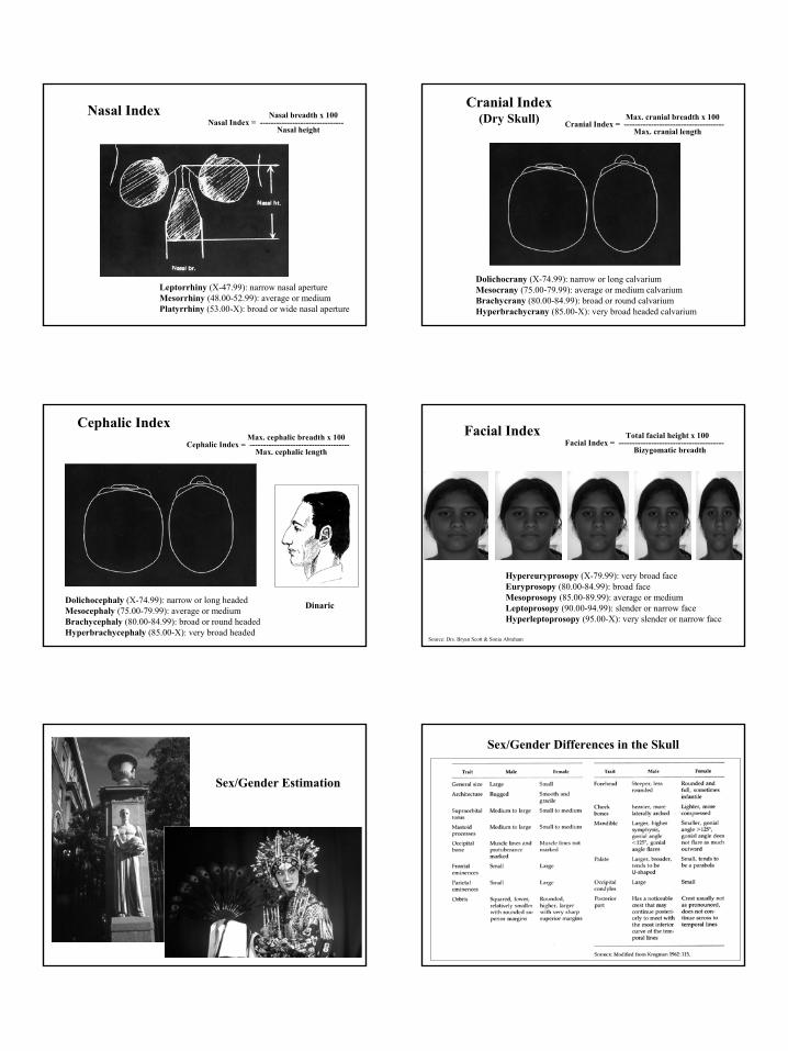
Nasal IndexNasal Index Nasal breadth x 100Nasal Index = -------------------------------
Nasal height
Leptorrhiny (X-47.99): narrow nasal apertureMesorrhiny (48.00-52.99): average or mediumPlatyrrhiny (53.00-X): broad or wide nasal aperture
Cranial IndexCranial Index(Dry Skull)(Dry Skull)
Dolichocrany (X-74.99): narrow or long calvariumMesocrany (75.00-79.99): average or medium calvariumBrachycrany (80.00-84.99): broad or round calvarium Hyperbrachycrany (85.00-X): very broad headed calvarium
Max. cranial breadth x 100Cranial Index = -------------------------------------
Max. cranial length
Cephalic IndexCephalic IndexMax. cephalic breadth x 100
Cephalic Index = -------------------------------------Max. cephalic length
Dolichocephaly (X-74.99): narrow or long headedMesocephaly (75.00-79.99): average or mediumBrachycephaly (80.00-84.99): broad or round headedHyperbrachycephaly (85.00-X): very broad headed
Dinaric
Facial IndexFacial Index Total facial height x 100Facial Index = ---------------------------------------
Bizygomatic breadth
Hypereuryprosopy (X-79.99): very broad faceEuryprosopy (80.00-84.99): broad faceMesoprosopy (85.00-89.99): average or mediumLeptoprosopy (90.00-94.99): slender or narrow faceHyperleptoprosopy (95.00-X): very slender or narrow face
Source: Drs. Bryan Scott & Sonia Abraham
Sex/Gender EstimationSex/Gender Estimation
Sex/Gender Differences in the SkullSex/Gender Differences in the Skull
7
Sexing the SkullSexing the Skull(Multiple Regression Analysis)(Multiple Regression Analysis)
Cephalometric Radiograph and Tracing (lateral view) Cephalometric Radiograph and Tracing (lateral view) Cephalometric LandmarksCephalometric Landmarks
9
Cephalometric Analysis: Columbia AnalysisCephalometric Analysis: Columbia Analysis Cephalometric Analysis: Finite Element AnalysisCephalometric Analysis: Finite Element Analysis
ReferencesReferences
Bass, W.M. (1987). Human Osteology: A Laboratory and Field Manual (3rd
edition). Special Publication No.2 of the Missouri Archeological Society. Columbia, Missouri: Missouri Archeologicall Soceity, Inc..
White, T.D. (2000). Human Osteology (2nd edition). San Diego, California: Acadmic Press.
Steele, D.G. & Bramblett, C.A. (1998). The Anatomy and Biology of the Human Skeleton. College Station, Texas: Texas A & M University Press
Krogman, W.M. & Iscan, M.Y. (1986). The Human Skeleton in Forensic Medicine. Springfield, Illinois: Charles C. Thomas Publisher.
AcknowledgmentsAcknowledgments
Thanks toThanks to
Professor Melvin MossProfessor Melvin MossProfessor Professor LettyLetty MossMoss--SalentijnSalentijn
Professor Professor Alfonso Alfonso SolimeneSolimeneProfessor Ralph L. HollowayProfessor Ralph L. Holloway
AndAndDr. Dr. ChristelChristel HummertHummert
Dr. Sonia AbrahamDr. Sonia AbrahamDr. Bryan ScottDr. Bryan Scott