INTRODUCTION INTRODUCTION GENERAL PRINCIPLES GENERAL PRINCIPLES Motion Motion Motion is a fundamental property of most animal Motion is a fundamental property of most animal life. In the simple unicellular animals, motion life. In the simple unicellular animals, motion and locomotion depend upon the contractility of and locomotion depend upon the contractility of protoplasm and the action of accessory organs protoplasm and the action of accessory organs such as cilia, flagella, etc. The lowest such as cilia, flagella, etc. The lowest multicellular animals possess rudimentary multicellular animals possess rudimentary neuromuscular mechanisms; in higher forms, motion neuromuscular mechanisms; in higher forms, motion is based upon the transmission of impulses from a is based upon the transmission of impulses from a receptor through an afferent neuron and ganglion receptor through an afferent neuron and ganglion cell to muscle. This same principle is found in cell to muscle. This same principle is found in the reflex arc of higher animals, including the reflex arc of higher animals, including humans, in whom the anterior spinal cord has humans, in whom the anterior spinal cord has developed into a central regulating mechanism,
Motion is a fundamental property of most animal life. In the simple Motion is a fundamental property of most animal life. In the simple unicellular animals, motion and locomotion depend upon the unicellular animals, motion and locomotion depend upon the contractility of protoplasm and the action of accessory organs contractility of protoplasm and the action of accessory organs such as cilia, flagella, etc. The lowest multicellular animals such as cilia, flagella, etc. The lowest multicellular animals possess rudimentary neuromuscular mechanisms; in higher forms, possess rudimentary neuromuscular mechanisms; in higher forms, motion is based upon the transmission of impulses from a receptor motion is based upon the transmission of impulses from a receptor through an afferent neuron and ganglion cell to muscle. This same through an afferent neuron and ganglion cell to muscle. This same principle is found in the reflex arc of higher animals, including principle is found in the reflex arc of higher animals, including humans, in whom the anterior spinal cord has developed into a humans, in whom the anterior spinal cord has developed into a central regulating mechanism, the brain, which is concerned with central regulating mechanism, the brain, which is concerned with initiating and integrating movements.initiating and integrating movements.
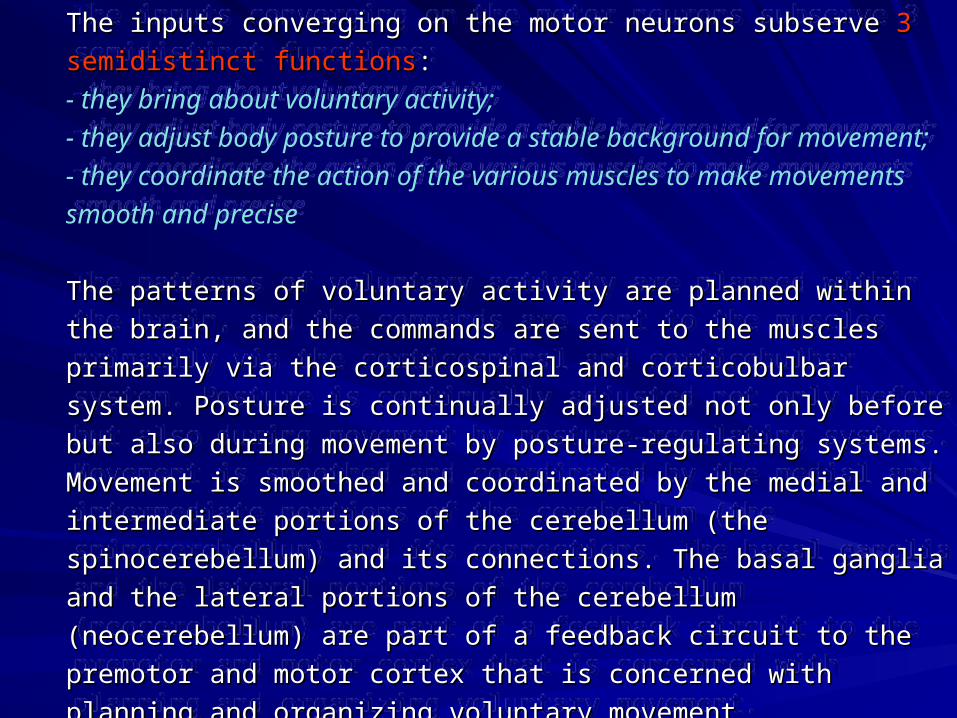
The inputs converging on the motor neurons subserve The inputs converging on the motor neurons subserve 3 semidistinct 3 semidistinct functionsfunctions::- they bring about voluntary activity; - they adjust body posture to provide a stable background for movement; - they coordinate the action of the various muscles to make movements smooth and precise
The patterns of voluntary activity are planned within the brain, and the The patterns of voluntary activity are planned within the brain, and the commands are sent to the muscles primarily via the corticospinal and commands are sent to the muscles primarily via the corticospinal and corticobucorticobullbar system. Posture is continually adjusted not only before but bar system. Posture is continually adjusted not only before but also during movement by posture-regulating systems. Movement is also during movement by posture-regulating systems. Movement is smoothed and coordinated by the medial and intermediate portions of the smoothed and coordinated by the medial and intermediate portions of the cerebellum (the spinocerebellum) and its connections. The basal ganglia cerebellum (the spinocerebellum) and its connections. The basal ganglia and the lateral portions of the cerebellum (neocerebellum) are part of a and the lateral portions of the cerebellum (neocerebellum) are part of a feedback circuit to the premotor and motor cortex that is concerned with feedback circuit to the premotor and motor cortex that is concerned with planning and organizing voluntary movement.planning and organizing voluntary movement.
The cortical areas from which The cortical areas from which the corticospinal and the corticospinal and corticobulbar system corticobulbar system originates are generally held to originates are generally held to be those where stimulation be those where stimulation produces prompt discrete produces prompt discrete movement. The best known is movement. The best known is the motor cortex in the the motor cortex in the precentral gyrus.precentral gyrus.
30% of the fibers making up the corticospinal and corticobulbar 30% of the fibers making up the corticospinal and corticobulbar tracts come from the motor cortex but 30% come from the tracts come from the motor cortex but 30% come from the premotor cortex and 40% from the parietal lobe, especially the premotor cortex and 40% from the parietal lobe, especially the somatic sensory area.somatic sensory area.
The various parts of the body are The various parts of the body are represented in the represented in the precentral gyrusprecentral gyrus, , with the feet at the top of the gyrus with the feet at the top of the gyrus and the face at the bottom. The facial and the face at the bottom. The facial area is represented bilaterally, but the area is represented bilaterally, but the rest of the representation is unilateral, rest of the representation is unilateral, the cortical motor area controlling the the cortical motor area controlling the musculature on the opposite side of musculature on the opposite side of the bodythe body. The cortical representation . The cortical representation of each body part is proportionate in of each body part is proportionate in size to the skill with which the part is size to the skill with which the part is used in fine, voluntary movement. The used in fine, voluntary movement. The areas involved in speech and hand areas involved in speech and hand movements are especially large in the movements are especially large in the cortex; use of the pharynx, lips, and cortex; use of the pharynx, lips, and tongue to form words and of the tongue to form words and of the fingers and opposable thumbsfingers and opposable thumbs toto
manipulate the environment are activities in which humans are especially skilled.manipulate the environment are activities in which humans are especially skilled.
The corticospinal tract originates as the The corticospinal tract originates as the axons of pyramidal neurons in the precentral axons of pyramidal neurons in the precentral gyrus. These neurons are especially large gyrus. These neurons are especially large cells, called cells, called Betz cells.Betz cells.
Deeper into the brain, all of these axons (slips Deeper into the brain, all of these axons (slips of white matter) merge to form one large body of white matter) merge to form one large body of axons, the of axons, the corona radiatacorona radiata, "radiating , "radiating crown".crown".
TractsTracts
As you get still deeper into the hemispheres, the As you get still deeper into the hemispheres, the corona radiata dives into the deep nuclei of the corona radiata dives into the deep nuclei of the brain, the caudate and putamen, splitting them in brain, the caudate and putamen, splitting them in two. At this point, all of these axons are called two. At this point, all of these axons are called the the internal capsuleinternal capsule..
The internal capsule is a major two-way The internal capsule is a major two-way highway, and very vulnerable to strokes. highway, and very vulnerable to strokes. Sensory information travels up it on the way Sensory information travels up it on the way from the thalamus to the cortex, and motor from the thalamus to the cortex, and motor information travels through on the way down to information travels through on the way down to the spine. In the horizontal sections at the the spine. In the horizontal sections at the beginning of the course, the internal capsule has beginning of the course, the internal capsule has an anterior and posterior limb. The motor and an anterior and posterior limb. The motor and somatosensory information travels through the somatosensory information travels through the posterior limbposterior limb..
In the In the medullamedulla, the fibers come together again as the , the fibers come together again as the pyramidspyramids. The pyramids were actually named as . The pyramids were actually named as landmarks on the surface of the brainstem - on a human landmarks on the surface of the brainstem - on a human brainstem you can clearly see them as two ridges brainstem you can clearly see them as two ridges running down the ventral midline. The pyramids run the running down the ventral midline. The pyramids run the entire length of the medulla, large uninterrupted axon entire length of the medulla, large uninterrupted axon tracts on the ventral surface.tracts on the ventral surface.
At the caudal end of the medulla, right about at the point At the caudal end of the medulla, right about at the point where you have to start calling it cervical spinal cord, where you have to start calling it cervical spinal cord, the the fibers in the pyramids crossfibers in the pyramids cross. The crossing event is called . The crossing event is called the the decussation of the pyramids.decussation of the pyramids.
The nerve fibres that cross the midline in the The nerve fibres that cross the midline in the medullary pyramids and form the medullary pyramids and form the lateral cortico lateral cortico spinal tractspinal tract make up about 80% of the fibres in make up about 80% of the fibres in the corticospinal pathway. The remaining 20% the corticospinal pathway. The remaining 20% make up the make up the anterior or ventral corticospinal anterior or ventral corticospinal tracttract, which does not cross the midline until the , which does not cross the midline until the level at which it synapses with motor neurons. level at which it synapses with motor neurons. In addition, this tract contains corticospinal In addition, this tract contains corticospinal neurons that end on the same side of the body. neurons that end on the same side of the body. The ventral pathway, which is the oldest The ventral pathway, which is the oldest phylogenetically, ends on neurons in the medial phylogenetically, ends on neurons in the medial portion of the ventral horn that control axial and portion of the ventral horn that control axial and proximal limb muscles. Conversely, the lateral proximal limb muscles. Conversely, the lateral cortico spinal pathway innervates lateral neurons cortico spinal pathway innervates lateral neurons in the ventral horn that are concerned with distal in the ventral horn that are concerned with distal limb muscles and hence with skilled limb muscles and hence with skilled movements. In humans, the neurons of this movements. In humans, the neurons of this phylogenetically new system end directly on the phylogenetically new system end directly on the lateral motor neurons.lateral motor neurons.
TractsTracts
The The corticobulbarcorticobulbar (or (or corticonuclearcorticonuclear)) tracttract is a white matter pathway connecting the is a white matter pathway connecting the cerebral cortex to the brainstem (the term cerebral cortex to the brainstem (the term "bulbar" referring to the brainstem). The 'bulb' "bulbar" referring to the brainstem). The 'bulb' is an archaic term for the medulla oblongata. is an archaic term for the medulla oblongata. The muscles of the face, head and neck are The muscles of the face, head and neck are controlled by the corticobulbar system, which controlled by the corticobulbar system, which terminates on motor neurons within terminates on motor neurons within brainstem motor nuclei. This is in contrast to brainstem motor nuclei. This is in contrast to the corticospinal tract, which connects the the corticospinal tract, which connects the cerebral cortex to spinal motor neurons, and cerebral cortex to spinal motor neurons, and controls movement of the torso, upper and controls movement of the torso, upper and lower limbs. The corticobulbar tract lower limbs. The corticobulbar tract innervates cranial motor nuclei bilaterally with innervates cranial motor nuclei bilaterally with the exception of the lower facial nucleus the exception of the lower facial nucleus which is innervated contralaterally. which is innervated contralaterally.
DISTURBANCES IN MOTOR POWERDISTURBANCES IN MOTOR POWER
Motor disturbances include weakness and paralysis.It may be result from lesions of:muscle,myoneural junction,peripheral nerve,CNS: lower motor neuron and upper motor neuron.
The lower motor neuron (final common path way) consists of a cell body located in the anterior gray column of the spinal cord or brain stem and an axon passing by way of the peripheral nerves to the motor end-plates of the muscles. It is the essential motor cell concerned with skeletal activity. It is called the "final common pathway" because it is the ultimate pathway through which neural impulses reach the muscle.
LesionsLesions of the lower motor neurons of the lower motor neurons may be located in the cells of the may be located in the cells of the ventral gray column of the spinal ventral gray column of the spinal cord or brain stem or in their axons, cord or brain stem or in their axons, which constitute the ventral roots of which constitute the ventral roots of the spinal nerves or the cranial the spinal nerves or the cranial nerves. Lesions may result from nerves. Lesions may result from trauma, toxins, infections, vascular trauma, toxins, infections, vascular disorders, degenerative processes, disorders, degenerative processes, neoplasms, or congenital neoplasms, or congenital malformations. Main signs of lower malformations. Main signs of lower motor neuron lesions - it is motor neuron lesions - it is flaccid flaccid paralysisparalysis of the involved muscles: of the involved muscles:
-muscle atrophy (with muscles -muscle atrophy (with muscles degeneration),degeneration),-diminished or absent of reflexes,-diminished or absent of reflexes, absent of pathologic reflexes. absent of pathologic reflexes.
The The upper motor neuronupper motor neuron – it is the – it is the nerve cell of the motor cortex with its nerve cell of the motor cortex with its process that passes through the process that passes through the internal capsule, brain stem, and internal capsule, brain stem, and spinal cord by way of the corticobulbar spinal cord by way of the corticobulbar or corticospinal tract to the lower motor or corticospinal tract to the lower motor neuron.neuron.LesionsLesions of the upper motor neuron of the upper motor neuron may be located in the cerebral cortex, may be located in the cerebral cortex, the internal capsule, the cerebral the internal capsule, the cerebral peduncles, the brain stem, or the peduncles, the brain stem, or the spinal cord. Signs of upper motor spinal cord. Signs of upper motor neuron lesions – it is neuron lesions – it is spastic paralysisspastic paralysis or or paresisparesis of the involved muscles: of the involved muscles:
-no muscle atrophy (probably atrophy -no muscle atrophy (probably atrophy dis use),dis use),-hyperactive deep reflexes, diminished -hyperactive deep reflexes, diminished or absent superficial reflexes,or absent superficial reflexes,-pathologic reflexes.-pathologic reflexes.
Types of Paralysis or Paresis Based on LocationTypes of Paralysis or Paresis Based on Location
HemiplegiaHemiplegia is a spastic or flaccid paralysis of one side of is a spastic or flaccid paralysis of one side of the body and extremities limited by the median line in front the body and extremities limited by the median line in front and in back. and in back. MonoplegiaMonoplegia is a paralysis of one extremity only. is a paralysis of one extremity only. DiplegiaDiplegia is a paralysis of any 2 corresponding extremities- is a paralysis of any 2 corresponding extremities-both lower or both upper extremities).both lower or both upper extremities).ParaplegiaParaplegia is a symmetric paralysis of both lower is a symmetric paralysis of both lower extremities. extremities. QuadriplegiaQuadriplegia, or , or tetraplegiatetraplegia, is a paralysis of all 4 , is a paralysis of all 4 extremities. extremities. Hemiplegia alternansHemiplegia alternans (crossed paralysis) is a paralysis of (crossed paralysis) is a paralysis of one or more ipsilateral cranial nerves and contralateral one or more ipsilateral cranial nerves and contralateral paralysis of the arm and leg.paralysis of the arm and leg.
ReflexesReflexes
Reflexes are inborn stimulus-response mechanisms. The instinctive behavior of lower animals is governed largely by reflexes; in humans, behavior is more a matter of conditioning, and reflexes are subordinated as basic defense mechanisms. The reflexes are, however, extremely important in the diagnosis and localization of neurologic lesions.
ANATOMY OF REFLEXES (The Reflex Arc)ANATOMY OF REFLEXES (The Reflex Arc)
The essential neural portion of a reflex includes a sensory and a motor neuron:
(1) A receptor, such as a special sense organ, cutaneous end organ, or neuromuscular spindle, stimulation of which initiates an impulse.(2) The afferent (or sensory) neuron, which transmits the impulse through a peripheral nerve to the CNS.(3) The efferent (or motor) neuron, which, passing outward in the nerve trunk, delivers the impulse to an effector. (4) An effector, such as a muscle or gland that produces the response.
TYPES OF REFLEXESTYPES OF REFLEXES
The reflexes that are of importance to the clinical neurologist may be divided into 4 groups:
• superficial (or skin and mucous membrane) reflexes, • deep reflexes• visceral (or organic) reflexes • pathologic (or abnormal) reflexes.
Reflexes may also be classified according to the level of their central representation, as spinal, bulbar (postural and righting reflexes), midbrain, or cerebellar reflexes.
1.1. Corneal (or conjunctival) reflexCorneal (or conjunctival) reflex – Blinking of the eye upon – Blinking of the eye upon gentle irritation of the cornea or conjunctiva with a small gentle irritation of the cornea or conjunctiva with a small piece of absorbent cotton. This reflex is lost in lesions of the piece of absorbent cotton. This reflex is lost in lesions of the fifth or seventh cranial nerves or their central connections in fifth or seventh cranial nerves or their central connections in the pons. Corneal ulcers will often result when the reflex is the pons. Corneal ulcers will often result when the reflex is not present; this is because of the absence of the protective not present; this is because of the absence of the protective mechanism.mechanism.
2.2. Pharyngeal (or gag) reflexPharyngeal (or gag) reflex – Retching or gagging when the – Retching or gagging when the pharynx is irritated is absent in lesions of the ninth or tenth pharynx is irritated is absent in lesions of the ninth or tenth cranial nerves or their nuclei and in hysteria.cranial nerves or their nuclei and in hysteria.
3.3. Uvular (or palatal) reflexUvular (or palatal) reflex – Raising of the uvula in – Raising of the uvula in phonation or upon irritation of its mucous membrane is also phonation or upon irritation of its mucous membrane is also dependent upon the ninth and tenth nerves.dependent upon the ninth and tenth nerves.
B.B. Skin ReflexesSkin Reflexes
1.1. Upper and lower abdominal reflexesUpper and lower abdominal reflexes – (Tested on each – (Tested on each side.) Tensing of the muscles beneath the skin area stroked side.) Tensing of the muscles beneath the skin area stroked usually causes the umbilicus to move in the direction of the skin usually causes the umbilicus to move in the direction of the skin area stimulated.area stimulated.
2.2. Cremasteric reflexCremasteric reflex – Elevation of the testicle upon stroking the – Elevation of the testicle upon stroking the inner aspect of the thigh.inner aspect of the thigh.
3.3. Plantar reflexPlantar reflex – Plantar flexion of the toes upon stroking the – Plantar flexion of the toes upon stroking the sole of the foot. In children there is usually also a retraction of sole of the foot. In children there is usually also a retraction of the foot.the foot.
4.4. Anal reflexAnal reflex – Contraction of the sphincter ani upon stroking the – Contraction of the sphincter ani upon stroking the perianal area or upon inserting a gloved finger into the rectum.perianal area or upon inserting a gloved finger into the rectum.
Deep ReflexesDeep ReflexesA. Important Deep ReflexesA. Important Deep Reflexes
1.1. Biceps reflexBiceps reflex – Flexion at the elbow when the biceps tendon – Flexion at the elbow when the biceps tendon is struck.is struck.
2.2. Triceps reflexTriceps reflex – Extension at the elbow when the triceps – Extension at the elbow when the triceps tendon is struck.tendon is struck.
3.3. Periosteoradial reflexPeriosteoradial reflex – Flexion and supination of the – Flexion and supination of the forearm upon striking the styloid process of the radius.forearm upon striking the styloid process of the radius.
4.4. Periosteoulnar reflexPeriosteoulnar reflex – Extension and ulnar abduction of the – Extension and ulnar abduction of the wrist when the styloid process of the ulna is struck.wrist when the styloid process of the ulna is struck.
5.5. Patellar (knee jerk) reflexPatellar (knee jerk) reflex – Extension at the knee when the – Extension at the knee when the patellar tendon is struck. patellar tendon is struck.
6.6. Achilles tendon reflexAchilles tendon reflex – Plantar flexion of the foot when the – Plantar flexion of the foot when the Achilles tendon is struck.Achilles tendon is struck.
Pathologic ReflexesPathologic Reflexes
In this group are found certain primitive defense responses that occur only with lesions of the upper motor neuron. Normally, they are suppressed by cerebral inhibition. When the lower motor neuron is separated from the influence of the higher centers, as in pyramidal tract lesions, they are released. Not in frequently, they can be elicited in normal infants up to age 5-7 months.
A. Lower ExtremityA. Lower Extremity
1.1. Babinski's signBabinski's sign – Extension of the large toe with fanning of the small – Extension of the large toe with fanning of the small toes upon stimulation of the plantar surface of the foot. toes upon stimulation of the plantar surface of the foot.
2.2. Chaddock's toe signChaddock's toe sign – Babinski response obtained by stroking the – Babinski response obtained by stroking the lateral malleolus.lateral malleolus.
3.3. Gordon's leg signGordon's leg sign - Babinski-like response upon squeezing the calf - Babinski-like response upon squeezing the calf muscle.muscle.
4.4. Oppenheim's signOppenheim's sign – Babinski - like response elicited by firm – Babinski - like response elicited by firm downward stroking of the tibia and tibialis anterior muscle.downward stroking of the tibia and tibialis anterior muscle.
5.5. Schaefer's signSchaefer's sign – Babinski-like response upon squeezing the Achilles – Babinski-like response upon squeezing the Achilles tendon.tendon.
6.6. Rossolimo's signRossolimo's sign - Flexion of the toes upon tapping the ball of the - Flexion of the toes upon tapping the ball of the foot.foot.
7.7. Mendel-Bechterew signMendel-Bechterew sign – Flexor movement of the 4 outer toes upon – Flexor movement of the 4 outer toes upon striking the dorsum of the foot over the cuboid bone.striking the dorsum of the foot over the cuboid bone.
8.8. Ankle clonusAnkle clonus – A continued rapid flexion and extension of the foot – A continued rapid flexion and extension of the foot obtained by forcibly and quickly dorsiflexing the foot while the leg is obtained by forcibly and quickly dorsiflexing the foot while the leg is held up by the examiner's other hand placed under the popliteal space. held up by the examiner's other hand placed under the popliteal space. A rapidly exhaustible clonus may be normal.A rapidly exhaustible clonus may be normal.
9.9. Patellar clonus (trepidation sign)Patellar clonus (trepidation sign) – A rapid up-and-down movement – A rapid up-and-down movement of the patella when it is forcibly depressed with a quick movement while of the patella when it is forcibly depressed with a quick movement while the leg is in extension and relaxed.the leg is in extension and relaxed.
Methods of testing for extensor- plantar Methods of testing for extensor- plantar reflexesreflexes
B. Upper ExtremityB. Upper Extremity
1.1. Finger flexion reflexFinger flexion reflex (Tromner's sign) – A (Tromner's sign) – A sharp tap on the palmar surface or the tips of sharp tap on the palmar surface or the tips of the middle 3 fingers produces prompt flexion of the middle 3 fingers produces prompt flexion of the fingers.the fingers.
2.2. Bechterew's signBechterew's sign – The patient flexes and – The patient flexes and then relaxes both forearms. The paralyzed then relaxes both forearms. The paralyzed forearm falls back more slowly and in a jerky forearm falls back more slowly and in a jerky manner, even when contractures are mild.manner, even when contractures are mild.
3.3. Mayer's signMayer's sign – Absence of adduction and – Absence of adduction and opposition of the thumb upon passive forceful opposition of the thumb upon passive forceful flexion of the proximal phalanges, especially of flexion of the proximal phalanges, especially of the third and fourth fingers, of the supinated the third and fourth fingers, of the supinated hand.hand.
C. HeadC. Head1.1. Babinski's platysma signBabinski's platysma sign - If resistance is offered to flexion of - If resistance is offered to flexion of
the chin against the chest or to opening the mouth, the the chin against the chest or to opening the mouth, the platysma on the sound side will contract, whereas that on the platysma on the sound side will contract, whereas that on the affected side will not.affected side will not.
2.2. McCarthy's signMcCarthy's sign (glabella reflex) - Percussion of the (glabella reflex) - Percussion of the supraorbital ridge results in a reflex contraction of the orbicularis supraorbital ridge results in a reflex contraction of the orbicularis oculi muscle.oculi muscle.
3.3. Snout reflexSnout reflex – Sharp tapping of the middle of the upper lip – Sharp tapping of the middle of the upper lip induces exaggerated reflex contraction of the lips.induces exaggerated reflex contraction of the lips.
4.4. Head retraction reflexHead retraction reflex – Sharp downward percussion upon the – Sharp downward percussion upon the upper lip with the head inclined slightly forward produces head upper lip with the head inclined slightly forward produces head bending followed by brisk head retraction.bending followed by brisk head retraction.