3488. JULY 5, 1890. Lectures ON SOME POINTS RELATING TO INJURIES TO THE HEAD. Delivered at the Royal College of Surgeons, BY WILLIAM HENRY BATTLE, F.R.C.S., HUNTERIAN PROFESSOR OF SURGERY AND PATHOLOGY, ROYAL COLLEGE OF SURGEONS ; ASSISTANT SURGEON TO THE ROYAL FREE HOSPITAL AND TO THE EAST LONDON HOSPITAL FOR CHILDREN ; FORMERLY RESIDENT ASSISTANT SURGEON AND SURGEON REGISTRAR AT ST. THOMAS’S HOSPITAL. LECTURE I. MR. PRESIDENT AND GENTLEMEN,-Permit me in the drat place, before entering on the more immediate subject of these lectures, to thank you for the honour which you have conferred upon me in electing me to the position which T occupy to-day. Believe me, I am not unmindful of the fact that with the honour there is also responsibility, .and trust that you may consider the result of my investiga- tion into the subject which I have chosen not unworthy of the theatre in which it is delivered. I have taken as a title for these lectures 11 Some Points relating to Eojuries to the Head," and purpose asking you to con- sider with me more especially the effect of injuries when fracture of the cranial base has been produced, with some of the complications which are only too liable to accompany these fractures or follow at a longer or shorter interval after the force producing them is expended. It would obviously take more time than that at my disposal to consider the whole subject of injuries to the head, including those of the vault; indeed, many of the cerebral and other complications which may arise even after fracture of the base will, I regret o say, remain unnoticed. It is acknowledged that a con- sideration of this subject will compel me to travel over much ground which has been well trodden by others, but where this is so I hope that the additional facts which I bring for- ward, though they be only in confirmation of established views, will prove of sufficient value to make the remarks of interest to those who are engaged in the treatment of head injuries. Surgery has done much in recent years to diminish the mortality formerly dependent on injuries of the cranium and its contents, and our literature has been much enriched by numerous contributions on the subject from the pens of able writers in this country and abroad; but this has been chiefly with regard to fractures of the cranial vault not usually extending into the base. It has seemed tome, how- ever, that the subject of fracture of the base of the skull has not received attention to an extent corresponding with its surgical interest when we consider the importance of the part and of the structures in the immediate neighbour- hood, or the high rate of mortality which still prevails after such injury. There are many reasons for this, and the chief of them is that during the course of a year’s surgical work at any of our large hospitals the number of cases which is presented for treatment to any one surgeon in which the base of the skull has been fractured is not large, and should any symptom of unusual character be manifested there is a tendency to wait for another and similar case before the attention of the profession is drawn to it, and if the case re- quired does not come the observation is lost. Isolated facts, too, are apt to fall out of mind and lose importance as the memory of them becomes less distinct. For several years in various official capacities I had the privilege of seeing the surgical practice of St. Thomas’s Hospital, and of closely obseiving amongst other cases many of injury to the head, and through the kindness of the members of the surgical staff of that hospital I am enabled to bring before you a series of the cases which were under care in that institution during a part of that time, and can thus present a compara- tively large number of examples for consideration. To these I have added the results of experience obtained at the Royal Free Hospital or elsewhere. I have made a summary of most of them, and it will be to cases in this group that my remarks will refer unless mention is made to the contrary. TABLE I. —NON-FATAL CASES OF FRACTURED BASE. TABLE II. -FATAL CASES OF FRACTURED BASE.
Transcript
3488.
JULY 5, 1890.
LecturesON
SOME POINTS RELATING TO INJURIESTO THE HEAD.
Delivered at the Royal College of Surgeons,
BY WILLIAM HENRY BATTLE, F.R.C.S.,HUNTERIAN PROFESSOR OF SURGERY AND PATHOLOGY, ROYAL
COLLEGE OF SURGEONS ; ASSISTANT SURGEON TO THE ROYALFREE HOSPITAL AND TO THE EAST LONDON HOSPITALFOR CHILDREN ; FORMERLY RESIDENT ASSISTANT
SURGEON AND SURGEON REGISTRAR ATST. THOMAS’S HOSPITAL.
LECTURE I.MR. PRESIDENT AND GENTLEMEN,-Permit me in the
drat place, before entering on the more immediate subjectof these lectures, to thank you for the honour which youhave conferred upon me in electing me to the position whichT occupy to-day. Believe me, I am not unmindful of thefact that with the honour there is also responsibility,.and trust that you may consider the result of my investiga-tion into the subject which I have chosen not unworthyof the theatre in which it is delivered. I have takenas a title for these lectures 11 Some Points relating toEojuries to the Head," and purpose asking you to con-sider with me more especially the effect of injuries whenfracture of the cranial base has been produced, with some ofthe complications which are only too liable to accompanythese fractures or follow at a longer or shorter interval afterthe force producing them is expended. It would obviouslytake more time than that at my disposal to consider thewhole subject of injuries to the head, including those of thevault; indeed, many of the cerebral and other complicationswhich may arise even after fracture of the base will, I regreto say, remain unnoticed. It is acknowledged that a con-
sideration of this subject will compel me to travel over muchground which has been well trodden by others, but wherethis is so I hope that the additional facts which I bring for-ward, though they be only in confirmation of establishedviews, will prove of sufficient value to make the remarks ofinterest to those who are engaged in the treatment of headinjuries. Surgery has done much in recent years to diminishthe mortality formerly dependent on injuries of the craniumand its contents, and our literature has been much enrichedby numerous contributions on the subject from the pens ofable writers in this country and abroad; but this has beenchiefly with regard to fractures of the cranial vault notusually extending into the base. It has seemed tome, how-ever, that the subject of fracture of the base of the skullhas not received attention to an extent corresponding withits surgical interest when we consider the importance ofthe part and of the structures in the immediate neighbour-hood, or the high rate of mortality which still prevails aftersuch injury. There are many reasons for this, and the chiefof them is that during the course of a year’s surgical workat any of our large hospitals the number of cases which ispresented for treatment to any one surgeon in which thebase of the skull has been fractured is not large, and shouldany symptom of unusual character be manifested there is atendency to wait for another and similar case before theattention of the profession is drawn to it, and if the case re-quired does not come the observation is lost. Isolated facts,too, are apt to fall out of mind and lose importance as thememory of them becomes less distinct. For several years invarious official capacities I had the privilege of seeing thesurgical practice of St. Thomas’s Hospital, and of closelyobseiving amongst other cases many of injury to the head,and through the kindness of the members of the surgicalstaff of that hospital I am enabled to bring before you aseries of the cases which were under care in that institutionduring a part of that time, and can thus present a compara-tively large number of examples for consideration. To theseI have added the results of experience obtained at the RoyalFree Hospital or elsewhere. I have made a summary ofmost of them, and it will be to cases in this group that myremarks will refer unless mention is made to the contrary.
TABLE I. —NON-FATAL CASES OF FRACTURED BASE.
TABLE II. -FATAL CASES OF FRACTURED BASE.
2
I do not propose to consider at any length the way in whichfractures of the base of the skull are produced, for this hasbeen very thoroughly dealt with by others. I think it right,however, to say a few words before proceeding to other partsof the subject. Fractures of the base of the skull may beproduced by direct or by indirect violence. Examples of theformer are afforded by (1) thrust wounds, usually of theorbit or nose; (2) a blow on the chin driving the condyle ofthe lower jaw through the glenoid fossa, as shown bya preparation in the Museum of St. George’s Hospital;(3) a blow on the nose, fracturing the bones which enterinto its formation and the cribriform plate of the ethmoid.The fracturing force may also be conveyed through thecondyles of the occipital bone, either from a fall on the ’,buttocks or feet or as the result of the impact of the bodyweight on the condyles in a fall on the head. Two opposingforces will also produce a fracture, sometimes limited to thecentral bones of the base. They may also be caused bycontrecoup, but are most commonly the result of extensionof a fissure from the vault into a fossa on the side to whichthe violence is applied. They may also be compound, eitheras the result of the force acting at the point struck, or fromtheir extension into cavities which communicate with theexternal air; for this reason the majority of fractures of thebase of the skull are compound when they are in the middlefossa, through implication of the middle ear, also a largenumber of those in the anterior fossa from extension of thefracture through the cribriform plate into the nose. As arule, when they extend into the base from a fissure of thevault, the fracture in the base retains that character, but itmay present some comminution, a not unusual circumstancewhen it has been produced by direct force.
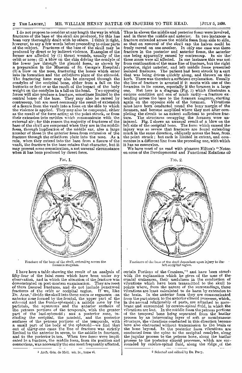
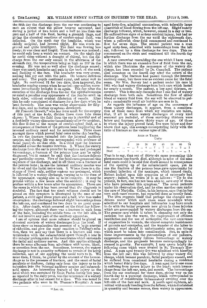
FiG. 1.
Fracture of the base of the skull, extending across theforamen mt,-;num.
I have here a table showing the result of an analysis offifty-four of the fatal cases which have been under myobservation, and in which the situation of the fracture wasdemonstrated on post-mortem examination. They are mostof them fissured fractures, and do not include puncturedfractures of the orbit or occipital region. If we, likeDr. Aran,’ divide the skull into three zones or segments-ananterior zone formed by the frontal, the upper part of theethmoid and the fronto-sphenoid; a middle zone by theparietals, the squamous and the anterior surfaces ofthe petrous portions of the temporals, with the greaterpart of the basi-sphenoid ; and a posterior zone, in-cluding the occipital, the mastoid, and the posteriorsurfaces of the petrous portions of the temporals, witha small part of the body of the sphenoid-we find thatout of thirty-one cases the line of fracture was strictlylimited to the anterior in seven, to the middle in fourteen,and to the posterior in ten. When two fossae were impli-cated in a fracture, the middle fossa, from its position andconnexions, was necessarily the one most frequently affected.
1 Arch. Gén. de Méd. sér. iv., tome vi.
Thus in eleven the middle and posterior fossae were involved,and in three the middle and anteiior. In two instances a.fracture extended across the middle fossa from side to side,,so that on removal of the skull cap the halves could befreely moved on one another. In only one case was there-fracture in the posterior and anterior fossae, the anterior-one being apparently caused by contrecoup. In six the-’three zones were all affected. In one instance this was not-,from continuation of the same line of fracture, but the rightposterior, right anterior, and left middle fcesse presented’isolated fractures. The patient had been struck by a cartthat was being driven quickly along, and thrown on thekerb. There was therefore a sufficient explanation. Usuallythe line of fracture is arrested if it meets with one of the.foramina in its course, especially if the foramen is a largeone. But here is a diagram (Fig. 1) which illustrates a.
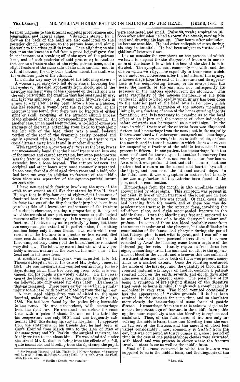
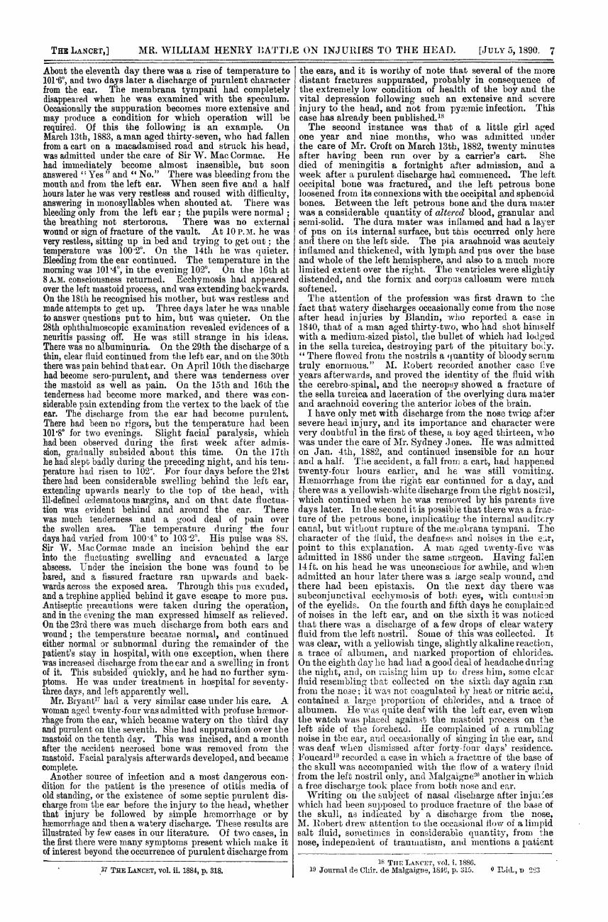
curious condition and one of much rarity-a fracture ex--tending across the base to the foramen magnum, startingagain on the opposite side of the foramen. Vibrations.must have been conducted round the bony margin of theforamen, and become amplified where they met after come-pleting the circuit to an extent sufficient to produce frac-ture. The structures occupying the foramen were un-injured. Fig. 2 shows an unusual result of a blow on theleft side of the occipital bone. The force which caused theinjury was so severe that fractures are found extendingmuch in the same direction, obliquely across the base, from.the point struck; but each is limited in extent, and como.mences in a different line from the preceding one, with whichit has no connexion.We have most of us read with pleasure Hilton’s "Notes.
on some of the Developmental and Functional Relations of;
FIG. 2.
Fractures of the base of the skull dependent upon injury to the-left occipital region.
certain Portions of the Cranium,"2 and have been strucbwith the explanation which he gives of the uses of the-cranial eminences, their usefulness in the conduction ofvibrations which have been transmitted to the skull to-
points where, from the nature of the surroundings, thesevibrations are least calculated to do harm by extension to,the brain. In the anterior fossa they are communicatedfrom the part struck to the anterior clinoid processes, which,,in the normal relationship of parts, are attached to mem-brane and surrounded by cerebro-spinal fluid, in which thevibrations are lost. In the middle fossa the petrous portionsof the temporal bone being separated from the basilarprocess by an intervening layer of soft or membranousstructure, the vibrations conducted in this direction become-here also obstructed without transmission to the brain orthe bone beyond. In the posterior fossa vibrations are-
conducted from the spine to the occipital condyles, alongthe jugular process to the petrous bone, along the basilarprocess to the posterior clinoid processes, which are sur-
rounded by cerebro-spinal fluid, along the ridge of the,
2 Selected and edited by Dr. Pavy.
3
foramen magnum to the internal occipital protuberance and’longitudinal and lateral ridges. Vibrations started by ablow on the back of the head are also conducted to theposterior clinoid processes, to the petrous bone, and alongthe vault to the crista galli in front. Thus alighting on the’feet or on the knees in a fall from a great height3 gave rise4n one instance to a breaking off of the apex of the petrousbone, and of both posterior clinoid processes; in anotherdnstance to a fracture also of the right petrous bone, and aslight fracture of the same side of the sella turcica; and, in.a third instance, the only bone broken about the skull wasthe cribriform plate of the ethmoid.
In a similar way may be explained the following cases :-A woman aged sixty-two fell down stairs, knocking her
eft eyebrow. She died apparently from shock, and at thenecropsy the lesser wing of the sphenoid on the left side wasthe only part within the cranium that had sustained injury,and that was splintered. A woman aged fifty-five died ina similar way after having been thrown from a hansom.She had received a wound over the eyebrow, and at the
necropsy it was found that there was no fracture of eitherspine or skull, excepting of the anterior clinoid processof the sphenoid on the side corresponding to the wound. Inanother case, a man aged twenty-nine, who died of extensivefractures of the vault, extending from back to front alongthe left side of the base, there was a small isolated,portion of the roof of the tympanic cavity loosened and.easily removed with the forceps. The main fracture ran,some distance away from it and in another direction.
With regard to the separation of szttqt2,es at the base, it was’!not uncommonly found that the line of fracture ran into one’of the sutures and caused separation, but at no post-mortemwas the fracture seen to be limited to a suture ; it alwaysextended into a bone beyond. The sutures between theoccipital and other bones were most commonly separated.in one case, that of a child aged three years and a half, who’had been run over, in addition to fracture of the middle- fossa there was separation of the squamo-parietal suture- on each side.
1 have not met with fracture involving the apex of the- orbit to an extent at all like that stated by Van Hiilder."He says that in fifty-four out of eighty-eight instances offractured base there was injury to the optic foramen, butin forty-two out of the fifty-four the injury had been from.gunshot; this still leaves twelve instances of the fractureof a simple character, making a proportion quite beyondwhat the records of our post-mortem rooms or pathologicalmuseums afford in this country. It is a recognised fact thatfractures of the base may unite perfectly by bone, but there.are many examples extant of imperfect union, the unitingmediuna being only fibrous tissue. Two cases which reco-vered from the fracture to a greater or less extent diedsome months afterwards from other causes, and in boththere was good bony union ; but the line of fracture remained’very distinct. The following cases illustrate what was pro-bably a second fracture of the base on the same side of the,head and in the same fossa:-A coachman aged twenty-six was admitted into St.
Thomas’s Hospital, under the care of Mr. Sydney Jones, in1881. He was insensible and vomited continuously for two’days, during which time free bleeding from both ears con-tinued, and the pupils were widely dilated. On the cessa-tion of the bleeding a thin watery discharge from the rightear followed, and only ceased six days later. Deafness in’that ear remained. Three years earlier he had had a similarunjury to the head, with profuse bleeding from the right ear.A man aged thirty-three was admitted to the same
’hospital, under the care of Mr. MacKellar, on July llth,’1886. He had been found by the police lying insensiblein the street. He was unconscious, with haemorrhagefrom the right ear. He remained unconscious for some’time with a pulse of about GO, and on the third day’Ms temperature was only 90 8°, and was frequently sub-mormal after this during his stay in hospital. It appearedfrom the statements of his friends that he had been in’Guy’s Hospital from March 30th to the llth of May ofthe same year; and Mr. Parkin, the surgical registrar, haskindly written to me about him. He was admitted underthe care of Mr. Durham suffering from the effects of a fall,quite insensible, and bleeding from the right ear; the pupils
3 Sir Prescott Hewett and Mr. Hulke in Holmes’ System of Surgery,vol. i., p. 587 ; Jour. de l’Éxper., 1843 ; Bull. de la Soc. Anat. de Paris,1848, pp. 193-258.
4 Berlin : Graefe, von Samisch.
were contracted and small. Pulse 80, weak; respiration 16.Soon after admission he had a convulsive attack, moving hishead and drawing his legs up. Four hours after admissionhe became sensible. He had other epileptic seizures duringhis stay in hospital. He had been subject to "attacks ofgiddiness" between times.Let us consider the symptoms on the presence of which
we have to depend for the diagnosis of fracture in one ormore of the fossae into which the base of the skull is sub-divided. The symptom most commonly met with, and theone on which we rely, more especially in those cases whichcome under our notice soon after the infliction of the injury,is haemorrhage from the seat of the fracture and its appear"ance in the neighbouring tissues, or its escape from thenose, the mouth, or the ear, and not unfrequently itspresence in the matters ejected from the stomach. Thegreat vascularity of the mucous membrane of the noserenders it liable to bleed readily when injury has been doneto the anterior part of the head by a fall or blow, whichmay have caused a laceration of the mucous membranelining it, or a fracture of some of the bones entering into itsformation ; and it is necessary to examine as to the localeffect of an injury and the presence of other indicationsbefore epistaxis can be regarded as a useful sign. In thecases in which fracture of the anterior fossa was diagnosedsixteen had heemorrhage from the nose ; but in the majoritythis was combined with other symptoms, such as hemorrhage,to a greater or less extent, into one or both orbits, or fromthe mouth, and in those instances in which there was reasonfor suspecting a fracture of the middle fossa also it waspresent in fifteen. In one patient the hemorrhage from thenose continued for three days ; in another it was very freewhen lying on the left side, and continued for four hours.As a rule, it was profuse at first and did not recur ; but onepatient had a return on the second and fourth days afterthe injury, and another on the fifth and seventh days. Inthe fatal cases it was a symptom in sixteen, but in onlynine was any fracture of the anterior fossa found at thepost-mortem examination.Haemorrhage from the mouth is also unreliable unless
accompanied by other signs. This symptom was present inten cases, in five of which fracture of the lower and in twofracture of the upper jaw was found. Of fatal cases, ninehad bleeding from the mouth, and of these one was de-pendent upon fracture in the anterior fossa, involving thecribriform plate, and eight accompanied fracture in themiddle fossa. Once the bleeding was free and appeared tobe arterial, for it was of a bright cherry-red colour andprofuse. In some of these the blood probably came fromthe mucous membrane of the pharynx, but the difficulty inexamination of the fauces and pharynx during the periodthat this symptom is met with is great, and I can offer nodefinite statement from personal observation. In a caserecorded by Aran5 the bleeding came from a rupture of theinternal jugular vein. Hardly separable from these twosigns, haemorrhage from the nose and mouth, is the appear-ance of blood in the vomit, and whenever this was sufficientto attract attention one or both of them was present, some-times to a marked extent. Once the vomiting continuedthe whole of the first day, and the quantity of blood in thevomited material was large; on another occasion a patientvomited blood on the sixth, seventh, and eighth days afteradmission without apparent cause. The possibility of itsbeing a symptom of pre-existing disease of the digestivetract must be borne in mind, though such a complication isundoubtedly very rare. The blood vomited occasionallyhas the appearance of "conee grounds" if it has beenretained in the stomach for some time, and so simulatesmore closely the haemorrhage of some forms of gastricdisease. Ilaemorrhage from the ears is acknowledged to bea most important sign of fracture in the middle fossa ; thisapplies more especially when the bleeding is copious andsustained. Thus, of the fatal cases of fracture only in-volving the middle fossa, there was bleeding from the earsin ten out of the thirteen, and the amount of blood lostvaried considerably; most commonly it trickled from theear, but was computed at thirty ounces in a short period oftime in the case of one patient whose clothes were saturatedwith blood, and was present in eleven where the fractureinvolved other fossa, as well as the middle fossa.Most of the cases recovered in which the fracture was
supposed to be in the middle fossa, and the diagnosis of the
5 Loc. cit.
4
position of the fracture was largely based on the occurrenceof haemorrhage from the ear. Thus, out of eighty-two,blood came from the right ear in thirty-two, from the leftin thirty-three, and from both ears in nine. Not infre-quently the haemorrhage recurred. This happened in dif-ferent patients as late as the fifth, sixth, and seventh daysrespectively. At no time did it last longer than a weekafter admission, but flowed freely for the whole of that timein one. In one of the patients it was considered advisableto examine the condition of the membrana tympani on thesecond day, to see whether it presented a rupture, but thenecessary removal of the clot which blocked the externalmeatus produced renewed haemorrhage, which went on forhours. In the main, these observations confirm the opinionthat immediate profuse and continued bleeding from the earis an important sign of fracture of the middle fossa, and of afracture passing through the tympanum associated with arupture of the membrana tympani, also that that fracturehas lacerated some of the vascular channels round thepetrous bone.Mr. Erichsen6 says that "copious haemorrhage from the
ear, to the extent of many ounces, has been known to occurfrom a fracture of the anterior and inferior part of themeatus auditorius externus, in consequence of the condyleof the lower jaw being forcibly driven up against it, thejaw itself having been fractured." And, in confirmation ofthe fact that this occasionally happens, I have here aspecimen removed by Mr. E. C. Stabb from the body of apatient who died in the Royal Free Hospital under the careof my senior colleague, Mr. Gant, to whom I am indebtedfor permission to bring the case forward. In this you willsee a very unusual association of fractures-fracture of thebony wall of the external meatus and also a fracture of thepetrous part of the temporal bone; but there was no ruptureof the membrana tympani; so that, although a fracture ofthe posterior and middle fossae of the skull was correctlydiagnosed, that in the latter position was supposed to beindicated by the bleeding from the ear, which in realitycame only from the external meatus. The patient, a
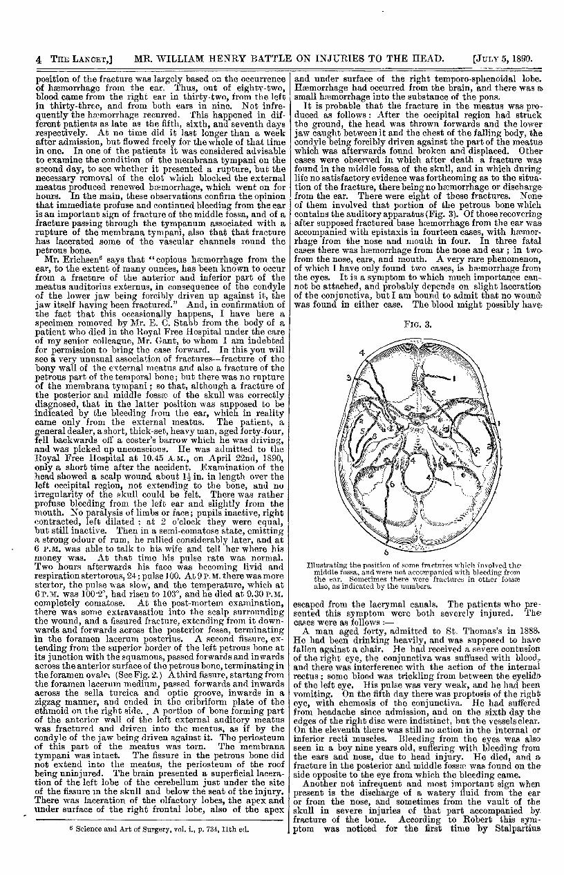
general dealer, a short, thick-set, heavy man, aged forty-four,fell backwards off a coster’s barrow which he was driving,and was picked up unconscious. He was admitted to theRoyal Free Hospital at 10.45 A.M., on April 22nd, 1890,only a short time after the accident. Examination of thehead showed a scalp wound about 1 in. in length over theleft occipital region, not extending to the bone, and noirregularity of the skull could be felt. There was ratherprofuse bleeding from the left ear and slightly from themouth. No paralysis of limbs or face; pupils inactive, rightcontracted, left dilated ; at 2 o’clock they were equal,but still inactive. Then in a semi-comatose state, emitting a strong odour of rum, he rallied considerably later, and at6 P.M. was able to talk to his wife and tell her where hismoney was. At that time his pulse rate was normal.Two hours afterwards his face was becoming livid andrespiration stertorous, 24; pulse 100. At 9 p. M. there was morestertor, the pulse was slow, and the temperature, which at6 P.M. was 100’2°, had risen to 103°, and he died at 9.30 P.M.completely comatose. At the post-mortem examination,there was some extravasation into the scalp surroundingthe wound, and a fissured fracture, extending from it down-wards and forwards across the posterior fossa, terminatingin the foramen lacerum posterius. A second fissure, ex-tending from the superior border of the left petrous bone atits junction with the squamous, passed forwards and inwardsacross the anterior surface of the petrous bone, terminating inthe foramen ovalp. (SeeFig.2.) A third fissure, starting fromthe foramen lacerum medium, passed forwards and inwardsacross the sella turcica and optic groove, inwards in azigzag manner, and ended in the cribriform plate of theethmoid on the right side.. A portion of bone forming partof the anterior wall of the left external auditory meatuswas fractured and driven into the meatus, as if by thecondyle of the jaw being driven against it. The periosteumof this part of the meatus was torn. The membranatympani was intact. The fissure in the petrous bone didnot extend into the meatus, the periosteum of the roofbeing uninjured. The brain presented a superficial lacera-tion of the left lobe of the cerebellum just under the siteof the fissure in the skull and below the seat of the injury.There was laceration of the olfactory lobes, the apex andunder surface of the right frontal lobe, also of the apex
6 Science and Art of Surgery, vol. i., p. 734, 11th ed.
and under surface of the right temporo-sphenoidal lobe.Haemorrhage had occurred from the brain, and there was asmall haemorrhage into the substance of the pons.
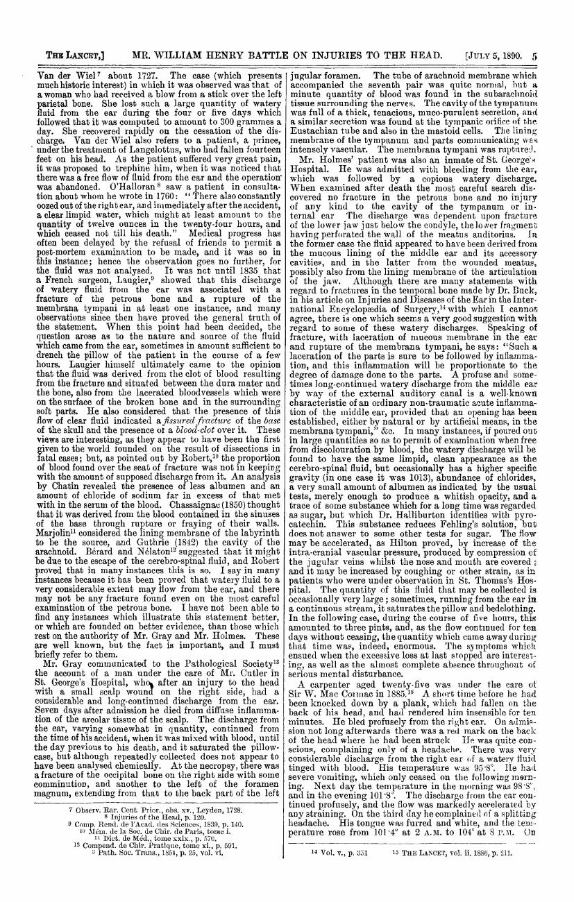
It is probable that the fracture in the meatus was pro-duced as follows: After the occipital region had struckthe ground, the head was thrown forwards and the lowerjaw caught between it and the chest of the falling body, thecondyle being forcibly driven against the part of the meatus’which was afterwards found broken and displaced. Othercases were observed in which after death a fracture wasfound in the middle fossa of the skull, and in which duringlife no satisfactory evidence was forthcoming as to the situa-tion of the fracture, there being no haemorrhage or dischargefrom the ear. There were eight of these fractures. Noneof them involved that portion of the petrous bone whichcontains the auditory apparatus (Fig. 3). Of those recoveringafter supposed fractured base haemorrhage from the ear was.accompanied with epistaxis in fourteen cases, with heemor-rhage from the nose and mouth in four. In three fatalcases there was haemorrhage from the nose and ear ; in two-from the nose, ears, and mouth. A very rare phenomenon,of which I have only found two cases, is haemorrhage fromthe eyes. It is a symptom to which much importance can-not be attached, and probably depends on slight lacerationof the conjunctiva, but I am bound to admit that no wound-was found in either case. The blood might possibly have-
FIG. 3.
Illustrating the position of some fractures which involved themiddle fossa, and were not accompanied with bleeding fromthe ear. Sometimes there were fractures in other fossaealso, as indicated by the numbers.
escaped from the lacrymal canals. The patients who pre-sented this symptom were both severely injured. Thecases were as follows :-A man aged forty, admitted to St. Thomas’s in 188&.
He had been drinking heavily, and was supposed to havefallen against a chair. He bad received a severe contusionof the right eye, the conjunctiva was suffused with blo@d"and there was interference with the action of the internalrectus; some blood was trickling from between the eyelidsof the left eye. His pulse was very weak, and he had beenvomiting. On the fifth day there was proptosis of the righteye, with chemosis of the conjunctiva. He had sufferedfrom headache since admission, and on the sixth day theedges of the right disc were indistinct, but the vessels clear.On the eleventh there was still no action in the internal orinferior recti muscles. Bleeding from the eyes was alsoseen in a boy nine years old, suffering with bleeding fromthe ears and nose, due to head injury. He died, and afracture in the posterior and middle fossse was found on the
side opposite to the eye from which the bleeding came.I Another not infrequent and most important sign whenpresent is the discharge of a watery fluid from the earor from the nose, and sometimes from the vault of theskull in severe injuries of that part accompanied by.fracture of the bone. According to Robert this sym-ptom was noticed for the first time by Stalpartius
5
Van der Wie17 about 1727. The case (which presentsmuch historic interest) in which it was observed was that ofa woman who had received a blow from a stick over the leftparietal bone. She lost such a large quantity of wateryfluid from the ear during the four or live days whichfollowed that it was computed to amount to 300 grammes aday. She recovered rapidly on the cessation of the dis-charge. Van der Wiel also refers to a patient, a prince,
under the treatment of Langelottus, who had fallen fourteenfeet on his head. As the patient suffered very great pain,it was proposed to trephine him, when it was noticed thatthere was a free flow of fluid from the ear and the operation’was abandoned. O’Halloran 8 saw a patient in consulta.tion about whom he wrote in 1760: " There also constantlyoozed out of the right ear, and immediately after the accident,a clear limpid water, which might at least amount to thequantity of twelve ounces in the twenty-four hours, andwhich ceased not till his death." Medical progress hasoften been delayed by the refusal of friends to permit apost-mortem examination to be made, and it was so inthis instance; hence the observation goes no further, forthe fluid was not analysed. It was not until 1835 thata French surgeon, Laugier, showed that this dischargeof watery fluid from the ear was associated with a
fracture of the petrous bone and a rupture of themembrana tympani in at least one instance, and manyobservations since then have proved the general truth ofthe statement. When this point had been decided, thequestion arose as to the nature and source of the fluidwhich came from the ear, sometimes in amount sufficient todrench the pillow of the patient in the course of a fewhours. Laugier himself ultimately came to the opinionthat the fluid was derived from the clot of blood resultingfrom the fracture and situated between the dura mater andthe bone, also from the lacerated bloodvessels which wereon the surface of the broken bone and in the surroundingsoft parts. He also considered that the presence of thisflow of clear fluid indicated a fissured fracture of the baseof the skull and the presence ot a blood-clot over it. Theseviews are interesting, as they appear to have been the firstgiven to the world founded on the result of dissections infatal cases; but, as pointed out by Robert," the proportionof blood found over the seat of fracture was not in keepingwith the amount of supposed discharge from it. An analysisby Chatin revealed the presence of less albumen and anamount of chloride of sodium far in excess of that metwith in the serum of the blood. Chassaignac (1850) thoughtthat it was derived from the blood contained in the sinusesof the base through rupture or fraying of their walls.Marjolin’1 considered the lining membrane of the labyrinthto be the source, and Guthrie (1842) the cavity of thearachnoid. Berard and Nélaton12 suggested that it mightbe due to the escape of the cerebro-spinal fluid, and Robertproved that in many instances this is so. I say in manyinstances because it has been proved that watery fluid to avery considerable extent may flow from the ear, and theremay not be any fracture found even on the most carefulexamination of the petrous bone. I have not been able tofind any instances which illustrate this statement better,or which are founded on better evidence, than those whichrest on the authority of Mr. Gray and Mr. Holmes. Theseare well known, but the fact is important, and I mustbriefly refer to them.Mr. Gray communicated to the Pathological Societyl3
the account of a man under the care of Mr. Cutler inSt. George’s Hospital, whOt after an injury to the headwith a small scalp wound on the right side, had a
considerable and long-continued discharge from the ear.
Seven days after admission he died from diffuse inflamma-tion of the areolar tissue of the scalp. The discharge fromthe ear, varying somewhat in quantity, continued fromthe time of his accident, when it was mixed with blood, untilthe day previous to his death, and it saturated the pillow-case, but although repeatedly collected does not appear tohave been analysed chemically. At the necropsy, there wasa fracture of the occipital bone on the right side with somecomminution, and another to the left of the foramenmagnum, extending from that to the back part of the left
7 Observ. Rar. Cent. Prior., obs. xv., Leyden, 1728.8 Injuries of the Head, p. 120.
9 Comp. Rend. de l’Acad. des Sciences, 1839, p. 140.10 Mém. de la Soc. de Chir. de Paris, tome i.
11 Dict. de Méd., tome xxix., p. 570.12 Compend. de Chir. Pratique, tome xi., p. 591.
3 Path. Soc. Trans., 1854, p. 25, vol. vi.
jugular foramen. The tube of arachnoid membrane whichaccompanied the seventh pair was quite normal, but aminute quantity of blood was found in the suba-rachnoidtissue surrounding the nerves. The cavity of the tympanumwas full of a thick, tenacious, muco-purulent secretion, anda similar secretion was found at the tympanic orifice of theEustachian tube and also in the mastoid cells. The liningmembrane of the tympanum and parts communicating wssintensely vascular. The membrana tympani was ruptured.Mr. Holmes’ patient was also an inmate of St. Georges
Hospital. He was admitted with bleeding from the ear,which was followed by a copious watery discharge.When examined after death the most careful search dis-covered no fracture in the petrous bone and no injuryof any kind to the cavity of the tympanum or in-ternal ear The discharge was dependent upon fractureof the lower jaw just below the condyle, the lower fragmenthaving perforated the wall of the meatus auditorius. Inthe former case the fluid appeared to have been derived fromthe mucous lining of the middle ear and its accessorycavities, and in the latter from the wounded meatus,possibly also from the lining membrane of the articulationof the jaw. Although there are many statements withregard to fractures in the temporal bone made by Dr. Buck,in his article on Injuries and Diseases of the Ear in the Inter-national Eccyclopsedia of Surgery,14 with which I cannotagree, there is one which seems a very good suggestion withregard to some of these watery discharges. Speaking offracture, with laceration of mucous membrane in the earand rupture of the membrana tympani, he says: "Such alaceration of the parts is sure to be followed by inflamma-tion, and this inflammation will be proportionate to thedegree of damage done to the parts. A profuse and some-times long-continued watery discharge from the middle earby way of the external auditory canal is a well-knowncharacteristic of an ordinary non-traumatic acute inflamma-tion of the middle ear, provided that an opening has beenestablished, either by natural or by artificial means, in themembrana tympani," &c. In many instances, if poured outin large quantities so as to permit of examination when freefrom discolouration by blood, the watery discharge will befound to have the same limpid, clean appearance as thecerebro-spinal fluid, but occasionally has a higher specificgravity (in one case it was 1013), abundance of chlorides,a very small amount of albumen as indicated by the usualtests, merely enough to produce a whitish opacity, and a,trace of some substance which for a long time was regardedas sugar, but which Dr. Halliburton identifies with pyro-catechin. This substance reduces Fehling’s solution, butdoes not answer to some other tests for sugar. The flowmay be accelerated, as Hilton proved, by increase of theintra-cranial vascular pressure, produced by compression ofthe jugular veins whilst the nose and mouth are covered ; 9and it may be increased by coughing or other strain, as inpatients who were under observation in St. Thomas’s Hos-pital. The quantity of this fluid that may be collected isoccasionally very large; sometimes, running from the ear ina continuous stream, it saturates the pillow and bedclothing.In the following case, during the course of five hours, thisamounted to three pints, and, as the flow continued for teadays without ceasing, the quantity which came away duringthat time was, indeed, enormous. The symptoms whichensued when the excessive loss at last stopped are interest,-ing, as well as the almost complete absence throughout ofserious mental disturbance.A carpenter aged twenty-five was under the care of
Sir W. Mac Corruac in 1885.]5 A short time before he hadbeen knocked down by a plank, which had fallen on theback of his head, and had rendered him insensible for tenminutes. He bled profusely from the right ear. On admis-sion not long afterwards there was a red mark on the backof the head where he had been struck He was quite con-scious, complaining only of a headache. There was veryconsiderable discharge from the right ear (tf a watery fluidtinged with blood. His temperature was 90 S°. lie hadsevere vomiting, which only ceased on the following morn-ing. Next day the temperature in the morning was 98’8’,and in the evening 101 ’8". The discharge from the ear con-tinued profusely, and the flow was markedly accelerated byany straining. On the third day he complained of a splittingheadache. His tongue was furred and white, and the ternperature rose from 101-4° at 2 A.M. to 104° at 8 P.M. On
14 Vol. v., p. 351 15 THE LANCET, vol. ii. 1886, p. 211
6
the fifth day the discharge from the ear was continuing tosuch .an extent that the pillow became saturated, andduring a period of two hours and a half no less than onepint and a half of this fluid, having a greenish tinge, andgiving the chemical reactions of cerebro-spinal fluid, wascollected. On the sixth day the headache, which waschiefly occipital, had become slight. He was much im-proved and quite intelligent. The fluid was flowing lessfreely; it was clear and limpid. Then deafness was noticed;he could only hear a watch on contact. On the ninth dayhe could sleep without a draught of chloral, but the dis-charge from the ear only ceased in the afternoon of theeleventh day, the temperature being as high as 103° in theevening. He was not so well after the discharge ceased;there was much headache and pain in the back of the neckand flushing of the face. The headache was very severe,making him cry out with the pain. He became deliriousand noisy. The pupils continued equal, and acted well tolight. He continued ill for two days, then improved, theheadache ceasing except when he moved ; any sudden move-ment immediately brought it on again. The day after thecessation of the discharge from the ear the ophthalmoscoperevealed a peculiar rosy appearance of the fundus oculi onboth sides, without evident dilatation of vessels. Afterthis he only complained of dizziness for a few days when hebent forwards. The man was under observation for fifty-five days, and no further symptoms developed.
Sir Prescott Hewett,16 who has done much for the surgeryof this part of the body, divides these cases into threeclasses: 1. Where the fluid from the ear is plentiful and ofa decidedly watery character immediately after the accident,this flow is clue to the escape of csrebro-spinal iluid, as aconsequence of fracture of the petrous bone implicating theinternal auditory canal and its membranes. Three cases. amongst those which proved fatal come under this heading.In two the fracture extended into the internal auditorymeatus, and during life was accompanied with evidence offacial paralysis on that side. In a third case the fractureextended across the meatus internus. 2. WTlzerp the waterydischarge from the ear is preceded by copious and prolonged Iihaemorrhage, a fracture of the petrous bone may be safely !,,diagnosed, hut it cannot be said that the fracture followsany particular course. Five of the fatal cases presented thissequence of the discharge, and in all there was a fracture ofthe petrous bone; in one the internal auditory meatus wasinvolved and the facial nerve torn. 3. Where there is a dis-charge of blood only, neither copious nor prolonged, whichis followed by a watery discharge, varying as to the time ofits appearance, varying also as to its quantity. It may bepresent within a few hours after the accident; it may beprofuse within a few hours after its appearance. These arethe cases in which it has been proved that the diagnosis isdoubtful. The fact that too much reliance should not beplaced on this symptom is illustrated by the case of onepatient, a child aged three years and a half, who was underobservation: the discharge followed slight haemorrhage fromthe left ear, and continued for two days in no quan-tity. After death, which occurred on the third day follow-ing the injury, although there was a, fracture in each fossaof the base, including the middle fossa en the lets side, itdid not involve any part of the auditory apparatus.
I am of opinion that more reliance should be placed onchemical analysis of watery discharges fro.a the head. Ifthese show on examination the presence of a large amountof chlorides, and give the sugar reaction to Fehling’s solu-tion, then we may say that there is a fracture and com-munication with the subarachnoid space tbroagh a tear inthe tubular prolongation of the membranes which surroundsthe facial and auditory nerves. And this applies althoughthere be some albumen from admixture with serum, blood,or secretion from the ear. Sometimes, bowever, the waterydischarge is not in quantity sufficient to permit of its beingcollected, and of our applying the usual tests to it. Wemust then, I think, be guided by the amount of the beinor-rhage as to the presence of fracture, and the onset of facialparalysis or deafness, dating only from the injury as to itsextension into the internal auditory meatus and subarach-noid space. An interesting feature of the injury to thehead which was sustained by Emin Pasha during last year,as reported in the daily press, was the recurrence of a waterydischarge after it had ceased for a time. This was noted intwo patients who were in St. Thomas’s Hospital: A man
16 Loc. cit.
aged forty-five, admitted unconscious, with tolerably largequantities of blood flow ing from the left ear. Slight waterydischarge followed, which, however, ceased in a day or two.He suffered from signs of serious cerebral injury, but had nofurther discharge from the ear until the eighteenth day,when a yellowish clear fluid commenced and continued toflow for seven days. The second patient was a managed sixty-four, admitted with haemorrhage from the leftear, followed by a thin discharge for two days. This re-commenced on the sixth and continued till the fourth dayafterwards.A case somewhat resembling the one which I have read,
in which there was an excessive flow of fluid from the ear,which also illustrates the recurrence of the watery dis-charge, has been recorded by Dr. Campbell. The patientdied comatose on the fourth day after the return of thedischarge. The fracture bad passed through the internalauditory canal, but there was no evident cause for the fatalcoma. Sir Wm. Savory had a patient under his care in1861 who had watery discharge from both ears which lastedfor nearly a month. The patient, a boy aged thirteen, re-covered. This is the only example that I can find of waterydischarge from both ears. Sometimes the flow of eitherblood or watery fluid from the ear will be observed to pul-sate ; occasionally small air bubbles are seen in it.As regards the influence of age on the occurrence of
these watery discharges, it appears, from a considera-tion of those cases from which my statistics are taken,that, if all in whom watery discharges of every kindoccurred are included, of those surviving thirteen werebelow and fourteen above thirty years of age. Of thosein whom the injury proved fatal, five were below and fourabove that age, this average corresponding fairly with theratio of fracture at the various ages of life.
That is to say, that out of thirty-six cases presenting thisphenomenon one-fourth died, although in none of the ninefatal cases could it be said that death ensued in consequenceof the opening up of the subarachnoid space, yet inone the fracture of the petrous bone gave opportunity forpurulent infection of the meninges, which caused death.Robert looked upon this symptom as of extremely badaugury; indeed, he writes of it as follows: "Up to thepresent, in fact, one has not been able to save any of thosewho have presented this symptom." The seven patientsunder his observation died, and he cites another case underthe care of Marjolin. Colles, in his lectures, says that he hasseen such cases recover, but regarded them as mostly fatal.The idea suggests itself that perhaps the changed con
ditions under which such cases come nowadays whenadmitted to our hospitals and infirmaries may have muchto do with the better prognosis now given in those injurieswhich are accompanied by watery discharges from the ear.The greater care which i6 taken in cleansing not only thepatients but also the wards, the employment of efficientventilation and the use of antiseptics, the rare occurrenceof suppuration in wounds, the almost complete banishmentof pyaemia and erysipelas, and the transfer of the latter toa special ward should it unfortunately arise, are thingswhich must be taken into consideration. But, in spite ofthese improvements in the surroundings of our patients,suppuration occasionally follows haemorrhage or waterydischarge, and the prognosis becomes correspondingly in-creased in gravity. For example, I may quote briefly thefollowing cases which were treated in St. Thomas’s Hos-pital. A man aged twenty-five was admitted with bleedingfrom the left ear; this was followed by clear watery dis-charge, which became purulent, facial paralysis ensued, andhe suffered from occasional headache during a residencewhich lasted thirty-five days. A man aged forty-one camein suffering from severe injury to the head ; much haemor-rhage from the left ear, nose, and mouth. The haemorrhagefrom the ear continued for three days, giving way on thefourth day to a purulent discharge, which corresponded tothe onset of considerable inflammatory swelling over theleft side of the face and neck. A youth aged eighteen, ad-mitted withmuch bleeding from the leftear, whiclidiminisheclin quantity and became serous, then watery in appearance.
7
About the eleventh day there was a rise of temperature to1016°, and two days later a discharge of purulent characterfrom the ear. The membrana tympani had completelydisappeared when he was examined with the speculum.Occasionally the suppuration becomes more extensive andmay produce a condition for which operation will berequired. Of this the following is an example. OnMarch 13th, 1883, a man aged thirty-seven, who had fallenfrom a cart on a macadamised road and struck his head,was admitted under the care of Sir W. Mac Cormac. Hehad immediately become almost insensible, but soon
answered "Yes" " and ’ No." There was bleeding from themouth and from the left ear. When seen five and a halfhours later lie was very restless and roused with difficulty,answering in monosyllables when shouted at. There wasbleeding only from the left ear; the pupils were normal ;the breathing not stertorous. There was no external iwound or sign of fracture of the vault. At 10 p. M. he wasvery restless, sitting up in bed and trying to get out ; the I,temperature was 1002°. On the 14th he was quieter. ’,Bleeding from the ear continued. The temperature in themorning was 1014°, in the evening 102°. On the 16th at8A.M. consciousness returned. Ecchymosis had appearedover the left mastoid process, and was extending backwards.On the 18th he recognised his mother, but was restless andmade attempts to get up. Three days later he was unableto answer questions put to him, but was quieter. On the28th ophthalmoscopic examination revealed evidences of aneuritis passing off. He was still strange in his ideas.There was no albuminuria. On the 29th the discharge of athin, clear fluid continued from the left ear, and on the 30ththere was pain behind that ear. On A prill0th the dischargehad become sero-purulent, and there was tenderness overthe mastoid as well as pain. On the 15th and 16th thetenderness had become more marked, and there was con-siderable p3.in extending from the vertex to the back of theear. The discharge from the ear had become purulent.There had been no rigors, but the temperature had been1018° for two evenings. Slight facial paralysis, whichhad been observed during the first week after admis-sion, gradually subsided about this time. On the 17thhe had slept badly during the preceding night, and his tem-perature had risen to 102°. For four days before the 21stthere had been considerable swelling behind the left ear,extending upwards nearly to the top of the head, withill-defined cedematous margins, and on that date fluctua-tion was evident behind and around the ear. Therewas much tenderness and a good deal of pain over
the swollen area. The temperature during the four
days had varied from 100 4° to 103’2°. His pulse was 88.Sir W. Mac Cormac made an incision behind the earinto the fluctuating swelling and evacuated a largeabscess. Under the incision the bone was found to bebared, and a fissured fracture ran upwards and back-wards across the exposed area. Through this pus exuded,and a trephine applied behind it gave escape to more pus.Antiseptic precautions were taken during the operation,and in the evening the man expressed himself as relieved.On the 23rd there was much discharge from both ears andwound; the temperature became normal, and continuedeither normal or subnormal during the remainder of thepatient’s stay in hospital, with one exception, when therewas increased discharge from the ear and a swelling in frontof it. This subsided quickly, and he had no further sym-ptoms. He was under treatment in hospital for seventy-three days, and left apparently well.Mr. Bl’yant17 had a very similar case under his care. A
woman aged twenty-four was admitted with profuse hæmor-rhage from the ear, which became watery on the third dayand purulent on the seventh. She had suppuration over themastoid on the tenth day. This was incised, and a monthafter the accident necrosed bone was removed from themastoid. Facial paralysis afterwards developed, and becamecomplete.Another source of infection and a most dangerous con-
dition for the patient is the presence of otitis media ofold standing, or the existence of some septic purulent dis-charge from the ear before the injury to the head, whetherthat injury be followed by simple haemorrhage or byhsemorrhage and then a watery discharge. These results areillustrated by few cases in our literature. Of two cases, inthe first there were many symptoms present which make itof interest beyond the occurrence of purulent discharge from
17 THE LANCET, vol. ii. 1884, p. 318.
the ears, and it is worthy of note that several of the moredistant fractures suppurated, probably in consequence ofthe extremely low condition of health of the boy and thevital depression following such an extensive and severeinjury to the head, and not from pyemic infection. Thiscase has already been published.I8The second instance was that of a little girl aged
one year and nine months, who was admitted underthe care of Mr. Croft on March 13th, 1882, twenty minutesafter having been run over by a carrier’s cart. Shedied of meningitis a fortnight after admission, and aweek after a purulent discharge had commenced. The left.occipital bone was fractured, and the left petrous boneloosened from its connexions with the occipital and sphenoidbones. Between the left petrous bone and the dura materwas a considerable quantity of altered blood, granular andsemi-solid. The dura mater was inflamed and had a layerof pus on its internal surface, but this occurred only hereand there on the left side. The pia arachnoid was acutelyinflamed and thickened, with lymph and pus over the baseand whole of the left hemisphere, and also to a much morelimited extent over the right. The ventricles were slightlydistended, and the fornix and corpus callosum were muchsoftened.The attention of the profession was first drawn to the
fact that watery discharges occasionally come from the noseafter head injuries by Blandin, who reported a case in1840, that of a man aged thirty-two, who had shot himselfwith a medium-sized pistol, the bullet of which had lodgedin the sella turcica, destroying part of the pituitary body." There flowed from the nostrils a quantity of bloody scrumtruly enormous." M. Robert recorded another case liveyears afterwards, and proved the identity of the fluid withthe cerebro-spinal, and the necropsy showed a fracture ofthe sella turcica and laceration of the overlying dura materand arachnoid covering the anterior lobes of the brain.
I have only met with discharge from the nose twice afsersevere head injury, and its importance and character werevery doubtful in the first of these, a boy aged thirteen, whowas under the care of Mr. Sydney Jones. He was admittedon Jan. 4th, 1882, and continued insensible for an hourand a half. The accident, a fall from a cart, had happenedtwenty-four hours earlier, and he was still vomiting.Haemorrhage from the right ear continued for a day, andthere was a yellowish-white discharge from the right nostril,which continued when he was removed by his parents fivedays later. In the second it is possible that there was a frac-ture of the petrous bone, implicating the internal auditcrycanal, but without rupture of the meJl,l1lrana tympani. Thecharacter of the fluid, the deafness and noises in the ear,point to this explanation. A man aged twenty-five wasadmitted in 1886 under the same surgeon. Having fallen14ft. on his head he was unconscions for awhile, and whenadmitted an hour later there was a iarge scalp wound, andthere had been epistaxis. On the next day there wassubconjunctival ecchymosis of both eyes, with contusionof the eyelids. On the fourth and fifth days he complainedof noises in the left ear, and on the sixth it was noticedthat there was a discharge of a few drops of clear wateryfluid from the left nostril. Some of this was collected. Itwas clear, with a yellowish tinge, slightly alkaline reaction,a trace of albumen, and marked proportion of chlorides.On the eighth day he had had a good deal of headache duringthe night, and, on raising him up to dress him, some clearfluid resembling that collected on the sixth day again ranfrom the nose; it was not coagulated lsy heat or nitric acid,contained a large proportion of chlorides, and a trace ofalbumen. He -kvas quite deaf with the left ear, even whenthe watch was placed against the mastoid process on theleft side of the forehead. He complained of 9. rumblingnoise in the ear, aud occasionally of singing in the ear, andwas deaf when dismissed after forty-foar days’ residence.Foucard19 recorded a case in which a fracture of the base ofthe skull was accompanied with the How of a watery fluidfrom the left nostril only, and Malgaïgne20 another in whicha free discharge took place from both nose and enr.Writing on the subject of nasal discharge after injuries
which had been supposed to produce fracture of the base ofthe skull, as indicated by a discharge from the nose,M. Robert drew attention to the occasional now of a limpidsalt fluid, sometimes in considerable quantity, from thenose, independent of traumatism, and mentions a patient
18 THE LANCET, vol. i. 1886.19 Journal de Chir. de Malgaigne, 1846, p. 315. 20 Ibid., p 283
8
who could till a wine-glass in the space of an hour, andsaturated twenty-five to thirty handkerchiefs in the courseof the day. He did not analyse the fluid.21As I am chiefly concerned now with those fractures whichinvolve the base of the skull, I do not purpose to considershe escape of cerebro-spinal fluid consequent on fracture ofthe vault. It may, however, be found under the scalp afterH. recent fracture of the subjacent bone, or later, forminga soft compressible tumour, over the site of the fracture.It flowed from the wound after a recent compound fractureof the skull, in which I found it necessary to remove frag-ments of bone from near the base, and in which there was:1, laceration of the underlying brain. It has flowed fromThe wound after trephining, and I have seen it coming inarge quantities in cases of hernia cerebri in which sloughingitf brain substance had opened up the lateral ventricle.Several instances of traumatic cephal-hydrocele have beenskûwn at the medical societies during the last few years,but as a recent condition I have only seen it once, and thatwas in the case of an infant under the care of Mr. R. W.Parker at the Children’s Hospital. The child presented acurious forcing outwards of the bones forming the posteriorIaart of a fracture in the right parietal region, the result ofconsiderable violence, and covering the fracture was a large;"oft fiuctuating swelling, which pulsated.
Protrusion of the brain substance after an injury to thehead which has caused a fracture of the base of the skull is Ian occurrence of very great rarity; in only one of the nfty-loar cases of fatal injury is it recorded, and then it was
through a fracture of the middle fossa. Indicating as ittbes a most severe injury to the base as well as to the brain,it is not surprising that such cases have usually had a fatalending. There are, however, instances on record in whichits escape from the ear or from the nose has been observed,and yet recovery, apparently complete in every respect,has ensued. I find an account given of the occurrence ofthe latter not followed by a fatal issue reported byMr. Luther Holden-a compound fracture of the skull in atiian of forty, in whom there was an escape of cerehralmatter from the wound and through the nostril. Thehacture, extending over the margin of the orbit, hadinvolved the frontal sinus, and thus allowed the escape of aportion of the brain through the right nostril on the dayfollowing the injury. Dr. Lockwood 22 reported a case inwhich a loss of cerebral substance from the ear followed afall on deck. The patient recovered.
ABSTRACT OF THE
Croonian LecturesON
CEREBRAL LOCALISATION.Delivered before the Royal College of Physicians of London,
BY DAVID FERRIER, M.D., LL.D., F.R.S.,PHYSICIAN TO KING’S COLLEGE HOSPITAL, AND TO THE NATIONAL
HOSPITAL FOR THE PARALYSED AND EPILEPTIC,QUEEN-SQUARE.
LECTURE V.
IN resuming the consideration of the paths traversed by3ensory impulses to the brain, Dr. Ferrier stated that therewas clinical and pathological evidence to show that thesensory tracts ran in the fillet and formatioreticularis, and thatthey were continued through thetegmen turn ofthecrus cerebriinto the posteriorpart of the internal capsule. With regard totheir subsequent course and the question of the localisationof a definite tactile centre, Dr. Ferrier reminded his audienceof his earlier researches, in which he observed that tactileand painful sensibility seemed to be entirely unaffected inmonkeys, notwithstanding the most extensive experimentallesions of every portion of the convex surface of the hemi-sphere, but that in several cases in which a post-mortemexamination showed a more or less extensive implication of thehippocampal region (including the cornu ammonis and gyrushippocampi) there was present impairment or abolition ofsensibility on the opposite side of the body. Dr. Ferrier,
21 See also the observations of Brodie and of Sir James Paget.22 American Journal of Medical Science, 1859, p. 254.
therefore, tried to destroy this region with the cautery, by
passing this instrument through the occipital lobe, with aslittle injury as possible, to the hippocampus. The superficialappearance of the lesion in one case, with the track of thesinus made by the cautery, have already been figured.!The result of this operation was total ansesthesia andanalgesia of the opposite side of the body. There was noloss of motor power, but considerable clumsiness andincoordination, so that the animal’s feet tended to slip offthe perch when its eyes were closed. Dr. Ferrier alsoinvestigated this subject later in conjunction withProfessor Gerald Yeo. The mode of experimenting was,in the main, the same. Ten experiments were made, infive of which the hippocampal region was removed onboth sides. The results entirely confirmed the previousobservations, and the conclusions drawn were that’’ the various forms of sensation embraced under the term’ common, or tactile sensibility,’ including cutaneous,musculo-cutaneous, and muscular, are capable of being pro-foundly impaired, or altogether abolished for the time, atleast, by destructive lesions of the hippocampal region, andthat the degree and duration of the anaesthesia varied withthe completeness of destruction of the area in question."The subject was next taken up by V. Horsley and Schafer,3but their early excisions were less extensive than thosedescribed above, and they were at first unable to corroborateDr. Ferrier’s results. In a later experiment, however, atwhich he assisted, the removal of one hippocampal regionwas followed on the next day by partial analgesia and com-plete insensibility to mere contact on the opposite side.Death unfortunately occurred on the second day, so that noconclusions as to duration were possible in this case. In asecond experiment the hippocampal region was again re-moved, and the incisions made so as to detach also thecalcarine fissure and the hippocampus minor. This animalwas profoundly anæsthetic on the opposite side, but theredid not appear to be absolute analgesia. Tactile anaesthesia,continued for several weeks without appreciable alteration,but a gradual improvement occurred, so that examinationat the end of six weeks revealed only some degree of im-pairment, as evidenced by less easily excited attention bygentle pricking, rubbing, &c., of the side opposite thelesion as compared with the other. Severe pricking, pinch-ing, or pungent heat, however, appeared to be well per-ceived. The gradual diminution of the anæsthesia inducedby extensive if not complete removal of the hippocampalregion led Dr. Ferrier to suggest similar experiments on thegyrus fornicatus, on the ground that the tactile centremight extend into the rest of the falciform lobe, of whichthe hippocampal gyrus is only a part. SubsequentlyHorsley and Schafer experimented on the gyrus fornicatus,and proved the accuracy of Broca’s anatomical view as tothe unity of the falciform lobe, by demonstrating thatlesions of the gyrus fornicatus caused similar symptoms tothose produced by destruction of the hippocampal region, ofperhaps even greater intensity and longer duration. In oneanimal the hippocampal region was removed on one side,and when the consequent ansesthesia had passed off themesial surface of the brain was again exposed, and thegyrus fornicatus excised along the whole length of thecorpus callosum. There resulted at first absolute anaesthesiaand analgesia of the opposite side of the body, and at theend of six weeks there was still absolute anaesthesia; and,although the analgesia had diminished, it was still manifestin some degree. The animal was in perfect health, andexhibited no sign of motor disorder except slight transientweakness of the opposite leg immediately after the opera-tion, owing to some injury of the parietal lobule and neigh-bourhood during the operations necessary to expose thegyrus fornicatus. In summing up their conclusions in theirMemoir in the Philosophical Transactions, ProfessorsHorsley and Sohafer conclude that "any extensive lesionof the gyrus fornicatus is followed by hemianaesthesia,more or less marked and persistent. In some cases
the anaesthetic condition has involved almost the wholeof the opposite side of the body ; in others it hasbeen localised to either the upper or the lower limband to particular parts of the trunk, but we have not yetsucceeded in establishing the relationship between specialregions of the body and the parts of the convolution whichhave been destroyed. Moreover, the anæsthesia was fre-quently very pronounced and general during the first three
1 Functions of the Brain, p. 329, fig. 105.2 Phil. Trans., Part II., 1884, Experiments 24 to 33, figs. 103-181.3 Functions of the Cerebral Cortex, B. 20, Phil. Trans., 1888.