5523 JULY 6, 1929. Lectures ON THE PHYSIOLOGY OF GLYCOGEN AND THE RÔLE OF INSULIN AND EPINEPHRINE IN CARBOHYDRATE METABOLISM. Delivered at the London Hospital BY J. J. R. MACLEOD, M.B., LL.D. ABERD., D.Sc. (HON.) TORONTO, F.R.S., REGIUS PROFESSOR OF PHYSIOLOGY IN THE UNIVERSITY OF ABERDEEN. LECTURE I. (Delivered on May 16th.) THE behaviour of the blood-sugar curve following the ingestion of soluble carbohydrates indicates that absorption must start at once. Under ordinary conditions none of this seems to occur in the stomach itself, as far as can be judged by chemical analysis, either of solutions of sugar left for some time in the viscus after ligation at the pylorus or of the chyme as this is discharged into the duodenum. Absorption of sugar takes place mainly in the upper levels of the small intestine, and different types of experiment have been employed to study the process. The oldest of these consists in placing known amounts of sugar in solution in isolated loops of intestine (Thiry-Vella) and then removing and analysing the contents after varying periods of time. The chief objection to this method depends on the fact that only an isolated portion of the entire gut is employed, an objection which is overcome in the method recently introduced by Cori and Cori,l in which the entire gastro-intestinal tract of small animals, such as the white rat, is used. The animals, kept for some time previously on a standard diet, are starved either for 24 or 48 hours, after which 2 or 3 c.cm. of a strong solution of sugar is placed in the stomach through a small flexible catheter passed by way of the mouth. The rate of absorption is ascertained by subtracting, from the amount of sugar that was given, the amount found by analysis of the gastro-intestinal contents after killing the animals at varying periods. Cori and Cori state that, when sufficient is given, the amount of sugar absorbed is constant from hour to hour, at least up to four hours, and they define as the coefficient of absorption that number of grammes of sugar absorbed per 100 g. of body-weight per hour. This is characteristic for each sugar, but it varies according to the time the animal was starved prior to giving the sugar. It is independent of the strength of the sugar solution. For the monosaccharides the coefficients are given as follows : After 48 hours’ starvation d-galactose 0-196 g., d-glucose 0-178 g., d-fructose 0-077 g., d-mannose 0-034 g., l-xylose 0-028 g., and Z-arabinose 0-016 g. After 24 hours’ starvation the coefficient for glucose is 0-266 g. These figures show a high degree of selectivity, dependent not at all upon molecular weight but markedly so on the structure of the molecule. Thus, the pentoses are absorbed at about ’, one-eighth the rate of the better-known hexoses, and the slight stereoisomeric difference between glucose and mannose makes a marked difference. These results stand out in sharp contrast with those obtained for the rate of absorption from the peritoneal cavity, as determined on mice by C. F. Cori and Goltz.2 In this case the sugar was absorbed at a diminishing rate with time, indicating that the concentration affects the process, and different sugars were absorbed at equal rates. When injected intravenously, different sugars were also found, by analysis of the so-called free sugar of the tissues, to be absorbed very rapidly and at equal rates into the liver and into the muscles, although it took a shorter time for equilibrium between blood and liver to be attained than between blood and muscles dependent probably on differences in the richness in blood-supply.3 I have only instanced a few of the numerous results which these workers have placed on record, but they are sufficient to show that the intestinal mucosa is endowed with a very highly specialised selective action, which is absent in the case of certain other absorbing membranes. It seems remarkable that bacterial decomposition should not account for a measurable fraction of the sugar which disappears from the intestine during four hours and that the concentration of the solution should not affect the rate of absorption. Cori and Cori state that no significant amount of glucose disappears when a solution of it is placed in the excised gastro-intestinal tract kept in isotonic saline solution at body tempera- ture for four hours. At my request Magee and Auchinochie have repeated this experiment and on one or two occasions have been able to recover all the glucose. On other occasions, however, there were considerable deficits which may have been due to bacterial destruction or to incomplete washing of the sugar out of the walls of the gut. Is it not possible also that bacterial action might occur in the intact intestine, in situ, but be retarded or absent in the excised one ? Corroboration or otherwise of the accuracy of the coefficients of absorption as.given by Cori and Cori for different sugars would be most important since their subsequent researches, in which they compare the amount of sugar absorbed, on the one hand, with the amount that can be accounted for by combustion and glycogen formation on the other, depends on it. One also wonders whether the higher coefficients of absorption observed for rats starved 24 hours, as compared with those starved 48 hours, may not have been due to greater bacterial destruction in the former case. It may be added that another of the Cori’s conclusions-namely, that the rate of absorption is independent, within wide limits of the sugar concentration, has been called in question by Pierce, Osgood, and Polansky. These workers found, on repeating Cori’s experiments, that the rate of absorption was less between the second and third hours than between the first and second. In a reply to this criticism, however, Cori, Cori and Goltz5- point out that Pierce, &c., did not feed enough sugar to let the absorption proceed for three hours. Although there may be reasons to doubt the reputed extreme accuracy of the coefficients, it is nevertheless certain that the striking differences observed cannot be mainly attributed to differences in the extent of destruction of the sugars by the bacteria of the intestine. That the hexoses are more readily absorbed than the pentoses has been confirmed by Magee and myself in experiments of a different type from those of the Cori’s. In these, we measured the rates of diffusion of equimolecular solutions of various sugars through the walls of isolated loops of rabbit’s intestine, kept alive by being suspended in oxygenated Tyrode’s solution. The different sugar solutions were placed in neighbouring loops of approximately equal weight and small portions of the outside solutions were pipetted off at intervals for determination of the reducing power by the Shaffer- Hartmann method. It was found in preliminary experi- ments that diffusion proceeds through pieces of gut which had previously been killed, either by heat or by the action of sodium fluoride, much more rapidly than through the living gut and that, in equimolecular concentration, pentoses diffuse more rapidly than hexoses, being dependent solely on the concentration of the sugar solution. Through living segments on the other hand, glucose diffused as quickly as pentose, or in some experiments more quickly. It is undoubted, therefore, that the intestinal mucosa absorbs hexoses more rapidly than pentoses, and I cannot resist wondering whether the former- undergo some change in the process. That the blood- sugar may consist of some form of glucose which differs in configuration from a mixture of the a- and &bgr;-glucoses, has been suggested by various authors, without any evidence being adduced to support the suggestion. It has, however, generally been assumed that this change, if it occurs, depends either on a. A
Transcript
5523
JULY 6, 1929.
LecturesON THE
PHYSIOLOGY OF GLYCOGENAND THE
RÔLE OF INSULIN AND EPINEPHRINE IN
CARBOHYDRATE METABOLISM.
Delivered at the London HospitalBY J. J. R. MACLEOD, M.B., LL.D. ABERD.,
D.Sc. (HON.) TORONTO, F.R.S.,REGIUS PROFESSOR OF PHYSIOLOGY IN THE UNIVERSITY OF
ABERDEEN.
LECTURE I.(Delivered on May 16th.)
THE behaviour of the blood-sugar curve followingthe ingestion of soluble carbohydrates indicates thatabsorption must start at once. Under ordinaryconditions none of this seems to occur in the stomachitself, as far as can be judged by chemical analysis,either of solutions of sugar left for some time in theviscus after ligation at the pylorus or of the chymeas this is discharged into the duodenum. Absorptionof sugar takes place mainly in the upper levels of thesmall intestine, and different types of experiment havebeen employed to study the process. The oldest ofthese consists in placing known amounts of sugar insolution in isolated loops of intestine (Thiry-Vella)and then removing and analysing the contents aftervarying periods of time. The chief objection to thismethod depends on the fact that only an isolatedportion of the entire gut is employed, an objectionwhich is overcome in the method recently introducedby Cori and Cori,l in which the entire gastro-intestinaltract of small animals, such as the white rat, is used.The animals, kept for some time previously on astandard diet, are starved either for 24 or 48 hours, afterwhich 2 or 3 c.cm. of a strong solution of sugar isplaced in the stomach through a small flexible catheterpassed by way of the mouth. The rate of absorptionis ascertained by subtracting, from the amount ofsugar that was given, the amount found by analysisof the gastro-intestinal contents after killing theanimals at varying periods. Cori and Cori state that,when sufficient is given, the amount of sugar absorbedis constant from hour to hour, at least up to four hours,and they define as the coefficient of absorption thatnumber of grammes of sugar absorbed per 100 g. ofbody-weight per hour. This is characteristic for eachsugar, but it varies according to the time the animalwas starved prior to giving the sugar. It is independentof the strength of the sugar solution.For the monosaccharides the coefficients are given
as follows : After 48 hours’ starvation d-galactose0-196 g., d-glucose 0-178 g., d-fructose 0-077 g.,d-mannose 0-034 g., l-xylose 0-028 g., and Z-arabinose0-016 g. After 24 hours’ starvation the coefficient forglucose is 0-266 g. These figures show a high degreeof selectivity, dependent not at all upon molecular weight but markedly so on the structure of themolecule. Thus, the pentoses are absorbed at about ’,one-eighth the rate of the better-known hexoses, andthe slight stereoisomeric difference between glucoseand mannose makes a marked difference. Theseresults stand out in sharp contrast with those obtainedfor the rate of absorption from the peritoneal cavity,as determined on mice by C. F. Cori and Goltz.2 Inthis case the sugar was absorbed at a diminishingrate with time, indicating that the concentrationaffects the process, and different sugars were absorbedat equal rates. When injected intravenously, differentsugars were also found, by analysis of the so-calledfree sugar of the tissues, to be absorbed very rapidlyand at equal rates into the liver and into the muscles,although it took a shorter time for equilibrium betweenblood and liver to be attained than between blood and
muscles dependent probably on differences in therichness in blood-supply.3
I have only instanced a few of the numerous resultswhich these workers have placed on record, but theyare sufficient to show that the intestinal mucosa isendowed with a very highly specialised selectiveaction, which is absent in the case of certain otherabsorbing membranes. It seems remarkable thatbacterial decomposition should not account for a
measurable fraction of the sugar which disappearsfrom the intestine during four hours and that theconcentration of the solution should not affect therate of absorption. Cori and Cori state that nosignificant amount of glucose disappears when asolution of it is placed in the excised gastro-intestinaltract kept in isotonic saline solution at body tempera-ture for four hours. At my request Magee andAuchinochie have repeated this experiment and onone or two occasions have been able to recover all theglucose. On other occasions, however, there wereconsiderable deficits which may have been due tobacterial destruction or to incomplete washing of thesugar out of the walls of the gut. Is it not possiblealso that bacterial action might occur in the intactintestine, in situ, but be retarded or absent in theexcised one ? Corroboration or otherwise of theaccuracy of the coefficients of absorption as.given byCori and Cori for different sugars would be mostimportant since their subsequent researches, in whichthey compare the amount of sugar absorbed, on theone hand, with the amount that can be accounted forby combustion and glycogen formation on the other,depends on it. One also wonders whether the highercoefficients of absorption observed for rats starved24 hours, as compared with those starved 48 hours,may not have been due to greater bacterial destructionin the former case. It may be added that anotherof the Cori’s conclusions-namely, that the rate ofabsorption is independent, within wide limits of thesugar concentration, has been called in question byPierce, Osgood, and Polansky. These workers found,on repeating Cori’s experiments, that the rate ofabsorption was less between the second and thirdhours than between the first and second. In a replyto this criticism, however, Cori, Cori and Goltz5-point out that Pierce, &c., did not feed enough sugarto let the absorption proceed for three hours.Although there may be reasons to doubt the reputed
extreme accuracy of the coefficients, it is neverthelesscertain that the striking differences observed cannotbe mainly attributed to differences in the extent ofdestruction of the sugars by the bacteria of theintestine. That the hexoses are more readily absorbedthan the pentoses has been confirmed by Magee andmyself in experiments of a different type from thoseof the Cori’s. In these, we measured the rates ofdiffusion of equimolecular solutions of various sugarsthrough the walls of isolated loops of rabbit’s intestine,kept alive by being suspended in oxygenated Tyrode’ssolution.The different sugar solutions were placed in neighbouring
loops of approximately equal weight and small portions ofthe outside solutions were pipetted off at intervals for
determination of the reducing power by the Shaffer-Hartmann method. It was found in preliminary experi-ments that diffusion proceeds through pieces of gut whichhad previously been killed, either by heat or by the actionof sodium fluoride, much more rapidly than through theliving gut and that, in equimolecular concentration, pentosesdiffuse more rapidly than hexoses, being dependent solelyon the concentration of the sugar solution. Through livingsegments on the other hand, glucose diffused as quickly aspentose, or in some experiments more quickly.
It is undoubted, therefore, that the intestinalmucosa absorbs hexoses more rapidly than pentoses,and I cannot resist wondering whether the former-undergo some change in the process. That the blood-sugar may consist of some form of glucose whichdiffers in configuration from a mixture of the a- and&bgr;-glucoses, has been suggested by various authors,without any evidence being adduced to support thesuggestion. It has, however, generally been assumedthat this change, if it occurs, depends either on a.
A
2
preliminary conversion of glucose into glycogen or onthe action of some hormone such as insulin, but is itnot possible, as has been hinted at by various writers,that some change actually occurs in the glucosemolecule while it is passing through the intestinalmucosa ?
INTERPRETATION OF CHANGES IN THE BLOOD-SUGAR.It is not my intention here to enter further into
the various interesting questions regarding intestinalabsorption which await investigation by these methods,but, confining ourselves to the sugars, it may be ofinterest if I refer to the significance of the resultswhich we have just been considering in connexionwith the behaviour of the blood-sugar followingcarbohydrate ingestion. When this is observed inman at short intervals, after moderately small amountsof sugar have been taken in the post-absorptive state,very constant results are obtained, and it is consideredthat any unusual heightening or lengthening of thecurve, which is obtained by plotting percentages ofblood-sugar against time, is indicative of a diabetictendency. Useful though this method may be as aclinical test, it should always be borne in mind thatnumerous factors enter in to influence the behaviourof the curves, for not only does this depend on therate of intestinal absorption of the sugar, which againis dependent on many factors, but also on the rate atwhich the tissues utilise the sugar that is absorbed.This disposal may be influenced in several ways, andthere is accumulating evidence to show that these arelargely controlled by the concentration of insulin inthe blood.How complicated the matter really is may be
illustrated by comparison of the blood-sugar curvesfollowing ingestion of fructose and glucose. Theformer sugar has a very small influence as comparedwith that of the latter and this is used clinically as atest of the efficiency of the liver, on the assumptionthat the difference depends on a rapid polymerisationof the fructose to glycogen in this organ. If Cori andCori’s results on rats apply in the case of man-namely,that fructose is absorbed at only half the rate of
glucose-then it is possible that this may in itselfaccount for the smaller influence which fructose has onthe curve. But the problem is really a complex one,since galactose was found by the Cori’s to be absorbedeven more rapidly than glucose, whereas this sugar,according to Folin and Berglund, has little influenceon the blood-sugar level.When the intestinal factor is eliminated, by injecting
intravenously at a slow continuous rate, the tolerancetowards sugar may be determined by observingwhether any is excreted in the urine. By this methodSansum, Woodyatt, and Wilder8 showed, several yearsago, that 0-85 g. of glucose could be tolerated per kg.body-weight and per hour, both in man and laboratoryanimals. In the white rat (under amytal anaesthesia.)and in the mouse, however, Cori and Cori state thatthe tolerance is three times as great (2-5 g. per kg.),except during the summer months when there is alowered tolerance (1-6 g. per kg.) and this was foundto be related to the development of a certain degree ofketonuria.
THE GLYCOGEN OF THE LIVER.
Physiologists have become so familiar with theview that the glycogen of the liver represents a
storage form of carbohydrate, that they have neglectedto take notice of many well-established facts whichindicate that it is also, in a sense, an internal secretion.As Cramer9 has pointed out, this was the originalconception of Claude Bernard, who believed glycogento be produced out of protein, not denying, however, that it is capable also of being formed out of absorbedcarbohydrate. And at the outset of a discussion of thesignificance to be attached to variations in the amountof glycogen which can be found in the organ afterdeath, it is important to recognise that this is only abalance between processes of production and loss andmay be no measure therefore of the rate at whichthese processes are proceeding during life ; as Cramerputs it, " one should look upon the glycogen of the
liver dynamically, and not, as has been done, staticallyas a mere store of excess of carbohydrate."When food is withheld, the amount of glycogen in .
the liver rapidly becomes decreased and then increasesagain. In the white rat, for example, Barbour,Chaikoff, Macleod, and Orr10 found the averagepercentage, in 24 hours after withdrawal of food, tobe 0-16 per cent. (average of 48 rats), whereas after48 hours it was 0-32 per cent., and these figures have,in general, been confirmed by Cori and Cori. Hersheyand Orr11 found that a steady increase occurs duringlater periods of starvation, although the values forindividual rats were observed to be less constant thanfor those killed after 24 or 48 hours. The increasecan be seen from the following results of an observationin which three rats were killed at each period.
Per cent.
After 72 hours’ starvation.... 0-21 and 0-21" 96 "
’
,, .... 0-65, 0-23 and 0-34" 120 ,, " .... 0-42, 0-23 and 0-92,, 144 ,, " .... 0-78, 0-78 and 0-78
These results confirm those previously obtained byPniiger on the dog in showing that a new formation ofglycogen, or gluconeogenesis, is occurring in the liverduring starvation. Protein and fat are the onlyknown sources for this glycogen, and there are manywho believe that the former is alone available, but thisquestion does not concern us here.Although it has been recognised for some time that
starvation alone does not cause all the liver glycogento disappear, it is usually taught that this occurs whenthe fasting animal is made to perform muscularexercise. But Hershey and Orr have been unable toconfirm this. Again using the white rat, these workersinduced muscular exercise, either by causing theanimals to work until exhausted on a treadmill, orby making them swim, with short intervals of rest,in cold water, every precaution being taken to seethat the time elapsing between termination of theexercise and removal of the liver was as short aspossible. The preliminary fasting period was 24 hoursand, in order to rule out all chances of error, a littermate, left fasting at rest, was used as a control ineach observation. The following are typical results :
Glycogenper cent.
After 2 hours’ exercise the liver contained.... 0-16And that of the control rat ......0-16
After 3 hours’ exercise the liver contained ....0-16And that of the control rat 0-20
After 6 hours’ exercise the liver contained.... 0-14And that of the control rat 0-14
The liver of a rat which ran on the treadmill for23 hours still contained 0-20 per cent. glycogen.
In none of these experiments was it possible toinduce any greater reduction in liver glycogen bymuscular exercise than by fasting alone. Correspond-ing results were obtained in the rats which were madeto swim in cold water. Thus, in two rats that were inthe water off and on for a period of 90 minutes,following a fast of 24 hours, the percentages of glycogenwere 0-17 and 0-18.
Before concludiug that it is impossible to rid theliver of glycogen by starvation combined with muscularexercise, it was decided to repeat the observations bywhat is usually considered to be an infallible methodfor this purpose-namely, by inducing strychnineconvulsions while the animal is under artificialrespiration. In three rabbits, deprived of food for48 hours and then injected with sufficient strychnineto induce convulsions for periods of 38, 44, and57 minutes, the liver contained 0-09, 0-06, and 0-08per cent. of glycogen respectively, and in otherobservations of a similar nature still larger amountswere found. These results show that the glycogenof the liver is of much greater metabolic significancethan is implied in the notion that it represents astorage form of carbohydrate.
In pathological conditions, also, it is questionable’whether all traces of glycogen ever disappear from the
3
liver ; at least we have always succeeded in demon-strating its presence in the liver of completelydepancreatised and fasting animals several days afterthey had been rendered diabetic by removal of insulin.Bordering on the pathological are those conditions established after repeated injection for a week or soof thyroid extract. Cramer and Krause9 found thatthe glycogen in the liver became so far reduced, evenwhen carbohydrate food was given during the thyroidinjections, that its amount could not be estimated bythe Pflüger-Bertrand method, and this has beenconfirmed by Kuriyamal2 and by Burn and Marks,13but probably some traces were present., It is importantthat it is only after the thyroid hormone has beenrepeatedly injected for some time that the glycogenalmost disappears. During earlier stages no changeoccurs in the actual amount of glycogen in the liver,although it appears to be more readily convertible intosugar, as judged from its behaviour after injectinginsulin or epinephrine. Thus, it requires very largedoses of insulin to lower the blood-sugar, but only verysmall ones of epinephiine to raise it. These resultssuggest that it is not alone the percentage of glycogenin the liver that determines the readiness with whichthis organ may discharge sugar into the blood, but alsothe sensitivity of the glycogenolytic mechanism.
Of course, it would be dangerous to affirm that theliver can never be entirely cleared of glycogen, butfor the present we must regard this substance as anessential intermediary in liver metabolism.
Considerable attention has in the past been givento the disappearance of glycogen from the liver whichoccurs immediately after death-the so-called post-mortem glycogenolysis-but it is not my intention toenter into this subject further than to allude to aninteresting difference between the process in the liverand that in the muscles. The actual amount of glycogenwhich disappears in a unit of time under similarconditions is very much alike in both cases, but the
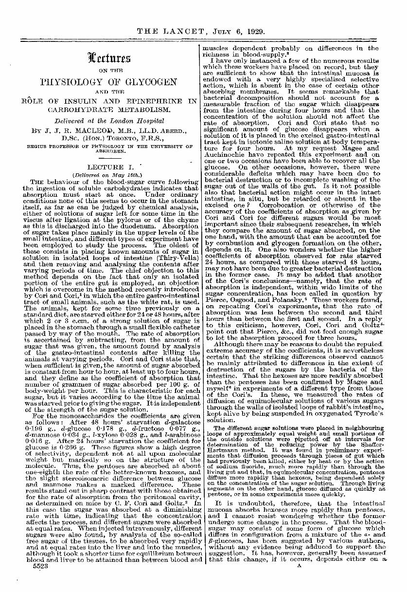
FIG. 1.
Showing disappearance of glycogen from the liver with increaseof free sugar and decrease of lactic acid. (Lactic acid inparts per 10,000 ; glycogen and sugar in parts per cent.) i
substances which are formed from it are different.As the glycogen disappears from the liver, free sugarincreases and there is no increase in lactic acid (seeFig. 1), whereas in muscle there is no increase in freesugar but lactic acid ultimately accumulates almostin proportion to the glycogen which has disappeared(Macleod and Simpsonl4). (Compare with Fig. 3.)We will again have occasion to refer to the significanceof these results.
Accumulation of Glycogen in the Liver.
Turning now to the conditions which cause glycogento accumulate in the liver, we again find that thestorage hypothesis is entirely inadequate. Perhapsthe results which show this most convincingly arethose obtained, first of all by Pollakls and later byMarkowitz,"6 as a result of injecting epinephrinerepeatedly into animals after the glycogen of the liverhad been reduced to a minimum, through fasting andstrychnine. Each injection of epinephrine was
followed by hyperglycaemia, and after the injections
had been repeated many times during several days,the liver was found to contain considerable amounts ofglycogen. This is shown in Table I.We shall see in the succeeding lecture that Cori and
Cori have attempted to explain this result as dependenton a transfer, or shift, of glycogen from muscles toliver, but even if this were to account for the glycogen
TABLE I.-Glycogen in Rabbits after Repeated Injections
..
of Fpinephrine.
I (a) Days on each of which from 3-5 c.cm. 1-1000 adrenalininjected in frequent doses.
accumulation following one injection of the hormone,it certainly could not do so for that remaining afterrepeated injections during several days.But although epinephrine stimulates the internal
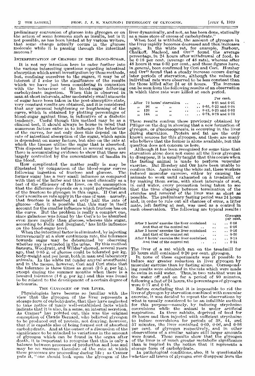
secretion of glycogen in fasting animals it is generallyregarded to do the opposite-namely, to hasten itsbreakdown when given to animals with large storesof glycogen in the liver. This latter belief is mainlybased on experiments by Ritzmann, but Olmsted andCoulthard15 found it very difficult in the spinal cat toobtain corroborative evidence. It was only when thepreparation was allowed to stand for some hoursafter decapitation, so as to get rid of all anaestheticfrom the blood, that they were able, by actual measure-ment of glycogen in small pieces of liver, todemonstrate that a decrease in percentage occurredas the blood-sugar rose. In view of these results, thishormone must be regarded as having a reversible actioncausing glycogen to be deposited in the liver whennone is there and to be broken down when abundanceis already present. The other hormone which mostdefinitely has an influence on carbohydrate meta-bolism-namely, insulin-exhibits similar reversibleeffects, although the procedures necessary to demon-strate them are different in detail. Thus, when insulinis given without food to fasting white rats it first of allcauses the glycogen content of the liver to becomereduced, but this is followed by an increase, provideda convulsive dose has not been given. This is shownin the accompanying chart (Fig. 2) and will be referredto again when we come to consider the action ofinsulin. Reversible effects of insulin are also seen bycomparing its action on normal and diabetic animals.When it is given along with food to normal animals,glycogen is always deposited in the liver muchmore slowly than after food alone, as will be shownlater, whereas there is an invariable and often amarked increase in liver glycogen when’ insulinis injected into diabetic animals, both when food isalso given and when it is Withheld.
Although the observations which we have justconsidered, as well as those of Cramer, show quiteclearly that glycogen must be produced in the livercells out of non-carbohydrate material, by some sort ofsecretory process, it must not be lost sight of that itcan also be deposited in them by condensation ofglucose, and other hexose monosaccharides, carriedto the viscus by the blood. At least, this is thesimplest way by which the gradual increase in glycogenpercentage can be explained when food rich incarbohydrate is fed to a previously fasted animal.In our observations of this type on white rats it wasnoted that the increase in glycogen was usually lessmarked during the first hour or so after feeding thanlater, the maximum, of about 4 per cent. being reachedin approximately four hours. This retarded onset inglycogen deposition may possibly depend upon anunpreparedness of the liver to condense the sugar ;
4
itcannot be due to inadequacy of a supply of blood-sugar, since this begins to mount within a few minutesof feeding the rats and, in our experiments, hadreached the peak in at least one hour after feeding,and probably much earlier.Many factors may come into play in the formation
of glycogen in the liver and some of these, such as
FIG. 2.
Influence on glycogen, in white rats, of varying amounts of insulin injected after48 hours’ fasting. Ordinates, per cent. glycogen ; abscissa, times of killing afterinjection; black columns, liver; cross-hatched columns, muscles. Numbers inbrackets give number of animals averaged in each case, and the unshaded portionof each column, the percentile error. Black dots. blood-sugar percentage.
the presence of insulin and epinephrine, we willdiscuss more fully later. Others concern the chemicalstructure of the carbohydrate present in the circulatingblood, but inasmuch as the observations have beenmainly restricted to the liver of cold-blooded animalsthey do not concern us here. A decided increase inglycogen occurs in the liver in the spinal cat, especiallyduring the first hour or so after decapitation. Olmstedand Coulthard,17 who discovered this, could give nosuggestion as to what the precursor of the glycogenmight be, although the blood-sugar, meanwhilefalling, was no doubt partly responsible. RecentlyCori and Cori,18 finding that d-lactic acid readilyforms glycogen in the liver when given to rats, byfeeding or injection, have attributed the result tod-lactic acid coming from the muscle glycogen. Theonly difficulty in this explanation is that the glycogenremains unchanged in the muscles of the spinal cat,according to Best, Hoet, and Marks.19
THE GLYCOGEN OF THE MUSCLES.
Turning now to the glycogen of muscle, which hascome to occupy so important a place in physiology,let us consider first of all the question of its formation. This seems to be mainly dependent on the sugar of theblood, which may be derived either from the break-down of liver glycogen or more directly from absorbedglucose. Some puzzling facts have recently beenbrought to light concerning the influence of the liver.After this organ has been removed in the dog themuscle glycogen steadily declines, Mann and Magath20 0finding that it was on an average 0-3 per cent. imme-diately after the hepatectomy, and 0-15 per cent.four hours later. Curiously enough, this diminutiondoes not occur when the liver is practically eliminatedfrom the circulation in the spinal cat, by ligation of theportal blood-vessels along with removal of theremaining abdominal viscera. In both groups ofexperiments the percentage of blood-sugar falls, butit is possible that it does so relatively less rapidly inthe eviscerated preparation, on account of a leakageof sugar from the liver through the patulous hepaticveins. But even if we allow for this leakage of sugar ,
it is difficult to account for the constancy in theglycogen of the muscles of the eviscerated preparationsince, though not visibly contracting, they are mean-while producing heat at the rate of at least 2 caloriesper kg. of body-weight and per hour. If the immediatefuel for this combustion is the glycogen of the muscles,as is the present-day teaching, then we must imagine
that glycogen is being built up ineach muscle at a rate which is justexactly the same as that at whichit is being broken down, and thatits source is, in large part, the sugarwhich is escaping into the bloodfrom the liver, since the totalamount of sugar originally presentin the blood is far from sufficient toaccount for the energy expended.
Best, Hoet, and Marks19 haveshown that when glucose is addedto the blood of the evisceratedpreparation, no glycogen is formedin the muscles unless when insulinis injected along with the sugar.On the other hand, Choi 21 hasshown that when the glucoseinjections are made into an animalfrom which the abdominal viscerahave not been removed, glycogenis formed in the muscles, this beingdependent upon the insulin secretedby the pancreas, since no glycogenformation occurs in animals fromwhich this organ, but not the liver,has been removed some hours pre-ceding hepatectomy. This is shownin Table II. It is no doubt forthep same reason that the fall inglycogen in the muscles ofhepatectomised dogs is prevented
when glucose is injected intravenously, but accord-ing to Mann and Magath20 much more glucose mustbe injected to accomplish this than the 0-25 g.per kg. per hour that suffices to maintain theblood-sugar at its normal level. Although theseresults show that glycogen formation can occur in themuscles in the presence of insulin, this does not meanthat insulin is essential for the process, since glycogenis formed in the muscles of diabetic (depancreatised)animals. Thus Chaikoff22 found the average percent-age of glycogen in the muscles of dogs on the third
-
TABLE IL-Effect of Injection of 3,5 g. Glucose onGlycogen of Muscles in Spinal Cat.
day of acute diabetes (caused by withdrawal ofinsulin from previously depancreatised animals) to be0-425 per cent., and on the fifth day 0-350 per cent.During this interval at least 2 calories per kg. anhour were used, so that a dog of 7 kg. (about theaverage of the animals used) would have expendedat least 2 x 7 x 48 = 672 calories, representing about160 g. of carbohydrate, in the two days, and forthis, large quantities of muscle glycogen must havebeen formed, unless we assume that other substancesthan glycogen can serve as the fuel for energy pro-duction in muscle. We cannot say what factor may-be responsible for glycogen formation in the muscleswhen there is no insulin available in the body, but it
would seem most likely that it depends in some wayon the liver. In other words, the material which is
5
used for glycogen formation in the muscles when noinsulin is present may be glucose that has passedthrough a glycogen stage in the liver or has beenmanufactured in this viscus, or even in the musclesthemselves, by a process of gluconeogenesis.
In so far as experimental evidence goes at present,however, the only way by which glycogen can be
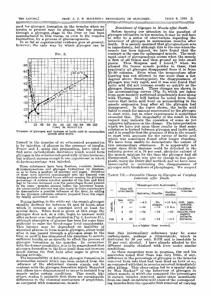
FIG. 3.
Decrease of glycogen and increase of lactic acid in mammalianmuscle after death.
formed in the muscles of an eviscerated preparationis by injection of glucose in the presence of insulin.Fraser and I, using this preparation, have tried tofind some carbohydrate derivative which would formglycogen in the muscles independently of this hormone,but without success except in one experiment in whichdi-hydroxyacetone was injected.
These substances have been fructose, r-sodium lactate,di-hydroxyacetone and glycogen hydrolysed by diastase,so as to form a mixture of dextrine and sugar. Solutionsof them were injected continuously into the femoral veinduring periods of several hours without causing the glycogenpercentage of certain selected muscles of the fore and hindlegs to become increased over the percentages found presentin the same muscles excised before the injections began.An unsuccessful attempt was also made in these experimentsto demonstrate a possible influence of the liver, by addingto the glycogen solutions extracts prepared from this organby faintly acidulated water.
During fasting, in the white rat the muscle glycogensteadily declines for between 24 and 48 hours, afterwhich it remains at a constant level at least forseveral days. When food is given at this stage theglycogen does not, as a rule., begin to increase untilafter an hour or so (as illustrated in Fig. 5, Lecture II.),although absorption of glucose has been for some timesufficient to raise the blood-sugar level considerably.This latency may be dependent on inability ofabsorbed glucose to form muscle glycogen, either untilafter it has passed through a glycogen stage in theliver or until it has stimulated the pancreas to secretesufficient insulin so as directly to affect the process ofglycogen formation in the muscles. In connexionwith the former possibility, it is to be remembered thatglycogen formation in the liver itself does not increasemuch during the first hour after sugar is given tofasting animals.The impossibility of detecting glycogen formation in
mammalian muscle which has been isolated from thenfluence of the abdominal viscera stands out incontrast with the glycogen formation which Meyerhofand others have demonstrated to occur in isolated frogmuscle under certain conditions. This result, likeothers, makes it probable that there are considerabledifferences in the chemical mechanism of amphibian,as compared with mammalian, muscle.
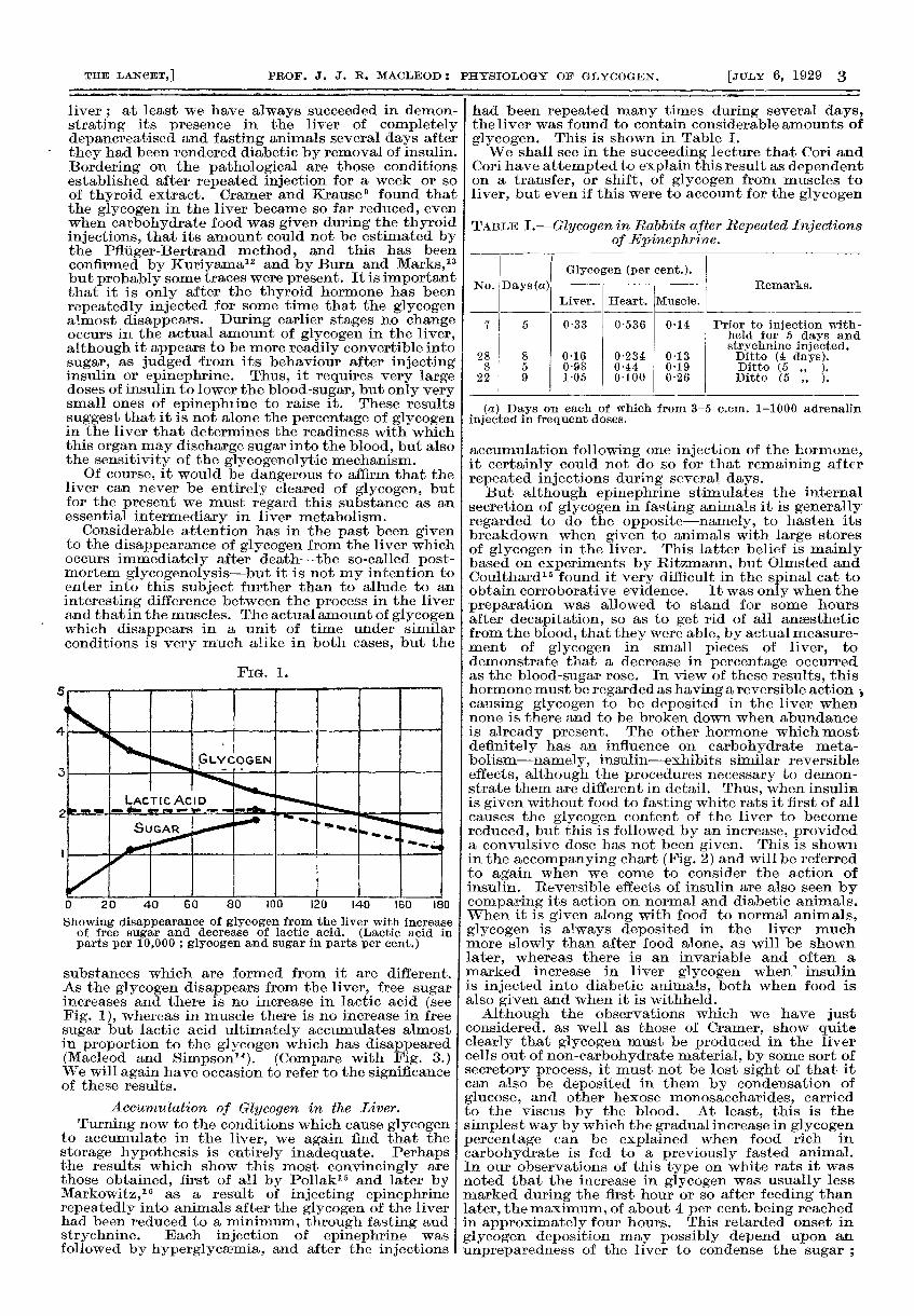
Breakdown of Glycogen in Muscle after Death.Before turning our attention to the question of
glycogen utilisation in the muscles, it may be well hereto refer to a series of observations regarding thebehaviour of glycogen in mammalian muscles afterdeath. It is usually assumed that glycogenolysis setsin immediately, but although this is the case when themuscle has been injured, we have found that thecontrary is the case for undamaged muscle. The mostrapid onset of glycogenolysis occurs when the muscleis first of all frozen and then ground up into smallpieces. Thus Simpson and I found,1 when weallowed the frozen muscle powder to thaw, thatpractically all glycogen disappeared from it in from20-30 minutes. Even when the temperature afterthawing was not allowed to rise more than a fewdegrees above freezing-point, the disappearance ofglycogen was very rapid, and it was also found thatlactic acid did not increase nearly so quickly as theglycogen disappeared. These changes are shown inthe accompanying curves (Fig. 3), which are takenfrom more recently performed experiments done alongwith Thomas. It will further be noted from thesecurves that lactic acid went on accumulating in themuscle suspension long after all the glycogen haddisappeared. In the curve shown, the lactic acidfailed to reach the amount represented by the glycogento start with, but in other experiments it sometimesexceeded this. The irregularity of the result in thisrespect may indicate the operation of some as yetunknown influences on the change. The interpretationwhich we have put upon these results is that somesubstance is formed between glycogen and lactic acid,and it is possible that the presence of this in the muscleto start with accounts for the excess of lactic acidwhich was sometimes found after longer standing.We have been unable to demonstrate the nature ofthis intermediary substance. It is apparently notsugar since little increase could be detected in thereducing power of a 70 per cent. alcoholic extract ofthe muscle mixture by the time the glycogen had alldisappeared. There was also no change in free phos-phate, using the Doisy-Bell method, and we have beenunsuccessful in correlating the disappearance ofglycogen with breakdown of phosphagen. We suspect
TABLE IIL-Percentile Change in Glycogen at VaryingIntervals after Death.
h.c. =higher than control.
that this intermediary substance may be some
carbohydrate, perhaps a trisaccharide, which isdestroyed by 30 per cent. KOH and is insoluble in70 per cent. alcohol. I have already alluded to thedifferent results obtained with liver under similarconditions.As these researches were in progress, Orr and his
associates noted that there was very little, if any,difference in the percentage of glycogen in the musclesremoved from rats that had been dead an hour or so,as compared with that in muscles removed immediatelyafter killing. This observation led to a special studyby Miss Mackay23 of the behaviour of glycogen inintact muscle, in which she compared the percentagesin certain muscles removed under ether anaesthesiafrom one limb (in rabbits) with those of the correspond-ing muscles from the opposite limb removed at varying
6
periods after death. The results are summarised inTable III. in which the changes in glycogen at theperiods indicated after death are expressed as a
percentage of the amounts present in the correspondingmuscles immediately before death.
It can be seen that post-mortem glycogenolysis inintact muscle is very slow compared with that occur-ring in pulverised muscle, that its rate varies consider-ably in different muscles, and that the glycogen,instead of decreasing, may actually increase in amount.Incidentally, it was observed by Miss Mackay thatthe irritability of the muscles towards electricalstimulation persisted longer in those which had beendissected free from the surrounding tissues and keptmoistened with gauze soaked in Ringer’s solutionthan in muscles left in situ without any dissection. Theonset of rigor was also delayed in the dissectedmuscles. The most noteworthy result was, however,the apparent increase in glycogen observed especiallyin the gastrocnemius muscles. Since this was notvery marked, however, it might signify nothing morethan a relative increase due to blood having drainedout of the muscle after death. In more recentexperiments which Fraser and I have performed, thispossibility has been ruled out, since we did not removethe muscles from the left leg until immediately afterdeath.
Rabbits killed either by chloroform or by stunning wereused. Certain muscles were then dissected from the leftleg, and after leaving the carcass for an hour at room-temperature, the corresponding muscles of the right legwere removed. Table IV. shows some of the results, andin this case these are given in actual percentages, as well asin percentile changes, ,
TABLE IV.
* The animals were killed by chloroform, but similar resultswere also obtained in those killed by stunning.
T.a. -tibialls anticus ; G. =gastrocnemius.
The usual variability, both in the glycogen contentof corresponding muscles in different animals and inthat of different muscles in the same animal, is seen,but in only one animal was the percentage less inone hour after death in both of the muscles examined.It is possible that corresponding muscles of the twosides did not contain the same percentages of glycogenat the time of death, and this will require furtherinvestigation. In the light of observations on otheranimals than the rabbit it has been assumed that thepercentages are alike. We also determined the
percentage of lactic acid, using for this purpose themodified permanganate oxidation method afterremoval of sugar from an alcoholic extract of themuscle, by means of copper sulphate and milk of lime.An increase is evident in all cases, but no relationshipcan be detected between either the glycogen percentageto start with or the extent of the post-mortem increasein glycogen and the increase in lactic acid.
These results, which it may be stated are notdependent upon any changes in the water content ofthe muscles, suggest the presence of some hithertounknown precursor from which both glycogen andlactic acid are derived. This must either break downflrst of all into glycogen, lactic acid being formed at anindependent rate out of the general store of glycogen,
or it must be a substance from which both glycogenand lactic acid are formed. In the former case, thesubstance would be a larger molecule than glycogen,whereas in the latter, it would be a smaller one andmight be a lower dextrine or some poly-saccharide,perhaps a tri-saccharide. We have so far beenunsuccessful in demonstrating any change in the freesugar of the muscle extractable by alcohol, but thismay be because we used strong alcohol in preparingthe extracts.
I have already referred to experiments by Simpson,Thomas, and myself in which it was shown thatglycogen disappears from pulverised frozen mam-malian muscle after thawing much more rapidly thanlactic acid accumulates, and from which it was
concluded that between the two some intermediarysubstance, possibly a tri-saccharide, must be formed.It is just possible that we are dealing with the samesubstance in both groups of experiments, but if thisbe the case then we must suppose that the agency forits conversion into glycogen becomes destroyed byfreezing and pulverising the muscle. May it bebecause of destruction of the architecture of themuscle-fibre by the fine ice crystals which form in itwhen it is suddenly frozen, that the power of glycogensynthesis disappears ?
Breakdown of Glycogen in Muscle during Life.With regard to the breakdown of glycogen in
mammalian muscle during life, very little is definitelyknown, at least for muscle which is under perfectlyphysiological conditions. That the percentage ofglycogen remains unchanged for at least an hour afterthe blood pressure has been reduced to zero makesone wonder whether its failure to rise in the muscles ofan eviscerated preparation, even when excess ofglucose is present in the blood, may not indicate thata part of the glycogen-forming powers have dis-appeared, although they can be stimualted in thepresence of an excess of both insulin and sugar.
It has been known for long that the glycogenbecomes reduced when the muscles contract, and,of the older experiments, I need only recall here thoseof Manché24 and Chandelon ’21 who compared itsamount in the muscles of one leg, paralysed by cuttingthe nerve-supply, with those of the opposite leg inwhich there had been more or less activity. Anexperiment of this type more recently performed isthat of Best, Hoet, and Marks,19 who, by injectingconvulsive doses of insulin into cats in which thesciatic nerve of one leg had been severed, found thatthe glycogen content of the paralysed muscles remainedhigh, while it became very low in those undergoingcontractions. Incidentally, it may be remarked,these results also show that the glycogen of one setof resting muscles cannot be drafted, by way of thecirculation, to provide glycogen for an active set,even although the demands of the latter may beurgent. In the light of these results and also thoseobtained on isolated frog muscle, it is to be expectedthat glycogen will decrease when mammalian muscleis stimulated. Some results obtained by Miss Mackaywill illustrate this.
In the decapitate cat three muscles were dissected fromthe left leg, and the corresponding ones of the right legwere then stimulated electrically, either through the nerveor directly. The extent of reduction in glycogen, calculatedas a percentage of the amount originally present in theresting muscles, is shown in Table V.Even a few moments of activity is seen to be associated
with a decided reduction in muscle glycogen, and thisbecomes more marked as activity is maintained.
In these experiments the liver was still intact andit contained a rich supply of glycogen, owing toprevious feeding of the animals with sugar. Had theliver been absent it is probable that the glycogenwould have become more rapidly reduced by activity,and it was the original intention of the research tostudy this possible influence of the liver. With thisin view Miss Mackay performed several experimentsin which the muscles in animals with the liver intactand containing much glycogen were left at rest for an
7
hour or so after stimulating them, but the extent towhich the glycogen reappeared, as judged by com-parison with the results shown in Table V., was verylimited, if any really occurred. I have formed theimpression, both from these experiments and fromthose more recently performed by Fraser and myself,that the failure to demonstrate decided glycogenformation in the muscles of the spinal preparationunless when insulin is present in excess is due to thelow blood pressure.
TABLE V.-Percentile Fall in Glycogen in StimulatedMuscle with its Blood-supply Intact.
* St. = Stimulated till muscle would contract no longer.
Finally, something must be said concerning thebehaviour of the muscle glycogen in white rats aftervarying degrees of exercise, either on a treadmill orby swimming in cold water. I have already alludedto the behaviour of the liver glycogen in these experi-ments. Table VI. shows the results for the muscles.
TABLE VI.-Glycogen (average per cent.) in Mzcscles ofFasting White Rats after Exercise (with Controls).Fasting Periods 24 Hours.
Sw. = Swimming. * Rat lost 4 g.
It is seen that even prolonged exercise in the ratdoes not depress the muscle glycogen very much, aresult which stands out in contrast with that obtainedby artificial stimulation of the muscles of a spinalpreparation. With the exception of those that weremade to swim in cold water off and on for 1 hours,the rats may be said to have been in a " steadystate " of activity without having been markedlyexhausted at the end of it. Nevertheless, runningon a treadmill for several hours is strenuous enoughwork for a rat. The results show that in the steadystate of muscular activity, not only are the oxygenrequirements of the acting muscles continuouslyprovided for but those of glycogen as well. Glucosederived from the glycogen of the liver no doubtfurnishes the raw material for the reconstruction ofmuch of this glycogen in the muscles, but it is possiblethat some is also manufactured in the muscles them-selves out of protein and fat. I can see no way forputting this possibility to the test of experiment,but it is significant that the percentage of blood-sugar,even after the longest periods of exercise, was practi-cally the same in the exercised rats as in the controls.It was certainly not lower. Taking the results ofboth groups of experiments together, it would appearthat it is only when muscular activity is so strenuousthat the oxygen-supply cannot keep pace with the
demands that the glycogen is significantly lowered.This would presumably be the case in muscles of thespinal cat when they are artificially stimulated andalso in those of intact animals injected with strychnineor with large doses of insulin. In seven rabbits givenconvulsive doses of strychnine, Hershey and Orr foundthat the muscle glycogen averaged 0-064 per cent.,the lowest value obtained after convulsions hadlasted for 57 minutes being 0-03 per cent. It isworthy of note that some glycogen was always foundpresent.
BIBLIOGRAPHY.1. Cori, C. F. : Jour. Biol. Chem., 1925, Ixvi., 691.2. Cori, C. F., and Goltz, H. L.: Proc. Soc. Exp. Biol. and
Med., 1925, xxiii., 122.3. Same Authors : Ibid., 1925, xxii., 124.4. Pierce, H. B., Osgood, H. S., and Polansky, J. B. : Ibid.,
1929, xxvi., 347 ; and Jour. of Nutrit., 1929, i., 247.5. Cori, C. F., and G. T., and Goltz, H. L. : Proc. Soc. Exp.
Biol. and Med., 1929, xxvi., 433.6. Magee, H. E., and Macleod, J. J. R. : Proc. Physiol. Soc.,
10. Barbour, A. D., Chaikoff, I. L., Macleod, J. J. R., and Orr,M. D. : Amer. Jour. Physiol., 1927, lxxx., 243.
11. Hershey, J. M., and Orr, M. D. : Trans. Roy. Soc. Can.,1928, Sec. v., 151.
12. Kuriyama, S. : Amer. Jour. Physiol., 1917, xliii., 481.13. Burn, J. H., and Marks, H. P.: Jour. Physiol., 1925, lx.,
131.14. Macleod, and Simpson, W. W. : Jour. Physiol., 1927, lxiv.,
256.15. Pollak, L.: Archiv f. exp. Path. u. Pharm., 1909, lxi.,
149 and 166.16. Markowitz, J. : Amer. Jour. Phys., 1925, lxxiv., 22.17. Olmsted, J. M. D., and Coulthard, H. S.: Ibid., 1928,
lxxxiv., 610.18. Cori, C. F., and G. T. : Jour. Biol. Chem., 1929.19. Best, C. H., Hoet, J. P., and Marks, H. P.: Proc. Roy.
Soc. B., 1926, c., 32.20. Mann, F. C.: Ergebnisse d. Physiol., 1925, xxiv., 379.21. Choi, Y. O. : Amer. Jour. Physiol., 1928, lxxxiii., 406.22. Chaikoff, I. L. : Jour. Biol. Chem., 1927, lxxiv., 203.23. MacKay, M. E. : Trans. Roy. Soc. Can., 1928, Sec. V., 329.24. Manché, E. : Zeit. f. Biol., xxv., 163.25. Chandelon, Th. : Archiv f. d. ges. Physiol. (Pflüger), xiii.,
626.
RADIUM TREATMENT BY THEIMPLANTATION OF SCREENED SEEDS.
BY JOSEPH MUIR, M.D.,NEW YORK CITY.
IT is now some two years since radium treatment bythe method of implanting screened seeds containingradon was introduced into England. The rapiditywith which this technique has made a place for itselfamong the longer established method of applyingradium is attested by the number of papers in this andother journals in which cases so treated are describedin considerable detail. In the Medical ResearchCouncil’s report on the work done in certain hospitalsduring the year 1927 we find but one reference to theimplantation of platinum radon seeds, but in Gask’sHunterian Lecture, delivered in January of last year,the method is described at length, as it is also inearlier publications of this speaker and a number ofothers, notably Gosse and Chester-Williams, andStanford Cade. The general plan of treatment andits results in a rather limited field are thus fairly familiarto readers of THE LANCET. It has, however, a farwider range of usefulness, and it has seemed possiblethat some suggestions along this line from its originatormight be of general interest to the British medicalprofession.
Advantages of the Screened Seed.The advantage of the implantation method as
compared with surface applications are too well knownto need any amplification here. Those which theseed screened with platinum possesses over theunscreened or " bare tube " and the gold container ofradon are perhaps less generally comprehended. Thebare tube offers no obstacle to the passage of caustic