RESEARCH ARTICLE Left Atrial Ligation Alters Intracardiac Flow Patterns and the Biomechanical Landscape in the Chick Embryo William J. Kowalski, 1 Nikola C. Teslovich, 1 Prahlad G. Menon, 1 Joseph P. Tinney, 1,2 Bradley B. Keller, 1,2 and Kerem Pekkan 1 * 1 Department of Biomedical Engineering, Carnegie Mellon University, Pittsburgh, Pennsylvania 2 Department of Pediatrics, Cardiovascular Innovation Institute, University of Louisville, Louisville, Kentucky Background: Hypoplastic left heart syndrome (HLHS) is a major human congenital heart defect that results in single ventricle physiology and high mortality. Clinical data indicate that intracardiac blood flow patterns during cardiac morphogenesis are a significant etiology. We used the left atrial ligation (LAL) model in the chick embryo to test the hypothesis that LAL immedi- ately alters intracardiac flow streams and the biomechanical environment, preceding morphologic and structural defects observed in HLHS. Results: Using fluorescent dye injections, we found that intracardiac flow patterns from the right common cardinal vein, right vitelline vein, and left vitelline vein were altered immediately following LAL. Furthermore, we quantified a significant ventral shift of the right common cardinal and right vitelline vein flow streams. We developed an in silico model of LAL, which revealed that wall shear stress was reduced at the left atrioventricular canal and left side of the common ventricle. Conclusions: Our results demonstrate that intracardiac flow patterns change immediately following LAL, supporting the role of hemodynamics in the progression of HLHS. Sites of reduced WSS revealed by computational modeling are commonly affected in HLHS, suggesting that changes in the biomechanical environment may lead to abnormal growth and remodeling of left heart structures. Developmental Dynamics 243:652–662, 2014. V C 2013 Wiley Periodicals, Inc. Submitted 30 May 2013; First Decision 16 September 2013; Accepted 20 September 2013; Published online 19 December 2013 Introduction Hypoplastic left heart syndrome (HLHS), is a rare but serious con- genital heart defect, occurring in 1 of every 5,000 births (Go et al., 2013). The hallmarks of HLHS are an underdeveloped and nonfunctioning left ventricle and hypoplastic ascending and transverse aorta in association with stenosis or atresia of the mitral and/or aortic valves, and intra-uterine compensatory enlargement of right sided cardiac structures (Friedman et al., 1951; Noonan and Nadas, 1958). A genetic component for HLHS is supported by studies that examined heritability, which show that HLHS is linked to chromosomes 10q and 6q and genetically related to bicuspid aortic valve (Hinton et al., 2007, 2009), although the strength of this relationship is unknown (McBride et al., 2009). The genetic basis of HLHS is still largely undeter- mined and no transgenic animal models have recapitulated the human HLHS phenotype (Sedmera et al., 2005). Clinical innova- tions and scientific research has significantly improved the out- look for infants born with HLHS from a fatality rate of over 95% in 1980 to our current projections that 70% of infants born with HLHS are expected to survive to adulthood (Feinstein et al., 2012). These advances in diagnostic and treatment strategies are remarkable; however, the pathogenesis of HLHS during embry- onic and fetal life remains poorly understood. Fetal interventions have become available with the goal of positively impacting fetal and post-natal cardiac growth and remodeling. For most of its history, HLHS has been classified as a “flow defect,” attributed to altered hemodynamic loading of the left heart structures, and fetal echocardiography has demonstrated that blood flow patterns have an important role in the develop- ment of HLHS (Grossfeld et al., 2009). An abnormally small or absent foramen ovale may be one key component, reducing flow to the left heart and impairing normal growth of left heart struc- tures (Chin et al., 1990; Feit et al., 1991; Rychik et al., 1999), and one study has shown a correlation between diameter of the fora- men ovale and relative right heart and/or left heart flow (Atkins et al., 1982). Obstructed inflow or outflow of the left ventricle due to valvular defects is more likely, however, as there is a strong correlation between the diameter of the left atrioventricu- lar junction and left ventricle or aortic root (Sedmera et al., 2005). While the initial insult causing HLHS, genetic or structural, is unknown, the resulting hemodynamic alterations are signifi- cant and progressive. A typical diagnostic scenario in the clinic is detection of normal left heart dimensions with reduced function at mid-gestation, which is later followed by progressive involu- tion of the left ventricle in the third trimester of pregnancy (McElhinney et al., 2010). One unifying hypothesis is that altered intracardiac flow patterns (ICFP) and altered mechanical loading DEVELOPMENTAL DYNAMICS Additional Supporting Information may be found in the online ver- sion of this article. Grant sponsor: NSF; Grant number: CAREER Award 0954465; Grant sponsor: Kosair Charities Pediatric Heart Research Program. *Correspondence to: Kerem Pekkan, Biomedical and Mechanical Engineer- ing, Carnegie Mellon University, 700 Technology Drive, Pittsburgh, PA 15219. E-mail: [email protected]Article is online at: http://onlinelibrary.wiley.com/doi/10.1002/dvdy. 24107/abstract V C 2013 Wiley Periodicals, Inc. DEVELOPMENTAL DYNAMICS 243:652–662, 2014 DOI: 10.1002/DVDY.24107 652
Transcript
a
RESEARCH ARTICLE
Left Atrial Ligation Alters Intracardiac Flow Patterns andthe Biomechanical Landscape in the Chick EmbryoWilliam J. Kowalski,1 Nikola C. Teslovich,1 Prahlad G. Menon,1 Joseph P. Tinney,1,2 Bradley B. Keller,1,2 and Kerem Pekkan1*
1Department of Biomedical Engineering, Carnegie Mellon University, Pittsburgh, Pennsylvania2Department of Pediatrics, Cardiovascular Innovation Institute, University of Louisville, Louisville, Kentucky
Background: Hypoplastic left heart syndrome (HLHS) is a major human congenital heart defect that results in single ventriclephysiology and high mortality. Clinical data indicate that intracardiac blood flow patterns during cardiac morphogenesis are asignificant etiology. We used the left atrial ligation (LAL) model in the chick embryo to test the hypothesis that LAL immedi-ately alters intracardiac flow streams and the biomechanical environment, preceding morphologic and structural defectsobserved in HLHS. Results: Using fluorescent dye injections, we found that intracardiac flow patterns from the right commoncardinal vein, right vitelline vein, and left vitelline vein were altered immediately following LAL. Furthermore, we quantified asignificant ventral shift of the right common cardinal and right vitelline vein flow streams. We developed an in silico model ofLAL, which revealed that wall shear stress was reduced at the left atrioventricular canal and left side of the common ventricle.Conclusions: Our results demonstrate that intracardiac flow patterns change immediately following LAL, supporting the roleof hemodynamics in the progression of HLHS. Sites of reduced WSS revealed by computational modeling are commonlyaffected in HLHS, suggesting that changes in the biomechanical environment may lead to abnormal growth and remodelingof left heart structures. Developmental Dynamics 243:652–662, 2014. VC 2013 Wiley Periodicals, Inc.
Submitted 30 May 2013; First Decision 16 September 2013; Accepted 20 September 2013; Published online 19 December 2013
Introduction
Hypoplastic left heart syndrome (HLHS), is a rare but serious con-genital heart defect, occurring in 1 of every 5,000 births (Goet al., 2013). The hallmarks of HLHS are an underdeveloped andnonfunctioning left ventricle and hypoplastic ascending andtransverse aorta in association with stenosis or atresia of themitral and/or aortic valves, and intra-uterine compensatoryenlargement of right sided cardiac structures (Friedman et al.,1951; Noonan and Nadas, 1958). A genetic component for HLHSis supported by studies that examined heritability, which showthat HLHS is linked to chromosomes 10q and 6q and geneticallyrelated to bicuspid aortic valve (Hinton et al., 2007, 2009),although the strength of this relationship is unknown (McBrideet al., 2009). The genetic basis of HLHS is still largely undeter-mined and no transgenic animal models have recapitulated thehuman HLHS phenotype (Sedmera et al., 2005). Clinical innova-tions and scientific research has significantly improved the out-look for infants born with HLHS from a fatality rate of over 95%in 1980 to our current projections that 70% of infants born withHLHS are expected to survive to adulthood (Feinstein et al.,2012). These advances in diagnostic and treatment strategies are
remarkable; however, the pathogenesis of HLHS during embry-onic and fetal life remains poorly understood. Fetal interventionshave become available with the goal of positively impacting fetaland post-natal cardiac growth and remodeling.
For most of its history, HLHS has been classified as a “flowdefect,” attributed to altered hemodynamic loading of the leftheart structures, and fetal echocardiography has demonstratedthat blood flow patterns have an important role in the develop-ment of HLHS (Grossfeld et al., 2009). An abnormally small orabsent foramen ovale may be one key component, reducing flowto the left heart and impairing normal growth of left heart struc-tures (Chin et al., 1990; Feit et al., 1991; Rychik et al., 1999), andone study has shown a correlation between diameter of the fora-men ovale and relative right heart and/or left heart flow (Atkinset al., 1982). Obstructed inflow or outflow of the left ventricledue to valvular defects is more likely, however, as there is astrong correlation between the diameter of the left atrioventricu-lar junction and left ventricle or aortic root (Sedmera et al.,2005). While the initial insult causing HLHS, genetic or structural,is unknown, the resulting hemodynamic alterations are signifi-cant and progressive. A typical diagnostic scenario in the clinic isdetection of normal left heart dimensions with reduced functionat mid-gestation, which is later followed by progressive involu-tion of the left ventricle in the third trimester of pregnancy(McElhinney et al., 2010). One unifying hypothesis is that alteredintracardiac flow patterns (ICFP) and altered mechanical loading
DE
VE
LO
PM
EN
TA
L D
YN
AM
ICS
Additional Supporting Information may be found in the online ver-sion of this article.Grant sponsor: NSF; Grant number: CAREER Award 0954465;Grant sponsor: Kosair Charities Pediatric Heart Research Program.*Correspondence to: Kerem Pekkan, Biomedical and Mechanical Engineer-ing, Carnegie Mellon University, 700 Technology Drive, Pittsburgh, PA15219. E-mail: [email protected]
Article is online at: http://onlinelibrary.wiley.com/doi/10.1002/dvdy.24107/abstractVC 2013 Wiley Periodicals, Inc.
conditions result in left ventricular hypoplasia due to the lack ofsufficient mechanical loading to stimulate cardiac growth andremodeling. This hypothesis has been applied as a rationale forfetal interventions, in which fetal balloon aortic valvuloplasty isperformed to restore normal antegrade aortic flow and left ven-tricular loading conditions (McElhinney et al., 2010).
A large number of transgenic animal models have revealed keyroles for signaling pathways and transcription factors in many ofthe events required for normal cardiovascular development,including outflow tract septation (Franz, 1989; Tallquist and Sor-iano, 2003), valve morphogenesis and remodeling (de la Pompaet al., 1998; Ranger et al., 1998; Dunker and Krieglstein, 2002;Hurlstone et al., 2003), and myocardial contraction (Bartmanet al., 2004). However, embryonic models that alter the mechani-cal environment in the setting of a normal genotype are limited.These studies are usually performed in avian embryos, mainlyrepresented by vitelline vein ligation (Rychter and Lemez, 1965;Hogers et al., 1997), conotruncal banding (Clark et al., 1989), andleft atrial ligation (Rychter and Lemez, 1958; Sedmera et al.,1999). Acquiring reliable, spatially resolved velocity measure-ments in the embryonic heart remains challenging, and manystudies lack a quantitative analysis of the biomechanical environ-ment after these perturbations. Newborn and juvenile models of
univentricular circulation have been developed in sheep to moni-tor cardiac function after repair of HLHS, although their long-term success and application is limited (Rodefeld et al., 2003;Myers et al., 2006). Further research using these existing mechan-ical models, and the development of novel models, is necessaryto understand the role of hemodynamics and optimize fetal inter-vention strategies (Pekkan and Keller, 2013).
The chick embryo has been widely used as a model for verte-brate cardiovascular development (Pexieder, 1986; Martinsen,2005). Left atrial ligation (LAL) in the chick embryo remains theonly long-term prenatal animal model of HLHS (Rychter, 1962).In the LAL model, tying off the presumptive left atrium as arecoverable procedure disrupts inflow to the left side of theembryonic ventricle. Measured as early as one hour after ligation,cardiac output is transiently reduced, returning to normal levelsby 32 hours post-LAL, while downstream cardiac pressuresremain normal throughout (Lucitti et al., 2005). Signs of remodel-ing of the ventricular myocardium can be observed 2 days afterligation, including decreased myocardial volume, accelerated tra-becular compaction, and delayed changes in transmural myofiberangle (Sedmera et al., 1999; Tobita et al., 2005). Circumferentialand longitudinal strain increases in both ventricles after LAL,while the onset of preferential circumferential strain patterns in
DE
VE
LO
PM
EN
TA
L D
YN
AM
ICS
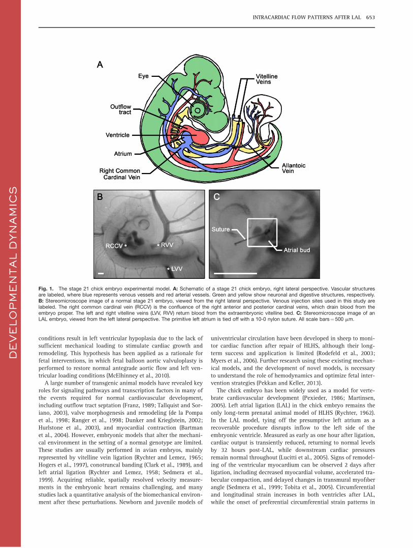
Fig. 1. The stage 21 chick embryo experimental model. A: Schematic of a stage 21 chick embryo, right lateral perspective. Vascular structuresare labeled, where blue represents venous vessels and red arterial vessels. Green and yellow show neuronal and digestive structures, respectively.B: Stereomicroscope image of a normal stage 21 embryo, viewed from the right lateral perspective. Venous injection sites used in this study arelabeled. The right common cardinal vein (RCCV) is the confluence of the right anterior and posterior cardinal veins, which drain blood from theembryo proper. The left and right vitelline veins (LVV, RVV) return blood from the extraembryonic vitelline bed. C: Stereomicroscope image of anLAL embryo, viewed from the left lateral perspective. The primitive left atrium is tied off with a 10-0 nylon suture. All scale bars¼ 500 mm.
INTRACARDIAC FLOW PATTERNS AFTER LAL 653
the right ventricle are accelerated and the preferential longitudi-nal strain patterns in the left ventricle are abolished (Tobita andKeller, 2000). Cellular changes after LAL include reduced prolifer-ation in the left ventricular compact layer and trabeculae,decreased expression of FGF-2 and PDGF-B throughout the heart,and an increased number of apopototic cells in the right atrioven-tricular cushions (Sedmera et al., 2002), as well as increasedmicrotubule density in the left ventricular compact layer(Schroder et al., 2002). Despite the limited number of studiesfocusing on vascular development compared with the ventricle,downstream vessels are also affected in LAL; flow distributionwithin the aortic arches is disrupted, and defects including aorticarch hypoplasia and interrupted aortic arch are observed as earlyas 32 hr after ligation (Hu et al., 2009).
These previous studies clearly demonstrate the morphologic,structural remodeling, and cellular consequences of LAL. How-ever, to date there has not been a quantitative analysis of changesin ICFP that occur following LAL. While hemodynamic anomaliessuch as reduced left ventricular filling and mitral, tricuspid, andaortic valve regurgitation have been recorded, these wereobserved 4 days post-LAL (deAlmeida et al., 2007). The immedi-ate effects of LAL (<1 hr after ligation) on flow patterns aretherefore unknown. Furthermore, quantitative analysis of hemo-dynamic loading after LAL has not been performed. In this study,we test the hypothesis that ICFP are altered immediately follow-ing LAL. Validation of this hypothesis would demonstrate thatchanges in ICFP precede changes in ventricular morphology andstructure, supporting the idea that flow patterns are a primaryfactor in the production of the HLHS phenotype after LAL.
We applied a quantitative analysis of fluorescent dye microinjec-tions to map the location of the intracardiac flow stream withinthe ventricle and outflow tract. We further developed a computa-tional fluid dynamics (CFD) model of the normal and LAL heartloop to estimate changes in wall shear stress (WSS) that mayoccur due to the occlusion of the left atrium. Our results showthat the position of the intracardiac flow stream is shifted signifi-cantly in two of the three venous injection sites and that WSS isreduced at the left atrioventricular canal and left ventricle. Thus,LAL immediately re-routes flow through the heart, likely causingreduced biomechanical loading and growth of left heart struc-tures and leading to impaired remodeling and eventual HLHS.
Results
Intracardiac Flow Streams in Normal and LAL Embryos
We performed LAL at Hamburger-Hamilton stage 21 (Hamburgerand Hamilton, 1951; Al Naieb et al., 2013). At stage 21, cardiaclooping is largely complete, with only minor migration and rota-tion of the outflow tract progressing through stage 24 (Manner,2000). The left and right atrial cavities have formed and ventricu-lar trabeculation is present, although neither septum has fullyformed (Martinsen, 2005). The dorsal and ventral atrioventricularcanal cushions have started to form (De la Cruz et al., 1983), andfour of the five primary outflow tract cushions are present(Qayyum et al., 2001). Blood enters the common atrium throughseveral venous sites including the vitelline veins (VV), whichdrain the vitelline bed, the right and left common cardinal veins
DE
VE
LO
PM
EN
TA
L D
YN
AM
ICS
Fig. 2. Representative images from the experimental fluorescent dye injections. Control embryos are on the left and LAL embryos are on theright. For each injection site, the overlay of the fluorescent and brightfield image is shown alongside the output of the image analysis to measurethe distance between the flow stream (pink) and the ventral (red lines) and dorsal (blue lines) walls of the heart. All scale bars¼ 250 mm.
654 KOWALSKI ET AL.
(CCV), which return blood from the embryo proper, and the allan-toic veins, which drain the allantois (Fig. 1). Blood is ejectedthrough the common outflow tract and symmetric, bilaterallypaired aortic arches. Most often three aortic arch pairs are presentat stage 21, although a variety of patterns have been documentedassociated with flow and vessel caliber mismatch (Kowalski et al.,2013). The stage 21 embryonic heart rate is 155 beats per minuteand cardiac output is 1.28 mm3/s (Hu and Clark, 1989).
To visualize flow streams, we injected embryos with fluores-cent dye at three different venous sites: (1) the right CCV (RCCV),(2) the right VV (RVV), and (3) the left VV (LVV) (Fig. 1). Eachembryo was injected once; sham and LAL embryos were injectedafter a reincubation period lasting less than 1 hr. We computed anormalized intracardiac stream position from the recorded injec-tions. Blood flow was laminar in all embryos. The heart contin-ued to beat at a constant rate for a minimum of one minute afterdye injection, allowing us to record multiple cardiac cycles. LowReynolds number flow in the embryonic ventricle does not allowflow stream mixing. After removing the needle, bloodhemorrhaged and the heart beat ceased rapidly afterward. Ourfluorescent dye injections and image analysis allowed us toquantitatively compute the position of the intracardiac flowstream within the common ventricle and outflow tract. Results ofour experimental dye injections are summarized in Figures 2 and3, and fluorescent overlay movies of representative injections areprovided in the Supplemental Material, which are availableonline. Our normalized stream position represents the distancefrom the dorsal wall of the embryonic heart, where 0 is exactlythe dorsal wall and 1 is exactly the ventral wall. For controlembryos, we found that dye injected at the RCCV took a slightlydorsal route, with an average stream location of 0.44 6 0.09(n¼ 12). The stream from the RVV was the most dorsal, with aposition of 0.41 6 0.11 (n¼ 17). The location of the LVV streamwas 0.49 6 0.07 (n¼ 13), directly through the midline of theheart. Sham embryos displayed similar ICFP, with no significantdifferences vs. the control group.
The response of LAL embryos to insertion of the micro needlewas similar to control embryos with no observable effect onhemodynamics or heart rate. For LAL embryos, our quantitative
analysis revealed that the RCCV stream changed significantly(P< 0.05), now taking a ventral course through the ventricle andoutflow tract, with an average location of 0.66 6 0.16 (n¼ 11).The RVV stream also shifted toward the ventral side of the heart(P <0.05), positioned at 0.54 6 0.10 (n¼ 7). The intracardiac pathof the LVV remained similar at 0.52 6 0.08 (n¼ 8, P >0.05). Inthe LAL embryos, the RCCV and RVV streams were ventral to theLVV stream, the reverse of the control ICFP. Of interest, all flowpatterns shifted ventrally, although only the RCCV and RVV weresignificantly changed compared with the controls. We alsoobserved some retrograde flow in the LAL embryos, where dyeinjected at the RVV and LVV would flow into the cardinal veins.These results demonstrate that LAL significantly alters ICFP inthe embryo, and that this change occurs immediately followingthe intervention.
In Silico LAL Model
We performed three-dimensional (3D) CFD simulations of steadyflow through control and LAL models of the stage 21 heart loop.Both models were created in the computer using our in-housesurgical planning suite (Pekkan et al., 2008; Dur et al., 2011).Blood flow was modeled using our in-house immersed boundarycardiovascular flow solver, which has previously been validatedfor models of the stage 11, 13, and 18 heart loop, comparingaccurately with the commercial second-order unstructured gridflow solver Fluent (version 6.3.26, ANSYS Inc., Canonsburg, PA)and experimental microfluidic models of the embryonic hearttube (Supp. Fig. S1; Pekkan et al., 2009). Mean total cardiac out-put was 1.28 mm3/s, equivalent to stage 21 Doppler flow meas-urements (Yoshigi et al., 2000), and rigid, no-slip walls wereassumed for simplicity, simulating mean quasi-steady flow con-ditions in the heart. We computed color-coded flow streamlinesoriginating from the right and left vitelline veins to observeICFP in our CFD models (Fig. 4). There were some subtle differ-ences, including an overall ventral shift of the flow streams inthe LAL model (Fig. 4). This movement of ICFP toward the ven-tral margin of the heart is similar to our experimental observa-tion. Flow from the RVV seemed to follow the inner curvatureof the heart more tightly in the LAL model as well. However, thevariation between the control and LAL CFD models is far lesspronounced than the experimental results, and we did notobserve the reversal of RVV and LVV stream positions (Fig. 4),prompting future improvements in computational model. Wecomputed the velocity profile at the outflow tract and foundthat it was slightly more skewed in the ventral and left direc-tions in the LAL vs. control model.
To examine changes in biomechanical loading that occur dueto removal of the left atrium, we calculated WSS from our CFDresults (Fig. 5). The maximum WSS levels in the heart were 3.5Pa, found at the CCV and VV inlets and the ventral side of theoutflow tract outlet, consistent with WSS values derived fromparticle image velocimetry in similar stage chick embryos(Poelma et al., 2010). Compared with the control model, WSS inthe LAL model was reduced at the left side of the atrioventricularcanal and left side of the common ventricle (Fig. 5). WSS wasalso slightly increased at the ventral surface of the distal outflowtract. These simplified, static CFD models show that redistributionof the left atrial volume is sufficient to alter ICFP and WSS load-ing, while the disparity with the experimental results supports the
DE
VE
LO
PM
EN
TA
L D
YN
AM
ICS
Fig. 3. Results of the intracardiac stream measurements. The averagelocation of the stream in control, sham, and LAL embryos is shown.Error bars represent the standard deviation. Numbers within each barindicate the sample size. * Indicates a statistically significant (P< 0.05)difference vs. the control group.
INTRACARDIAC FLOW PATTERNS AFTER LAL 655
additional roles of the endocardial cushions and dynamic ventric-ular suction during relaxation in determining ICFP.
Discussion
Qualitative description of normal ICFP in the chick embryo hasbeen taken up by several authors, giving rise to some contradic-tions (Bremer, 1932; Rychter and Lemez, 1965; Jaffee, 1967;Yoshida et al., 1983; Hogers et al., 1995). Early studiesattempted to follow red blood cells as they coursed through theheart, and led to the widely perceived notion that two separatestreams spiraled around each other through the outflow tract(Bremer, 1932; Jaffee, 1967). The presence of these streams wasthought to shape the spiral aortopulmonary septum, with anom-alous spiraling proposed as a major cause of septation defects(Jaffee, 1965). This notion was challenged when researchersbegan injecting dyes or India ink and did not observe spiralingoutflow streams (Yoshida et al., 1983; Hogers et al., 1995). Intheir comprehensive study of chick embryos between stages 14and 22, Yoshida et al. observed two types of intracardiacstreams which depended on both injection site and developmen-
tal age (Yoshida et al., 1983). These streams did not spiralaround one another, but rather followed a dorsal or ventral paththrough the ventricle and outflow tract. These patterns werelater confirmed by Hogers et al. using India ink injections ofembryos between stages 12 and 17 (Hogers et al., 1995). Theirstudy revealed distinct ICFP for blood emanating from differentyolk sac regions, further demonstrating the significance ofvenous return sites and the presence of multiple simultaneousICFP. Our ICFP observed in control stage 21 embryos matchedthose of Yoshida et al. (1983), with blood from the RVV taking amore dorsal path compared with blood from the LVV. No studyhas examined either CCV. In no cases of these embryonic did weobserve evidence of spiraling streams through the outflow tract.The work of Yoshida et al. also suggested that blood from theRVV and LVV perfused only the ipsilateral aortic arches at stage21 (Yoshida et al., 1983). This pattern was contradicted by otherstudies, which showed that the distribution through the aorticarches was not a simple left and right current (Rychter andLemez, 1965; Hogers et al., 1995; Hu et al., 2009). While we didnot examine ICFP beyond the outflow tract in this study, ourprevious investigations dedicated to the aortic arches
DE
VE
LO
PM
EN
TA
L D
YN
AM
ICS
Fig. 4. Intracardiac flow patterns in CFD models of control and in silico LAL embryonic cardiac geometries. The control model is on the left andthe LAL model is on the right. In the upper figures, color coded streamlines are shown, with blue representing flow emanating from the RVV andyellow depicting flow from the LVV. Red arrows give the forward flow direction. The bottom panels show cross-sections at each of the siteslabeled in the control flow stream figure. Control cross-sections are on the left and LAL on the right. The cross-sections reveal a slight overall ven-tral shift of flow paths in the LAL model. The velocity profile at the outflow tract is shown for cross-section 4, where a slight ventral shift of thepeak flow location is observed in the LAL model.
656 KOWALSKI ET AL.
corresponds with a complex perfusion pattern (Wang et al.,2009; Kowalski et al., 2013).
Yoshida et al. observed nearly the exact reverse ICFP inembryos stage 17–18: blood from the RVV took a more ventralroute compared with blood from the LVV, and the two streamsperfused the contralateral aortic arches (Yoshida et al., 1983).These patterns transitioned at around stage 19, and the authorsattributed the dramatic reversal to the changes in the vitellinevenous system. Our data show that in LAL stage 21 embryos,blood from the RVV courses more ventrally compared with bloodfrom the LVV, similar to the stage 17–18 patterns described byYoshida et al. (Fig. 2). It may be that obstruction of the left atriumprevents the change in RVV and LVV patterns or returns ICFP totheir earlier state. Before stage 18, the atrial cavities are smalland hardly distinguishable from the heart tube. Between stages18 and 21, however, the atria significantly increase in size, par-ticularly the left atrium, which becomes larger than the right(Manner, 2000; Kim et al., 2011; van den Berg and Moorman,2011; Yalcin et al., 2011). This expansion of atrial volume andthe atrial contribution to ventricular filling may relate to thechange in ICFP, which occurs during the same interval. As theexpansion of the left atrium is more pronounced than the right,its ligation may return the cardiac geometry to resemble stage 17,causing ICFP to revert. Ventricular trabeculation may also have arole in the changing flow patterns at stage 19. Beginning at stage16/17, trabeculation progresses rapidly during this period, and islargely responsible for increases in myocardial mass (Sedmeraet al., 2000). Another key morphologic event occurring duringthe period between stages 17 and 25 involves substantial growthand remodeling of the endocardial cushions lining the atrioven-tricular canal and outflow tract, brought on by the invasion ofcells that begin to undergo epithelial to mesenchymal transfor-
mations at stage 17 (Noden, 1991; Butcher et al., 2007). LAL mayinterfere with these events by preventing growth of the leftatrium, limiting the expansion of the atrioventricular canal, ordisplacing the endocardial cushions.
Our numerical control vs. LAL CFD models confirmed the vari-ation in the location of the flow streams originating from theRVV and LVV, but these are less pronounced that the in vivoinjections (Fig. 4). A major difference is that we do not see thedramatic ventral shift of the RVV flow stream after LAL. Pres-ently, our CFD model did not incorporate the endocardial cush-ions or ventricular trabeculae and applied rigid, no-slip walls andsteady, unidirectional flow, ignoring the AV synchronous con-tractions and pulsatile flow environment. Furthermore, the exter-nal boundary conditions specified at the vessel inlet and outlets,as well as the neglected wall movement, may suppress the in vivoobserved internal flow differences. These limitations influencethe numerical model of ICFP and mask key elements of the bio-mechanical environment such as oscillatory WSS. Effects of flowpulsatility may be less important, illustrated in our previous pul-satile CFD models of the stage 18 heart loop, which revealed thatWomersely numbers were low (0.44), suggesting that the effectsof pulsatile flow are small (Pekkan et al., 2009). The assumptionsdescribed above were necessary to reduce computational timeand produce a solvable model. Differences between the computa-tional model and experiment may indicate that those aspectsabsent from the model, such as cardiac contractions, endocardialcushions, and ventricular trabeculae, are vital to establishingICFP. Even without these features, however, our models showthat simply removing the left atrium can alter the course of bloodand WSS levels in the heart realistically. In particular, WSS wasreduced at the left side of the common ventricle and left wall ofthe atrioventricular canal and slightly increased on the ventral
DE
VE
LO
PM
EN
TA
L D
YN
AM
ICS
Fig. 5. WSS computed from CFD models of control and in silico LAL embryonic cardiac geometries. The control model is on the left and theLAL model is on the right. Reduced WSS can be seen at the left side of the atrioventricular canal and left region of the common ventricle.Increased WSS is located at the ventral side of the distal outflow tract. To display a broader range of WSS, a peak WSS of 1.0 Pa is shown.
INTRACARDIAC FLOW PATTERNS AFTER LAL 657
surface of the outflow tract (Fig. 5). WSS differed by 0.5 Pa atthese sites, which is above the known threshold to initiate anendothelial response (Egorova et al., 2011). These areas are com-monly affected in HLHS, suggesting that LAL may redirect flowto unload the presumptive left ventricle and its inflow. The veloc-ity profile change in the in silico LAL model may indicate a rea-son for altered aortic arch perfusion and defects in LAL embryos(Hu et al., 2009). However, our recent simulations of aortic archflow using various velocity profile shapes at the outflow tract donot show significant changes in flow distribution or WSS, againdemonstrating the importance of the dynamic contractions(Kowalski et al., 2013).
Morphogenesis of the veno-atrial region is ongoing at stage21, and the veno-atrial connections may differ between embryos(Manner and Merkel, 2007; van den Berg and Moorman, 2011).Our model geometry includes proximal separation of the rightand left vitelline veins, a centrally located venous sinus, andsymmetric cardinal veins, all of which are characteristic of theearly state of veno-atrial morphogenesis (Fig. 4). By stage 24,however, the proximal vitelline veins have fused, the sinus con-nection is limited to the right atrium, and the left cardinal vein iselongated. As this transformation likely influences ICFP, wemodified our model geometry to correspond with this later statevenous anatomy and repeated our control and LAL flow models(Supp. Fig. S2). As with our original geometry, flow patterns inthe LAL model were not substantially different from the control.This later state model, therefore, reinforces our previous reason-ing that those features not incorporated, i.e. cardiac contractions,endocardial cushions, and ventricular trabeculae, have a role indirecting intracardiac blood flow. We did find, however, that theoutflow velocity profile was more skewed in the ventral directionafter changing the venous morphology (Fig. 4 and Supp. Fig. S2).These two models suggest that morphogenesis of the sinuatrialregion can influence ICFP and may provide an explanation forthe changes in flow patterns that occur in normal embryos fromstages 18 to 24 (Yoshida et al., 1983).
Static CFD models of flow in the human embryonic heart showthe presence of multiple simultaneous intracardiac streams, withpatterns similar to those observed in the chick embryo, demon-strating the relevance of these animal models to human develop-ment (DeGroff et al., 2003). While success has been made inreconstructing static embryonic cardiac geometries from poly-meric casting (Wang et al., 2009; Yalcin et al., 2011), there is noexisting model of the dynamic embryonic heart. Recent techni-ques have simulated blood flow in the contractile outflow tract(Liu et al., 2011), although this work is in nascent stages and hasyet to incorporate the upstream heart and downstream vessels.Validation of such models will require information of 3D velocityprofile shapes in vivo. We have recently explored a combinedparticle image velocimetry and optical coherence tomographytechnique to acquire velocity measurements noninvasively,which can be applied to future studies (Chen et al., 2012).
Numerous studies using chick (Gessner, 1966; Hogers et al.,1997, 1999; Reckova et al., 2003; Lucitti et al., 2006) and zebra-fish (Hove et al., 2003; Chen et al., 2011; Corti et al., 2011)embryos support the role of hemodynamics in modifying cardio-vascular growth, morphogenesis, and remodeling. It is well estab-lished that WSS is a key environmental factor affectingcardiovascular development in the embryo and adaptation in theadult (Rodbard, 1975; Langille and O’Donnell, 1986; Girerd et al.,1996; Bayer et al., 1999; Culver and Dickinson, 2010). Using an
RVV ligation model, Hogers et al. found that ICFP emanatingfrom four yolk sac regions followed altered routes through theheart and outflow tract, leading to defects such as small to largeventricular septal defects, bicuspid aortic valve, and aortic archanomalies (Hogers et al., 1997). This ligation model was furthershown to cause spatial and temporal changes in the transcrip-tional profile of genes encoding Kr€uppel-like factor (increased),endothelin-1 (decreased), and endothelial nitric oxide synthase(decreased), suggesting increased WSS (Groenendijk et al., 2005).The increased WSS in the outflow tract was also associated withhigher Tgfb/Alk5 signaling, a phenomenon which was sensitiveto the level of WSS (Egorova et al., 2011). This mechanosensitiv-ity of endocardial cells, coupled with the changed ICFP after LAL,likely results in abnormal morphogenesis and remodeling of theatrioventricular and outflow tract cushions, leading to valvedefects normally present in HLHS. Although our technique doesnot capture total flow through the atrioventricular canal, thequantitative measurements through the outflow tract show a sig-nificant ventral shift of the RVV and RCCV streams (Fig. 3). Inthe RVV ligation model of Hogers et al., flow streams from twoof the four yolk sac regions saw a similar dorsal to ventral shift(Hogers et al., 1997). It is interesting that the outlet of the leftventricle is dorsal to that of the right ventricle in the maturechick. The lack of a dominant dorsal stream after LAL may causereduced WSS on the dorsal cushions present at stage 21, impair-ing the formation of the left ventricle outflow tract and aorticvalve.
Our experiments support the hypothesis that LAL immediatelyalters ICFP in the chick embryo. We quantitatively showed a sig-nificant ventral shift in both the RVV and RCCV flow streams.While the LVV flow stream did not change significantly, the LVVand RVV streams switched relative positions in LAL comparedwith control hearts, resembling earlier stage 18 patterns (Yoshidaet al., 1983). Our static CFD model of the embryonic heart loopfurther revealed that removing the left atrium volume was suffi-cient to alter WSS at critical locations within the left heart. Theseresults suggest that changes in ICFP following LAL precede struc-tural defects such as bicuspid aortic valve and the characteristicundersized left ventricle. The concordance between control andsham embryos suggests that the manipulations required to per-form LAL do not cause a change in flow patterns. However, theligation itself may cause structural damage to the heart, whichcan affect its further development. While we take great care andreject any embryos that show bleeding after LAL, we cannot ruleout this trauma as a cause for HLHS defects. Therefore, we con-clude that the change in ICFP occurs early in development ofHLHS after LAL, but may not be the only or initial insult.
Experimental Procedures
Experimental Measurement of IntracardiacStream Position
Embryo preparation and injection
Fertile white Leghorn chicken eggs were incubated blunt end upin a forced draft incubator at 37
�C and 50% humidity for 84 hr to
Hamburger-Hamilton stage 21 (Hamburger and Hamilton, 1951;Al Naieb et al., 2013). To access the developing embryo, wegently cracked the eggshell above the air sac and peeled back theshell and membrane to form a 1-cm2 window. We then removed
DE
VE
LO
PM
EN
TA
L D
YN
AM
ICS
658 KOWALSKI ET AL.
the overlying extraembryonic membrane using microforceps(Dumont M5, WPI, Sarasota, FL). Limb size, spinal curvature, andthe presence of body tissues were used to stage embryos, and anythat were dysmorphic, such as lacking retina, displaying axialtwisting, or left side up were excluded. To perform LAL, we posi-tioned embryos in ovo under a stereomicroscope (Leica M165F,Leica Microsystems GmbH, Germany), dissected the amnioticmembrane, and flipped the embryo to reveal the presumptive leftatrium. We used microforceps to make a slit-like opening in thethoracic wall above the atrium and tightened an overhand knotformed from 10-0 nylon suture around the primitive left atrium,decreasing its effective volume (Fig. 1) (Tobita et al., 2005). Werepositioned the embryo to its original right-side up orientation,sealed the window with Parafilm (Pechiney Plastic Packaging,Menasha, WI), and reincubated for 1 hr to restore normal heartrhythms. Control embryos had only the overlying membraneremoved and were injected immediately with no reincubation.Sham embryos had a slit made in the thoracic wall above the leftatrium and were reincubated for one hour.
To inject embryos, we pulled needles from 1.0 mm inner diam-eter glass capillary pipettes to 15 mm inner diameter and a 30
�
bevel (PC-10 Puller, EG-44 Microgrinder, Narishige Inc., Japan).Needles were connected by means of polyethylene tubing (PE100,Braintree Scientific, Braintree, MA) to a 10 ml glass Hamiltongastight syringe (1701LT, Fisher Scientific, Waltham, MA). Thesyringe was mounted on a three-axis mechanical micromanipula-tor (Leica Microsystems GmbH, Germany). We primed thesyringe, tubing, and needle with chick ringer’s solution and thendrew up 1 ml of Cy5-diethyl fluorescent dye diluted 1:1 in phos-phate buffered saline (excitation wavelength 626 nm, emissionwavelength 666 nm). Cy5 dyes were prepared at the MolecularBiosensor Imaging Center at CMU (Mujumdar et al., 1993). Weinjected embryos at three different venous sites: (1) RCCV, (2)RVV, and (3) LVV, with all embryos viewed in the right side uporientation (Fig. 1). Each embryo was placed under the stereomi-croscope and the yolk sac membrane (RVV, LVV sites) or amnion(RCCV site) was removed above the injection site. The needle wasinserted into the selected vein and a brightfield movie wasrecorded using a low light monochrome digital camera (LeicaDFC 350FX, Leica Microsystems, GmbH, Germany). We theninjected 0.3 ml of dye, visualized using an external fluorescentlight source (Leica EL6000, Leica Microsystems, GmbH, Germany)and recorded the flow streams. This volume of injected dye hasbeen shown to minimally affect normal hemodynamics, resultingin less than a 10% change in stroke volume and mean arterialpressure (Wagman et al., 1990). A total of 84 embryos wereinjected: 28 RCCV (12 control, 5 sham, 11 LAL), 29 RVV (17 con-trol, 5 sham, 7 LAL), and 27 LVV (13 control, 6 sham, 8 LAL).Movies of each injection site for representative control and LALembryos are provided in the . All injections were performed inovo and only one site was injected per embryo.
Quantitative measurement of the intracardiac flow stream
We applied a quantitative approach to determine the location ofthe intracardiac streams. End-diastolic still images (696 � 520pixels) were extracted from the recorded brightfield movies andused to manually identify the ventral and dorsal boundaries ofthe ventricle and outflow tract. The manually selected pointswere used to form polynomial expressions defining the shape ofthe ventral and dorsal margins, giving two continuous functions.
We used the horizontal position as the independent variable, androtated images, if necessary, so that the long axis of the heartwas approximately horizontal. We then extracted still framesfrom the fluorescent recordings of the injections, which showedthe flow stream as it passed through the ventricle and outflowtract. The centerline of the flow stream was manually identifiedand used to form a third polynomial expression. Any rotationsapplied to the brightfield image were applied exactly to the fluo-rescent image. For each point along the flow stream, we werethen able to mathematically calculate the two points of intersec-tion between the line normal to the stream and the ventral anddorsal walls of the heart. Using these intersection points, we thencalculated the Euclidean distance between the centerline of thestream and the ventral (e) and dorsal (d) walls. The normalizedintracardiac stream position was defined as d/(eþd). We com-puted the position for 10 evenly distributed points along thestream centerline and took the mean value as the final intracar-diac stream position (Fig. 2). The entire process was performedusing a semi-automated ad hoc program developed in MATLAB(R2011a, Mathworks, Natick, MA). We performed the analysisonce per embryo.
The brightfield movie was recorded after insertion of the needleinto the vessel, but before injection of the dye. The injectionmovie was then recorded without moving the embryo or chang-ing the zoom level to facilitate direct overlapping. As a check, wecan clearly observe that the dye within the needle seen in the flu-orescent image directly overlaps with the bore of the needle visi-ble in the brightfield image (Fig. 2). When performing ouranalysis, we rejected any overlays that did not meet this registra-tion test. In addition, as the aortic arches, dorsal aorta, and extra-embryonic vessels were filled with dye, their patterns directlyoverlapped with these vessels in the brightfield image, furtherdemonstrating that no movement occurred between the recordingof the brightfield and fluorescent data. When manually selectingthe ventral and dorsal boundaries, we chose points along theouter surface of the heart, as it was easier to identify. Althoughthese boundaries are not the inner lumen, we were consistent inour selection, allowing for a one to one comparison between con-trol and LAL embryos.
We performed two-tailed, unpaired t-tests to determine differ-ences in ICFP between groups. A p-value of less than 0.05 wasconsidered significant.
Computational Fluid Dynamics Model of an In SilicoLAL Stage 21 Heart
Creation of geometric heart loop models
We based the geometry and proportions of our control stage 21heart loop model on a variety of anatomical imaging studiesincluding serial histology (van den Berg and Moorman, 2011),scanning electron microscopy (Manner, 2000), micro computedtomography (Kim et al., 2011), magnetic resonance imaging (Yel-buz et al., 2003), and optical coherence tomography (Davis et al.,2008). The general “looped tube” structure was designed in Pro-Engineer (Parametric Technology Corp., Needham, MA) based onprevious sketches and models (Patten, 1920; Manner, 2004).Atrial and ventricular bulbs, outflow tract tapering, and thevenous inlets (RVV, LVV, and both CCV) were added using ourin-house sketch-based anatomical editing tool, SketchCAD. Theheart geometry was modeled at end diastole. The model was
DE
VE
LO
PM
EN
TA
L D
YN
AM
ICS
INTRACARDIAC FLOW PATTERNS AFTER LAL 659
completed and made CFD-ready using Geomagic Studio 10 (Geo-magic Inc., Durham, NC). An in silico LAL surgery was performedby removing the presumptive left atrium volume and creating asmoothed surface to connect the left wall of the atrioventricularcanal to the remaining right atrium, using our in-house surgicalplanning suite (Pekkan et al., 2008; Dur et al., 2011). The finishedcontrol and LAL models were rendered in Geomagic Studio 10(Geomagic Inc., Durham, NC) and exported into GAMBIT (ANSYSInc., Cannonsburg, PA), to create a surface triangular mesh. Thetriangular mesh was imported into our in-house CFD preproces-sor, which generates an unstructured 3D Cartesian immersedboundary mesh. The immersed boundary grid had a spatial reso-lution of 14 mm for both the control and LAL models.
Computational fluid dynamics
Blood flow through the embryonic heart was modeled using ourin-house immersed boundary cardiovascular flow solver incorpo-rating a validated second-order accurate multi-grid artificialcompressibility numerical method, described in our previousstudies (Payli et al., 2007; Pekkan et al., 2009; Menon et al.,2012). Flow was simulated in terms of inlet normalized spatio-temporal units, on a high-resolution unstructured Cartesianimmersed boundary grid with finite-difference numerical treat-ment. The objective of the CFD model was to determine if thegeometric change of removing the left atrial volume was suffi-cient to alter ICFP and WSS. Therefore, we performed a steady-state simulation with fixed walls and no slip boundary condi-tions. The mean cardiac output of the stage 21 chick embryo,1.28 mm3/s (Yoshigi et al., 2000), was used to compute inflowboundary conditions at each of the four venous inlets. As reliabledata for the venous flow rates was not available, we prescribedan inflow distribution of 25/75 between the blood returned fromthe embryo by the CCV and blood returned from the vitelline bedby the VV (Hu et al., 1996). Furthermore, a 50/50 split was speci-fied between the cumulative inflows from the laterals. The Reyn-olds number (Re) of each of the CCV inlets was therefore 0.25, theRe of the VV inlets was 0.76, and the Re at the outflow tract was2. A plug flow profile was assigned to all inlets. An instantaneousmass-flow preserving outflow boundary condition was adopted.Blood was treated as a Newtonian fluid with constant hemody-namic properties (r¼ 1,060 kg/m3, m¼ 3.71 � 10�3 Pa.s) (Al-Roubaie et al., 2011). In the case of the later stage model, whichhas only three inlets, the cardiac output remained the same, with75% from the vitelline vein and 12.5% from each of the CCV. Allother boundary conditions and assumptions were unchanged.
CFD simulations were conducted at Pittsburgh SupercomputingCenter’s Blacklight supercomputing cyber-infrastructure. Eachsimulation cost an average of 22 hr at 32 core parallelism. Conver-gence of the steady inflow CFD solution was monitored using therunning average velocity field, which converged to steady staterelatively quickly.
ReferencesAl Naieb S, Happel CM, Yelbuz TM. 2013. A detailed atlas of chick
heart development in vivo. Ann Anat 195:324–341.Al-Roubaie S, Jahnsen ED, Mohammed M, Henderson-Toth C,
Jones EA. 2011. Rheology of embryonic avian blood. Am JPhysiol Heart Circ Physiol 301:H2473–H2481.
Atkins DL, Clark EB, Marvin WJ Jr. 1982. Foramen ovale/atrial sep-tum area ratio: a marker of transatrial blood flow. Circulation 66:281–283.
Bartman T, Walsh EC, Wen KK, McKane M, Ren J, Alexander J,Rubenstein PA, Stainier DY. 2004. Early myocardial functionaffects endocardial cushion development in zebrafish. PLoS Biol2:E129.
Bremer JL. 1932. The presence and influence of two spiral streamsin the heart of the chick embryo. Am J Anat 49:409–440.
Butcher JT, McQuinn TC, Sedmera D, Turner D, Markwald RR.2007. Transitions in early embryonic atrioventricular valvularfunction correspond with changes in cushion biomechanicsthat are predictable by tissue composition. Circ Res 100:1503–1511.
Chen CY, Menon PG, Kowalski WJ, Pekkan K. 2012. Time-resolvedOCT-mPIV: a new mPIV technique applied to noninvasive anddepth-resolved pulsatile flow measurements in microchannelsand chick embryos. Exp Fluids 54:1496.
Chen CY, Patrick MJ, Corti P, Kowalski W, Roman BL, Pekkan K. 2011.Analysis of early embryonic great-vessel microcirculation in zebra-fish using high-speed confocal mPIV. Biorheology 48:305–321.
Chin AJ, Weinberg PM, Barber G. 1990. Subcostal two-dimensional echocardiographic identification of anomalousattachment of septum primum in patients with left atrioventricularvalve underdevelopment. J Am Coll Cardiol 15:678–681.
Clark EB, Hu N, Frommelt P, Vandekieft GK, Dummett JL, TomanekRJ. 1989. Effect of increased pressure on ventricular growth instage 21 chick embryos. Am J Physiol 257:H55–H61.
Corti P, Young S, Chen CY, Patrick MJ, Rochon ER, Pekkan K,Roman BL. 2011. Interaction between alk1 and blood flow in thedevelopment of arteriovenous malformations. Development 138:1573–1582.
Culver JC, Dickinson ME. 2010. The effects of hemodynamic forceon embryonic development. Microcirculation 17:164–178.
Davis AM, Rothenberg FG, Shepherd N, Izatt JA. 2008. In vivospectral domain optical coherence tomography volumetric imag-ing and spectral Doppler velocimetry of early stage embryonicchicken heart development. J Opt Soc Am A Opt Image Sci Vis25:3134–3143.
De la Cruz MV, Gimenez-Ribotta M, Saravalli O, Cayre R. 1983.The contribution of the inferior endocardial cushion of the atrio-ventricular canal to cardiac septation and to the development ofthe atrioventricular valves: study in the chick embryo. Am J Anat166:63–72.
de la Pompa JL, Timmerman LA, Takimoto H, Yoshida H, Elia AJ,Samper E, Potter J, Wakeham A, Marengere L, Langille BL,Crabtree GR, Mak TW. 1998. Role of the NF-ATc transcriptionfactor in morphogenesis of cardiac valves and septum. Nature392:182–186.
deAlmeida A, McQuinn T, Sedmera D. 2007. Increased ventri-cular preload is compensated by myocyte proliferation in normaland hypoplastic fetal chick left ventricle. Circ Res 100:1363–1370.
DeGroff CG, Thornburg BL, Pentecost JO, Thornburg KL, GharibM, Sahn DJ, Baptista A. 2003. Flow in the early embryonichuman heart: a numerical study. Pediatr Cardiol 24:375–380.
Dunker N, Krieglstein K. 2002. Tgfbeta2 -/- Tgfbeta3 -/- doubleknockout mice display severe midline fusion defects and earlyembryonic lethality. Anat Embryol (Berl) 206:73–83.
Dur O, Coskun ST, Coskun KO, Frakes D, Kara LB, Pekkan K.2011. Computer-aided Patient-specific coronary artery graftdesign improvements using CFD coupled shape optimizer. Cardi-ovasc Eng Technol 2:35–47.
Egorova AD, Van der Heiden K, Van de Pas S, Vennemann P,Poelma C, DeRuiter MC, Goumans MJ, Gittenberger-de GrootAC, ten Dijke P, Poelmann RE, Hierck BP. 2011. Tgfbeta/Alk5signaling is required for shear stress induced klf2 expression inembryonic endothelial cells. Dev Dyn 240:1670–1680.
Feinstein JA, Benson DW, Dubin AM, Cohen MS, Maxey DM,Mahle WT, Pahl E, Villafane J, Bhatt AB, Peng LF, Johnson BA,Marsden AL, Daniels CJ, Rudd NA, Caldarone CA, Mussatto KA,Morales DL, Ivy DD, Gaynor JW, Tweddell JS, Deal BJ, FurckAK, Rosenthal GL, Ohye RG, Ghanayem NS, Cheatham JP,Tworetzky W, Martin GR. 2012. Hypoplastic left heart syndrome:
DE
VE
LO
PM
EN
TA
L D
YN
AM
ICS
660 KOWALSKI ET AL.
current considerations and expectations. J Am Coll Cardiol 59:S1–S42.
Feit LR, Copel JA, Kleinman CS. 1991. Foramen ovale size in thenormal and abnormal human fetal heart: an indicator of transat-rial flow physiology. Ultrasound Obstet Gynecol 1:313–319.
Franz T. 1989. Persistent truncus arteriosus in the Splotch mutantmouse. Anat Embryol (Berl) 180:457–464.
Friedman S, Murphy L, Ash R. 1951. Aortic atresia with hypoplasiaof the left heart and aortic arch. J Pediatr 38:354–368.
Gessner IH. 1966. Spectrum of congenital cardiac anomalies pro-duced in chick embryos by mechanical interference with cardio-genesis. Circ Res 18:625–633.
Girerd X, London G, Boutouyrie P, Mourad JJ, Safar M, Laurent S.1996. Remodeling of the radial artery in response to a chronicincrease in shear stress. Hypertension 27:799–803.
Go AS, Mozaffarian D, Roger VL, Benjamin EJ, Berry JD, BordenWB, Bravata DM, Dai S, Ford ES, Fox CS, Franco S, FullertonHJ, Gillespie C, Hailpern SM, Heit JA, Howard VJ, Huffman MD,Kissela BM, Kittner SJ, Lackland DT, Lichtman JH, Lisabeth LD,Magid D, Marcus GM, Marelli A, Matchar DB, McGuire DK,Mohler ER, Moy CS, Mussolino ME, Nichol G, Paynter NP,Schreiner PJ, Sorlie PD, Stein J, Turan TN, Virani SS, Wong ND,Woo D, Turner MB. 2013. Heart disease and stroke statistics–2013 update: a report from the American Heart Association. Cir-culation 127:e6–e245.
Groenendijk BC, Hierck BP, Vrolijk J, Baiker M, Pourquie MJ,Gittenberger-de Groot AC, Poelmann RE. 2005. Changes inshear stress-related gene expression after experimentally alteredvenous return in the chicken embryo. Circ Res 96:1291–1298.
Grossfeld P, Ye M, Harvey R. 2009. Hypoplastic left heart syn-drome: new genetic insights. J Am Coll Cardiol 53:1072–1074.
Hamburger V, Hamilton HL. 1951. A series of normal stages in thedevelopment of the chick embryo. J Morphol 88:49–92.
Hinton RB Jr, Martin LJ, Tabangin ME, Mazwi ML, Cripe LH,Benson DW. 2007. Hypoplastic left heart syndrome is heritable.J Am Coll Cardiol 50:1590–1595.
Hinton RB, Martin LJ, Rame-Gowda S, Tabangin ME, Cripe LH,Benson DW. 2009. Hypoplastic left heart syndrome links to chro-mosomes 10q and 6q and is genetically related to bicuspid aor-tic valve. J Am Coll Cardiol 53:1065–1071.
Hogers B, DeRuiter MC, Baasten AM, Gittenberger-de Groot AC,Poelmann RE. 1995. Intracardiac blood flow patterns related tothe yolk sac circulation of the chick embryo. Circ Res 76:871–877.
Hogers B, DeRuiter MC, Gittenberger-de Groot AC, Poelmann RE.1997. Unilateral vitelline vein ligation alters intracardiac bloodflow patterns and morphogenesis in the chick embryo. Circ Res80:473–481.
Hogers B, DeRuiter MC, Gittenberger-de Groot AC, Poelmann RE.1999. Extraembryonic venous obstructions lead to cardiovascu-lar malformations and can be embryolethal. Cardiovasc Res 41:87–99.
Hove JR, Koster RW, Forouhar AS, Acevedo-Bolton G, Fraser SE,Gharib M. 2003. Intracardiac fluid forces are an essential epige-netic factor for embryonic cardiogenesis. Nature 421:172–177.
Hu N, Christensen DA, Agrawal AK, Beaumont C, Clark EB,Hawkins JA. 2009. Dependence of aortic arch morphogenesison intracardiac blood flow in the left atrial ligated chick embryo.Anat Rec (Hoboken) 292:652–660.
Hu N, Clark EB. 1989. Hemodynamics of the stage 12 to stage 29chick embryo. Circ Res 65:1665–1670.
Hu N, Ngo TD, Clark EB. 1996. Distribution of blood flow betweenembryo and vitelline bed in the stage 18, 21 and 24 chickembryo. Cardiovasc Res 31 Spec No:E127–E131.
Hurlstone AF, Haramis AP, Wienholds E, Begthel H, Korving J, VanEeden F, Cuppen E, Zivkovic D, Plasterk RH, Clevers H. 2003.The Wnt/beta-catenin pathway regulates cardiac valve formation.Nature 425:633–637.
Jaffee OC. 1965. Hemodynamic factors in the development of thechick embryo heart. Anat Rec 151:69–75.
Jaffee OC. 1967. The development of the arterial outflow tract inthe chick embryo heart. Anat Rec 158:35–42.
Kim JS, Min J, Recknagel AK, Riccio M, Butcher JT. 2011. Quanti-tative three-dimensional analysis of embryonic chick morphogen-
esis via microcomputed tomography. Anat Rec (Hoboken) 294:1–10.
Kowalski WJ, Dur O, Wang Y, Patrick MJ, Tinney JP, Keller BB,Pekkan K. 2013. Critical transitions in early embryonic aorticarch patterning and hemodynamics. PLoS One 8:e60271.
Langille BL, O’Donnell F. 1986. Reductions in arterial diameter pro-duced by chronic decreases in blood flow are endothelium-dependent. Science 231:405–407.
Liu A, Nickerson A, Troyer A, Yin X, Cary R, Thornburg K, Wang R,Rugonyi S. 2011. Quantifying blood flow and wall shear stressesin the outflow tract of chick embryonic hearts. Comput Struct89:855–867.
Lucitti JL, Tobita K, Keller BB. 2005. Arterial hemodynamics andmechanical properties after circulatory intervention in the chickembryo. J Exp Biol 208:1877–1885.
Manner J. 2000. Cardiac looping in the chick embryo: a morpho-logical review with special reference to terminological and bio-mechanical aspects of the looping process. Anat Rec 259:248–262.
Manner J. 2004. On rotation, torsion, lateralization, and handed-ness of the embryonic heart loop: new insights from a simulationmodel for the heart loop of chick embryos. Anat Rec A DiscovMol Cell Evol Biol 278:481–492.
Manner J, Merkel N. 2007. Early morphogenesis of the sinuatrialregion of the chick heart: a contribution to the understanding ofthe pathogenesis of direct pulmonary venous connections to theright atrium and atrial septal defects in hearts with right isomer-ism of the atrial appendages. Anat Rec (Hoboken) 290:168–180.
Martinsen BJ. 2005. Reference guide to the stages of chick heartembryology. Dev Dyn 233:1217–1237.
McBride KL, Zender GA, Fitzgerald-Butt SM, Koehler D,Menesses-Diaz A, Fernbach S, Lee K, Towbin JA, Leal S,Belmont JW. 2009. Linkage analysis of left ventricular outflowtract malformations (aortic valve stenosis, coarctation of theaorta, and hypoplastic left heart syndrome). Eur J Hum Genet17:811–819.
McElhinney DB, Tworetzky W, Lock JE. 2010. Current status offetal cardiac intervention. Circulation 121:1256–1263.
Menon PG, Sotiropoulos F, Pekkan K. 2012. CFD challenge: com-putational hemodynamics analysis of patient-specific internalcarotid artery aneursym using an in-house finite difference cardi-ovascular flow solver. In: ASME Summer Bioengineering Confer-ence. Fajardo, Puerto Rico.
Mujumdar RB, Ernst LA, Mujumdar SR, Lewis CJ, Waggoner AS.1993. Cyanine dye labeling reagents: sulfoindocyanine succini-midyl esters. Bioconjug Chem 4:105–111.
Myers CD, Mattix K, Presson RG Jr, Vijay P, Maynes D, Litwak KN,Brown JW, Rodefeld MD. 2006. Twenty-four hour cardiopulmo-nary stability in a model of assisted newborn Fontan circulation.Ann Thorac Surg 81:264–270; discussion 270–271.
Noden DM. 1991. Origins and patterning of avian outflow tractendocardium. Development 111:867–876.
Noonan JA, Nadas AS. 1958. The hypoplastic left heart syndrome;an analysis of 101 cases. Pediatr Clin North Am 5:1029–1056.
Patten BM. 1920. The early embryology of the chick. Philadelphia:P. Blakiston’s Son and Co. 167 p.
Payli R, Pekkan K, Zelicourt D, Frakes D, Sotiropoulos F,Yoganathan A. 2007. High performance clinical computing onthe TeraGrid: patient-specific hemodynamic analysis and surgicalplanning. In: TeraGrid 2007 Conference Madison, WI.
Pekkan K, Dur O, Zelicourt D, Payli R, Sotiropoulos F, KowalskiWJ, Chen C-Y, Patrick MJ, Kara L, Keller BB. 2009. Embryonicintra-cardiac flow fields at three idealized ventricular morpholo-gies. In: 62nd Annual Meeting of the APS Division of FluidDynamics. Minneapolis, MN.
Pekkan K, Keller BB. 2013. Guest editorial: special issue on fetalhemodynamics. Cardiovasc Eng Technol:1–3.
fluid dynamics optimization through free-form haptic anatomy edit-ing tool (SURGEM). Med Biol Eng Comput 46:1139–1152.
Pexieder T. 1986. Standardized method for study of normal andabnormal cardiac development in chick, rat, mouse, dog andhuman embryos. Teratology 33:91C–92C.
Poelma C, Van der Heiden K, Hierck BP, Poelmann RE,Westerweel J. 2010. Measurements of the wall shear stress dis-tribution in the outflow tract of an embryonic chicken heart. J RSoc Interface 7:91–103.
Qayyum SR, Webb S, Anderson RH, Verbeek FJ, Brown NA,Richardson MK. 2001. Septation and valvar formation in the out-flow tract of the embryonic chick heart. Anat Rec 264:273–283.
Ranger AM, Grusby MJ, Hodge MR, Gravallese EM, de la BrousseFC, Hoey T, Mickanin C, Baldwin HS, Glimcher LH. 1998. Thetranscription factor NF-ATc is essential for cardiac valve forma-tion. Nature 392:186–190.
Reckova M, Rosengarten C, deAlmeida A, Stanley CP, Wessels A,Gourdie RG, Thompson RP, Sedmera D. 2003. Hemodynamics isa key epigenetic factor in development of the cardiac conductionsystem. Circ Res 93:77–85.
Potter AW, Brown JW. 2003. Cavopulmonary assist: circulatorysupport for the univentricular Fontan circulation. Ann ThoracSurg 76:1911–1916.
Rychik J, Rome JJ, Collins MH, DeCampli WM, Spray TL. 1999.The hypoplastic left heart syndrome with intact atrial septum:atrial morphology, pulmonary vascular histopathology and out-come. J Am Coll Cardiol 34:554–560.
Rychter Z. 1962. Experimental morphology of the aortic archesand the heart loop in chick embryos. Adv Morphog 2:333–371.
Rychter Z, Lemez L. 1958. Acta Universitatis Carolinae: Medica.Supplementum 5:299–309.
Rychter Z, Lemez L. 1965. Changes in localization in aortic archesof laminar blood streams of main venous trunks to heart afterexclusion of vitelline vessels on second day of incubation. FedProc Transl Suppl 24:815–820.
Schroder EA, Tobita K, Tinney JP, Foldes JK, Keller BB. 2002.Microtubule involvement in the adaptation to altered mechanicalload in developing chick myocardium. Circ Res 91:353–359.
Sedmera D, Cook AC, Shirali G, McQuinn TC. 2005. Current issuesand perspectives in hypoplasia of the left heart. Cardiol Young15:56–72.
Sedmera D, Hu N, Weiss KM, Keller BB, Denslow S, ThompsonRP. 2002. Cellular changes in experimental left heart hypoplasia.Anat Rec 267:137–145.
Sedmera D, Pexieder T, Rychterova V, Hu N, Clark EB. 1999.Remodeling of chick embryonic ventricular myoarchitectureunder experimentally changed loading conditions. Anat Rec 254:238–252.
Sedmera D, Pexieder T, Vuillemin M, Thompson RP, Anderson RH.2000. Developmental patterning of the myocardium. Anat Rec258:319–337.
Tallquist MD, Soriano P. 2003. Cell autonomous requirement forPDGFRalpha in populations of cranial and cardiac neural crestcells. Development 130:507–518.
Tobita K, Garrison JB, Liu LJ, Tinney JP, Keller BB. 2005. Three-dimensional myofiber architecture of the embryonic left ventricleduring normal development and altered mechanical loads. AnatRec A Discov Mol Cell Evol Biol 283:193–201.
Tobita K, Keller BB. 2000. Right and left ventricular wall deforma-tion patterns in normal and left heart hypoplasia chick embryos.Am J Physiol Heart Circ Physiol 279:H959–H969.
van den Berg G, Moorman AF. 2011. Development of the pulmo-nary vein and the systemic venous sinus: an interactive 3D over-view. PLoS One 6:e22055.
Wagman AJ, Hu N, Clark EB. 1990. Effect of changes in circulatingblood-volume on cardiac-output and arterial and ventricularblood-pressure in the stage-18, stage-24, and stage-29 chick-embryo. Circ Res 67:187–192.
Wang Y, Dur O, Patrick MJ, Tinney JP, Tobita K, Keller BB, PekkanK. 2009. Aortic arch morphogenesis and flow modeling in thechick embryo. Ann Biomed Eng 37:1069–1081.
Yalcin HC, Shekhar A, McQuinn TC, Butcher JT. 2011. Hemody-namic patterning of the avian atrioventricular valve. Dev Dyn240:23–35.
Yelbuz TM, Zhang X, Choma MA, Stadt HA, Zdanowicz M,Johnson GA, Kirby ML. 2003. Images in cardiovascular medi-cine. Approaching cardiac development in three dimensions bymagnetic resonance microscopy. Circulation 108:e154–e155.
Yoshida H, Manasek F, Arcilla RA. 1983. Intracardiac flow pat-terns in early embryonic life. A reexamination. Circ Res 53:363–371.
Yoshigi M, Knott GD, Keller BB. 2000. Lumped parameter estima-tion for the embryonic chick vascular system: a time-domainapproach using MLAB. Comput Methods Programs Biomed 63:29–41.