Page 1
Zurich Open Repository andArchiveUniversity of ZurichMain LibraryStrickhofstrasse 39CH-8057 Zurichwww.zora.uzh.ch
Year: 2018
Legionella-Containing Vacuoles Capture PtdIns(4)-Rich Vesicles Derivedfrom the Golgi Apparatus
Weber, Stephen ; Steiner, Bernhard ; Welin, Amanda ; Hilbi, Hubert
Abstract: Legionella pneumophila is the causative agent of a pneumonia termed Legionnaires’ disease.The facultative intracellular bacterium employs the Icm/Dot type IV secretion system (T4SS) and aplethora of translocated “effector” proteins to interfere with host vesicle trafficking pathways and es-tablish a replicative niche, the Legionella-containing vacuole (LCV). Internalization of the pathogenand the events immediately ensuing are accompanied by host cell-mediated phosphoinositide (PI) lipidchanges and the Icm/Dot-controlled conversion of the LCV from a PtdIns(3)P-positive vacuole into aPtdIns(4)P-positive replication-permissive compartment, which tightly associates with the endoplasmicreticulum. The source and formation of PtdIns(4)P are ill-defined. Using dually labeled Dictyosteliumdiscoideum amoebae and real-time high-resolution confocal laser scanning microscopy (CLSM), we showhere that nascent LCVs continuously capture and accumulate PtdIns(4)P-positive vesicles from the hostcell. Trafficking of these PtdIns(4)P-positive vesicles to LCVs occurs independently of the Icm/Dot sys-tem, but their sustained association requires a functional T4SS. During the infection, PtdIns(3)P-positivemembranes become compacted and segregated from the LCV, and PtdIns(3)P-positive vesicles traffic tothe LCV but do not fuse. Moreover, using eukaryotic and prokaryotic PtdIns(4)P probes (2×PHFAPP-green fluorescent protein [2×PHFAPP-GFP] and P4CSidC-GFP, respectively) along with Arf1-GFP, weshow that PtdIns(4)P-rich membranes of the trans-Golgi network associate with the LCV. Intriguingly,the interaction dynamics of 2×PHFAPP-GFP and P4CSidC-GFP are spatially separable and reveal thespecific PtdIns(4)P pool from which the LCV PI originates. These findings provide high-resolution real-time insights into how L. pneumophila exploits the cellular dynamics of membrane-bound PtdIns(4)P forLCV formation.
DOI: https://doi.org/10.1128/mBio.02420-18
Posted at the Zurich Open Repository and Archive, University of ZurichZORA URL: https://doi.org/10.5167/uzh-168522Journal ArticlePublished Version
The following work is licensed under a Creative Commons: Attribution 4.0 International (CC BY 4.0)License.
Originally published at:Weber, Stephen; Steiner, Bernhard; Welin, Amanda; Hilbi, Hubert (2018). Legionella-Containing Vac-uoles Capture PtdIns(4)-Rich Vesicles Derived from the Golgi Apparatus. mBio, 9(6):pii: e02420-18.
Page 2
DOI: https://doi.org/10.1128/mBio.02420-18
2
Page 3
Legionella-Containing Vacuoles Capture PtdIns(4)P-RichVesicles Derived from the Golgi Apparatus
Stephen Weber,a Bernhard Steiner,a Amanda Welin,a Hubert Hilbia
aInstitute of Medical Microbiology, University of Zürich, Zürich, Switzerland
ABSTRACT Legionella pneumophila is the causative agent of a pneumonia termed
Legionnaires’ disease. The facultative intracellular bacterium employs the Icm/Dot
type IV secretion system (T4SS) and a plethora of translocated “effector” proteins to
interfere with host vesicle trafficking pathways and establish a replicative niche, the
Legionella-containing vacuole (LCV). Internalization of the pathogen and the events
immediately ensuing are accompanied by host cell-mediated phosphoinositide (PI)
lipid changes and the Icm/Dot-controlled conversion of the LCV from a PtdIns(3)P-
positive vacuole into a PtdIns(4)P-positive replication-permissive compartment,
which tightly associates with the endoplasmic reticulum. The source and formation
of PtdIns(4)P are ill-defined. Using dually labeled Dictyostelium discoideum amoebae
and real-time high-resolution confocal laser scanning microscopy (CLSM), we show
here that nascent LCVs continuously capture and accumulate PtdIns(4)P-positive ves-
icles from the host cell. Trafficking of these PtdIns(4)P-positive vesicles to LCVs oc-
curs independently of the Icm/Dot system, but their sustained association requires a
functional T4SS. During the infection, PtdIns(3)P-positive membranes become com-
pacted and segregated from the LCV, and PtdIns(3)P-positive vesicles traffic to the
LCV but do not fuse. Moreover, using eukaryotic and prokaryotic PtdIns(4)P probes
(2�PHFAPP-green fluorescent protein [2�PHFAPP-GFP] and P4CSidC-GFP, respectively)
along with Arf1-GFP, we show that PtdIns(4)P-rich membranes of the trans-Golgi
network associate with the LCV. Intriguingly, the interaction dynamics of 2�PHFAPP-
GFP and P4CSidC-GFP are spatially separable and reveal the specific PtdIns(4)P pool
from which the LCV PI originates. These findings provide high-resolution real-time
insights into how L. pneumophila exploits the cellular dynamics of membrane-bound
PtdIns(4)P for LCV formation.
IMPORTANCE The environmental bacterium Legionella pneumophila causes a life-
threatening pneumonia termed Legionnaires’ disease. The bacteria grow intracellu-
larly in free-living amoebae as well as in respiratory tract macrophages. To this end,
L. pneumophila forms a distinct membrane-bound compartment called the
Legionella-containing vacuole (LCV). Phosphoinositide (PI) lipids are crucial regulators
of the identity and dynamics of host cell organelles. The PI lipid PtdIns(4)P is a hall-
mark of the host cell secretory pathway, and decoration of LCVs with this PI is re-
quired for pathogen vacuole maturation. The source, dynamics, and mode of accu-
mulation of PtdIns(4)P on LCVs are largely unknown. Using Dictyostelium amoebae
producing different fluorescent probes as host cells, we show here that LCVs rapidly
acquire PtdIns(4)P through the continuous interaction with PtdIns(4)P-positive host
vesicles derived from the Golgi apparatus. Thus, the PI lipid pattern of the secretory
pathway contributes to the formation of the replication-permissive pathogen com-
partment.
KEYWORDS Amoeba, Dictyostelium, Golgi apparatus, Legionella, effector protein,
host-pathogen interaction, live-cell imaging, pathogen vacuole, phosphoinositide
lipid, type IV secretion, vesicle trafficking
Received 1 November 2018 Accepted 6
November 2018 Published 11 December
2018
CitationWeber S, Steiner B, Welin A, Hilbi H.
2018. Legionella-containing vacuoles capture
PtdIns(4)P-rich vesicles derived from the Golgi
apparatus. mBio 9:e02420-18. https://doi.org/
10.1128/mBio.02420-18.
Editor Philippe J. Sansonetti, Pasteur Institute
Copyright © 2018 Weber et al. This is an open-
access article distributed under the terms of
the Creative Commons Attribution 4.0
International license.
Address correspondence to Hubert Hilbi,
[email protected] .
This article is a direct contribution from a
Fellow of the American Academy of
Microbiology. Solicited external reviewers: Jean
Celli, Washington State University; Tomoko
Kubori, Gifu University.
RESEARCH ARTICLE
Host-Microbe Biology
crossm
November/December 2018 Volume 9 Issue 6 e02420-18 ® mbio.asm.org 1
on M
arc
h 8
, 2019 b
y g
uest
http
://mbio
.asm
.org
/D
ow
nlo
aded fro
m
Page 4
The causative agent of a life-threatening pneumonia called Legionnaires’ disease,
Legionella pneumophila, is a natural parasite of environmental protozoa, including
Acanthamoeba and Dictyostelium spp. (1–4). L. pneumophila is a facultative intracellular
pathogen, which in amoebae as well as in mammalian macrophages replicates in a
dedicated compartment, the Legionella-containing vacuole (LCV) (5–7). LCV formation
is a complex process depending on the bacterial Icm/Dot type IV secretion system
(T4SS) (8), which translocates a plethora of T4SS substrates termed “effector” proteins
into host cells, where they subvert critical processes (9–11).
The LCV avoids fusion with bactericidal lysosomes but extensively communicates
with the endocytic, secretory, and retrograde trafficking pathways and eventually is
tightly engulfed by the endoplasmic reticulum (ER) (6, 12, 13). Small GTPases of the Arf
(14, 15), Rab (6, 16), Ran (17), and Rap (18) families regulate organelle and cell dynamics
and play important roles for L. pneumophila-host cell interactions. Furthermore, large
GTPases implicated in membrane fusion and fission events contribute to L. pneumo-
phila infection. The ER tubule-resident large GTPase atlastin3 (Atl3/Sey1) promotes ER
remodeling around LCVs, pathogen vacuole expansion, and intracellular replication
(19), and the large dynamin1-like GTPase Dnm1l mediates L. pneumophila-induced
mitochondrial fragmentation and inhibition of respiration (20).
Another crucial class of regulators of membrane dynamics comprises the phospho-
inositide (PI) lipids. These mono- or polyphosphorylated derivatives of phosphatidyl-
inositol (PtdIns) are present in low abundance in all cell membranes and codetermine
organelle identity and vesicle trafficking routes (21, 22). The turnover of PI lipids is
tightly controlled in a spatiotemporal manner by PI kinases and phosphatases. Seven
naturally occurring PI lipids exist, among which PtdIns(4,5)P2 governs the connection of
the cytoskeleton to the plasma membrane, and PtdIns(3)P or PtdIns(4)P represent
pivotal regulators of the endocytic or secretory (anterograde) trafficking pathway,
respectively. PtdIns(4)P is the key PI lipid component of the Golgi apparatus (23) but is
also present at the plasma membrane and (late) endosomes (22, 24).
Live-cell imaging of the spatiotemporal PI pattern in L. pneumophila-infected D.
discoideum revealed that a PtdIns(3,4,5)P3-rich cup is formed during uptake, immedi-
ately followed by the formation of a PtdIns(3,4,5)P3-rich macropinosome (25). Regard-
less of whether the compartment contains virulent L. pneumophila or an Icm/Dot
mutant strain, PtdIns(3,4,5)P3 disappears within a minute, and PtdIns(4,5)P2 is regen-
erated at the site of uptake. Up to 30 min after uptake, LCVs harboring virulent or
Icm/Dot mutants accumulate PtdIns(3)P, the volume of the macropinosome lumen
concomitantly decreases, and the LCV appears tight. While LCVs harboring Icm/Dot
mutants remain PtdIns(3)P-positive, LCVs harboring wild-type L. pneumophila gradually
lose PtdIns(3)P, which still decorates about 20% of the vacuoles at 2 h postinfection
(p.i.). Beyond 2 h, the LCV continues to expand, and PtdIns(3)P becomes undetectable.
Remarkably, LCVs steadily acquire PtdIns(4)P, and the PI remains on the pathogen
vacuole membrane throughout the infection (25, 26). At 2 h p.i., nearly all LCVs are
positive for PtdIns(4)P and appear spherical with a very intense ring of this PI. Of note,
the LCVs acquire PtdIns(4)P prior to and independently of the ER, and the two
membranes remain distinct over a long period of time during the infection (25). Except
for a weak and transient localization of plasma membrane-derived PtdIns(4)P, this PI is
not present on the tight vacuoles harboring Icm/Dot mutant L. pneumophila.
The PI conversion from PtdIns(3)P to PtdIns(4)P is a hallmark of LCV maturation (19,
25–27). PtdIns(4)P is bound by a number of L. pneumophila effectors, which, due to
different catalytic activities and host targets, further promote the maturation of the
pathogen vacuole. PtdIns(4)P-binding Icm/Dot substrates include SidC (26, 28, 29) and
SidM (alias DrrA) (30–34). However, the source, dynamics, and mode of accumulation of
PtdIns(4)P on LCVs are ill-defined. To address this issue, we used high-resolution
live-cell imaging of L. pneumophila-infected, dually labeled D. discoideum amoebae.
Here, we reveal that nascent LCVs continuously capture and accumulate PtdIns(4)P-
positive, Golgi-derived vesicles from the host cell. While the interaction of pathogen
Weber et al. ®
November/December 2018 Volume 9 Issue 6 e02420-18 mbio.asm.org 2
on M
arc
h 8
, 2019 b
y g
uest
http
://mbio
.asm
.org
/D
ow
nlo
aded fro
m
Page 5
vacuoles with PtdIns(4)P-positive vesicles occurs independently of the bacterial Icm/
Dot T4SS, the sustained association of the vesicles with LCVs requires a functional T4SS.
RESULTS
Early LCVs capture host-derived PtdIns(4)P-rich vesicles. The secretory pathway
PI lipid, PtdIns(4)P, was previously shown to visibly accumulate on the LCV around
30 min p.i. (25). However, it is not known whether the PI lipid is formed directly on the
LCV by phosphorylation or dephosphorylation of a PI precursor molecule or whether it
accumulates on the LCV by interaction with PtdIns(4)P-rich membranes. To address this
issue, we used D. discoideum amoebae producing P4CSidC-GFP, a PtdIns(4)P-specific
probe comprising the PI-binding domain of the L. pneumophila Icm/Dot substrate SidC
(28, 35). The amoebae were infected with red fluorescent L. pneumophila and analyzed
by real-time three-dimensional (3D) resonant confocal laser scanning microscopy
(CLSM). A fast capture rate of 5 frames per second revealed the speed and dynamics of
PtdIns(4)P trafficking to the LCV, and 3D capture over time allowed the visualization of
compartment lumen above and below the standard plane of focus in 2D (Fig. 1; see also
Movies S1 and S2).
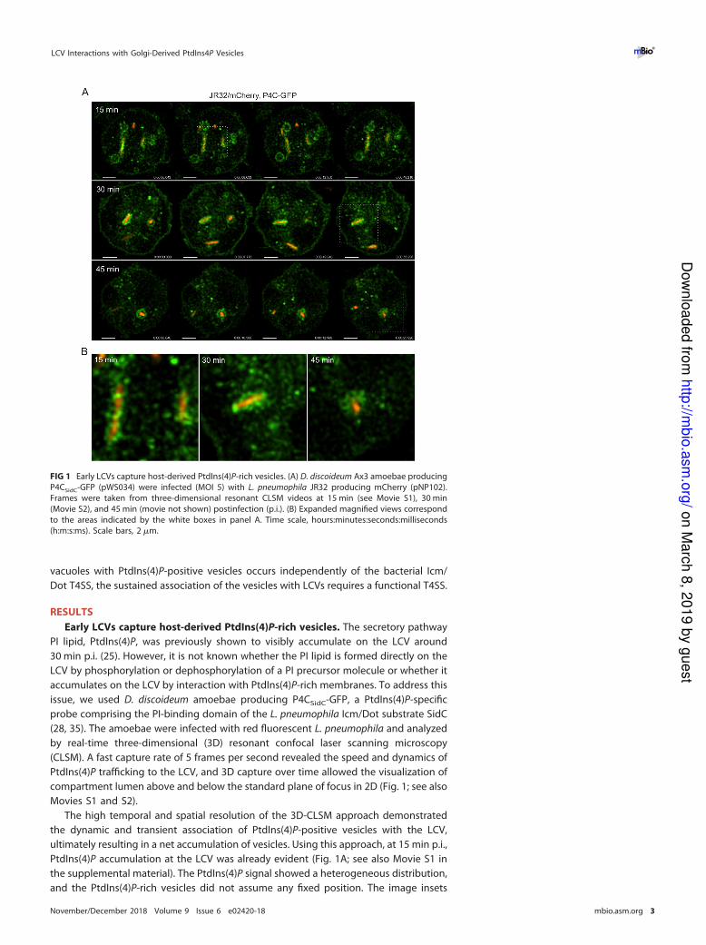
The high temporal and spatial resolution of the 3D-CLSM approach demonstrated
the dynamic and transient association of PtdIns(4)P-positive vesicles with the LCV,
ultimately resulting in a net accumulation of vesicles. Using this approach, at 15 min p.i.,
PtdIns(4)P accumulation at the LCV was already evident (Fig. 1A; see also Movie S1 in
the supplemental material). The PtdIns(4)P signal showed a heterogeneous distribution,
and the PtdIns(4)P-rich vesicles did not assume any fixed position. The image insets
FIG 1 Early LCVs capture host-derived PtdIns(4)P-rich vesicles. (A) D. discoideum Ax3 amoebae producing
P4CSidC-GFP (pWS034) were infected (MOI 5) with L. pneumophila JR32 producing mCherry (pNP102).
Frames were taken from three-dimensional resonant CLSM videos at 15 min (see Movie S1), 30 min
(Movie S2), and 45 min (movie not shown) postinfection (p.i.). (B) Expanded magnified views correspond
to the areas indicated by the white boxes in panel A. Time scale, hours:minutes:seconds:milliseconds
(h:m:s:ms). Scale bars, 2 �m.
LCV Interactions with Golgi-Derived PtdIns4P Vesicles ®
November/December 2018 Volume 9 Issue 6 e02420-18 mbio.asm.org 3
on M
arc
h 8
, 2019 b
y g
uest
http
://mbio
.asm
.org
/D
ow
nlo
aded fro
m
Page 6
demonstrate the vesicular nature of the PtdIns(4)P association (Fig. 1B). At 30 min p.i.,
net accumulation of PtdIns(4)P-rich vesicles was obvious, increasingly giving the ap-
pearance that the PtdIns(4)P around the LCV was a continuous membrane (Fig. 1; see
also Movie S2). However, this was not the case; individual vesicles could still be
resolved, and the vesicle association did not show a continuous elliptical curvature, as
typically observed with longer exposure times.
By 45 min p.i., the LCV took on the classic spherical appearance. The LCV membrane
comprised a collection of slightly larger PtdIns(4)P-positive vesicles, compared to the
previous time points (Fig. 1) (movie not shown). Importantly, the 45-min time series
clearly illustrates that the PtdIns(4)P association is vesicular, as the vesicles could be
observed to change position and deviate from the limiting LCV membrane, rather than
forming a continuous PtdIns(4)P-positive membrane. The image inset of the final frame
poignantly confirms these observations, as the individual PtdIns(4)P vesicle lumens
became resolvable in their dynamic repositioning. In summary, the use of real-time 3D
high-resolution resonant CLSM allowed the observation of the net accumulation of
PtdIns(4)P-rich vesicles on LCVs. At around 45 min, vesicle lumens were still resolvable,
and LCVs were not uniformly coated with a continuous PtdIns(4)P membrane. Rather,
vesicles “stagnated” on most LCVs, thus apparently leading to a net accumulation of the
PtdIns(4)P lipid.
Host- and T4SS-dependent association of PtdIns(4)P vesicles with LCVs. Apply-
ing real-time CLSM, we used dually labeled D. discoideum strains producing in tandem
P4CSidC-mCherry and the PtdIns(3)P probe GFP-2�FYVE to analyze the PI patterns
underlying the formation of vacuoles harboring L. pneumophila JR32 or the T4SS-
deficient strain ΔicmT. The high-resolution approach revealed that vesicles positive for
PtdIns(4)P or PtdIns(3)P both simultaneously and independently of one another inter-
acted with the bacterial compartments, while the morphological appearances of the
vesicles were similar (Fig. 2; see also Movies S3 to S6).
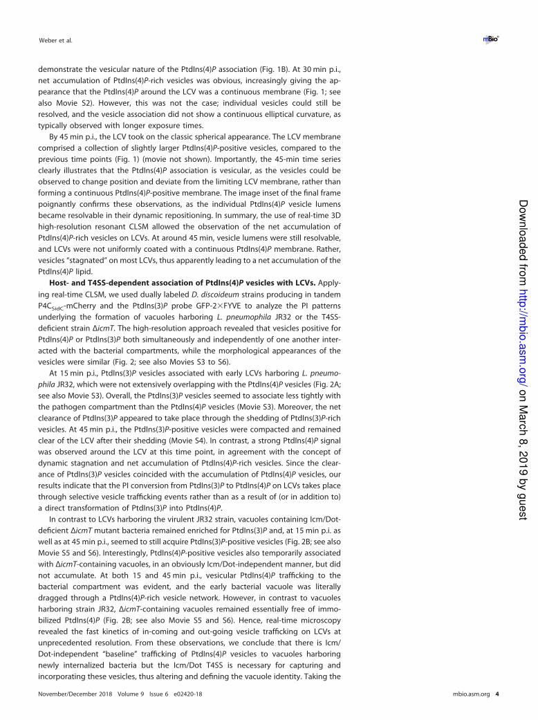
At 15 min p.i., PtdIns(3)P vesicles associated with early LCVs harboring L. pneumo-
phila JR32, which were not extensively overlapping with the PtdIns(4)P vesicles (Fig. 2A;
see also Movie S3). Overall, the PtdIns(3)P vesicles seemed to associate less tightly with
the pathogen compartment than the PtdIns(4)P vesicles (Movie S3). Moreover, the net
clearance of PtdIns(3)P appeared to take place through the shedding of PtdIns(3)P-rich
vesicles. At 45 min p.i., the PtdIns(3)P-positive vesicles were compacted and remained
clear of the LCV after their shedding (Movie S4). In contrast, a strong PtdIns(4)P signal
was observed around the LCV at this time point, in agreement with the concept of
dynamic stagnation and net accumulation of PtdIns(4)P-rich vesicles. Since the clear-
ance of PtdIns(3)P vesicles coincided with the accumulation of PtdIns(4)P vesicles, our
results indicate that the PI conversion from PtdIns(3)P to PtdIns(4)P on LCVs takes place
through selective vesicle trafficking events rather than as a result of (or in addition to)
a direct transformation of PtdIns(3)P into PtdIns(4)P.
In contrast to LCVs harboring the virulent JR32 strain, vacuoles containing Icm/Dot-
deficient ΔicmT mutant bacteria remained enriched for PtdIns(3)P and, at 15 min p.i. as
well as at 45 min p.i., seemed to still acquire PtdIns(3)P-positive vesicles (Fig. 2B; see also
Movie S5 and S6). Interestingly, PtdIns(4)P-positive vesicles also temporarily associated
with ΔicmT-containing vacuoles, in an obviously Icm/Dot-independent manner, but did
not accumulate. At both 15 and 45 min p.i., vesicular PtdIns(4)P trafficking to the
bacterial compartment was evident, and the early bacterial vacuole was literally
dragged through a PtdIns(4)P-rich vesicle network. However, in contrast to vacuoles
harboring strain JR32, ΔicmT-containing vacuoles remained essentially free of immo-
bilized PtdIns(4)P (Fig. 2B; see also Movie S5 and S6). Hence, real-time microscopy
revealed the fast kinetics of in-coming and out-going vesicle trafficking on LCVs at
unprecedented resolution. From these observations, we conclude that there is Icm/
Dot-independent “baseline” trafficking of PtdIns(4)P vesicles to vacuoles harboring
newly internalized bacteria but the Icm/Dot T4SS is necessary for capturing and
incorporating these vesicles, thus altering and defining the vacuole identity. Taking the
Weber et al. ®
November/December 2018 Volume 9 Issue 6 e02420-18 mbio.asm.org 4
on M
arc
h 8
, 2019 b
y g
uest
http
://mbio
.asm
.org
/D
ow
nlo
aded fro
m
Page 7
results together, vesicular trafficking largely contributes to both the Icm/Dot-
dependent removal and segregation of PtdIns(3)P as well as the accumulation of
PtdIns(4)P on LCVs.
PtdIns(3)P-positive vesicles interact with but do not fuse with PtdIns(4)P-
positive LCVs. LCVs harboring wild-type L. pneumophila shed their PtdIns(3)P identity
early during the infection process through the net loss of PtdIns(3)P-positive vesicles
(Fig. 2). To assess vesicle dynamics at later stages of LCV maturation, we infected dually
labeled D. discoideum strains producing P4CSidC-mCherry and GFP-2�FYVE in tandem
with the virulent JR32 strain and imaged the infection after 18 h. At that time point, all
observed LCVs harboring several bacteria were exclusively PtdIns(4)P-positive. Under
those conditions, PtdIns(3)P-rich vesicles still trafficked to PtdIns(4)P-positive LCVs but
did not fuse or accumulate on the LCV membrane at all (Fig. 3A). Moreover, in heavily
FIG 2 Host- and T4SS-dependent association of PtdIns(4)P vesicles with LCVs. D. discoideum Ax3
amoebae producing GFP-2�FYVE (pHK95) and P4CSidC-mCherry (pWS032) were infected (MOI 5) with (A)
L. pneumophila JR32 (Movie S3 and S4) or (B) ΔicmT (Movie S5 and S6) producing mCerulean (pNP099).
Resonant CLSM videos were taken at 15 min (Movie S3 and S5) or 45 min p.i. (Movie S4 and S6). Time
scale, h:m:s:ms. Scale bars, 2 �m.
LCV Interactions with Golgi-Derived PtdIns4P Vesicles ®
November/December 2018 Volume 9 Issue 6 e02420-18 mbio.asm.org 5
on M
arc
h 8
, 2019 b
y g
uest
http
://mbio
.asm
.org
/D
ow
nlo
aded fro
m
Page 8
infected amoebae, PtdIns(3)P-positive vesicles also interacted with PtdIns(4)P-negative
(likely newly formed) pathogen vacuoles but also did not fuse with these compart-
ments (Fig. 3B).
In general, at a given late point during infection, PtdIns(3)P-positive vesicles were
still vividly trafficking along microtubules and overall vesicle trafficking seemed intact
(Fig. 3; see also Movie S7). These observations indicated that the infection with L.
pneumophila was relatively stealthy and did not severely compromise crucial cellular
trafficking pathways. In contrast, the trafficking of PtdIns(4)P-rich vesicles was no longer
observed at late stages of infection, likely because the probe was tied up on the
massively PtdIns(4)P-positive LCVs at this time point. In summary, at late stages of
infection, PtdIns(3)P-positive vesicles still interact with but do not fuse with PtdIns(4)P-
positive LCVs, and the trafficking of these vesicles as well as vesicle trafficking in general
does not seem to be substantially compromised by the infection with L. pneumophila.
LCVs interact with PtdIns(4)P from the trans-Golgi network. To characterize the
cellular compartment source of the PtdIns(4)P-positive vesicles accumulating on LCVs,
we employed D. discoideum strains producing the well-characterized PtdIns(4)P/Golgi
probe 2�PHFAPP-GFP (24, 36). In keeping with the reported probe localization in
mammalian cells, 2�PHFAPP-GFP principally localizes to the trans-Golgi network (TGN),
with weak plasma membrane localization also in D. discoideum (Fig. 4A). Upon infection
of D. discoideum producing 2�PHFAPP-GFP with L. pneumophila JR32, the 2�PHFAPP-
GFP probe not only labeled the PtdIns(4)P-positive filaments of the Golgi apparatus but
also accumulated on the limiting membrane of LCVs. Projections of the Golgi apparatus
labeled by 2�PHFAPP-GFP made contact with and began to associate with the LCV
around 15 min p.i. (Fig. 4B) and robustly enveloped the pathogen vacuole 30 min p.i.
over the course of several minutes (Fig. 4C). In contrast, in D. discoideum infected with
ΔicmTmutant bacteria, the probe still robustly labeled the PtdIns(4)P-positive filaments
of the Golgi apparatus but did not localize to or accumulate on the membrane of
vacuoles harboring the avirulent bacteria (Fig. 4A).
To validate the observed interactions of LCVs with Golgi membranes, we used an
unrelated Golgi marker, golvesin (37). D. discoideum amoebae producing in parallel
2�PHFAPP-mCherry and the specific Golgi core probe Δ(1–75;119–579)golvesin-GFP
were infected with L. pneumophila JR32 or ΔicmT mutant bacteria for 1 h (Fig. 4D).
Vacuoles harboring strain JR32 robustly stained positive for this set of Golgi markers,
FIG 3 PtdIns(3)P-positive vesicles interact with but do not fuse with PtdIns(4)P-positive LCVs. D. discoideum Ax3 amoebae
producing GFP-2�FYVE (pHK95) and P4CSidC-mCherry (pWS032) were infected (MOI 5, 18 h) with L. pneumophila JR32
producing mCerulean (pNP099). Arrows indicate (A) vesicle trafficking events or (B) sustained PtdIns(3)P vesicle association
with a PtdIns(4)P-negative LCV. Resonant CLSM video was taken at 18 h p.i. (Movie S7). Time scale, h:m:s:ms. Scale bars, 2 �m.
Weber et al. ®
November/December 2018 Volume 9 Issue 6 e02420-18 mbio.asm.org 6
on M
arc
h 8
, 2019 b
y g
uest
http
://mbio
.asm
.org
/D
ow
nlo
aded fro
m
Page 9
corroborating that the PtdIns(4)P decorating LCVs originated from a Golgi-derived
source. In contrast, vacuoles containing ΔicmT mutant bacteria were totally devoid of
either of the two Golgi markers. Taking the results together, the mammalian PtdIns(4)P
probe 2�PHFAPP-GFP also labels Golgi PtdIns(4)P and LCVs in D. discoideum, and the D.
discoideum Golgi marker golvesin accumulates on LCVs, indicating that PtdIns(4)P-rich
Golgi membranes associate with LCVs.
The Icm/Dot T4SS determines sustained association of LCVs with the Golgi
apparatus. Next, we sought to assess the contribution of the Icm/Dot T4SS to the
accumulation of Golgi-derived PtdIns(4)P-positive vesicles on LCVs. To this end, we
employed D. discoideum strains producing in tandem 2�PHFAPP-mCherry and Arf1-GFP.
The Golgi-associated small GTPase Arf1 regulates Golgi-ER trafficking as well as intra-
Golgi transport (38) and is recruited to LCVs by the Icm/Dot translocated effector
protein RalF (14).
FIG 4 LCVs interact with PtdIns(4)P from the trans-Golgi network. (A) D. discoideum Ax3 amoebae
producing 2�PHFAPP-GFP (pWS033) were infected (MOI 5, 2 h) with mCherry-producing L. pneumophila
JR32 or ΔicmT (pNP102) or left uninfected, and localization of the probe to the Golgi pool of PtdIns(4)P
and LCVs was observed by CLSM. (B and C) Filaments labeled by 2�PHFAPP-GFP in D. discoideum (B)
began to associate with the LCV around 15 min p.i. and (C) robustly enveloped the LCV 30 min p.i. (D)
D. discoideum Ax3 amoebae producing 2�PHFAPP-mCherry (pWS035) and the specific Golgi core probe
Δ(1–75;119–579)golvesin-GFP (pWS037) were infected (MOI 5, 1 h) with L. pneumophila JR32 or ΔicmT
producing mCerulean (pNP099). Scale bars, 2 �m.
LCV Interactions with Golgi-Derived PtdIns4P Vesicles ®
November/December 2018 Volume 9 Issue 6 e02420-18 mbio.asm.org 7
on M
arc
h 8
, 2019 b
y g
uest
http
://mbio
.asm
.org
/D
ow
nlo
aded fro
m
Page 10
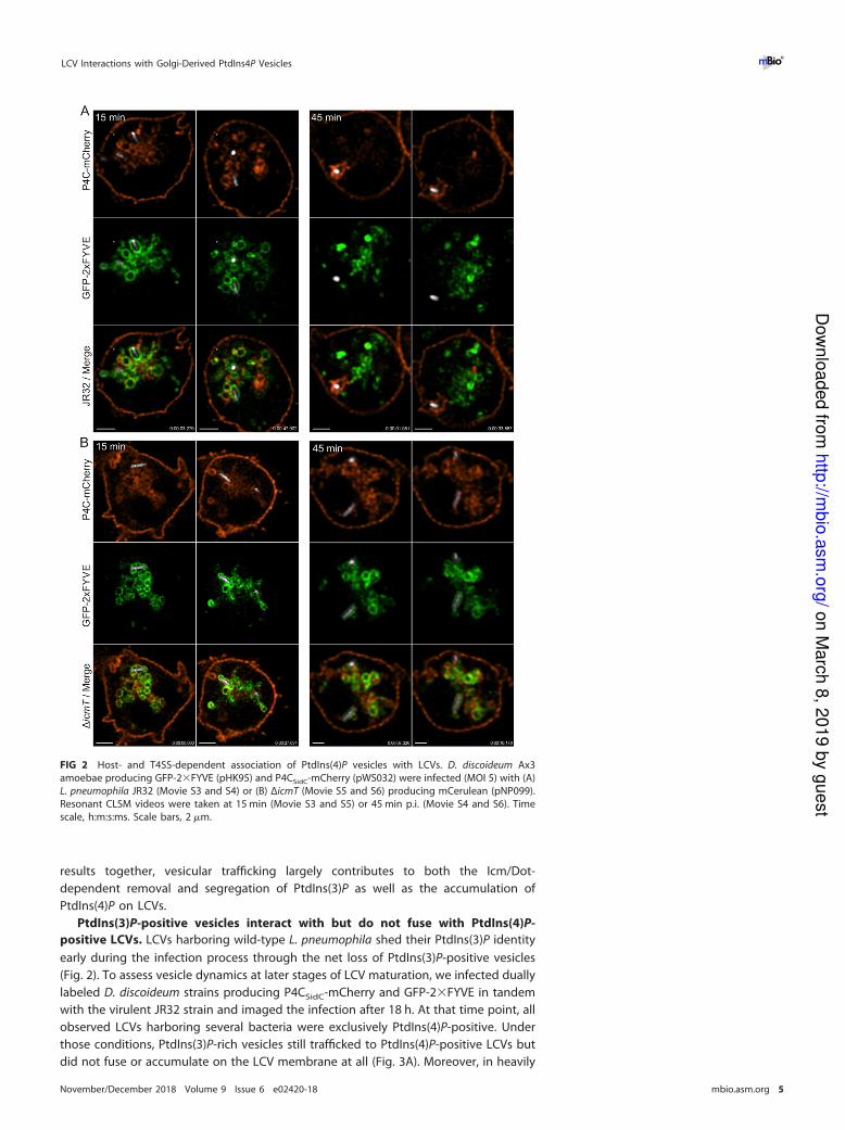
Upon infection of the dually labeled D. discoideum strain with L. pneumophila JR32,
both 2�PHFAPP-mCherry and Arf1-GFP associated with LCVs in a sustained manner, but
the two probes did not strictly overlap and showed distinct accumulation kinetics
(Fig. 5A; see also Movie S8). While the amount of 2�PHFAPP-mCherry increased from 30
to 60 min p.i., Arf1-GFP association did not appear to intensify during this period. In
contrast, upon infection of D. discoideum producing 2�PHFAPP-mCherry and Arf1-GFP
with L. pneumophila ΔicmT, the Golgi membranes were inevitably brought into prox-
imity of the compartment harboring the bacteria but did not engage in sustained
interactions (Fig. 5B; see also Movie S9). The video frames 30 min p.i. showed what
appears to be co-localization of the ΔicmT-containing compartment and both Golgi
probes, but approximately 700 ms later, the Golgi membranes were entirely clear of the
compartment. Thus, the Golgi does not sustainably associate with the vacuole con-
FIG 5 The Icm/Dot T4SS determines sustained association of LCVs with the Golgi apparatus. D.
discoideum Ax3 amoebae producing Arf1-GFP (pWS036) and 2�PHFAPP-mCherry (pWS035) were infected
(MOI 5) with L. pneumophila (A) JR32 (Movie S8), (B) ΔicmT (Movie S9), or (C) ΔralF producing mCerulean
(pNP099). Sustained and/or transient interactions of both probes simultaneously with bacterium-
containing vacuoles were recorded by resonant CLSM at 30 min (movie not shown) and 60 min (Movie
S8 and S9) p.i. Time scale, h:m:s:ms. Scale bars, 2 �m.
Weber et al. ®
November/December 2018 Volume 9 Issue 6 e02420-18 mbio.asm.org 8
on M
arc
h 8
, 2019 b
y g
uest
http
://mbio
.asm
.org
/D
ow
nlo
aded fro
m
Page 11
taining avirulent L. pneumophila. Finally, upon infection of D. discoideum producing
2�PHFAPP-mCherry and Arf1-GFP with L. pneumophila ΔralF, the PtdIns(4)P probe
labeled LCVs harboring the mutant strain to the same extent as LCVs harboring the
parental strain, while Arf1-GFP was not observable on pathogen vacuoles (Fig. 5C).
These findings are in agreement with the notion that Golgi-derived PtdIns(4)P accu-
mulates on LCVs independently of RalF-mediated Arf1 recruitment. In summary, the use
of 2�PHFAPP-mCherry and Arf1-GFP revealed that the Icm/Dot T4SS determines sus-
tained association of LCVs with the Golgi apparatus in an Arf1-independent manner.
The PtdIns(4)P probes, 2�PHFAPP and P4CSidC show distinct LCV interaction
dynamics. Based on the different spatiotemporal localization of 2�PHFAPP-mCherry
and Arf1-GFP on LCVs, we decided to simultaneously assess the localization dynamics
of the eukaryotic and bacterial PtdIns(4)P probes, 2�PHFAPP and P4CSidC, respectively.
In D. discoideum producing in parallel 2�PHFAPP-GFP and P4CSidC-mCherry, the former
predominantly labels the Golgi apparatus, while the latter in addition to the Golgi
primarily localizes to the plasma membrane and (endosomal) vesicles surrounding the
Golgi (Fig. 6A). Hence, aside from the plasma membrane where P4CSidC-mCherry
localization is dominant, there is little obvious spatial overlap between the two probes
recognizing the same PI lipid.
Upon infection of D. discoideum producing 2�PHFAPP-GFP and P4CSidC-mCherry
with L. pneumophila JR32, the LCVs were marked by PtdIns(4)P-positive vesicles as
indicated by P4CSidC-mCherry, but were also entangled by a dynamic meshwork of TGN
labeled by 2�PHFAPP-GFP (Fig. 6B; see also Movie S10). Noteworthy, while P4CSidCexclusively labeled the limiting LCV membrane, thus defining its identity, 2�PHFAPP not
only labeled the LCV membrane (as seen in Fig. 4 and 5), but also extended into the
TGN. The kinetics of LCV labeling of both probes, P4CSidC-mCherry and 2�PHFAPP-GFP,
were very similar (80% to 90% positive LCVs 1 to 2 h p.i.), and the probes maintained
their distinct labeling patterns throughout the infection with L. pneumophila from 2 h
p.i. to 16 h p.i. (Fig. 6C).
Upon infection of D. discoideum producing 2�PHFAPP-GFP and P4CSidC-mCherry
with ΔicmT mutant bacteria, the bacterial compartment was transiently labeled by the
PtdIns(4)P probes (representing “baseline” PtdIns(4)P levels; see Fig. 1), but did not
stably interact with the Golgi PtdIns(4)P pool (Fig. 6D). Taking the results together, the
PtdIns(4)P probes 2�PHFAPP-GFP and P4CSidC-mCherry showed distinct and robust
interaction dynamics with vacuoles harboring L. pneumophila JR32 (but not ΔicmT
mutant bacteria), suggesting that LCVs accumulate Golgi-derived rather than plasma
membrane-derived PtdIns(4)P.
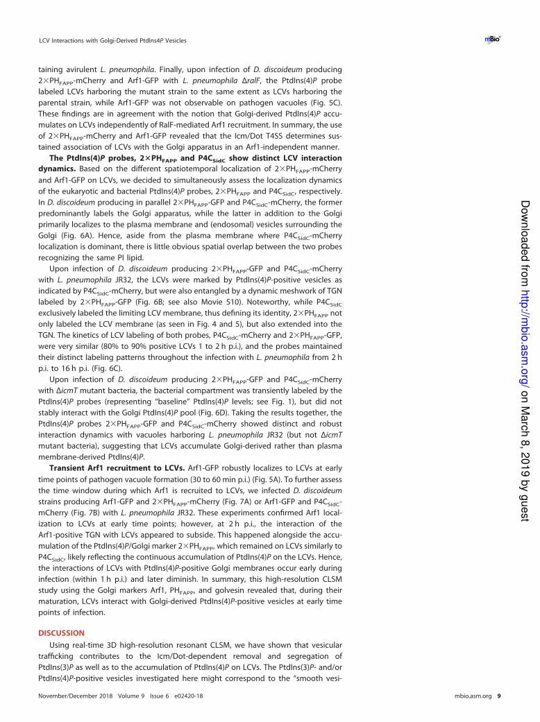
Transient Arf1 recruitment to LCVs. Arf1-GFP robustly localizes to LCVs at early
time points of pathogen vacuole formation (30 to 60 min p.i.) (Fig. 5A). To further assess
the time window during which Arf1 is recruited to LCVs, we infected D. discoideum
strains producing Arf1-GFP and 2�PHFAPP-mCherry (Fig. 7A) or Arf1-GFP and P4CSidC-
mCherry (Fig. 7B) with L. pneumophila JR32. These experiments confirmed Arf1 local-
ization to LCVs at early time points; however, at 2 h p.i., the interaction of the
Arf1-positive TGN with LCVs appeared to subside. This happened alongside the accu-
mulation of the PtdIns(4)P/Golgi marker 2�PHFAPP, which remained on LCVs similarly to
P4CSidC, likely reflecting the continuous accumulation of PtdIns(4)P on the LCVs. Hence,
the interactions of LCVs with PtdIns(4)P-positive Golgi membranes occur early during
infection (within 1 h p.i.) and later diminish. In summary, this high-resolution CLSM
study using the Golgi markers Arf1, PHFAPP, and golvesin revealed that, during their
maturation, LCVs interact with Golgi-derived PtdIns(4)P-positive vesicles at early time
points of infection.
DISCUSSION
Using real-time 3D high-resolution resonant CLSM, we have shown that vesicular
trafficking contributes to the Icm/Dot-dependent removal and segregation of
PtdIns(3)P as well as to the accumulation of PtdIns(4)P on LCVs. The PtdIns(3)P- and/or
PtdIns(4)P-positive vesicles investigated here might correspond to the “smooth vesi-
LCV Interactions with Golgi-Derived PtdIns4P Vesicles ®
November/December 2018 Volume 9 Issue 6 e02420-18 mbio.asm.org 9
on M
arc
h 8
, 2019 b
y g
uest
http
://mbio
.asm
.org
/D
ow
nlo
aded fro
m
Page 12
cles” associating with LCVs originally observed by EM (39). At early time points (�1 h
p.i.) LCVs were not uniformly coated with a continuous PtdIns(4)P membrane, and the
lumen of PtdIns(4)P-positive vesicles was still resolvable. The association of small
PtdIns(4)P-positive vesicles with LCVs correlates with the punctate PtdIns(4)P and SidC
staining observed previously (26, 28). The PtdIns(4)P-positive vesicles appeared to
“stagnate” on the LCVs, thus leading to a net accumulation of the PI lipid. This process
likely involves tethering and immobilization of PtdIns(4)P-positive vesicles on the LCVs,
followed by fusion of the vesicle and the pathogen vacuole membrane. At present, the
putative host and pathogen factors promoting the tethering of and interactions with
PtdIns(4)P-positive vesicles are unknown.
The Golgi protein FAPP1 binds both PtdIns(4)P and Arf1 (24, 36, 40). Producing
2�PHFAPP-mCherry and Arf1-GFP or 2�PHFAPP-GFP and P4CSidC-mCherry, respectively,
FIG 6 The PtdIns(4)P probes, 2�PHFAPP and P4CSidC, show distinct LCV interaction dynamics. D.
discoideum Ax3 amoebae producing 2�PHFAPP-GFP (pWS033) and P4CSidC-mCherry (pWS032) were (A)
left uninfected or infected (MOI 5) for the time indicated with (B and C) L. pneumophila JR32 or with (D)
ΔicmT mutant bacteria producing mCerulean (pNP099). PHFAPP predominantly labels the TGN, while
P4CSidC predominantly localizes to the plasma membrane and to cytoplasmic vesicles surrounding the
Golgi, as well as to LCVs. High-resolution video capture of resonant CLSM is shown (B; Movie S10). Time
scale, h:m:s:ms. Scale bars, 2 �m.
Weber et al. ®
November/December 2018 Volume 9 Issue 6 e02420-18 mbio.asm.org 10
on M
arc
h 8
, 2019 b
y g
uest
http
://mbio
.asm
.org
/D
ow
nlo
aded fro
m
Page 13
in D. discoideum indicated that the LCVs associate with the Golgi apparatus and
accumulate Golgi-derived rather than plasma membrane-derived PtdIns(4)P. Most of
the cellular PtdIns(4)P is found in the Golgi apparatus, the secretory vesicles, and the
plasma membrane (22, 24), but there are additional pools of this lipid found in (late)
endosomes (41, 42), which might contribute to the acquisition of vesicle-bound
PtdIns(4)P by nascent LCVs. However, the fact that LCVs deviate from the endosomal
route early during formation, together with the accumulation on LCVs of the Golgi-
specific probes 2�PHFAPP-mCherry and Arf1-GFP, strongly suggests that the PtdIns(4)P-
positive vesicles interacting with LCVs are indeed derived from the Golgi apparatus.
Overall, these results also emphasize the importance of performing live-cell rather than
fixed-sample experiments and strengthen the notion of the LCV as a dynamic com-
partment (co)defined by the frequency and/or duration of vesicular interactions. From
a technological standpoint, the speed of the resonant scans and of multi-Z-plane
imaging allowed us to decipher these processes.
At later stages of infection, the PtdIns(4)P-positive LCVs still interacted but did not
fuse with PtdIns(3)P-positive vesicles. The Icm/Dot-translocated effector VipD shows
Rab5-activated phospholipase A1 activity, removes PtdIns(3)P from endosomal mem-
branes, and reduces Rab5 levels on early LCVs (43). Thus, VipD might contribute to limit
the interactions of LCVs with endosomes throughout pathogen vacuole maturation.
The putative Icm/Dot substrates promoting the observed early interactions of LCVs
with PtdIns(4)P-positive vesicles and their sustained accumulation on the pathogen
vacuole are unknown. In any case, the Icm/Dot substrate RalF is dispensable for the
accumulation of PtdIns(4)P on LCVs. While Arf1-GFP was not observable on pathogen
vacuoles harboring L. pneumophila ΔralF (Fig. 5C), as published previously for mam-
FIG 7 Transient Arf1 recruitment to LCVs. D. discoideum Ax3 amoebae producing Arf1-GFP (pWS036) and
(A) 2�PHFAPP-mCherry (pWS035) or (B) P4CSidC-mCherry (pWS032) were infected (MOI 5, 0.5 h or 2 h) with
L. pneumophila JR32 producing mCerulean (pNP099).
LCV Interactions with Golgi-Derived PtdIns4P Vesicles ®
November/December 2018 Volume 9 Issue 6 e02420-18 mbio.asm.org 11
on M
arc
h 8
, 2019 b
y g
uest
http
://mbio
.asm
.org
/D
ow
nlo
aded fro
m
Page 14
malian cells (14), the accumulation of 2�PHFAPP-mCherry and, hence, PtdIns(4)P was
not compromised. These results also indicate that Arf1, which recruits PI 4-kinase (see
below), is dispensable for the accumulation of PtdIns(4)P on LCVs.
PI modulation during L. pneumophila infection and LCV formation is a complex
process, likely involving the vesicle trafficking processes described here as well as L.
pneumophila effectors. Several L. pneumophila Icm/Dot-translocated effectors have
been described which might contribute to PI lipid metabolism directly on LCVs (27, 44).
LepB, originally characterized as a Rab1 GTPase activating protein (GAP) (45–48), also
exhibits PI 4-kinase activity and converts PtdIns(3)P to PtdIns(3,4)P2 (49). Furthermore,
L. pneumophila produces the PI 3-phosphatases SidF (50), which preferentially hydro-
lyzes PtdIns(3,4)P2 and PtdIns(3,4,5)P3 in vitro, and SidP (51), which preferentially
hydrolyzes PtdIns(3)P and PtdIns(3,5)P2 in vitro. LepB and SidF have been shown to
contribute to the formation of PtdIns(4)P on LCVs in L. pneumophila-infected cells (49,
50), using the localization of the PtdIns(4)P-binding Icm/Dot substrate SidC as a readout
(28). Interestingly, a novel family of translocated PtdIns 3-kinases which generate
PtdIns(3)P from PtdIns (52) has recently been identified in Francisella (OpiA) as well as
in L. pneumophila (LegA5). Finally, L. pneumophila produces an Icm/Dot-translocated
phytase (inositol hexakisphosphate phosphatase), which produces PtdIns(4)P from the
polyphosphorylated PI lipids PtdIns(3,4)P2, PtdIns(4,5)P2, and PtdIns(3,4,5)P3 in vitro (53).
Although LppA appeared an ideal candidate to generate PtdIns(4)P on LCVs, no
evidence was obtained to demonstrate that the phytase indeed modulates the patho-
gen vacuole PI pattern. In summary, a plausible sequence of events regarding the
contribution of some L. pneumophila effectors to PI conversion on LCVs is as follows:
The PI 3-kinase LegA5 and the PI 4-kinase LepB phosphorylate PtdIns and PtdIns(3)P,
respectively, to produce PtdIns(3,4)P2, which is converted by the PI 3-phosphatase SidP
to PtdIns(4)P.
Further adding to the complexity of the process, a number of host PI-metabolizing
enzymes have been implicated in the production of PtdIns(4)P on the LCV membrane.
The PI 4-kinase class III� (PI4K III�) is recruited by the small GTPase Arf1 and promotes
traffic along the secretory pathway (54). Both Arf1 and PI4K III� promote accumulation
of SidC on the LCV, suggesting that these host factors contribute to PtdIns(4)P accu-
mulation (30, 55). Arf1 localizes to LCVs (14), but the association of PI4K III� with the
pathogen vacuole remains to be assessed. Another host factor potentially involved in
shaping the LCV PI pattern is the PI 5-phosphatase Oculocerebrorenal syndrome of
Lowe (OCRL), which localizes to the TGN and endosomes and regulates retrograde
trafficking between the two compartments (56). OCRL promotes intracellular replica-
tion of L. pneumophila (57) and determines LCV composition, including Rab1 and
retrograde trafficking components (58). The PI 5-phosphatase preferentially dephos-
phorylates PtdIns(4,5)P2 and also PtdIns(3,4,5)P3, yielding PtdIns(4)P and PtdIns(3,4)P2.
Based on the SidC localization assay, OCRL produces PtdIns(4)P on LCVs (57). Moreover,
the PI 3-phosphatase effector SidF possibly cooperates with OCRL to produce
PtdIns(4)P from PtdIns(3,4)P2.
Taken together, the available data are in agreement with a model stipulating that
LCV PI conversion involves host factors as well as pathogen factors and is the sum of
processes occurring in trans (at a distance from the LCV) and others occurring in cis (on
the LCV directly). As documented in this study, vesicle identity and trafficking in trans
seem to set the stage and determine early events of LCV formation. The L. pneumophila
PI-modulating effectors appear to preferentially act in cis. Yet the issue of whether
some of these effectors also act in trans, like several other L. pneumophila effectors,
modifying, e.g., ribosomes, mitochondria, or histones (9, 10), has not been addressed.
The work presented here provides an outline to address these issues and to search
among the more than 250 uncharacterized L. pneumophila Icm/Dot substrates for
effectors modulating early steps of LCV formation by interfering with host cell vesicle
trafficking.
Weber et al. ®
November/December 2018 Volume 9 Issue 6 e02420-18 mbio.asm.org 12
on M
arc
h 8
, 2019 b
y g
uest
http
://mbio
.asm
.org
/D
ow
nlo
aded fro
m
Page 15
MATERIALS AND METHODS
Bacteria, cells, and growth conditions. Bacterial strains and cell lines used are listed in Table 1. L.
pneumophila strains were grown for 2 to 3 days on charcoal yeast extract (CYE) agar plates, buffered with
N-(2-acetamido)-2-aminoethane sulfonic acid (ACES), at 37°C. Liquid cultures in ACES yeast extract (AYE)
medium were inoculated at an optical density at 600 nm (OD600) of 0.1 and grown at 37°C for 16 to 21 h
to the early stationary phase (2 � 109 bacteria/ml). Chloramphenicol (Cam; 5 �g/ml) was added for
plasmid retention.
D. discoideum Ax3 amoebae were cultivated in HL-5 medium (ForMedium) at 23°C in the dark. Cells
were maintained every 2 to 3 days by rinsing once with fresh HL-5, washing off cells with 10 ml HL-5, and
transferring 10% to 20% of the volume to a new T75 flask containing 10 ml medium. Cells were strictly
maintained at between 30% and 90% confluence.
Plasmid cloning. All plasmids used are listed in Table 1. The pEGFP-N1-PHFAPP1-mCherry template
was originally obtained from Ari Helenius and was cloned into pSW102 (pDXA-MCS-gfp), yielding
pRM010. For plasmid pSE002 (pDXA-2�PHFAPP-GFP), the PHFAPP gene was duplicated by two PCR
amplifications using pRM010 as a template and the primer pairs oRM17 (5=-AAAAACGCGGTACCAAGGA
GGGGGTGTTGTACAAGTGGAC-3=)/oSE003 (5=-AAAAACGCGGATCCTTGTCCTTGTGCTTTGGAGCTCCCCAGA
GCGACCAGCCACC-3=) and oSE004 (5=-AAAAACGCGGATCCAAGGAGGGGGTGTTGTACAAGTGGAC-3=)/
oSE002 (5=-AAAAACGCCTCGAGATGCTTTGGAGCTCCCCAGAGCGAC-3=). The fragments were cut with
BamHI, ligated, digested with KpnI and XhoI, and inserted into pSW102 cut with the same enzymes. To
construct plasmids pWS033 (2�PHFAPP-GFP) and pWS035 (2�PHFAPP-mCherry), the tandem PHFAPP
domain was amplified from pSE002 using primers oWS41 (5=-TCAGATCCCAAGCTAGATCTATGGATGGTA
CC-3=) and oWS42 (5=-CGCCCTTGCTCACCATACTAGTAGATGCTTTG-3=). The PCR fragment was cloned
with BglII/SpeI into vectors pDM323 and pDM1044, respectively. To construct plasmid pWS036, Arf1
(ArfA) was PCR amplified from purified D. discoideum Ax3 cDNA (NBRP Nenkin, Tsukuba, Japan) using
primers oWS53 (5=-TTTGGATCCATGGGTCTCGCTTTTGGTAAAC-3=) and oWS54 (5=-AAAACTAGTTTTTGAGG
AGCTTGTTAAGGTATTTG-3=). The product was cloned with BamHI/SpeI into pDM323. To construct
pWS038 [Δ(1–75;119–579)golvesin-GFP], the golvesin core fragment was amplified from template
pGolvesin-GFP using primers oWS55 (5=-AAAAAGATCTATGTCAAATACAGGTAAAATATATTTAAG-3=) and
oWS26 (5=-AAAAACTAGTATCAAATGGTAAACTAAAAACTAC-3=). The PCR fragment was cloned with BglII/
SpeI into pDM323. All new vectors were transformed into Escherichia coli TOP10 for amplification and
then sequenced.
Transformation of Dictyostelium discoideum. The D. discoideum parental strain Ax3 was grown to
approximately 70% confluence. The HL-5 medium was discarded, and the flask was rinsed with 5 ml
electroporation buffer (EB; 10 mM KH2PO4, 50 mM sucrose [pH 6.1], filter sterilized and stored at 4°C)
without disturbing the cells. The rinse buffer was replaced with 5 ml fresh EB, and the cells were
dislodged by the use of a 5-ml serological pipette. A 1-ml volume of cell suspension was added to each
4-mm-gap electroporation cuvette (Bio-Rad), and 4 to 5 �g of a given vector was mixed into the cuvette.
For dually fluorescent strains, the two vectors were added to the cuvette simultaneously. Electroporation
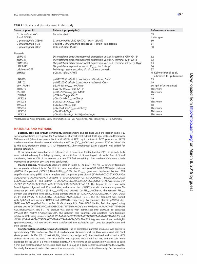
TABLE 1 Strains and plasmids used in this study
Strain or plasmid Relevant property(ies)a Reference or source
D. discoideum Ax3 Parental strain 59
E. coli TOP10 Invitrogen
L. pneumophila GS3011 L. pneumophila JR32 icmT3011::Kanr (ΔicmT) 60
L. pneumophila JR32 Virulent L. pneumophila serogroup 1 strain Philadelphia 61
L. pneumophila CR02 JR32 ralF::Kanr (ΔralF) 30
Plasmids
pDM317 Dictyostelium extrachromosomal expression vector, N-terminal GFP, G418r 62
pDM323 Dictyostelium extrachromosomal expression vector, C-terminal GFP, G418r 62
pDM1044 Dictyostelium extrachromosomal expression vector, C-terminal mCherry, Hygr 63
pDXA-HC Dictyostelium expression vector, Pact15, Neor, Ampr 64
pGolvesin-GFP Full-length gene encoding D. discoideum golvesin 37
pHKB95 pDM317-gfp-2�FYVE H. Koliwer-Brandl et al.,
submitted for publication
pNP099 pMMB207-C, ΔlacIq (constitutive mCerulean), Camr 19
pNP102 pMMB207-C, ΔlacIq (constitutive mCherry), Camr 19
pPH_FAPP1 pEGFP-N1-PHFAPP1-mCherry 36 (gift of A. Helenius)
pRM010 pSW102-PHFAPP1-gfp, G418r This work
pSE002 pDXA-2�PHFAPP1-gfp, G418r This work
pSW102 pDXA-MCS-gfp, G418r 57
pWS032 pDM1044-P4CSidC-mCherry 19
pWS033 pDM323-2�PHFAPP1-gfp This work
pWS034 pDM323-P4CSidC-gfp 58
pWS035 pDM1044-2�PHFAPP1-mCherry This work
pWS036 pDM323-Arf1-gfp This work
pWS038 pDM323-�(1–75;119–579)golvesin-gfp This work
aAbbreviations: Amp, ampicillin; Cam, chloramphenicol; Hyg, hygromycin; Kan, kanamycin; G418, Geneticin.
LCV Interactions with Golgi-Derived PtdIns4P Vesicles ®
November/December 2018 Volume 9 Issue 6 e02420-18 mbio.asm.org 13
on M
arc
h 8
, 2019 b
y g
uest
http
://mbio
.asm
.org
/D
ow
nlo
aded fro
m
Page 16
was performed with 2 pulses of 1 ms and 1 mV separated by a 5-s gap. Directly after electroporation, cells
were transferred into a T75 flask containing 10 ml HL-5. At between 12 and 24 h after electroporation, the
medium was replaced with fresh HL-5 and the required selection antibiotics were added. The medium
was changed 72 h later. Upon the obvious appearance of several microcolonies (usually 6 to 7 days after
transformation), cells were dislodged into fresh medium and transferred to a new flask.
Sample preparation for microscopy. D. discoideum amoebae producing the desired fluorescent
probes were harvested from approximately 70%-confluent cultures. HL-5 medium was removed, and
cultures were washed with 5 ml LoFlo medium (ForMedium) and resuspended in fresh LoFlo medium.
The cells were seeded (300 �l) at a density of 2.5 � 105/ml to 4 � 105/ml in eight-well �-slides (Ibidi).
Cells were allowed to adhere for 1 h, after which the LoFlo medium was replaced. Infections (at a
multiplicity of infection [MOI] of 5) with early stationary-phase cultures of L. pneumophila JR32 harboring
pNP099 (mCerulean) or pNP102 (mCherry) were initiated in �-slides already in position for imaging.
Confocal laser scanning fluorescence microscopy setup. All imaging was performed with living
cells, carried out with a Leica TCS SP8 X CLSM with the following setup: white-light laser (WLL), 442-nm
diode, HyD hybrid detectors for each channel used, HC PL APO CS2 63�/1.4 oil objective with Leica type
F immersion oil, Leica LAS X software. mCerulean was excited at 442 nm and detected at around 469 nm.
Enhanced GFP (EGFP) was excited at 487 nm and detected at around 516 nm. mCherry was excited at
587 nm and detected at around 622 nm. The microscope stage thermostat was set to hold the
temperature at between 22°C and 25°C. Images were captured with a pinhole at between 0.6 and 0.9 Airy
units (AU) and with a pixel/voxel size at or close to the instrument’s Nyquist criterion of approximately
39.5 � 39.5 � 118 nm (xyz).
Resonant scanning at 8,000 Hz (bidirectional scan) was used to capture videos corresponding to
Fig. 1, 2, 3, 5, and 6B. Capture rates for 2 scans with 2 to 8 line averages were between approximately
2.5 and 5 frames per second. For Fig. 1, four Z-slices with 110-nm spacing were captured per time
interval. Standard scanning at frequencies between 200 and 600 Hz (bidirectional scan with 2 to 3 line
averages) was used to capture images and videos corresponding to Fig. 4, 6A, C, and D, and 7.
Video and image processing. All images were deconvolved with Huygens Professional version
17.10 (Scientific Volume Imaging, The Netherlands) using the CMLE algorithm with 40 iterations and a
0.05 quality threshold. Signal-to-noise ratios were estimated from the photons counted for a given
image. Video captures and their snapshots were finalized with Imaris 9.1.0 software (Bitplane, Switzer-
land). Still images were finalized and exported with ImageJ software (https://imagej.nih.gov/ij/).
SUPPLEMENTAL MATERIAL
Supplemental material for this article may be found at https://doi.org/10.1128/mBio
.02420-18.
MOVIE S1, MOV file, 2.7 MB.
MOVIE S2, MOV file, 1.8 MB.
MOVIE S3, MOV file, 6.6 MB.
MOVIE S4, MOV file, 3.2 MB.
MOVIE S5, MOV file, 3.5 MB.
MOVIE S6, MOV file, 1.5 MB.
MOVIE S7, MOV file, 2.4 MB.
MOVIE S8, MOV file, 3.3 MB.
MOVIE S9, MOV file, 4.7 MB.
MOVIE S10, MOV file, 0.9 MB.
ACKNOWLEDGMENTS
We thank Ari Helenius for plasmid pEGFP-N1-FAPP1-PH-mCherry and A. Leoni Swart,
Roger Meier, and Sabrina Engelhardt for help with cloning.
Research in the laboratory of H.H. was supported by the Swiss National Science
Foundation (SNF; 31003A_153200 and 31003A_175557), the OPO Foundation, and the
Novartis Foundation for Medical-Biological Research. Confocal laser scanning micros-
copy was performed using equipment of the Center of Microscopy and Image Analysis,
University of Zürich. A.W. was supported by a grant from the Swedish Research Council
(2014-396). The funders had no role in study design, data collection and analysis,
decision to publish, or preparation of the manuscript.
REFERENCES
1. Newton HJ, Ang DK, van Driel IR, Hartland EL. 2010. Molecular
pathogenesis of infections caused by Legionella pneumophila. Clin
Microbiol Rev 23:274 –298. https://doi.org/10.1128/CMR.00052
-09.
2. Hilbi H, Hoffmann C, Harrison CF. 2011. Legionella spp. outdoors: colo-
nization, communication and persistence. Environ Microbiol Rep
3:286–296. https://doi.org/10.1111/j.1758-2229.2011.00247.x.
3. Hoffmann C, Harrison CF, Hilbi H. 2014. The natural alternative: protozoa
as cellular models for Legionella infection. Cell Microbiol 16:15–26.
https://doi.org/10.1111/cmi.12235.
Weber et al. ®
November/December 2018 Volume 9 Issue 6 e02420-18 mbio.asm.org 14
on M
arc
h 8
, 2019 b
y g
uest
http
://mbio
.asm
.org
/D
ow
nlo
aded fro
m
Page 17
4. Swart AL, Harrison CF, Eichinger L, Steinert M, Hilbi H. 2018. Acanth-
amoeba and Dictyostelium as cellular models for Legionella infection.
Front Cell Infect Microbiol 8:61. https://doi.org/10.3389/fcimb.2018
.00061.
5. Asrat S, de Jesus DA, Hempstead AD, Ramabhadran V, Isberg RR. 2014.
Bacterial pathogen manipulation of host membrane trafficking. Annu
Rev Cell Dev Biol 30:79–109. https://doi.org/10.1146/annurev-cellbio
-100913-013439.
6. Sherwood RK, Roy CR. 2016. Autophagy evasion and endoplasmic retic-
ulum subversion: the yin and yang of Legionella intracellular infection.
Annu Rev Microbiol 70:413–433. https://doi.org/10.1146/annurev-micro
-102215-095557.
7. Steiner B, Weber S, Hilbi H. 2018. Formation of the Legionella-containing
vacuole: phosphoinositide conversion, GTPase modulation and ER dy-
namics. Int J Med Microbiol 308:49–57. https://doi.org/10.1016/j.ijmm
.2017.08.004.
8. Kubori T, Nagai H. 2016. The type IVB secretion system: an enigmatic
chimera. Curr Opin Microbiol 29:22–29. https://doi.org/10.1016/j.mib
.2015.10.001.
9. Finsel I, Hilbi H. 2015. Formation of a pathogen vacuole according to
Legionella pneumophila: how to kill one bird with many stones. Cell
Microbiol 17:935–950. https://doi.org/10.1111/cmi.12450.
10. Escoll P, Mondino S, Rolando M, Buchrieser C. 2016. Targeting of host
organelles by pathogenic bacteria: a sophisticated subversion strategy.
Nat Rev Microbiol 14:5–19. https://doi.org/10.1038/nrmicro.2015.1.
11. Qiu J, Luo ZQ. 2017. Legionella and Coxiella effectors: strength in diver-
sity and activity. Nat Rev Microbiol 15:591–605. https://doi.org/10.1038/
nrmicro.2017.67.
12. Isberg RR, O’Connor TJ, Heidtman M. 2009. The Legionella pneumophila
replication vacuole: making a cosy niche inside host cells. Nat Rev
Microbiol 7:13–24. https://doi.org/10.1038/nrmicro1967.
13. Personnic N, Bärlocher K, Finsel I, Hilbi H. 2016. Subversion of retrograde
trafficking by translocated pathogen effectors. Trends Microbiol 24:
450–462. https://doi.org/10.1016/j.tim.2016.02.003.
14. Nagai H, Kagan JC, Zhu X, Kahn RA, Roy CR. 2002. A bacterial guanine
nucleotide exchange factor activates ARF on Legionella phagosomes.
Science 295:679–682. https://doi.org/10.1126/science.1067025.
15. Goody RS, Itzen A. 2013. Modulation of small GTPases by Legionella. Curr
Top Microbiol Immunol 376:117–133. https://doi.org/10.1007/82_2013
_340.
16. Hoffmann C, Finsel I, Otto A, Pfaffinger G, Rothmeier E, Hecker M,
Becher D, Hilbi H. 2014. Functional analysis of novel Rab GTPases
identified in the proteome of purified Legionella-containing vacuoles
from macrophages. Cell Microbiol 16:1034–1052. https://doi.org/10
.1111/cmi.12256.
17. Rothmeier E, Pfaffinger G, Hoffmann C, Harrison CF, Grabmayr H, Repnik
U, Hannemann M, Wölke S, Bausch A, Griffiths G, Müller-Taubenberger A,
Itzen A, Hilbi H. 2013. Activation of Ran GTPase by a Legionella effector
promotes microtubule polymerization, pathogen vacuole motility and
infection. PLoS Pathog 9:e1003598. https://doi.org/10.1371/journal.ppat
.1003598.
18. Schmölders J, Manske C, Otto A, Hoffmann C, Steiner B, Welin A, Becher
D, Hilbi H. 2017. Comparative proteomics of purified pathogen vacuoles
correlates intracellular replication of Legionella pneumophila with the
small GTPase Ras-related protein 1 (Rap1). Mol Cell Proteomics 16:
622–641. https://doi.org/10.1074/mcp.M116.063453.
19. Steiner B, Swart AL, Welin A, Weber S, Personnic N, Kaech A, Freyre C,
Ziegler U, Klemm RW, Hilbi H. 2017. ER remodeling by the large GTPase
atlastin promotes vacuolar growth of Legionella pneumophila. EMBO Rep
18:1817–1836. https://doi.org/10.15252/embr.201743903.
20. Escoll P, Song OR, Viana F, Steiner B, Lagache T, Olivo-Marin JC, Impens
F, Brodin P, Hilbi H, Buchrieser C. 2017. Legionella pneumophila modu-
lates mitochondrial dynamics to trigger metabolic repurposing of in-
fected macrophages. Cell Host Microbe 22:302–316. https://doi.org/10
.1016/j.chom.2017.07.020.
21. Di Paolo G, De Camilli P. 2006. Phosphoinositides in cell regulation
and membrane dynamics. Nature 443:651–657. https://doi.org/10
.1038/nature05185.
22. Schink KO, Tan KW, Stenmark H. 2016. Phosphoinositides in control of
membrane dynamics. Annu Rev Cell Dev Biol 32:143–171. https://doi
.org/10.1146/annurev-cellbio-111315-125349.
23. Graham TR, Burd CG. 2011. Coordination of Golgi functions by phos-
phatidylinositol 4-kinases. Trends Cell Biol 21:113–121. https://doi.org/
10.1016/j.tcb.2010.10.002.
24. Varnai P, Gulyas G, Toth DJ, Sohn M, Sengupta N, Balla T. 2017. Quan-
tifying lipid changes in various membrane compartments using lipid
binding protein domains. Cell Calcium 64:72–82. https://doi.org/10
.1016/j.ceca.2016.12.008.
25. Weber S, Wagner M, Hilbi H. 2014. Live-cell imaging of phosphoinositide
dynamics and membrane architecture during Legionella infection. mBio
5:e00839-13. https://doi.org/10.1128/mBio.00839-13.
26. Weber SS, Ragaz C, Reus K, Nyfeler Y, Hilbi H. 2006. Legionella pneumo-
phila exploits PI(4)P to anchor secreted effector proteins to the replica-
tive vacuole. PLoS Pathog 2:e46. https://doi.org/10.1371/journal.ppat
.0020046.
27. Haneburger I, Hilbi H. 2013. Phosphoinositide lipids and the Legionella
pathogen vacuole. Curr Top Microbiol Immunol 376:155–173. https://
doi.org/10.1007/82_2013_341.
28. Ragaz C, Pietsch H, Urwyler S, Tiaden A, Weber SS, Hilbi H. 2008. The
Legionella pneumophila phosphatidylinositol-4 phosphate-binding type
IV substrate SidC recruits endoplasmic reticulum vesicles to a
replication-permissive vacuole. Cell Microbiol 10:2416–2433. https://doi
.org/10.1111/j.1462-5822.2008.01219.x.
29. Luo X, Wasilko DJ, Liu Y, Sun J, Wu X, Luo ZQ, Mao Y. 2015. Structure of the
Legionella virulence factor, SidC reveals a unique PI(4)P-specific binding
domain essential for its targeting to the bacterial phagosome. PLoS Pathog
11:e1004965. https://doi.org/10.1371/journal.ppat.1004965.
30. Brombacher E, Urwyler S, Ragaz C, Weber SS, Kami K, Overduin M, Hilbi
H. 2009. Rab1 guanine nucleotide exchange factor SidM is a major
phosphatidylinositol 4-phosphate-binding effector protein of Legionella
pneumophila. J Biol Chem 284:4846–4856. https://doi.org/10.1074/jbc
.M807505200.
31. Schoebel S, Blankenfeldt W, Goody RS, Itzen A. 2010. High-affinity
binding of phosphatidylinositol 4-phosphate by Legionella pneumophila
DrrA. EMBO Rep 11:598–604. https://doi.org/10.1038/embor.2010.97.
32. Zhu Y, Hu L, Zhou Y, Yao Q, Liu L, Shao F. 2010. Structural mechanism of
host Rab1 activation by the bifunctional Legionella type IV effector
SidM/DrrA. Proc Natl Acad Sci U S A 107:4699–4704. https://doi.org/10
.1073/pnas.0914231107.
33. Del Campo CM, Mishra AK, Wang YH, Roy CR, Janmey PA, Lambright
DG. 2014. Structural basis for PI(4)P-specific membrane recruitment
of the Legionella pneumophila effector DrrA/SidM. Structure 22:
397–408. https://doi.org/10.1016/j.str.2013.12.018.
34. Hubber A, Arasaki K, Nakatsu F, Hardiman C, Lambright D, De Camilli P,
Nagai H, Roy CR. 2014. The machinery at endoplasmic reticulum-plasma
membrane contact sites contributes to spatial regulation of multiple
Legionella effector proteins. PLoS Pathog 10:e1004222. https://doi.org/
10.1371/journal.ppat.1004222.
35. Dolinsky S, Haneburger I, Cichy A, Hannemann M, Itzen A, Hilbi H. 2014.
The Legionella longbeachae Icm/Dot substrate SidC selectively binds
phosphatidylinositol 4-phosphate with nanomolar affinity and promotes
pathogen vacuole-endoplasmic reticulum interactions. Infect Immun
82:4021–4033. https://doi.org/10.1128/IAI.01685-14.
36. Godi A, Di Campli A, Konstantakopoulos A, Di Tullio G, Alessi DR, Kular
GS, Daniele T, Marra P, Lucocq JM, De Matteis MA. 2004. FAPPs control
Golgi-to-cell-surface membrane traffic by binding to ARF and PtdIns(4)P.
Nat Cell Biol 6:393–404. https://doi.org/10.1038/ncb1119.
37. Schneider N, Schwartz JM, Kohler J, Becker M, Schwarz H, Gerisch G.
2000. Golvesin-GFP fusions as distinct markers for Golgi and post-Golgi
vesicles in Dictyostelium cells. Biol Cell 92:495–511. https://doi.org/10
.1016/S0248-4900(00)01102-3.
38. Arakel EC, Schwappach B. 2018. Formation of COPI-coated vesicles at a
glance. J Cell Sci https://doi.org/10.1242/jcs.209890.
39. Horwitz MA. 1983. Formation of a novel phagosome by the Legion-
naires’ disease bacterium (Legionella pneumophila) in human mono-
cytes. J Exp Med 158:1319–1331. https://doi.org/10.1084/jem.158.4
.1319.
40. Levine TP, Munro S. 2002. Targeting of Golgi-specific pleckstrin homol-
ogy domains involves both PtdIns 4-kinase-dependent and
-independent components. Curr Biol 12:695–704. https://doi.org/10
.1016/S0960-9822(02)00779-0.
41. Hammond GR, Machner MP, Balla T. 2014. A novel probe for phospha-
tidylinositol 4-phosphate reveals multiple pools beyond the Golgi. J Cell
Physiol 205:113–126. https://doi.org/10.1083/jcb.201312072.
42. Jeschke A, Zehethofer N, Lindner B, Krupp J, Schwudke D, Haneburger I,
Jovic M, Backer JM, Balla T, Hilbi H, Haas A. 2015. Phosphatidylinositol
4-phosphate and phosphatidylinositol 3-phosphate regulate phagolyso-
LCV Interactions with Golgi-Derived PtdIns4P Vesicles ®
November/December 2018 Volume 9 Issue 6 e02420-18 mbio.asm.org 15
on M
arc
h 8
, 2019 b
y g
uest
http
://mbio
.asm
.org
/D
ow
nlo
aded fro
m
Page 18
some biogenesis. Proc Natl Acad Sci U S A 112:4636–4641. https://doi
.org/10.1073/pnas.1423456112.
43. Gaspar AH, Machner MP. 2014. VipD is a Rab5-activated phospho-
lipase A1 that protects Legionella pneumophila from endosomal fu-
sion. Proc Natl Acad Sci U S A 111:4560–4565. https://doi.org/10
.1073/pnas.1316376111.
44. Pizarro-Cerdá J, Kühbacher A, Cossart P. 2015. Phosphoinositides and
host-pathogen interactions. Biochim Biophys Acta 1851:911–918.
https://doi.org/10.1016/j.bbalip.2014.09.011.
45. Ingmundson A, Delprato A, Lambright DG, Roy CR. 2007. Legionella
pneumophila proteins that regulate Rab1 membrane cycling. Nature
450:365–369. https://doi.org/10.1038/nature06336.
46. Gazdag EM, Streller A, Haneburger I, Hilbi H, Vetter IR, Goody RS, Itzen
A. 2013. Mechanism of Rab1b deactivation by the Legionella pneumo-
phila GAP LepB. EMBO Rep 14:199–205. https://doi.org/10.1038/embor
.2012.211.
47. Mishra AK, Del Campo CM, Collins RE, Roy CR, Lambright DG. 2013. The
Legionella pneumophila GTPase activating protein LepB accelerates Rab1
deactivation by a non-canonical hydrolytic mechanism. J Biol Chem
288:24000–24011. https://doi.org/10.1074/jbc.M113.470625.
48. Yu Q, Hu L, Yao Q, Zhu Y, Dong N, Wang DC, Shao F. 2013. Structural
analyses of Legionella LepB reveal a new GAP fold that catalytically
mimics eukaryotic RasGAP. Cell Res 23:775–787. https://doi.org/10.1038/
cr.2013.54.
49. Dong N, Niu M, Hu L, Yao Q, Zhou R, Shao F. 2016. Modulation of
membrane phosphoinositide dynamics by the phosphatidylinositide
4-kinase activity of the Legionella LepB effector. Nat Microbiol 2:16236.
https://doi.org/10.1038/nmicrobiol.2016.236.
50. Hsu F, Zhu W, Brennan L, Tao L, Luo ZQ, Mao Y. 2012. Structural basis for
substrate recognition by a unique Legionella phosphoinositide phospha-
tase. Proc Natl Acad Sci U S A 109:13567–13572. https://doi.org/10.1073/
pnas.1207903109.
51. Toulabi L, Wu X, Cheng Y, Mao Y. 2013. Identification and structural
characterization of a Legionella phosphoinositide phosphatase. J Biol
Chem 288:24518–24527. https://doi.org/10.1074/jbc.M113.474239.
52. Ledvina HE, Kelly KA, Eshraghi A, Plemel RL, Peterson SB, Lee B, Steele S,
Adler M, Kawula TH, Merz AJ, Skerrett SJ, Celli J, Mougous JD. 2018. A
phosphatidylinositol 3-kinase effector alters phagosomal maturation to
promote intracellular growth of Francisella. Cell Host Microbe 24:
285–295. https://doi.org/10.1016/j.chom.2018.07.003.
53. Weber S, Stirnimann CU, Wieser M, Frey D, Meier R, Engelhardt S, Li X,
Capitani G, Kammerer RA, Hilbi H. 2014. A type IV translocated Legionella
cysteine phytase counteracts intracellular growth restriction by phytate.
J Biol Chem 289:34175–34188. https://doi.org/10.1074/jbc.M114.592568.
54. Godi A, Pertile P, Meyers R, Marra P, Di Tullio G, Iurisci C, Luini A, Corda
D, De Matteis MA. 1999. ARF mediates recruitment of PtdIns-4-OH
kinase-beta and stimulates synthesis of PtdIns(4,5)P2 on the Golgi com-
plex. Nat Cell Biol 1:280–287. https://doi.org/10.1038/12993.
55. Urwyler S, Nyfeler Y, Ragaz C, Lee H, Mueller LN, Aebersold R, Hilbi H.
2009. Proteome analysis of Legionella vacuoles purified by magnetic
immunoseparation reveals secretory and endosomal GTPases. Traffic
10:76–87. https://doi.org/10.1111/j.1600-0854.2008.00851.x.
56. Mehta ZB, Pietka G, Lowe M. 2014. The cellular and physiological func-
tions of the Lowe syndrome protein OCRL1. Traffic 15:471–487. https://
doi.org/10.1111/tra.12160.
57. Weber SS, Ragaz C, Hilbi H. 2009. The inositol polyphosphate
5-phosphatase OCRL1 restricts intracellular growth of Legionella, lo-
calizes to the replicative vacuole and binds to the bacterial effector
LpnE. Cell Microbiol 11:442–460. https://doi.org/10.1111/j.1462-5822
.2008.01266.x.
58. Welin A, Weber S, Hilbi H. 2018. Quantitative imaging flow cytometry of
Legionella-infected Dictyostelium amoebae reveals the impact of retro-
grade trafficking on pathogen vacuole composition. Appl Environ Mi-
crobiol 84:e00158-18. https://doi.org/10.1128/AEM.00158-18.
59. Loovers HM, Kortholt A, de Groote H, Whitty L, Nussbaum RL, van
Haastert PJ. 2007. Regulation of phagocytosis in Dictyostelium by the
inositol 5-phosphatase OCRL homolog Dd5P4. Traffic 8:618–628.
https://doi.org/10.1111/j.1600-0854.2007.00546.x.
60. Segal G, Shuman HA. 1998. Intracellular multiplication and human mac-
rophage killing by Legionella pneumophila are inhibited by conjugal
components of IncQ plasmid RSF1010. Mol Microbiol 30:197–208.
https://doi.org/10.1046/j.1365-2958.1998.01054.x.
61. Sadosky AB, Wiater LA, Shuman HA. 1993. Identification of Legionella
pneumophila genes required for growth within and killing of human
macrophages. Infect Immun 61:5361–5373.
62. Veltman DM, Akar G, Bosgraaf L, Van Haastert PJM. 2009. A new set of
small, extrachromosomal expression vectors for Dictyostelium discoi-
deum. Plasmid 61:110–118. https://doi.org/10.1016/j.plasmid.2008.11
.003.
63. Barisch C, Paschke P, Hagedorn M, Maniak M, Soldati T. 2015. Lipid
droplet dynamics at early stages of Mycobacterium marinum infection in
Dictyostelium. Cell Microbiol 17:1332–1349. https://doi.org/10.1111/cmi
.12437.
64. Manstein DJ, Schuster HP, Morandini P, Hunt DM. 1995. Cloning vectors
for the production of proteins in Dictyostelium discoideum. Gene 162:
129–134. https://doi.org/10.1016/0378-1119(95)00351-6.
Weber et al. ®
November/December 2018 Volume 9 Issue 6 e02420-18 mbio.asm.org 16
on M
arc
h 8
, 2019 b
y g
uest
http
://mbio
.asm
.org
/D
ow
nlo
aded fro
m