12
Leica TCS MP5 Optimized for Multiphoton Imaging
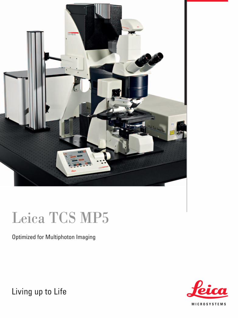
Leica TCS MP5Optimized for Multiphoton Imaging

2
• Dedicated multiphoton platform – perfect for intravital imaging
• Follow fast cellular dynamics at video rate scan speed
• Combine up to seven channels for multimodal imaging
• Effi ciently excite red fl uorophores with up to 1300 nm
• Discover details from deeper tissue sections with prechirped femtosecond lasers
Leica Design by Christophe Apothéloz
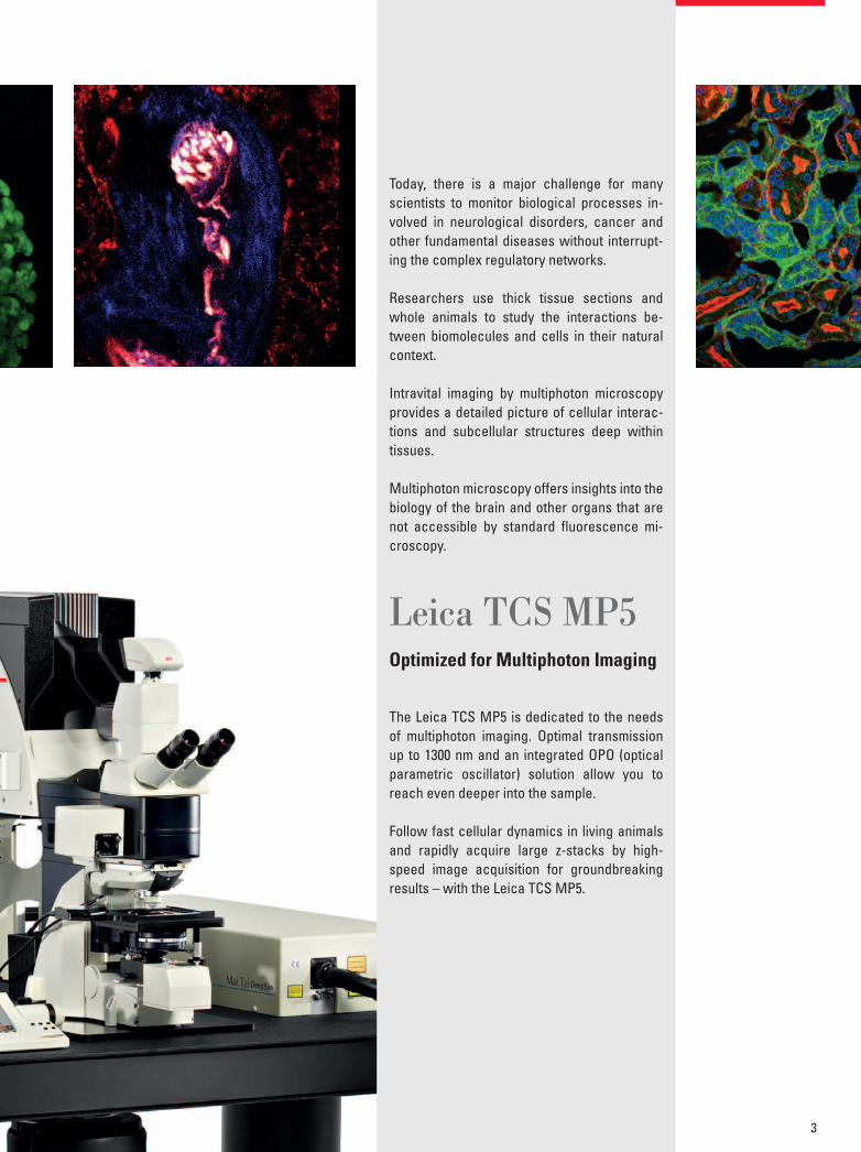
Today, there is a major challenge for many scientists to monitor biological processes in-volved in neurological disorders, cancer and other fundamental diseases without interrupt-ing the complex regulatory networks.
Researchers use thick tissue sections and whole animals to study the interactions be-tween biomolecules and cells in their natural context.
Intravital imaging by multiphoton microscopy provides a detailed picture of cellular interac-tions and subcellular structures deep within tissues.
Multiphoton microscopy offers insights into the biology of the brain and other organs that are not accessible by standard fl uorescence mi-croscopy.
Leica TCS MP5Optimized for Multiphoton Imaging
The Leica TCS MP5 is dedicated to the needs of multiphoton imaging. Optimal transmission up to 1300 nm and an integrated OPO (optical parametric oscillator) solution allow you to reach even deeper into the sample.
Follow fast cellular dynamics in living animals and rapidly acquire large z-stacks by high-speed image acquisition for groundbreaking results – with the Leica TCS MP5.
3
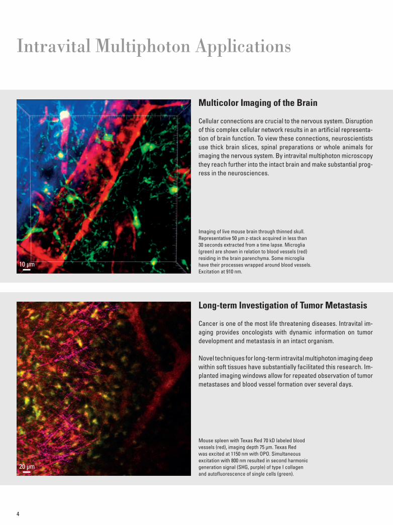
Long-term Investigation of Tumor Metastasis
Cancer is one of the most life threatening diseases. Intravital im-aging provides oncologists with dynamic information on tumor development and metastasis in an intact organism. Novel techniques for long-term intravital multiphoton imaging deep within soft tissues have substantially facilitated this research. Im-planted imaging windows allow for repeated observation of tumor metastases and blood vessel formation over several days.
Intravital Multiphoton Applications
4
Multicolor Imaging of the Brain
Cellular connections are crucial to the nervous system. Disruption of this complex cellular network results in an artifi cial representa-tion of brain function. To view these connections, neuroscientists use thick brain slices, spinal preparations or whole animals for imaging the nervous system. By intravital multiphoton microscopy they reach further into the intact brain and make substantial prog-ress in the neurosciences.
Imaging of live mouse brain through thinned skull. Representative 50 µm z-stack acquired in less than 30 seconds extracted from a time lapse. Microglia (green) are shown in relation to blood vessels (red) residing in the brain parenchyma. Some microglia have their processes wrapped around blood vessels. Excitation at 910 nm.
Mouse spleen with Texas Red 70 kD labeled blood vessels (red), imaging depth 75 µm. Texas Red was excited at 1150 nm with OPO. Simultaneous excitation with 800 nm resulted in second harmonic generation signal (SHG, purple) of type I collagen and autofl uorescence of single cells (green).
10 µm
20 µm

5
Dynamics of Anti-viral Response in Spleen
Our immune system constantly defends us against attacks by viruses and other microorganisms. If it fails to respond quickly, microorganisms can cause fatal diseases. Immunologists use video rate intravital multiphoton microscopy to follow infections and the response of the immune system in real time deep within tissues.
Ultra-rapid Imaging of Embryonic Blood Flows
During embryonic development biological fl uid fl ows play an im-portant role. To investigate how cells interpret physical informa-tion from their environment, high-speed intravital multiphoton imaging is used to study fl uid fl ows with minimal photodamage to the developing organism.
Blood cells labeled with DsRed (red). 167 frames/second at 512 x 64 pixels with Resonant Scanner.
Multiphoton excitation at 1100 nm with OPO.
3D reconstruction of a 50 µm z-stack from a time lapse captured in the spleen 7 days following
infection with lymphocytic choriomeningitis virus (LCMV). Anti-viral CD8-CFP (red) and CD4-YFP (green)
T cells excited with 910 nm. Spectral unmixing was performed using Leica LAS AF software.
Zebrafi sh embryonic heart,100 µm deep. Blood cells labeled with DsRed (red),SHG of muscle (gray)
20 µm20 µm
t = 0.105 sec
t = 0.111 sec
t = 0.117 sec
t = 0.122 sec
t = 0.128 sec
t = 0.134 sec
50 µm
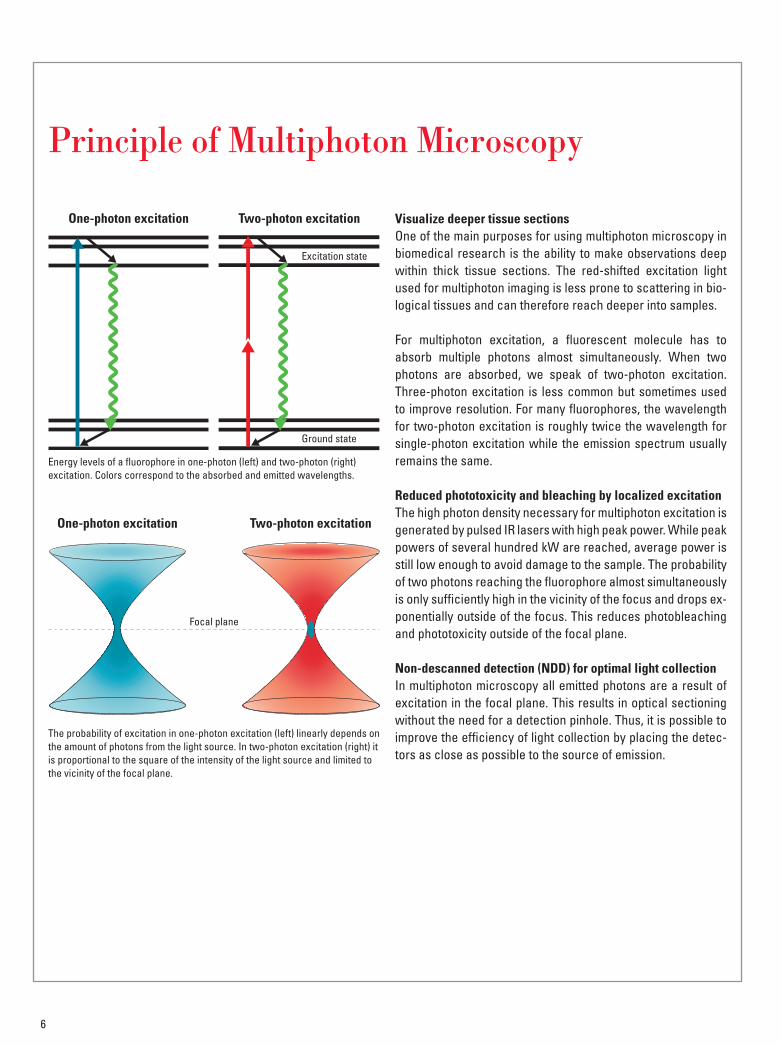
Visualize deeper tissue sectionsOne of the main purposes for using multiphoton microscopy in biomedical research is the ability to make observations deep within thick tissue sections. The red-shifted excitation light used for multiphoton imaging is less prone to scattering in bio-logical tissues and can therefore reach deeper into samples.
For multiphoton excitation, a fl uorescent molecule has to absorb multiple photons almost simultaneously. When two photons are absorbed, we speak of two-photon excitation. Three-photon excitation is less common but sometimes used to improve resolution. For many fl uorophores, the wavelength for two-photon excitation is roughly twice the wavelength for single-photon excitation while the emission spectrum usually remains the same.
Reduced phototoxicity and bleaching by localized excitationThe high photon density necessary for multiphoton excitation is generated by pulsed IR lasers with high peak power. While peak powers of several hundred kW are reached, average power is still low enough to avoid damage to the sample. The probability of two photons reaching the fl uorophore almost simultaneously is only suffi ciently high in the vicinity of the focus and drops ex-ponentially outside of the focus. This reduces photobleaching and phototoxicity outside of the focal plane.
Non-descanned detection (NDD) for optimal light collectionIn multiphoton microscopy all emitted photons are a result of excitation in the focal plane. This results in optical sectioning without the need for a detection pinhole. Thus, it is possible to improve the effi ciency of light collection by placing the detec-tors as close as possible to the source of emission.
6
Energy levels of a fl uorophore in one-photon (left) and two-photon (right) excitation. Colors correspond to the absorbed and emitted wavelengths.
The probability of excitation in one-photon excitation (left) linearly depends on the amount of photons from the light source. In two-photon excitation (right) it is proportional to the square of the intensity of the light source and limited to the vicinity of the focal plane.
Principle of Multi photon Microscopy
One-photon excitation
One-photon excitation
Two-photon excitation
Two-photon excitation
Excitation state
Ground state
Focal plane
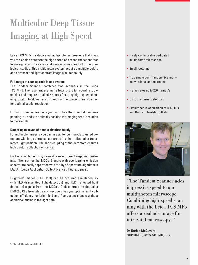
“The Tandem Scanner adds impressive speed to ourmultiphoton microscope. Combining high-speed scan-ning with the Leica TCS MP5 offers a real advantage for intravital microscopy.”
Dr. Dorian McGavernNIH/NINDS, Bethesda, MD, USA
Leica TCS MP5 is a dedicated multiphoton microscope that gives you the choice between the high speed of a resonant scanner for following rapid processes and slower scan speeds for morpho-logical studies. This multiphoton system acquires multiple colors and a transmitted light contrast image simultaneously.
Full range of scan speeds in one systemThe Tandem Scanner combines two scanners in the Leica TCS MP5. The resonant scanner allows users to record fast dy-namics and acquire detailed z-stacks faster by high-speed scan-ning. Switch to slower scan speeds of the conventional scanner for optimal spatial resolution.
For both scanning methods you can rotate the scan fi eld and use panning in x and y to optimally position the imaging area in relation to the sample.
Detect up to seven channels simultaneouslyFor multicolor imaging you can use up to four non-descanned de-tectors with large photo sensor areas in either refl ected or trans-mitted light position. The short coupling of the detectors ensures high photon collection effi cency.
On Leica multiphoton systems it is easy to exchange and custo-mize fi lter set for the NDDs. Signals with overlapping emission spectra are easily separated with the Dye Separation algorithm in LAS AF (Leica Application Suite Advanced Fluorescence).
Brightfi eld images (DIC, Dodt) can be acquired simultaneously with TLD (transmitted light detection) and RLD (refl ected light detection) signals from the NDDs*. Dodt contrast on the Leica DM6000 CFS fi xed stage microscope gives you optimal light coll-ection effi ciency for brightfi eld and fl uorescent signals without additional prisms in the light path.
* not available on Leica DMI6000
7
• Freely confi gurable dedicatedmultiphoton microscope
• Small footprint
• True single point Tandem Scanner – conventional and resonant
• Frame rates up to 250 frames/s
• Up to 7 external detectors
• Simultaneous acquisition of RLD, TLDand Dodt contrast/brightfi eld
Multicolor Deep TissueImaging at High Speed
You want to see as deeply into your sample as possible. Leica Micro systems offers you the integration of the most advanced pulsed IR laser sources for multiphoton imaging. With pulses of less than 70 femtoseconds or wavelengths up to 1300 nm, the system is perfectly prepared for your most challenging samples.
Expand into the red for multicolor applicationsExcitation up to 1300 nm with an optical parametric oscillator (OPO) allows you to explore the full range of fl uorescent proteins and dyes.
With Leica’s OPO solution, simultaneous multiphoton excitation of green and red fl uorophores is possible. The longer wavelengths of the OPO can reach deeper into tissue and are less harmful to samples.
Leica Microsystems offers an OPO solution that is fully integrated into the software interface of LAS AF for easy tuning and setup of laser wavelengths. High transmission, up to 1300 nm, of the Leica TCS MP5 scan optics brings all the power from the OPO to the sample.
Precompensation for maximum powerPrecompensation of the dispersive effects of glass ensures that the narrow pulse width of ultrashort light pulses is maintained.The maximum peak power is delivered at the sample for brightest images.
Modulation of laser power with minimal pulse broadeningIn addition to a polarizing fi lter wheel, a continuously adjustable electro-optical modulator (EOM) attenuates the laser power. The EOM is highly effi cient over the complete excitation range and causes minimal pulse broadening.
The rapid switching time of less than a microsecond is optimal for fast ROI (region of interest) scans.
Exciting a Broader Range of Fluorophores
8
• Optimal transmission up to 1300 nm
• Fully integrated OPO solution
• Femtosecond and precompensatedIR lasers with pulse widths < 70 fs
• Rapid modulation by EOM
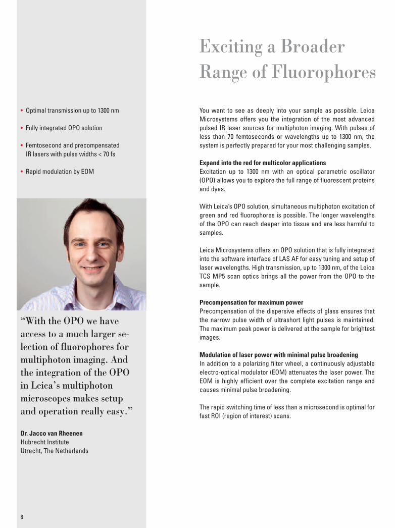
“With the OPO we have access to a much larger se-lection of fluorophores for multiphoton imaging. And the integration of the OPO in Leica’s multiphoton microscopes makes setupand operation really easy.”
Dr. Jacco van Rheenen Hubrecht InstituteUtrecht, The Netherlands
The Leica DM6000 CFS provides the best mechanical and elec-tronic stability for sensitive experiments. The new motorized 2-position objective changer allows smooth and vibration-free switching and dipping of objectives – all controlled remotely.
Dipping objectives for high resolution and optimal accessThe Leica HCX IRAPO L 25x/0.95 W objective provides maximum clearance around the specimen with an access angle of 41° and a free working distance of 2.5 mm. A complete range of dipping lenses and high resolution objectives is available to match your needs.
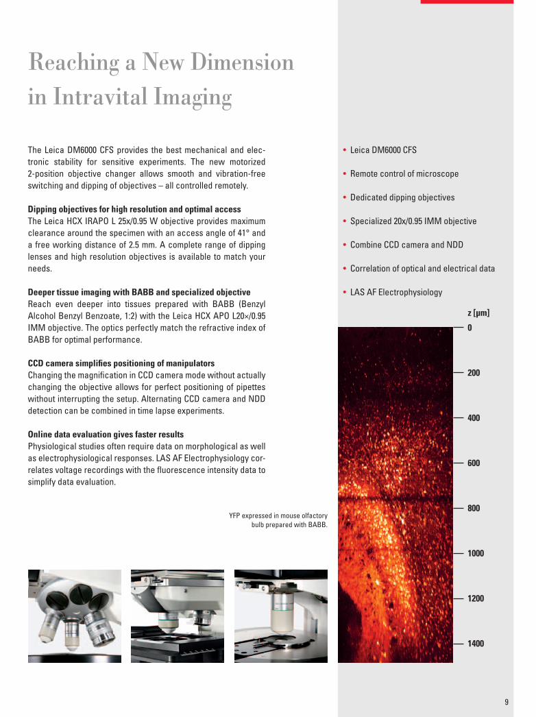
Deeper tissue imaging with BABB and specialized objectiveReach even deeper into tissues prepared with BABB (Benzyl Alcohol Benzyl Benzoate, 1:2) with the Leica HCX APO L20×/0.95 IMM objective. The optics perfectly match the refractive index of BABB for optimal performance.
CCD camera simplifi es positioning of manipulatorsChanging the magnifi cation in CCD camera mode without actually changing the objective allows for perfect positioning of pipettes without interrupting the setup. Alternating CCD camera and NDD detection can be combined in time lapse experiments.
Online data evaluation gives faster resultsPhysiological studies often require data on morphological as well as electrophysiological responses. LAS AF Electrophysiology cor-relates voltage recordings with the fl uorescence intensity data to simplify data evaluation.
9
• Leica DM6000 CFS
• Remote control of microscope
• Dedicated dipping objectives
• Specialized 20x/0.95 IMM objective
• Combine CCD camera and NDD
• Correlation of optical and electrical data
• LAS AF Electrophysiology
Reaching a New Dimension in Intravital Imaging
YFP expressed in mouse olfactorybulb prepared with BABB.
0
200
400
600
800
1000
1200
1400
z [µm]
10
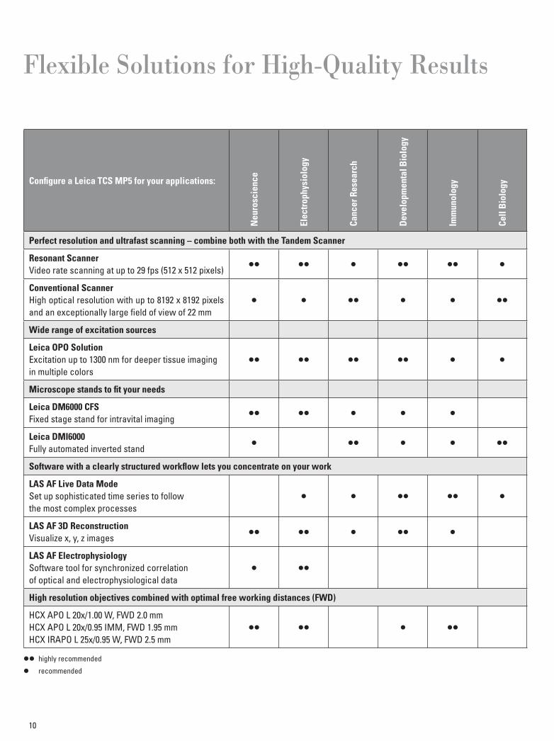
Flexible Solutions for High-Quality Results
Confi gure a Leica TCS MP5 for your applications:
Neu
rosc
ienc
e
Elec
trop
hysi
olog
y
Canc
er R
esea
rch
Dev
elop
men
tal B
iolo
gy
Imm
unol
ogy
Cell
Bio
logy
Perfect resolution and ultrafast scanning – combine both with the Tandem Scanner
Resonant ScannerVideo rate scanning at up to 29 fps (512 x 512 pixels)
Conventional ScannerHigh optical resolution with up to 8192 x 8192 pixels and an exceptionally large fi eld of view of 22 mm
Wide range of excitation sources
Leica OPO SolutionExcitation up to 1300 nm for deeper tissue imagingin multiple colors
Microscope stands to fi t your needs
Leica DM6000 CFSFixed stage stand for intravital imaging
Leica DMI6000Fully automated inverted stand
Software with a clearly structured workfl ow lets you concentrate on your work
LAS AF Live Data ModeSet up sophisticated time series to followthe most complex processes
LAS AF 3D ReconstructionVisualize x, y, z images
LAS AF ElectrophysiologySoftware tool for synchronized correlationof optical and electrophysiological data
High resolution objectives combined with optimal free working distances (FWD)
HCX APO L 20x/1.00 W, FWD 2.0 mmHCX APO L 20x/0.95 IMM, FWD 1.95 mmHCX IRAPO L 25x/0.95 W, FWD 2.5 mm
highly recommended
recommended
Acknowledgements
We gratefully acknowledge the following scientists for providing images, data, samples and valuable support:
Page2/3 (from left to right)Mouse mammary gland. Blood vessels labeled with 70 kD-Texas Red (red),SHG of type I collagen (purple), autofl uorescence of single cells (green).Courtesy of Evelyne Beerling, Dr. Jacco van Rheenen, Hubrecht Institute, Utrecht, The Netherlands
Lymph node. CD8-CFP (red) and CD4-GFP (green) T cells. SHG of collagen (cyan). Representative z-stack of 3D time lapse series. Courtesy of Dr. Bernd Zinselmeyer and Dr. Dorian McGavern, NIH/NINDS, Bethesda, MD, U.S.A.
Embryonic day 16 kidney from a HoxB7-eGFP mouse.HoxB7 labels the ureteric bud.Courtesy of Prof. Deborah Hyink, Mount Sinai School of Medicine, New York, NY, U.S.A.
Lymph node. Alexa 488 (green), PE (red), SHG (blue).Courtesy of Dr. Pierre Bourdoncle, Institut Cochin, Paris, France
Mouse kidney section. Wheat germ agglutinin-Alexa 488 (green), phalloidin-Alexa 568 (red) and DAPI (blue). Courtesy of Dr. Jacco van Rheenen, Hubrecht Institute, Utrecht, The Netherlands
Page 4Top: Microglia labeled with GFP (green), 655 nm quantum dots injected in brain vasculature (red), SHG of skull bone (blue). Representative z-stack of 3D time lapse series.Courtesy of Dr. Debasis Nayak, Dr. Bernd Zinselmeyer and Dr. Dorian McGavern, NIH/NINDS, Bethesda, MD, U.S.A.
Bottom: Mouse kidney. Blood vessels labeled with 70 kD-Texas Red (red), SHG of Type I collagen (purple), autofl uorescence of single cells (green).Courtesy of Evelyne Beerling, Dr. Jacco van Rheenen, Hubrecht Institute, Utrecht, The Netherlands
Page 5Top: Zebrafi sh embryo, blood cells labeled with DsRed.Courtesy of Dr. Julien Vermot, IGBMC, Strasbourg-Illkirch, France
Bottom: Spleen. Anti-viral CD8-CFP (red) and CD4-GFP (green) T cells.Representative z-stack of 3D time lapse series.Courtesy of Dr. Bernd Zinselmeyer and Dr. Dorian McGavern, NIH/NINDS, Bethesda, MD, U.S.A.
Page 9Mouse olfactory bulb labeled with venusYFP. Sample cleared with BABB.Courtesy of Jan Thomas Herb, Dr. Andreas Schäfer, Annemarie Scherbarth,Dr. Günter Giese and Prof. Winfried Denk, MPI for Medical Research, Heidelberg, Germany
11
The statement by Ernst Leitz in 1907, “with the user, for the user,” describes the fruitful collaboration with end users and driving force of innovation at Leica Microsystems. We have developed fi ve brand values to live up to this tradition: Pioneering, High-end Quality, Team Spirit, Dedication to Science, and Continuous Improvement. For us, living up to these values means: Living up to Life.
Active worldwide Australia: North Ryde Tel. +61 2 8870 3500 Fax +61 2 9878 1055
Austria: Vienna Tel. +43 1 486 80 50 0 Fax +43 1 486 80 50 30
Belgium: Groot Bijgaarden Tel. +32 2 790 98 50 Fax +32 2 790 98 68
Canada: Richmond Hill/Ontario Tel. +1 905 762 2000 Fax +1 905 762 8937
Denmark: Ballerup Tel. +45 4454 0101 Fax +45 4454 0111
France: Nanterre Cedex Tel. +33 811 000 664 Fax +33 1 56 05 23 23
Germany: Wetzlar Tel. +49 64 41 29 40 00 Fax +49 64 41 29 41 55
Italy: Milan Tel. +39 02 574 861 Fax +39 02 574 03392
Japan: Tokyo Tel. +81 3 5421 2800 Fax +81 3 5421 2896
Korea: Seoul Tel. +82 2 514 65 43 Fax +82 2 514 65 48
Netherlands: Rijswijk Tel. +31 70 4132 100 Fax +31 70 4132 109
People’s Rep. of China: Hong Kong Tel. +852 2564 6699 Fax +852 2564 4163
Portugal: Lisbon Tel. +351 21 388 9112 Fax +351 21 385 4668
Singapore Tel. +65 6779 7823 Fax +65 6773 0628
Spain: Barcelona Tel. +34 93 494 95 30 Fax +34 93 494 95 32
Sweden: Kista Tel. +46 8 625 45 45 Fax +46 8 625 45 10
Switzerland: Heerbrugg Tel. +41 71 726 34 34 Fax +41 71 726 34 44
United Kingdom: Milton Keynes Tel. +44 800 298 2344 Fax +44 1908 246312
USA: Buffalo Grove/lllinois Tel. +1 847 405 0123 Fax +1 847 405 0164 and representatives in more than 100 countries
Leica Microsystems operates globally in four divi sions, where we rank with the market leaders.
• Life Science DivisionThe Leica Microsystems Life Science Division supports the imaging needs of the scientifi c community with advanced innovation and technical expertise for the visualization, measurement, and analysis of microstructures. Our strong focus on understanding scientifi c applications puts Leica Microsystems’ customers at the leading edge of science.
• Industry DivisionThe Leica Microsystems Industry Division’s focus is to support customers’ pursuit of the highest quality end result. Leica Microsystems provide the best and most innovative imaging systems to see, measure, and analyze the micro-structures in routine and research industrial applications, materials science, quality control, forensic science inves-tigation, and educational applications.
• Biosystems DivisionThe Leica Microsystems Biosystems Division brings his-topathology labs and researchers the highest-quality, most comprehensive product range. From patient to pa-thologist, the range includes the ideal product for each histology step and high-productivity workfl ow solutions for the entire lab. With complete histology systems fea-turing innovative automation and Novocastra™ reagents, Leica Microsystems creates better patient care through rapid turnaround, diagnostic confi dence, and close cus-tomer collaboration.
• Medical DivisionThe Leica Microsystems Medical Division’s focus is to partner with and support surgeons and their care of pa-tients with the highest-quality, most innovative surgi cal microscope technology today and into the future.
“With the user, for the user”Leica Microsystems
www.leica-microsystems.com
Orde
r no.
: Eng
lish
1593
0020
15 •
IV/1
1/AX
/Br.H
. • C
opyr
ight
© b
y Le
ica
Mic
rosy
stem
s CM
S Gm
bH, M
annh
eim
, Ger
man
y, 2
011
LEIC
A an
d th
e Le
ica
Logo
are
regi
ster
ed tr
adem
arks
of L
eica
Mic
rosy
stem
s IR
Gm
bH.


