Leishmania Leishmania: a large group of kinetoplastid parasites causing a variety of syndromes Phosphoglycans: important molecules for parasite development and pathogenesis Classic model for polarized T-cell response (Th1/Th2, will be covered later in the immunology part of this course)
Transcript
Leishmania
Leishmania: a large group of kinetoplastid parasites causing a variety of syndromes
Phosphoglycans: important molecules for parasite development and pathogenesis
Classic model for polarized T-cell response (Th1/Th2, will be covered later in the immunology part of this course)
Leishmania belong to the order kinetoplastida
Group of flagellates at the basis of the eukaryotic tree
Harbor name-giving mitochondrion with large genome which is always associated with the basal body of the single flagellum
Trypanosoma & Leishmania are the medically important and best studied genera in this group
trypomastigote
epimastigote
promastigote
amastigote
Leishmania parasites exist as pro- and amastigotes
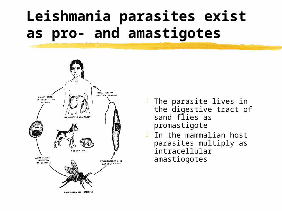
The parasite lives in the digestive tract of sand flies as promastigote
In the mammalian host parasites multiply as intracellular amastiogotes
Leishmania infects and thrives in macrophages
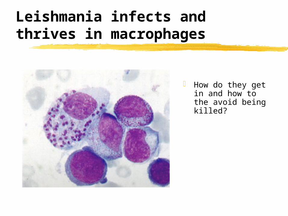
How do they get in and how to the avoid being killed?
Leishmania infects and thrives in macrophages
Uptake of Leishmania amazonensis metacyclic promastigote by a mouse macrophage. The parasite is phagocytosed with the cell body entering first and through the formation of a long tubular pseudopod. Images were captured every 0.5 seconds over the course of 367 seconds. Courret et al. 2002 http://jcs.biologists.org/cgi/content/full/115/11/2303
Leishmania infects and thrives in macrophages
Phagocytosis of a Leishmania amazonensis metacyclic promastigote by a mouse macrophage. The parasite binds to the macrophage plasma membrane by the tip of the flagellum. It then turns around and is finally ingested via the cell body. Images were
captured every 0.5 seconds over the course of 125 seconds. Courret et al. 2002 http://jcs.biologists.org/cgi/content/full/115/11/2303
Leishmania infects and thrives in macrophages
Leishmania stimulates this process by binding elements of the complement system to its surface
Binding of complement can destroy pathogens but also tags them for phagocytosis (opsonization: pathogen bound 3Cb is a potent ‘eat me’ signal for macrophages & neutrophils)
However, the parasite prevents the formation of the fully functional membrane attack complex
A molecule on the surface of the parasite seems to be responsible both for complement activation and prevention of the final attack
Leishmania infects and thrives in macrophages
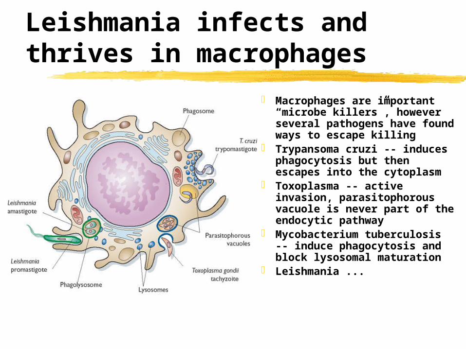
Macrophages are important “microbe killers”, however several pathogens have found ways to escape killing
Trypansoma cruzi -- induces phagocytosis but then escapes into the cytoplasm
Toxoplasma -- active invasion, parasitophorous vacuole is never part of the endocytic pathway
Mycobacterium tuberculosis -- induce phagocytosis and block lysosomal maturation
Leishmania ...
Leishmania infects and thrives in macrophages
… Leishmania just doesn’t seem to care
Amastigotes thrive in what looks like a fully matured lysosome with acidic pH and abundant lysosomal hydrolases
Amastigotes rapidly divide and will infect new macrophages after rupture of host cell
The dense surface coat covering Leishmania seems to protect the parasite from the action of the lytic enzymes
However, with help from T cells macrophages can be stimulated to kill the parasite
A TH1 response is required for parasite control and healing
Stimmulation with different cytokines leads to the development of two types of T-cells specialized for different immune responses
Th1 and Th2 strongly downregulate each other
This polarization has important consequences for the downstream response and can spell life or death
Non healing Leishmania infections are characterized by a strong TH2 response (remember this was the response useful to get rid of worms by antibody and hypersensitivity)
Healing infections are characterized by TH1 The parasites seems to manipulate this
balance in his favor, we don’t understand yet how that is done
Leishmania is transmitted by sand flies (Phlebotomidae)
Sand flies are minute diptera (only females bite)
They do not fly well and stay close to the ground
In the wild sand flies often breed in rodent burrows
Old world Phlebotomus, new world Lutzomya
Can also transmit Bartonella bacilliformis and Papatsi virus
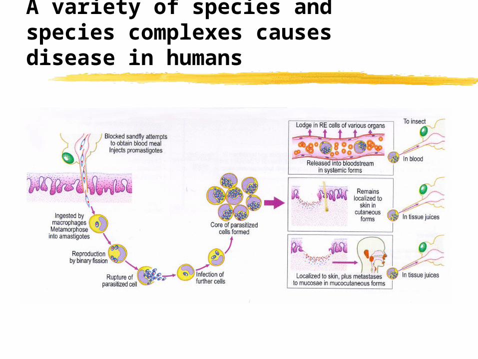
A variety of species and species complexes causes disease in humans
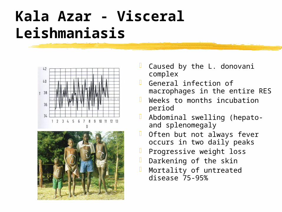
Kala Azar - Visceral Leishmaniasis
Caused by the L. donovani complex General infection of macrophages in
the entire RES Weeks to months incubation period Abdominal swelling (hepato- and
splenomegaly Often but not always fever occurs in
two daily peaks Progressive weight loss Darkening of the skin Mortality of untreated disease 75-
95%
Cutaneous Leishmaniasis is usually self-limiting
Old world oriental sore is caused by parasites of the L. tropica complex. (similar disease in the new world is caused by L. mexicana)
A chronic but self-limiting dry ulceration at the site of the bite
Ulceration start months after infection
Parasites are not found outside the lesion
Nearly absolute resistance to reinfection (however, there is long term persistence and persistence is required for resistance)
Espundia -- Mucocutaenous Leishmaniasis
Caused by L. braziliensis ~20% of infected patients
develop ulcers of the oral and nasal mucosa
Progression of the ulceration is slow but steady, ultimately destroying all soft parts of the nose, the lips, the soft and the soft palate
Death occurs usually through secondary bacterial infection
Leishmania produce a unique glycoconjugate
Leishmania parasites can be labeled at surprizing efficiency with radioactive sugars and inositol
Label is incorporated into a large glycoconjugate which is acid labile and has a lipid anchor
This lipophosphoglycan (LPG) completely covers the surface of the the promastigote
Lipophosphoglycans share structural features with GPIs
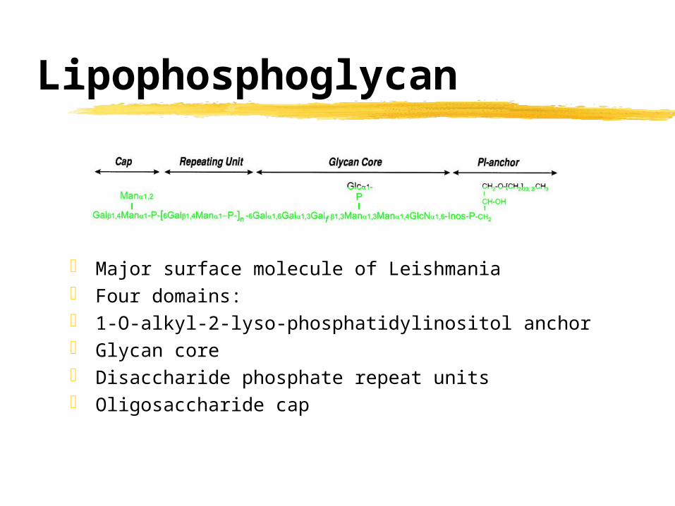
Lipophosphoglycan
Major surface molecule of Leishmania Four domains: 1-O-alkyl-2-lyso-phosphatidylinositol anchor Glycan core Disaccharide phosphate repeat units Oligosaccharide cap
LPG shows structural variation among Leishmania species
Biosynthesis of LPG
What does LPG “not” do?
LPG protects parasites in the sand fly midgut LPG attaches parasites to the sand fly midgut
epithelium LPG protects against complement attack LPG enhances uptake into macrophages LPG interferes with macrophage signaling
preventing oxidative burst LPG protect from toxic macrophage products
Is LPG a pathogenesis factor?Stan Falkow’s virulence postulates
Pathogenesis is reasonably associated with the expression of a certain virulence factor
Inactivation of the gene should result in loss of virulence
Restoration of the gene should fully restore virulence
Identification of LPG genes by genetic complementation
Mutants in LPG biosynthesis as tools to study LPG enzymology & function
To proof the third postulate has been a challenge
Lpg- mutants show significant loss in virulence both in in vitro macrophage infections as well as in in vivo experiments
But: Leishmania tends to loose virulence in culture anyway, and expression of the WT gene did not always fully restore virulence
Chemical mutants might be ‘over-mutated’ and carry multiple mutations (some might not be related to LPG but affect virulence)
Presence of several LPG related molecules further complicate the issue
A number of Leishmania glycoconjugates share structural features with LPG
Knock outs by gene targeting as a cleaner way to obtain mutants
Promastigotes attach to and metacyclics detach from the midgut epithelium
procyclics and metacyclics
Infected macrophages are taken up with the blood meal and amastigotes released by digestion transform into procyclic promastigotes which attach to the midgut epithelium
Attached promastigotes divide rapidly
Metacylcic promastigote detach and pass forward into the pharynx from where they are regurgitated into the bite site
Structural modification of LPG during the sand fly cycle
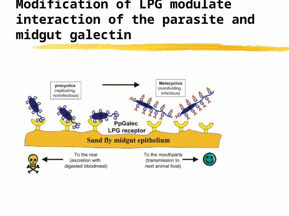
LPG is structurally modified during metacylogenesis
LPG in metacylics has 2-3 times the number of repeat units
Side chains with terminal galactose are down-regulated in favor of chains with terminal arabino-pyranose
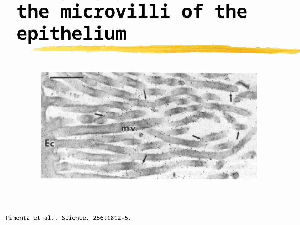
Only phosphoglycans from procyclics attach to the midgut
Opened midguts were incubated with PG from procyclics (A/B) and metacyclics (C/D) and detected with an antibody
Pimenta et al., Science. 256:1812-5.
Phosphoglycan binds to the microvilli of the epithelium
Pimenta et al., Science. 256:1812-5.
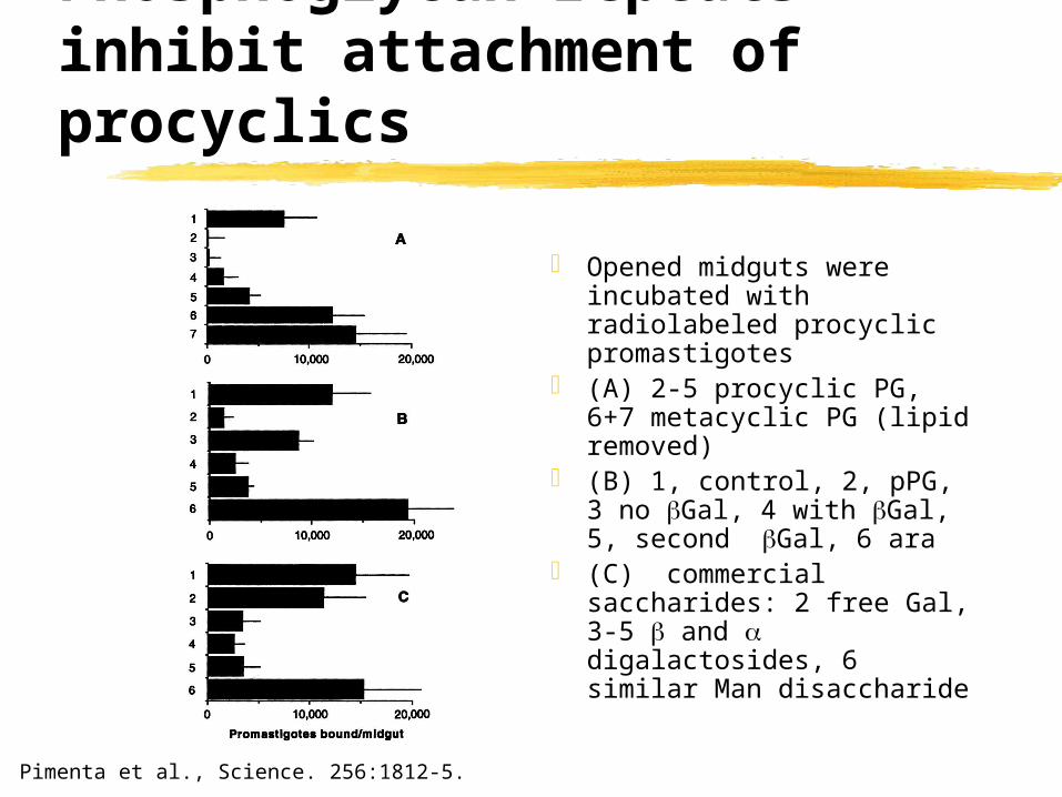
Phosphoglycan repeats inhibit attachment of procyclics
Opened midguts were incubated with radiolabeled procyclic promastigotes
(B) 1, control, 2, pPG, 3 no Gal, 4 with Gal, 5, second Gal, 6 ara
(C) commercial saccharides: 2 free Gal, 3-5 and digalactosides, 6 similar Man disaccharide
Pimenta et al., Science. 256:1812-5.
LPG is not essential for early survival but for retention
(A + B) number of promastigotes present in midgut upon disection (in B 5x higher inoculum is used)
Sacks et al., PNAS 97: 406-411
LPG binds to a species specific galectin in the sandfly midgut
A gene for an abundantly expressed galactose binding protein or lectin (galectin) was identified in a sandfly sequencing project
A specific antibody against the protein encoded by this gene reacts with the midgut of the sandfly species from which it was isolated (but not from other species)
High resolution microscopy shows that the protein(red in lower panel) is found on the luminal side of the midgut epithelium
Cell 119:329-41
LPG binds to a species specific galectin in the sandfly midgut
Galectin (labeled with a fluorescent dye) binds specifically to procyclic Leishmania major parasites
However little binding is detected when incubated with metacyclic parasites (the stage that detaches and moves to the the proboscis to infect the mammalian host, V1met)
There is little binding to a mutant parasite (Spock) which lacks LPG
(B lower) anti-galectin also blocks binding of procyclic parasites to the midgut epithelium
Cell 119:329-41
Modification of LPG modulate interaction of the parasite and midgut galectin
Summary
Leishmania species cause three clinical syndromes depending on the spread of the infection in the body
Leishmania ‘provoke’ phagocytosis by macrophages and develop intracellular in an fully acidified lysosome
LPG-galectin interaction and modification of LPG regulate attachment and detachment of parasites in the sandfly host


![Anti-Parasitic Activities of Allium sativum and Allium ... · Trypanosomiasis (HAT) [15]. Leishmaniasis is a disease caused by the protozoan parasite Leishmania, which results in](https://static.documents.pub/doc/80x56/5f9e9234ee0f1e0741090a0d/anti-parasitic-activities-of-allium-sativum-and-allium-trypanosomiasis-hat.jpg)


