65
UNCLASSIFIED WRAIR- GEIS 'Operational Clinical Infectious Disease' Course Leishmaniasis

UNCLASSIFIED
WRAIR- GEIS 'Operational Clinical Infectious Disease' Course
Leishmaniasis
LTC James E. Moon, MD
Chief, Sleep Trials Branch
Walter Reed Army Institute of Research
CDR Ramiro L. Gutierrez, MD
Chief, Enterics Department
Naval Medical Research Center
JULY 2015
Acknowledgments
The views expressed in this presentation
are those of the speaker and authors,
and do not reflect the official policy of the
Department of Army, Department of the
Navy, Department of Defense, or the U.S.
Government
Disclaimer

Leishmaniasis
• Diverse group of diseases caused by infection from protozoan parasites of the genus Leishmania
• Designated one of the five most important diseases worldwide by the WHO
– 1.5 to 2 million new cases/year
• Leishmaniasis threatens over 350 million individuals in 88 countries, and directly impacts US service members abroad
www.yourarticlelibrary.com
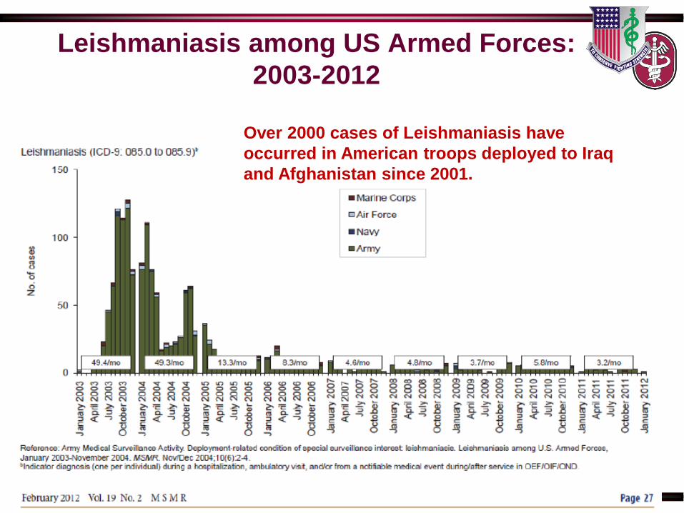
Leishmaniasis among US Armed Forces:
2003-2012
Over 2000 cases of Leishmaniasis have
occurred in American troops deployed to Iraq
and Afghanistan since 2001.
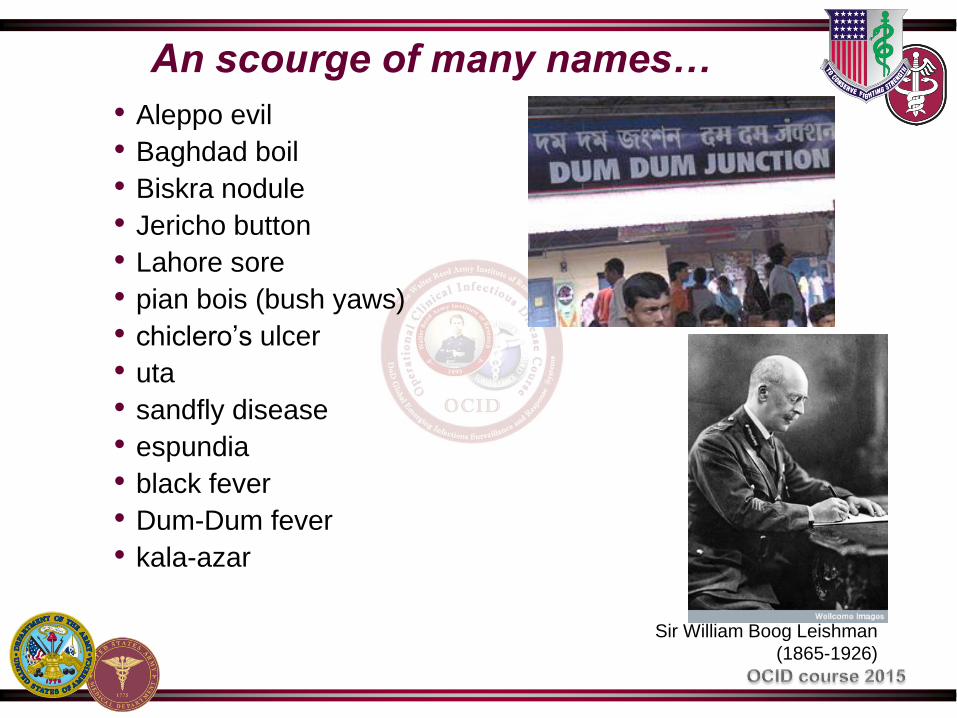
An scourge of many names…
• Aleppo evil
• Baghdad boil
• Biskra nodule
• Jericho button
• Lahore sore
• pian bois (bush yaws)
• chiclero’s ulcer
• uta
• sandfly disease
• espundia
• black fever
• Dum-Dum fever
• kala-azar
Sir William Boog Leishman
(1865-1926)

Vector
- Female Sand fly
-Lutzomyia in the Americas
-Phlebotomus elsewhere
- Poor flyers, remain near ground
- World wide distribution
- Bites at exposed areas and clothing
lines
Reservoirs
- Humans
- Dogs
- Rodents

Transmission* and Lifecycle
http://www.niaid.nih.gov/topics/leishmanias
is *Also transmitted by blood transfusion!
Disease
• Three clinical syndromes:
– Cutaneous (skin)
• Localized, diffuse, Leishmania recidivans, post kala-azar
dermal leish.
– Mucocutaneous (mouth, nose, also called “espundia”)
– Visceral (internal organs, also called “kala-azar”)
• Each syndrome can be caused by multiple different
Leishmania species, and many species can cause multiple
different syndromes
– Determined by species of parasite, location of infected
macrophages, and individual immune response.

Highly Endemic Areas
• 90% of cutaneous leishmaniasis occur in Afghanistan,
Brazil, Iran, Peru, Saudi Arabia, and Syria.
• 90% mucocutaneous leishmaniasis occur in Bolivia, Brazil,
and Peru
• 90% of all visceral leishmaniasis cases occur in Bangladesh,
Brazil, India, Nepal, and Sudan
WHO Leishmaniasis: Burden of Disease
Cutaneous Leishmaniasis (CL)
• Overwhelming majority of Leishmaniasis
– 1 to 1.5 million cases/year
• Endemic in widely scattered regions throughout the world
• Generally not life-threatening, but potentially permanently
disfiguring
• Wide spectrum of clinical presentations that differs
somewhat between New and Old World due to regional
Leishmania species

Old World Cutaneous Leishmaniasis
PPID, 8th ed., pg. 3093

New World Cutaneous Leishmaniasis
PPID, 8th ed., pg. 3093
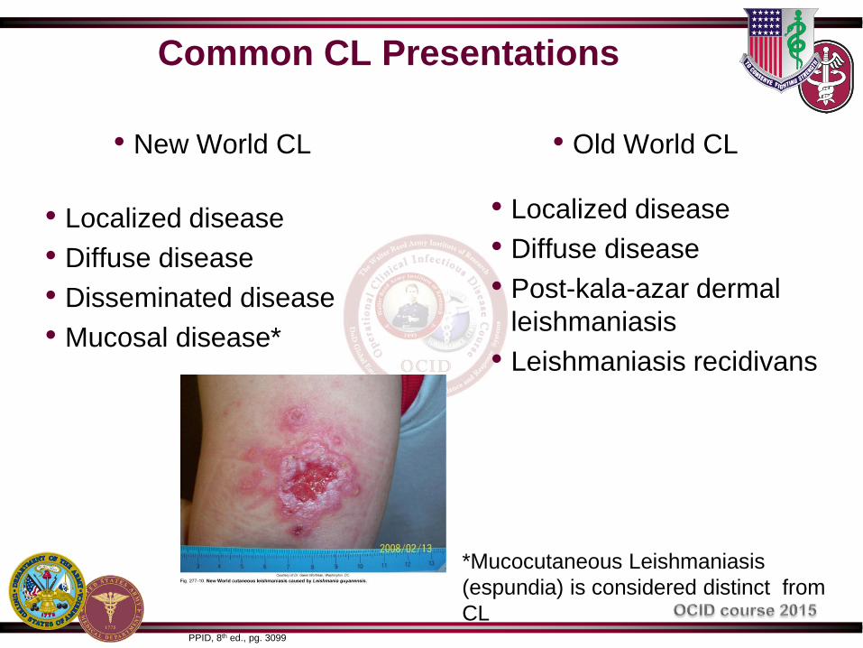
Common CL Presentations
• New World CL
• Localized disease
• Diffuse disease
• Disseminated disease
• Mucosal disease*
• Old World CL
• Localized disease
• Diffuse disease
• Post-kala-azar dermal
leishmaniasis
• Leishmaniasis recidivans
*Mucocutaneous Leishmaniasis
(espundia) is considered distinct from
CL PPID, 8th ed., pg. 3099
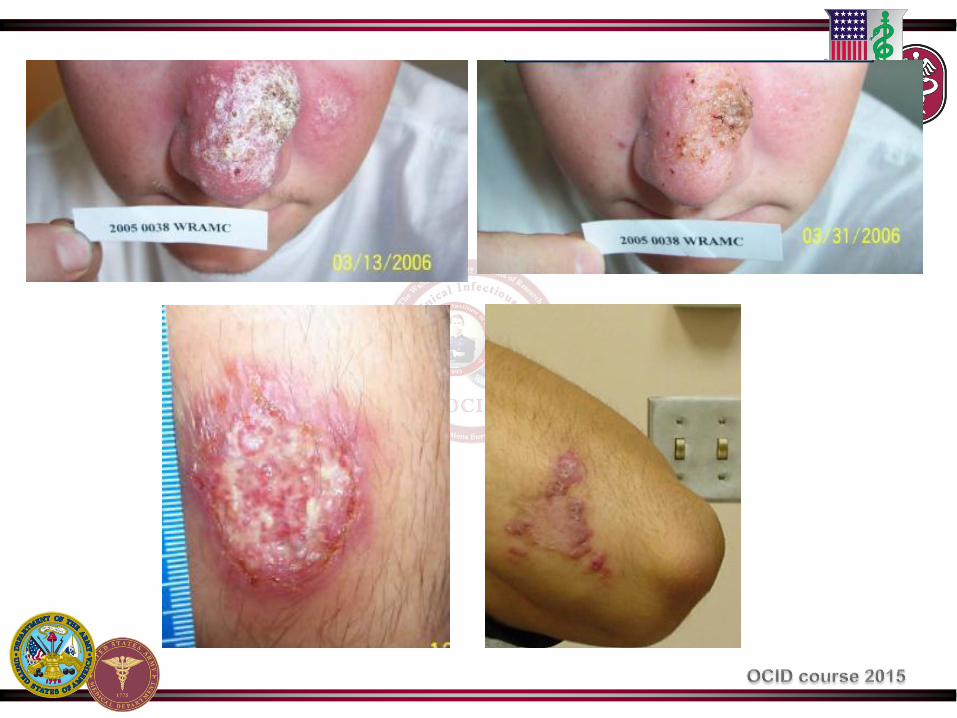
Localized Cutaneous Leishmaniasis
• Single or multiple lesions, appearance varies
• Nodules develop, expand, ulcerate over weeks
• Incubation time ~ 40 days (days – years)
• Usually painless or minimally painful
• Persists months to years, eventually heals with burnlike scar
– L. major most common causative species

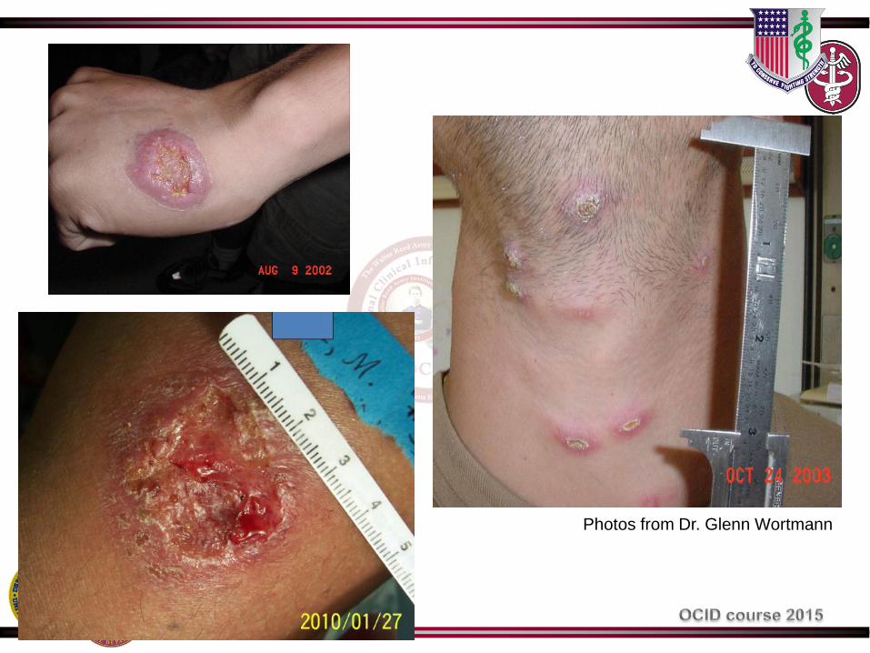
Photos from Dr. Glenn Wortmann

Chiclero’s Ulcer
• Localized cutaneous leishmaniasis (ear)
• Majority of cases caused by L. mexicana
• Chicleros – men who collect the chicle latex from which
chiclets chewing gum is made
Photo: Dr. Jason Blaylock
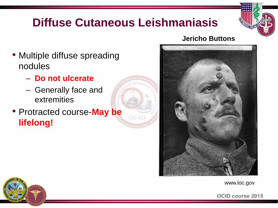
Diffuse Cutaneous Leishmaniasis
• Multiple diffuse spreading
nodules
– Do not ulcerate
– Generally face and
extremities
• Protracted course-May be
lifelong!
Jericho Buttons
www.loc.gov
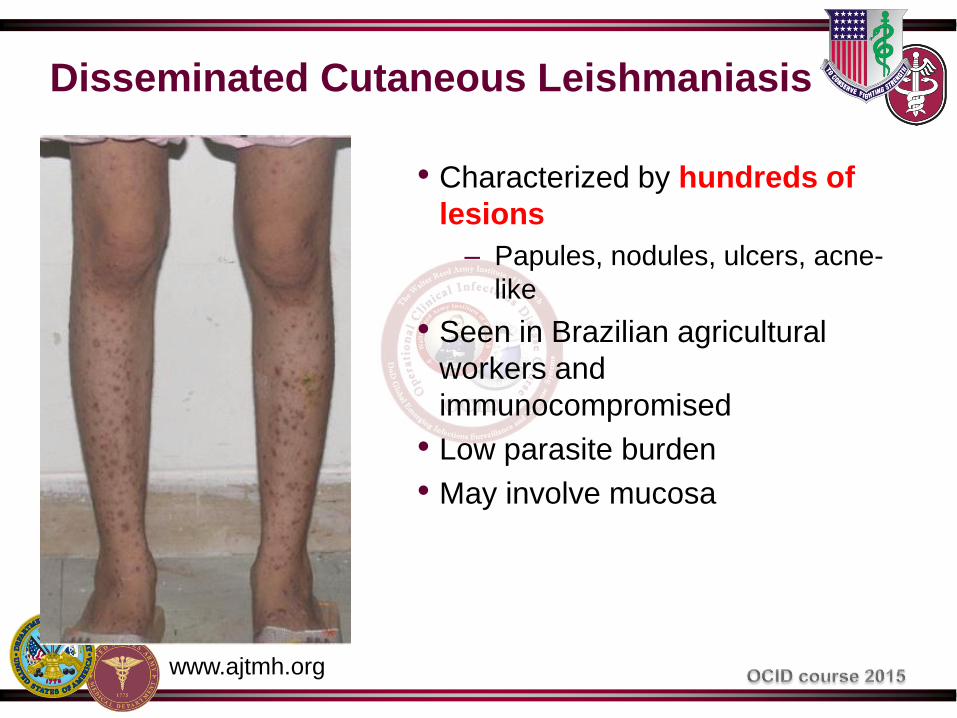
Disseminated Cutaneous Leishmaniasis
• Characterized by hundreds of
lesions
– Papules, nodules, ulcers, acne-
like
• Seen in Brazilian agricultural
workers and
immunocompromised
• Low parasite burden
• May involve mucosa
www.ajtmh.org
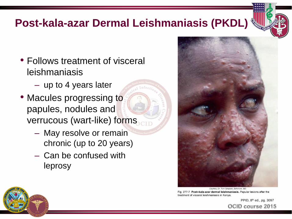
Post-kala-azar Dermal Leishmaniasis (PKDL)
• Follows treatment of visceral
leishmaniasis
– up to 4 years later
• Macules progressing to
papules, nodules and
verrucous (wart-like) forms
– May resolve or remain
chronic (up to 20 years)
– Can be confused with
leprosy
PPID, 8th ed., pg. 3097

PKDL
USUHS teaching slides
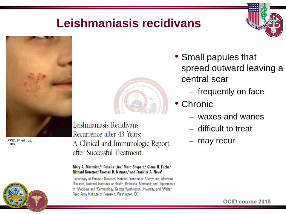
Leishmaniasis recidivans
• Small papules that
spread outward leaving a
central scar
– frequently on face
• Chronic
– waxes and wanes
– difficult to treat
– may recur PPID, 8th ed., pg.
3100

Mucucutaneous Leishmaniasis (ML)
• 2-5% of persons with New World CL develop mucous
membrane involvement
– Nose, oral cavity, pharynx, larynx
– Concurrent or months to decades after CL resolves (can
also be primary presentation)
– Can be severely mutilating and life-threatening
www.who.int

Prof. Luis A. Leon
LAB. LEON Quito-Ecuador
Manson-Bahr, 1972
Long-standing cases PPID, 8th ed., pg. 3099

Visceral Leishmaniasis (VL)
• Leishmanial infection of the internal organs
• Unlike CL, generally similar in all regions
• Incubation: 2-8 months (10 days to >1 year)
• Wide spectrum of presentations
– majority asymptomatic (6.5-18:1 ratio)
– asymptomatic to subacute to severe multi-organ disease
– can spontaneously resolve over weeks to months, or
progress to fatality if not treated
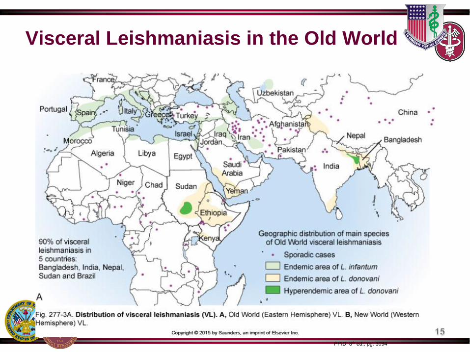
Visceral Leishmaniasis in the Old World
PPID, 8th ed., pg. 3094
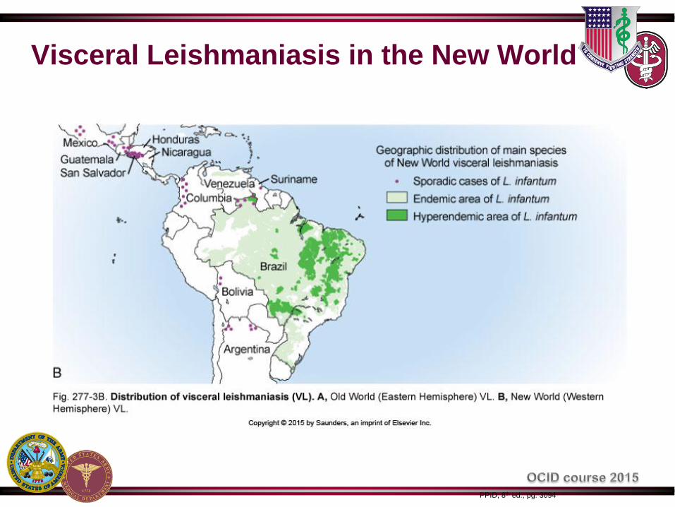
Visceral Leishmaniasis in the New World
PPID, 8th ed., pg. 3094

kala-azar (black or fatal sickness)
• Severe VL
– Classic pentad: prolonged
fever, weight loss,
hepatosplenomegaly,
pancytopenia,
hypergammaglobulinemia
• Progressive (variable rates)
• > 90% mortality within first two
years
• Can be opportunistic infection in
immunocompromised state
PPID, 8th ed., pg. 3095

Viscerotropic Leishmaniasis from Desert Storm
(L. tropica)
• Rare, low-grade, syndrome first identified in 8 patients returning from Operation Desert Storm
– Fevers: 6 of 8
– Weight loss: 2 of 8
– Nausea, vomiting, low-grade watery diarrhea: 2 of 8
– Lymphadenopathy: 2 of 8
– Hepatosplenomegly: 2 of 8
– Anemia: 3 of 8
– Leukopenia or thrombocytopenia: 0 of 8
– Elevated liver enzymes: 6 of 8
– No symptoms: 1 of 8 • Similar syndromes since found in Brazil and Italy
Magill et al, NEJM 1993:328(19)

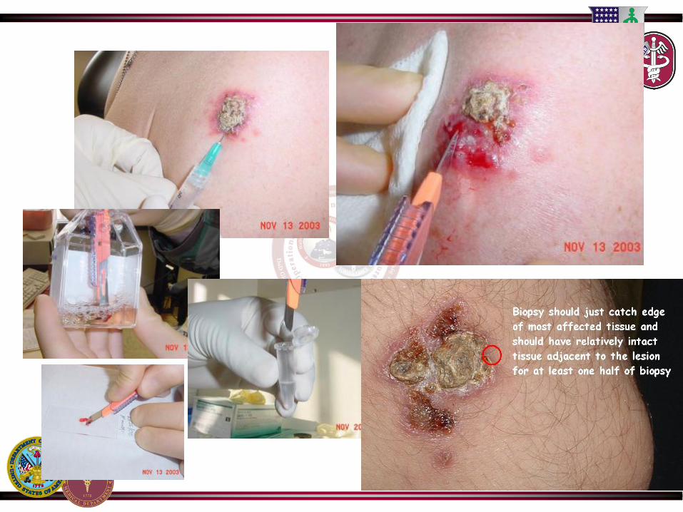
Diagnosis
• Clinical Diagnosis
• Cutaneous Leishmaniasis
– Biopsy/Aspiration/Scraping
– Amastigotes in a smear
– Promastigotes in culture
– PCR of sample (DNA/RNA)
• Visceral Leishmaniasis
– Biopsy of Bone Marrow or Spleen
•Touch Prep, PCR, Culture
– Immunologic
• rK39 leish. antigen direct agglutination test
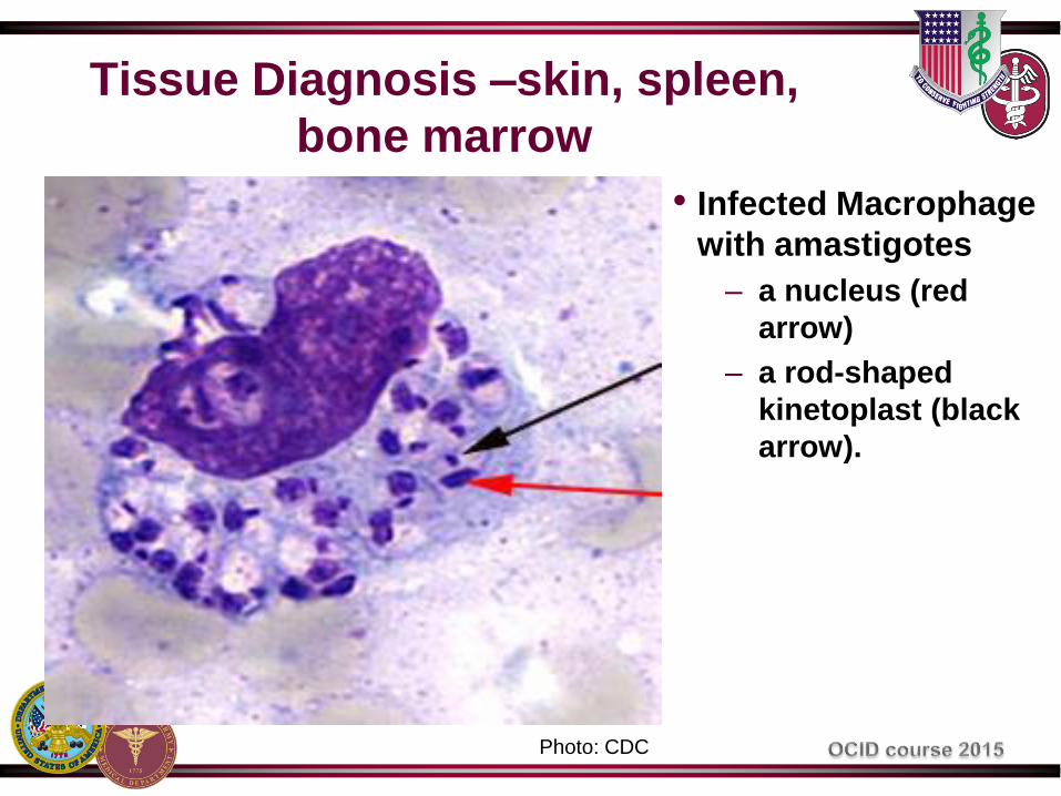
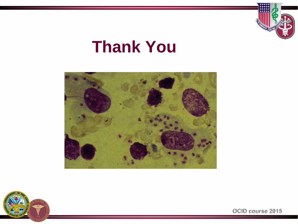
Tissue Diagnosis –skin, spleen,
bone marrow
• Infected Macrophage
with amastigotes
– a nucleus (red
arrow)
– a rod-shaped
kinetoplast (black
arrow).
Photo: CDC

Diagnosis - culture
Photo: CDC
Promastigotes
Photo: www.msu.edu
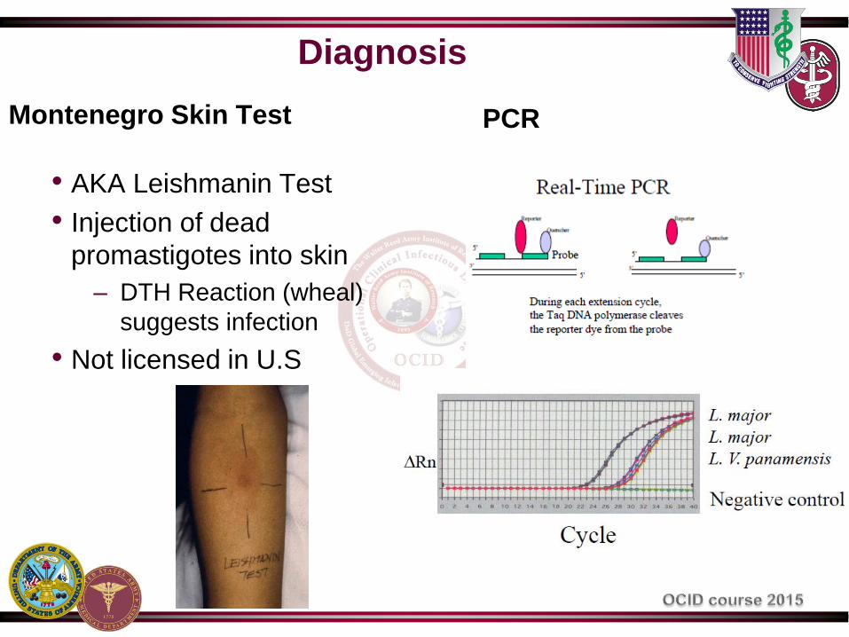
Diagnosis
Montenegro Skin Test
• AKA Leishmanin Test
• Injection of dead
promastigotes into skin
– DTH Reaction (wheal)
suggests infection
• Not licensed in U.S
PCR

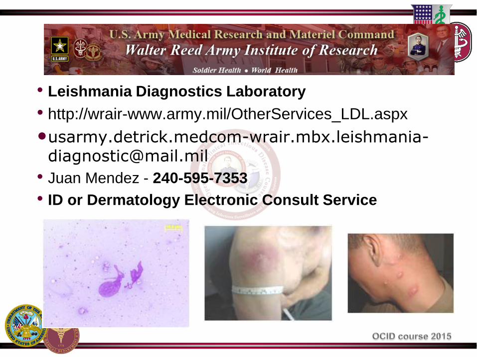
• Leishmania Diagnostics Laboratory
• http://wrair-www.army.mil/OtherServices_LDL.aspx
• Juan Mendez - 240-595-7353
• ID or Dermatology Electronic Consult Service
Treatment
• Treatment is not standardized
– What works on one species and clinical presentation
may fail in another
– Must adapt to regional experience
– Much is anecdotal and off-label
• In general, treatments result in clinical cure, but not
parasitical cure
– Lifelong potential for reactivation in
immunocompromised
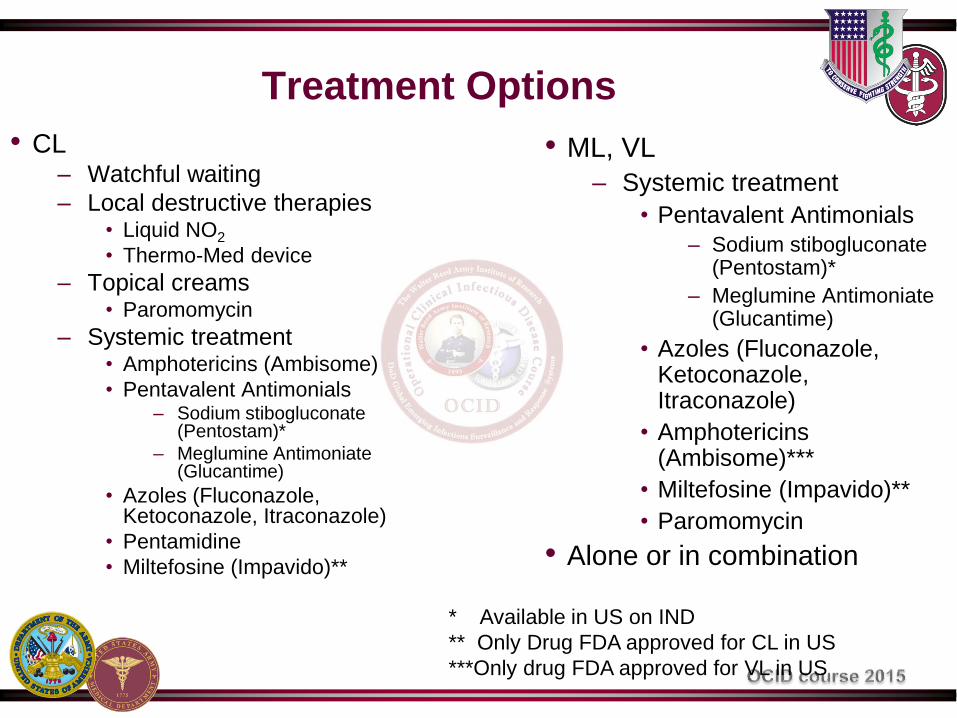
Treatment Options
• CL – Watchful waiting
– Local destructive therapies • Liquid NO2
• Thermo-Med device
– Topical creams • Paromomycin
– Systemic treatment • Amphotericins (Ambisome)
• Pentavalent Antimonials – Sodium stibogluconate
(Pentostam)*
– Meglumine Antimoniate (Glucantime)
• Azoles (Fluconazole, Ketoconazole, Itraconazole)
• Pentamidine
• Miltefosine (Impavido)**
• ML, VL
– Systemic treatment
• Pentavalent Antimonials
– Sodium stibogluconate (Pentostam)*
– Meglumine Antimoniate (Glucantime)
• Azoles (Fluconazole, Ketoconazole, Itraconazole)
• Amphotericins (Ambisome)***
• Miltefosine (Impavido)**
• Paromomycin
• Alone or in combination
* Available in US on IND
** Only Drug FDA approved for CL in US
***Only drug FDA approved for VL in US

CL-When to consider doing nothing
CRITERIA FAVORS NO TREATMENT TREATMENT USUALLY
INDICATED
Age and direction of
healing
Clearly improving compared to prior month Worsening lesions
Number of lesions One or a few >5 and in different locations
Complexity Uncomplicated Restricts movement or wearing of
clothes, cosmetic concerns
Size of lesion(s) Small (<1 cm) Very large (>5cm)
Immune status Immunocompetent Immunocompromised
Mucosal involvement None Yes
Location Nonexposed skin Exposed skin, especially facial
L. brazilensis? No or unlikely Yes or likely*
How bothersome to
patient and family?
No or little concern Very concerned or preoccupied
*If Bolivia, Brazil, Peru, should be treated with systemic therapy due
to risk of mucosal involvement
Adapted from PPID, 8th ed., pg. 3104
UNCLASSIFIED Slide 41
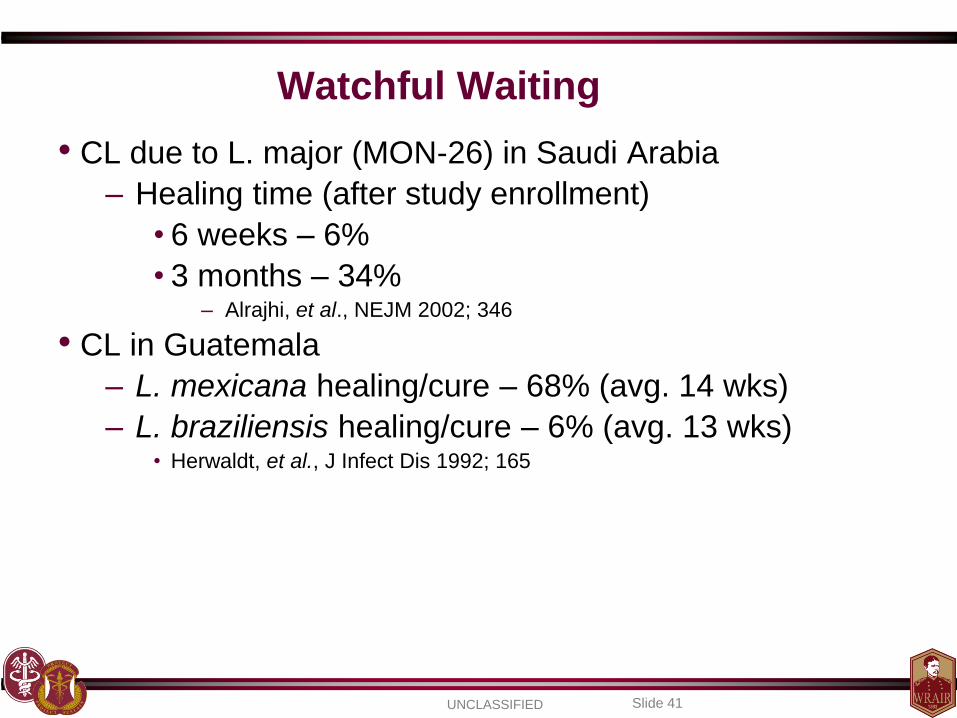
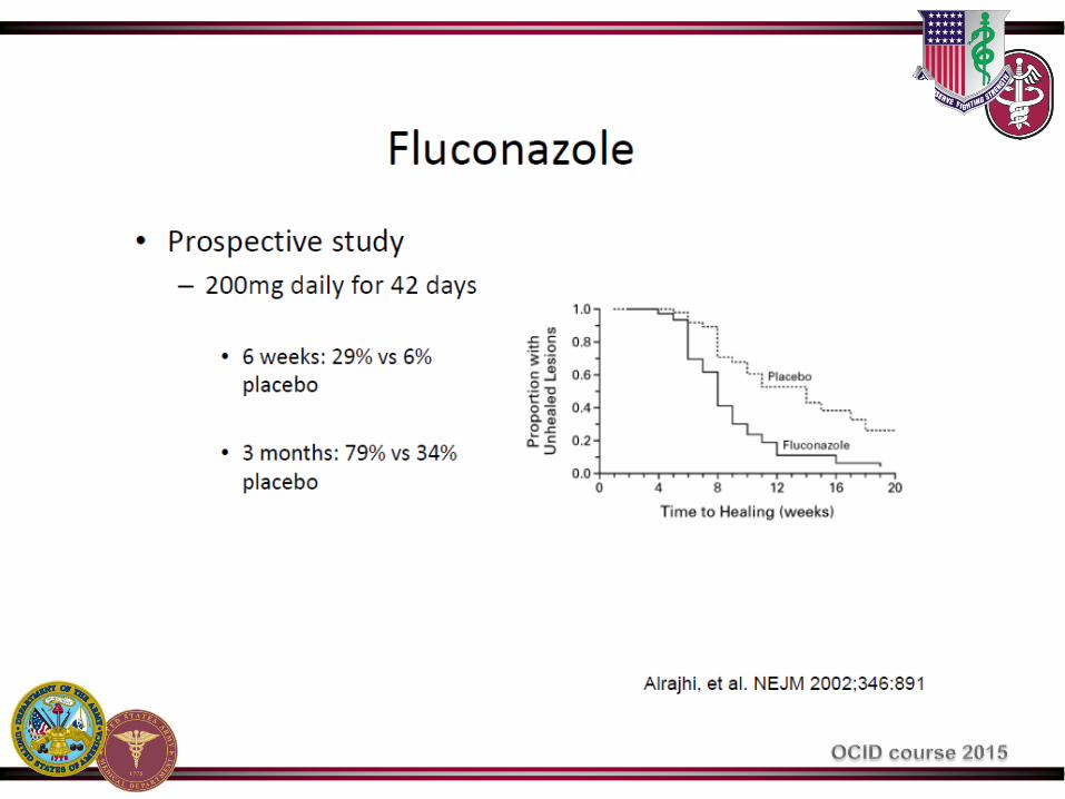
• CL due to L. major (MON-26) in Saudi Arabia
– Healing time (after study enrollment)
• 6 weeks – 6%
• 3 months – 34% – Alrajhi, et al., NEJM 2002; 346
• CL in Guatemala
– L. mexicana healing/cure – 68% (avg. 14 wks)
– L. braziliensis healing/cure – 6% (avg. 13 wks) • Herwaldt, et al., J Infect Dis 1992; 165
Watchful Waiting

No Treatment

Locally Destructive Therapies
LNO2
• Freezes lesions to kill
parasites
• May cause
hypopigmentation
• Not standardized
– Cyroprobe suggested
• Painful / blister formation
ThermoMed
• Heats lesions to kill
parasites
• ~ 70 % efficacy in CL
caused by L. major in Iraq
and L. tropica in
Afghanistan
Reithinger, et al CID 2005
Aronson, et al PloS Negl Trop Dis 2010
Photo: Dr. Glenn Wortmann

Topical Cream: Paromomycin
• Aminoglycoside
• Compounded
– 15% paromomycin
– +/- 0.5% gentamicin
• Apply to affected area twice
a day x 28 days
• 81% cure -L. major
N Engl J Med. 2013 Feb
7;368(6):524-32
mrmc.amedd.army.mil
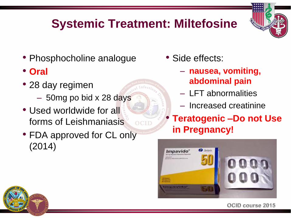
Systemic Treatment: Miltefosine
• Phosphocholine analogue
• Oral
• 28 day regimen
– 50mg po bid x 28 days
• Used worldwide for all
forms of Leishmaniasis
• FDA approved for CL only
(2014)
• Side effects:
– nausea, vomiting,
abdominal pain
– LFT abnormalities
– Increased creatinine
• Teratogenic –Do not Use
in Pregnancy!

Systemic Treatment: Azoles
• Fluconazole
• Ketoconazole, Itraconazole
• Limited data
• Variable regimens
– Oral
– 4-6 weeks or longer
– Weight based
• Variable efficacy
Systemic Treatment: Antimonials
• Sodium stribogluconate (Pentostam)
– Investigational New Drug
– Available from the CDC for civilians
– Available from Walter Reed for military
– Regimen
• CL: intravenous 20mg/kg/day for 10-20 days
– Outside US is often given intra-lesionally
• VL: intravenous 20mg/kg/day for 28 days
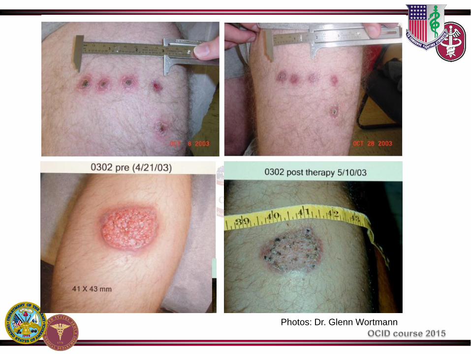
Photos: Dr. Glenn Wortmann

UNCLASSIFIED Slide 53
DO NOT USE IN PREGNANCY
• Toxicities
– Elevated amylase/lipase ~95%
– Elevated liver enzymes ~50%
– Arthralgias/myalgias ~65%
– Rare significant EKG changes/cytopenias
– Dermatological ~10%
• Wide range of presentations
• Herpes zoster virus (shingles)
Pentostam®

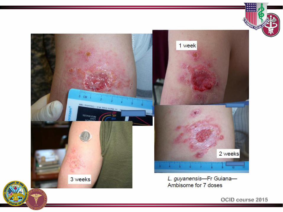
Systemic Treatment: Amphotericins
• Liposomal Amphotericin B (Ambisome)
– Drug of choice for VL
– Regimen (IV)
• Immunocompetent
– 3 mg/kg/day on days 1-5, 14, and 21
• Immunocompromised
– 4 mg/kg/day on days 1-5, 10, 17, 24, 31, 38
– No established regimen for CL
– Extensive side-effect profile
Systemic Treatment: Pentamidine
• No longer recommended for VL due to high toxicity
• One indication only
– Short course (2 IM injections of 4mg/kg) has been
found to be effective for CL caused by L.
guyananensis in French Guyana and Surinam
only

Systemic Dosing Summary
• Cutaneous Leishmaniasis
– Pentostam – 20 mg/kg IV x 10 -20 days
– Ambisome (liposomal amphotericin B) • 3 mg/kg on days 1-5, 14, & 21
– Fluconazole – 8 mg/kg/day (4 – 12 weeks)
– Miltefosine –50mg po twice a day x 28 days
• Visceral Leishmaniasis
– Ambisome (liposomal amphotericin B) 3 mg/kg on days 1-5, 14, & 21
– Pentostam – 20 mg/kg IV x 28 days – Miltefosine –50mg po twice a day x 28 days
Prevention
• Sandflies bite and are active at night (warmer months)
• Stay indoors between dusk and dawn
• Keep dogs and susceptible animals indoors at night
• Use fans - sandflies are poor fliers deterred by wind
• Sandflies are small and can get through mesh netting if
not extremely fine
• House construction and modification; sandflies breed in
cracks of houses
• Insecticides on people and animals
• Help from entomologists
• Dog vaccine available in Brazil
http://www.cfsph.iastate.edu/Factsheets/pdfs/leishmaniasis.pdf

Sandfly Habitat
Volume 28, Issue 12, December 2012, Pages 531–538
Summary – Leishmaniasis
• Worldwide distribution
• Many species with different disease presentations
• Cutaneous form may be self-limited
• Think about mucocutaneous disease, especially in South
America
• Resources available for diagnosis (WRAIR)
• Treatment response varies with species and host
Thank You
Extra Slides

Classification
• Old World, Cutaneous Disease: – L. tropica; L. major; L. aethiopica
– L. tropica can cause visceral disease
• Old World, Visceral Disease: – L. donovani complex with 3 species (L. donovani, L. infantum, and L.
chagasi)
• New World, Cutaneous disease: – L. mexicana complex with 3 main species (L. mexicana, L.
amazonensis, and L. venezuelensis)
• New World, Cutaneous and Mucocutaneous disease – Subgenus Viannia with 4 main species (L. (V.) braziliensis,
L. (V.) guyanensis, L. (V.) panamensis, and L. (V.) peruviana)
• New World, Visceral Disease – L. chagasi
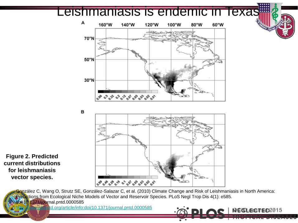
Figure 2. Predicted
current distributions
for leishmaniasis
vector species.
González C, Wang O, Strutz SE, González-Salazar C, et al. (2010) Climate Change and Risk of Leishmaniasis in North America:
Predictions from Ecological Niche Models of Vector and Reservoir Species. PLoS Negl Trop Dis 4(1): e585.
doi:10.1371/journal.pntd.0000585
http://www.plosntd.org/article/info:doi/10.1371/journal.pntd.0000585
Leishmaniasis is endemic in Texas
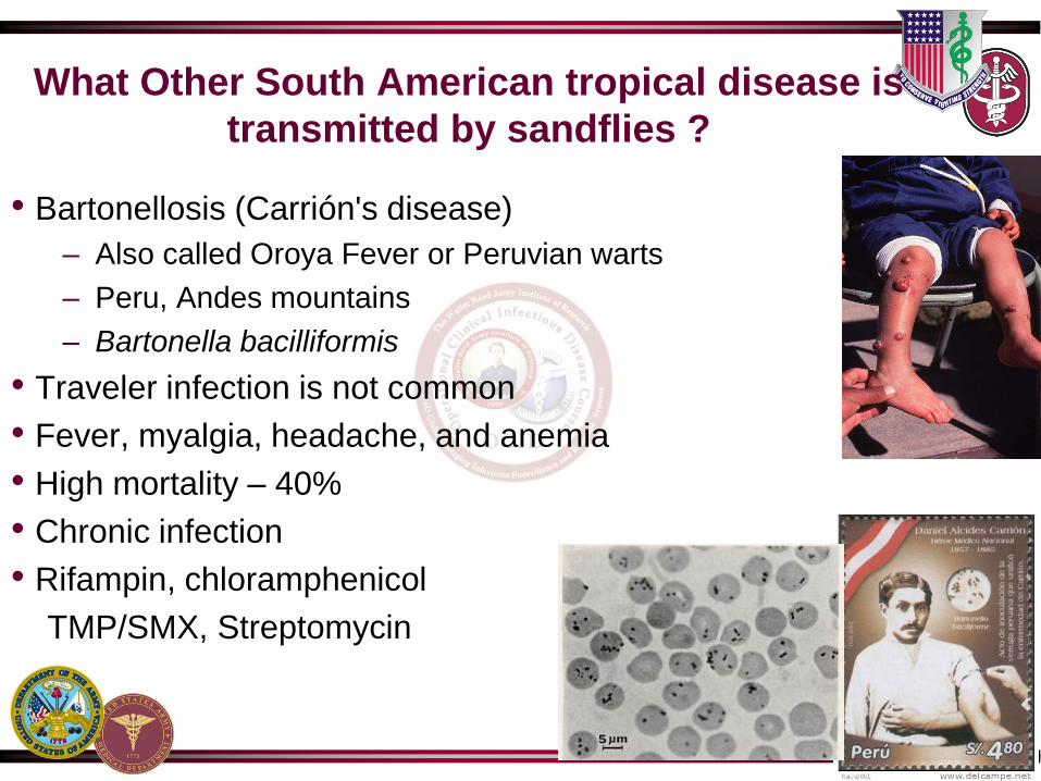
What Other South American tropical disease is
transmitted by sandflies ?
• Bartonellosis (Carrión's disease)
– Also called Oroya Fever or Peruvian warts
– Peru, Andes mountains
– Bartonella bacilliformis
• Traveler infection is not common
• Fever, myalgia, headache, and anemia
• High mortality – 40%
• Chronic infection
• Rifampin, chloramphenicol
TMP/SMX, Streptomycin


