K S I N G H Department of Dermatology & Venereology , All India Institute of
Medical Sciences, Ansari Nagar , New Delhi- l l0029, India*
Accepted for publication 1 5 April 1 986
Summary A 45-year-old male patient suffering from borderl ine lepromatous leprosy with reaction, developed round or i rregular, we\ 1 defined, large tense bu\1ae on exist ing leprosy lesions. There was deposit ion of I gG , IgM, IgA and fibrin along the basement membrane. It was not a bu\ 1ous drug eruption due to ei ther rifampicin , dapsone or clofazimine, but a component of leprosy reaction . Difficulties in classifying as either Type I or Type I I reaction are discussed .
Bullous eruptions are rarely observed in leprosy patients . Generalized bullous drug eruptions due to rifampicin I and DDS2 have been reported . Vesicles and bullae have been observed in purpuric, tender erythematous plaques of Lucio phenomenon and in the evanascent , tender erythematous skin nodules called erythema nodosum leprosum (ENL) lesions which may occur as a part of Type I I leprosy reactions . 3
Type I leprosy reactions occur in borderline leprosy . Existing lesions become more red and oedematous . Systemic disturbances are rare, though the patients may develop oedema of hands, feet or face. On the other hand in Type I I reactions, seen i n borderline lepromatous (BL) and lepromatous leprosy (LL), systemic disturbances are usual and ENL lesions may occur, but there is no change whatsoever in the appearance of leprosy lesions . 3
Case report
A 45-year-old, male, gave a 4-year history of a progressively enlarging hypopigmented hypo-aesthetic patch over the left side of the chest and a
• Present address: Farasan General H ospital, Farasan, Gizan, K ingdom of Saudi Arabia
stationary patch over the left thigh . Two months earlier, he had suddenly developed multiple erythematous plaq ues over the body, paraesthesia over hands, forearms, shins and face, oedema of hands and feet and pain in wrist and ankle j oints . This was not associated with fever, malaise or drug intake.
On examination, there were numerous, asymmetrically, distributed, rai sed erythematous, plaques with smooth stretched surface and grad ually sloping borders. The margins were indistinct in places . Two of these plaques were large, being 1 2 cm in diameter, whereas most others were smal l , varying between I and 3 cm in diameter . Hair loss and hypo-aesthesia were noted only in the larger lesions . There was glove and stocking type of anesthesia . Ulnar, lateral popli teal and radial nerves on both sides were thickened and tender. Axil lary and inguinal lymph nodes were enlarged , discrete, mobile and non-tender. Oedema of hands and feet was gross. Systemic examination was unremarkable.
I nvestigations revealed Hb 1 3 · 2 gm dl - I , total leukocyte count 5200 cu mm - I , neutrophils 60% , lymphocytes 3 2 % , eosinophils 4 % , monocytes 4% and erythrocyte sedimentation rate by Westergrens method was 23 mm in one hour. No abnormality was detected in urine and stool examination . Blood chemistry revealed random glucose 60 g d l - I , urea 1 6 mg dl - I , creatinine 1 -4 mg dl - I , sodium 1 40 mEq L - I , potassium 3 - 4 mEq L - I , albumin 3 · 6 g dl - I , globulin 4 · 3 g dl - I , total bil irubin 1 · 6 mg dl - I , alkaline phosphatase 3 · 3 I U dl - I , glutamic oxaloacetic transaminase 60 units ml - I and glutamic pyruvic transaminase 45 units ml - I . Chest X-ray examination was normal . Histopathology of lesions revealed normal epidermis, a Grenz zone and loose foamy macrophage granulomas scattered throughout the oedematous dermis . There were numerous lymphocytes and occasional foreign body giant cel ls . Nerves had onion-skin perineurium. Ziehl -Neelsen staining showed numerous (5 + ) granular acid-fast baci l l i (AFB) .
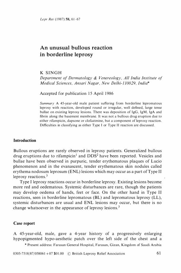
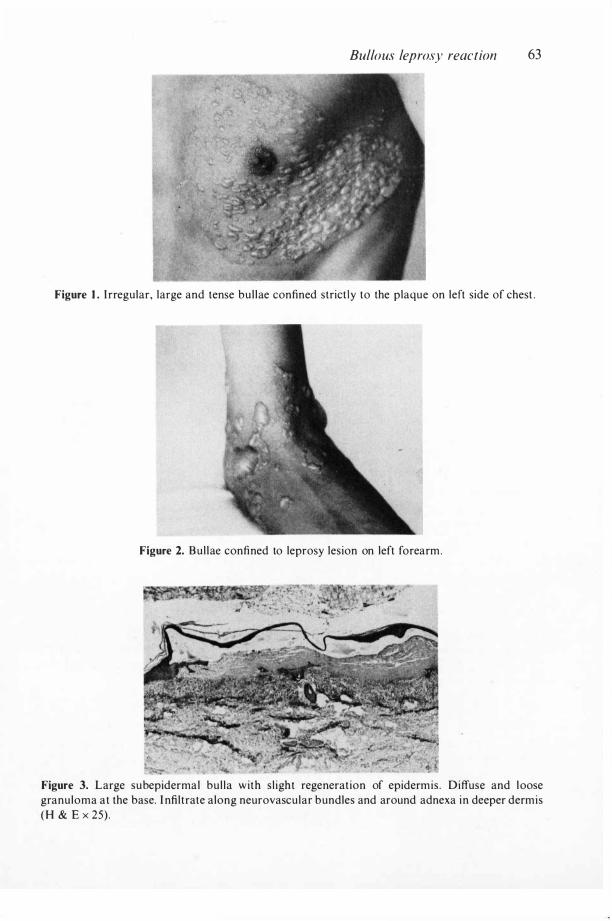
At this stage the patient was thought to have BL leprosy in Type I reaction. He was put on multidrug regimen with rifampicin 600 mg, clofazimine 300 mg, DDS 1 00 mg and prednisolone 30 mg daily . The reaction subsided mildly over the first few days, but worsened between the 1 2th to 1 4th day. At this time numerous, round to i rregular, well defined, tense bullae measuring 5-20 mm in diameter appeared over all the leprosy lesions ( Figures 1 and 2). M ost of them contained clear fluid though some were haemorrhagic . A few small lesions occurred over soft palate.
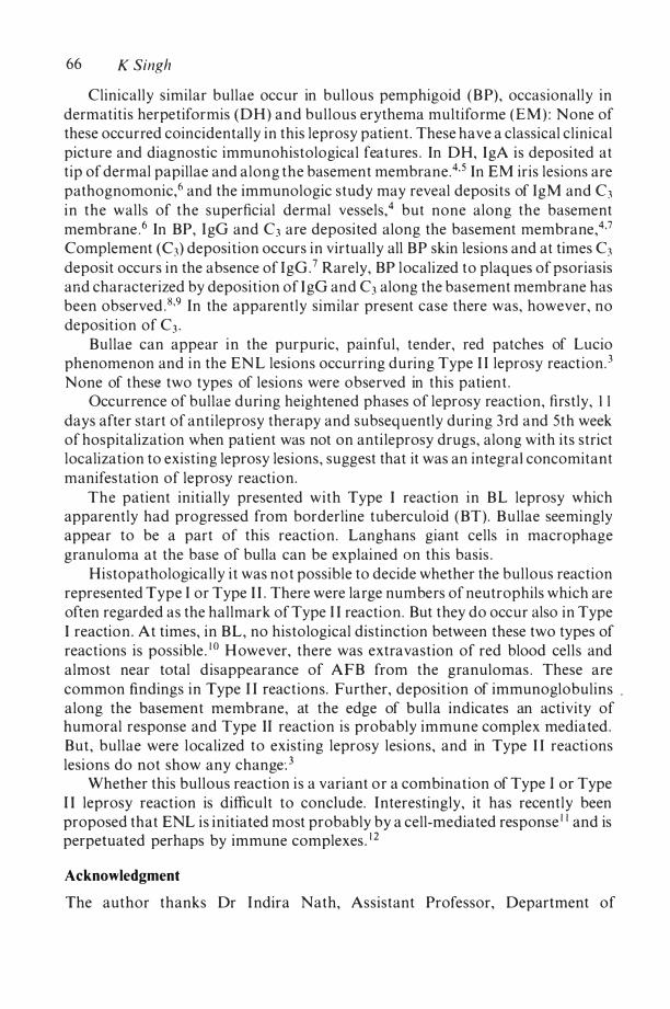
The patient was hospitalized and repeat haemogram and blood chemistry done at this time were essential ly similar to that at presentation. However improvement of liver function tests was observed . The bul la fluid contained polymorphs and occasional eosinophil s . Acantholytic cell s and AFB were not seen . Histopathology of the bulla (Figures 3-6) showed a location in the subepidermis . It contained fibrin , neutrophil s , red blood cells ( RBC) and occasionally plasma cells and eosinophils but no acantholytic cel l s . At the base of bul la and in the dermis , there were foamy macrophage granulomas, with a few
Bullous leprosy reaction 63
Figure 1 . I rregular, large and tense bul lae confined strict ly to the plaque on left side of chest.
Figure 2. Bul lae confined to leprosy lesion on left forearm .
Figure 3. Large subepidermal bul la with s l ight regeneration of epidermis . Diffuse and loose granuloma at the base. I nfiltrate along neurovascular bundles and around adnexa in deeper dermis ( H & E x 25) .
64 K Singh
Figure 4. Marked oedema of upper dermis and fibrin deposit ion . Dilated vascular spaces . Dense infiltrate with few langhan giant ce\1s (H & E x 72 · 5) .
Figure 5. Dilated vessels, extravasation of R BC, neutrophiles, n uclear dust , lymphocytes, langhan gian t ce\ 1s , vacuolated macrophages and oedema of dermis at the base of bu\ 1a (H&E x 250) .
epithelioid cells and lymphocytes, occasional Langhans and foreign body giant cel ls . Significantly the granuloma was heavily infiltrated by neutrophils, some showing karyorrhexis , with a few eosinophils and plasma cel ls . There was mild extravasatio n of RBCs. AFB were 2 + and fragmented . At the edge of bulla, indirect immunofluorescence of cryostat sections showed positive staining for IgM, IgG, I gA and fibrin but no C3 along the basement membrane .
At this time, antileprosy drugs were stopped, while prednisolone was
Bullous leprosy reaction 65
Figure 6. I nfiltrate of vacuolated macrophages, lymphocytes, neutrophi ls along neurovascular bundles i n lower dermis (H&E x 250) .
increased to �O mg per day. Within 3 days the bullous eruption was control led and the patient improved grad ually . By 4 weeks prednisolone could be reduced to 1 0 mg/day. I n the 3 rd and 5th week the patient developed occasional small bullae along with mild exacerbation of reactio n. They subsided spontaneously without any further treatment . Histopathology o f bullae were identical to that examined at the time of hospitalization .
During the 6th week provocation tests were conducted . Rifampicin 600 mg/ day was introduced fol lowed 3 days later by DDS 1 00 mg/day. Though there were no drug eruptions, including bullous eruption, the patient developed arthralgia and swell ing of vestibule and inferior turbinate leading to difficulty in breathing. Investigations including histopathology were essentially similar to that observed at time of presentation. H owever, the foam cell granulomas had scant lymphocytes, mild fibrosis and 2 + A F B .
Rifampicin w a s withdrawn and prednisolone w a s increased to 6 0 mg/day, whereupon there was improvement . On reintroduction of rifampicin the patient developed similar symptoms within 24 hours, and thereafter rifampicin was avoided . I ntroduction of clofazimine 300 mg/day led to moderate improvement. , Chloroquine phosphate 250 mg twice daily was introduced in the 1 2th week . Over the succeeding 3 weeks there was rapid improvement and the steroids were withdrawn without further deterioration in the clinical state .
Discussion
Large, tense, histologically subepidermal bullae with deposition of I gM , I gG, IgA and fibrin but no complement along the basement membrane, were confined almost exclusively to leprosy lesions . It was not a drug eruption as provocative tests with rifampicin, clofazimine and DDS did not reproduce bullae.
66 K Singh Clinical ly s imilar bullae occur in bul lous pemphigoid ( B P) , occasionally in
dermati t is herpetiformis ( D H ) and bul lous erythema mult ifo rme ( E M ) : N one of these occurred coincidentally in this leprosy patient . These have a classical cl inical pict ure and diagnostic immunohistological feat u res . In DH, I gA is deposi ted at t ip of dermal papillae and a long the basemen t membrane.4, 5 In E M ir is lesions are pathognomonic,6 and the immunologic s tudy may reveal deposi ts of I g M and C3 i n the walls of the superficial dermal vessels,4 but none along the basement membrane .6 In BP, I gG and C3 a re deposi ted along the basement membra ne,4,7 Complement (C3) deposi t ion occurs i n virtual ly all B P skin lesions and at times C3 deposit occurs in the absence of IgG . 7 Rarely, BP l ocalized t o plaq ues of psoriasis and characterized by deposi t ion of I gG and C3 along the basement membrane has been observed . 8,9 In the apparently similar present case there was, however, no deposit ion of C3 .
Bul lae can appear i n the purpuric, painful , tender, red patches of Lucio phenomenon and i n the EN L lesions occurring d u ring Type I I leprosy react ion . 3
N one of these two types of lesions were observed in this patien t . Occurrence of bul lae duri ng heightened phases of leprosy reacti on, fi rstly, I I
days after start of ant i leprosy therapy and subseq uently during 3 rd a nd 5th week of hospitalization when pa tient was not on anti leprosy drugs, along with i ts s trict localization to existing leprosy lesions, suggest that it was an integra l concomitant manifestat ion of leprosy reacti o n .
T h e patient init ial ly presented w i t h Type I react ion i n B L leprosy which apparently had progressed from borderline tuberculoid ( BT) . B u llae seemi ngly appear to be a part of this reactio n . Langhans giant cells i n macrophage granuloma at the base of bulla can be explai ned on this basis .
Histopathologically i t was n o t possible to decide whether the bul lous reaction represented Type I or Type I I . There were la rge numbers of neutrophils which are often regarded as the hal lmark of Type I I reacti o n . But t hey do occur also in Type I reactio n . At times, in B L , no histological distinction between these two types of reactions is possible . l O H owever, there was extravastion of red blood cel ls and almost near total d isappearance of A F B from the granulomas . These are common findings i n Type I I reactions . Further, deposit ion of immunoglobul ins , along the basemen t membrane, at the edge of bul la indicates an activity of humoral response and Type I I react ion is probably immune complex mediated . But , bul lae were l ocalized t o existing leprosy les ions, and in Type I I reacti ons lesions do not show any change: 3
Whether this bul lous react ion i s a variant o r a combination of Type l or Type I I leprosy reaction is difficult to conclude. I nterestingly, it has recently been proposed that ENL is i n itiated most probably by a cell-mediated response l l and is perpetuated perhaps by immune complexes . l 2
Acknowledgment
The author thanks Dr I ndira Nath, Assistant Professor, Department o f
Bullous leprosy reaction 67
Pa thology, A I M S, for her invaluable assistance and guidance. H e also thanks Prof U N Bhuyan, Depa rtment of Pathology, A I I M S for immunofluorescent eva l uations .
References
I Nigam P, Daya l SG, Goyal B M . Eryt hema mul t iforme bul losum due to ri fampicin . Lepr India,
1 979; 5 1 : 249-5 J . 2 Dutta R K , Erythema mul t iforme bul losum Que to dapsone. Lepr India, 1 980; 52: 306-9 . 3 Jopl ing W H o Leprosy reactions ( Reactional states) . I n : Handbook of Leprosy. London: Wil l iam
Heinemann Medical Books Ltd (2nd ed . ) , 1 978; 66-74. 4 Lever WF, Schaumburg- Lever G. Non infectious vesicular and bul lous diseases. I n : Histopatho
logy of the skin . Philadelphia : J B Lippincott Company, 1 983 ; 92- 1 35 . l Katz S I , Marks J M . Dermatit is herpetiformis . I n : Dermatology in General Medicine . Fitzpatrick
TB, Eiren AZ, Wolff et al. (eds), New York : McGraw-Hi l i Book Company (2nd ed . ) , 1 979; 326-30.
6 White JW, Jr . Hypersensit ivi ty and miscel laneous inflammatory disorders. In: Dermatology.
Moschel la SL, Hurley HJ (eds), Philadelphia : W B Saunders Company (2nd ed . ), 1 985 ; 464-98.
7 Jorden RE . Bul lous pemphigoid, cicatricial pemphigoid and chronic bul lous dermatosis of chi ldhood . In: Dermatology in General Medicine . Fitzpatrick TB, Eiren AZ, Wolff K et al .
(eds), New York : McGraw-Hi l i Book Company (2nd ed . ) , 1 979; 3 1 8-23 . 8 Abel EA, Bennett A . Bul lous pemphigoid . Occurrence in psoriasis treated with psoralens plus
long-wave ul traviolet radiation . A rch Dermatol, 1 979; 1 1 5: 988-9. 9 Person JR , Rogers I I I RS . Bul lous pemphigoid and psoriasis : does subcl in ical bul lous
pemphigoid exist? Brit J Derm, 1 976; 95: 535-40. 1 0 Ridley OS. React ions, I n : Sk in Biopsy in Leprosy. Basic: Documenta Geigy, 1 977; 47-52 . I I Laal S, Bhutani LK, Nath I. The natural emergence of ant igen react ive T cel ls in lepromatous
leprosy pat ients during erythema nodosum leprosy. Infec Immunity, accepted for publicat ion . 1 2 Mshana RN. Hypothesis : Erythema nodosum leprosum is precipita ted by an imbalance of T