25
Lesson 7 – Cell Membranes and the Cytoskeleton
| Date post: | 24-Dec-2015 |
| Category: |
Documents |
| Upload: | dora-morrison |
| View: | 223 times |
| Download: | 3 times |

Lesson 7 – Cell Membranes and the Cytoskeleton

Hank – in da club
Crash Course Biology - Cell membranes

Cell membranes
On your desks draw out the structure of the plasma membrane of a eukaryotic cell.

Cell MembraneFrom now on we will refer to the exterior cell membrane as the plasma membrane as it is more descriptive of its nature.
The plasma membrane separates the contents of the cell from its environment. Membranes within the cell are also used to create compartments
Functions of MembranesThere are 6 main functions of membranes in cells:
1. Providing selectively permeable barriers2. Compartmentalisation
3. Localising reactions in the cell
4. Transport of solutes
5. Signal Transduction
6. Cell-Cell Recognition
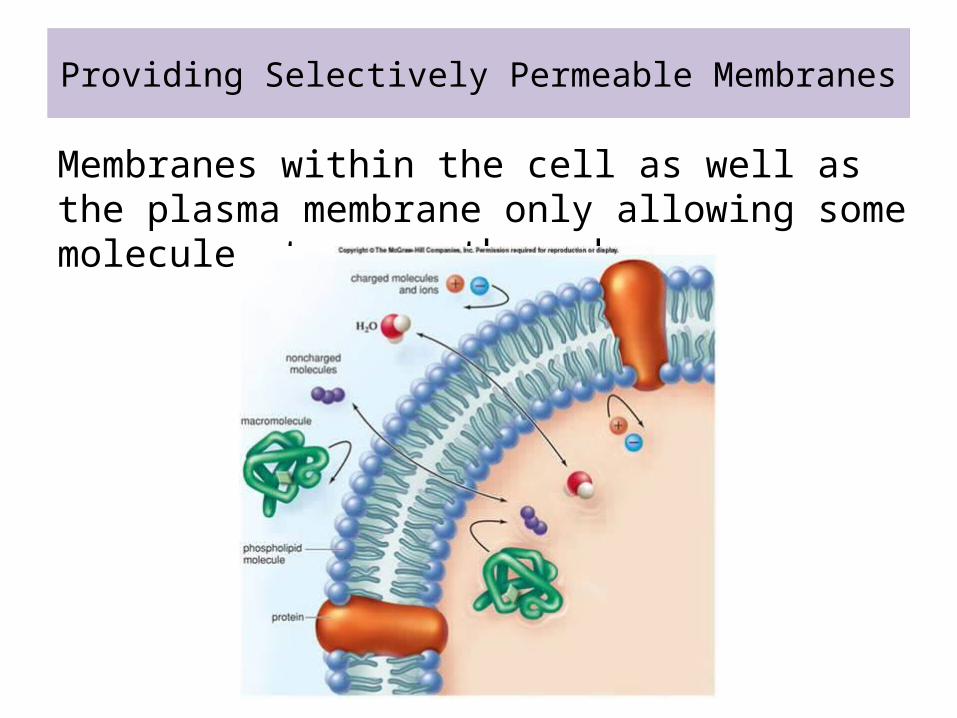
Providing Selectively Permeable Membranes
Membranes within the cell as well as the plasma membrane only allowing some molecules to pass through.

Compartmentalisation
Membranes help to divide up areas within eukaryotic cells. Membranes are used for the nuclear envelope, endoplasmic reticulum, Golgi apparatus, mitochondria, and chloroplasts.
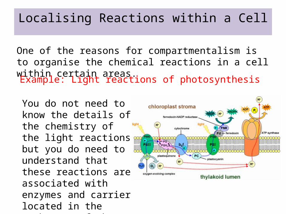
Localising Reactions within a Cell
One of the reasons for compartmentalism is to organise the chemical reactions in a cell within certain areas.Example: Light reactions of photosynthesis
You do not need to know the details of the chemistry of the light reactions but you do need to understand that these reactions are associated with enzymes and carrier located in the membranes of the chloroplast.
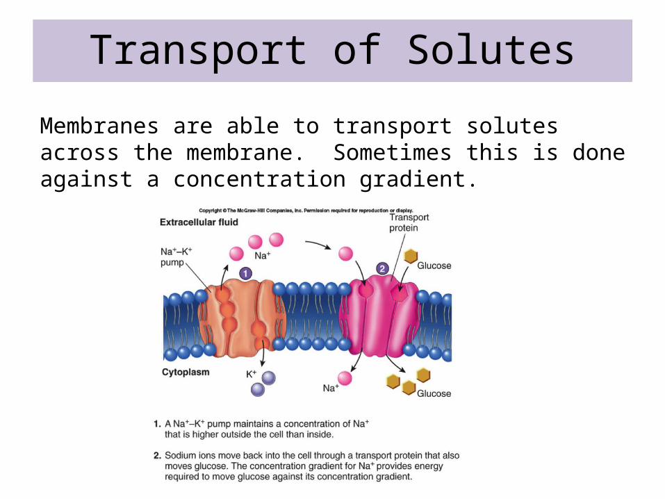
Transport of Solutes
Membranes are able to transport solutes across the membrane. Sometimes this is done against a concentration gradient.

Signal TransductionThere are receptors found on the surface of the cell membrane that can recognise and respond to the presence of specific molecules.

Cell-Cell Recognition
Molecules on the surface of the cell are unique to the cell and can be used for cell-cell recognition.
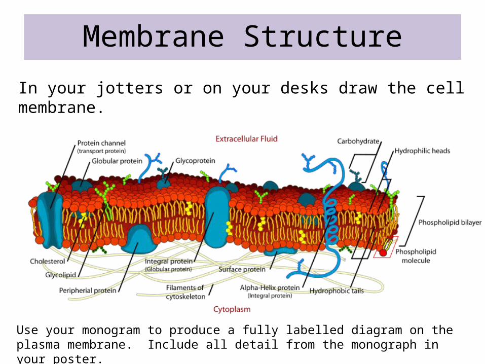
Membrane Structure
In your jotters or on your desks draw the cell membrane.
Use your monogram to produce a fully labelled diagram on the plasma membrane. Include all detail from the monograph in your poster.
Function of Membrane Proteins
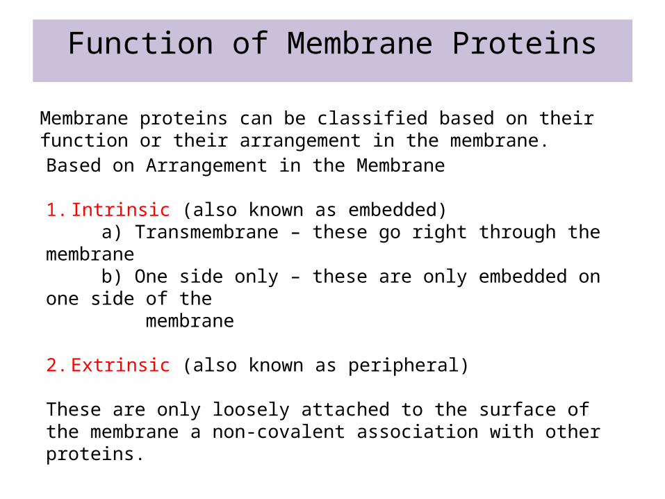
Membrane proteins can be classified based on their function or their arrangement in the membrane.Based on Arrangement in the Membrane
1. Intrinsic (also known as embedded) a) Transmembrane – these go right through the membrane b) One side only – these are only embedded on one side of the membrane
2. Extrinsic (also known as peripheral)
These are only loosely attached to the surface of the membrane a non-covalent association with other proteins.

Extrinsic Proteins
Function of Membrane Proteins
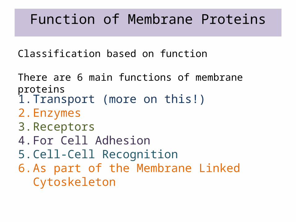
Classification based on function
There are 6 main functions of membrane proteins
1. Transport (more on this!)2. Enzymes3. Receptors4. For Cell Adhesion5. Cell-Cell Recognition6. As part of the Membrane Linked
Cytoskeleton
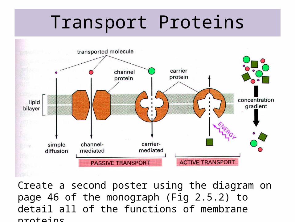
Transport Proteins
Create a second poster using the diagram on page 46 of the monograph (Fig 2.5.2) to detail all of the functions of membrane proteins.

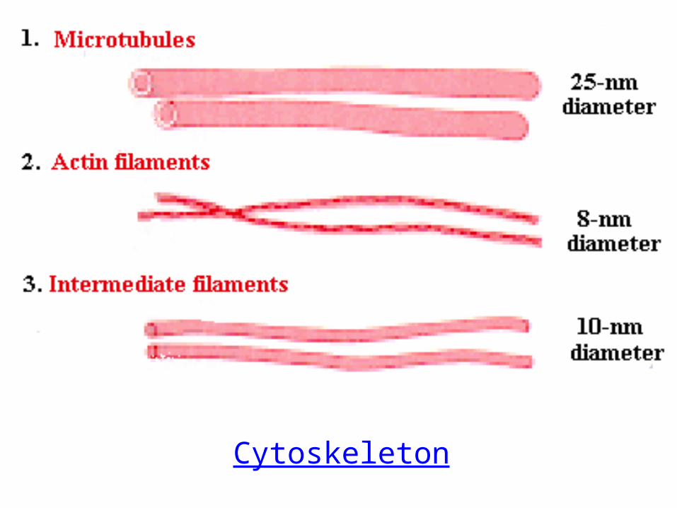
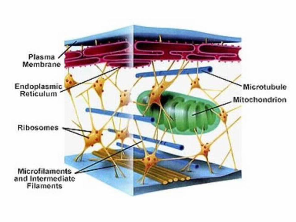
The Cytoskeleton
In eukaryotes there is an internal structure consisting of actin filaments, intermediate filaments and microtubules that support the internal structure of the cell.

Microtubules
Microtubules are hollow tubes made up of the protein tubulin. Heterodimers (one α-tubulin and one β-tubulin per dimer) are arranged as a set of 13 protofilaments to make a microtubule.
You will recall the importance of microtubules are important as they make up the spindle fibre network crucial in cell division. They are also responsible for moving components around in the cell.
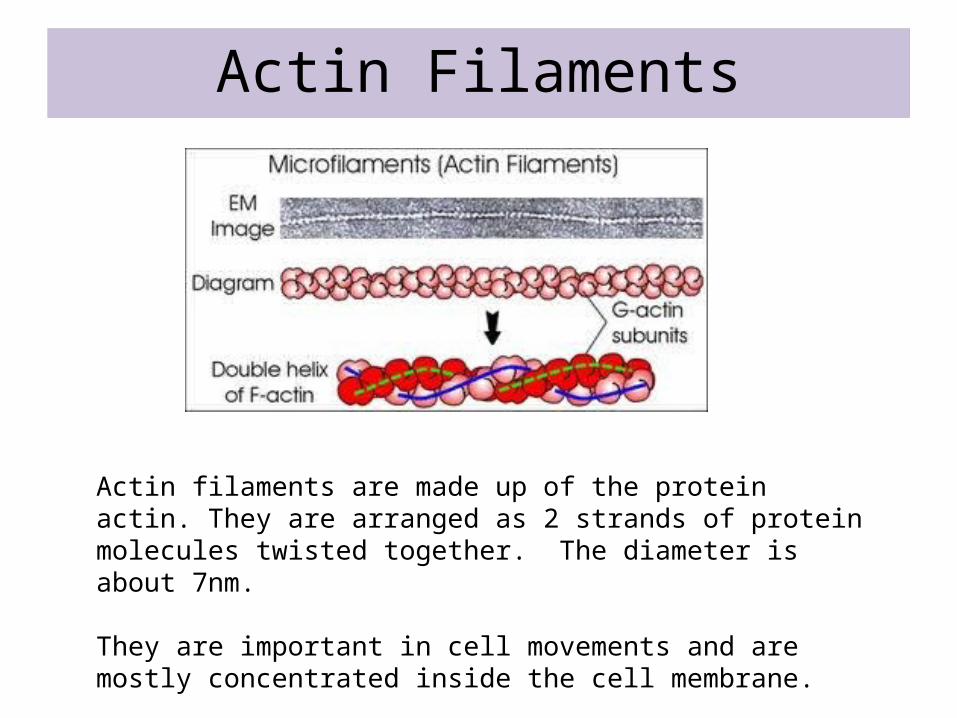
Actin Filaments
Actin filaments are made up of the protein actin. They are arranged as 2 strands of protein molecules twisted together. The diameter is about 7nm.
They are important in cell movements and are mostly concentrated inside the cell membrane.
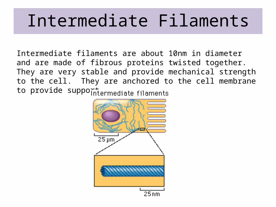
Intermediate Filaments
Intermediate filaments are about 10nm in diameter and are made of fibrous proteins twisted together. They are very stable and provide mechanical strength to the cell. They are anchored to the cell membrane to provide support.

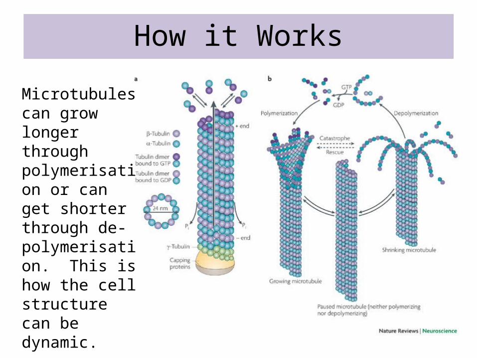
How it Works
Microtubules can grow longer through polymerisation or can get shorter through de-polymerisation. This is how the cell structure can be dynamic.
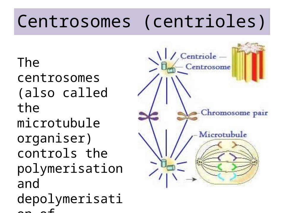
Centrosomes (centrioles)
The centrosomes (also called the microtubule organiser) controls the polymerisation and depolymerisation of microtubules.