The upper respiratory system includes the nasal cavity and paranasal sinuses, pharynx and larynx. This system allows the passage of both air and food to their respective destinations (an aerodiges- tive system). The main function of the larynx, besides producing voice, is to prevent the entry of food intro the trachea and lung through multiple sphincter mechanisms. Tumors of the aerodigestive tract are grouped together because of their common etiologic fac- tors, histologic profile and lines of treatment. As a group, they constitute about 55% of head and neck cancer (pharynx 25%, larynx 25% and si- nonasal structures 5%). The sinonasal structures include the nasal cavity and the paranasal sinuses. The nasal cavity extends from the nose anteriorly to about the end of hard palate posteriorly where it communicates with nasopharynx through the choanae (Fig 9-1). The landmarks of the three main regions of the pharynx are demonstrated in (Fig 9-1). The nasopharynx (postnasal space or epipharynx) lies above the soft palate which forms its floor. The oropharynx (mesopharynx) extends from the lower border of soft palate to the lingual surface of epiglottis. On each side of the glossoepiglot- tic fold lies the vallecula. The hypopharynx (laryngopharynx) extends from the tip of epiglot- tis to the lower border of cricoid cartilage opposite the body of the sixth cervical vertebra where the esophagus begins. The regional lymph nodes of the head include 4 main groups, namely: the buccal, parotid, occipi- tal and retropharyngeal (Fig 9-2). The latter group is commonly forgotten due to its hidden impalpa- ble condition. Fig 9-1 Anatomic landmarks of the three components of upper respiratory system: the sinonasal structures, pharynx and larynx. Fig 9-2 Regional lymph nodes of head and neck. Head: (1) buccal, (2) parotid (3) occipital and (4) retropharyngeal. Neck lymph nodes: IA submental IB submandibular, II upper jugular, III midjugular, IV lower jugular, V posterior triangle, (A) upper and (B) lower, VI anterior central, and VII upper mediastinal. Pathology of Cancer El Bolkainy et al 5th edition, 2016
Transcript
The upper respiratory system includes the nasal cavity and paranasal sinuses, pharynx and larynx. This system allows the passage of both air and food to their respective destinations (an aerodiges-tive system). The main function of the larynx, besides producing voice, is to prevent the entry of food intro the trachea and lung through multiple sphincter mechanisms.
Tumors of the aerodigestive tract are grouped together because of their common etiologic fac-tors, histologic profile and lines of treatment. As a group, they constitute about 55% of head and neck cancer (pharynx 25%, larynx 25% and si-nonasal structures 5%).
The sinonasal structures include the nasal cavity and the paranasal sinuses. The nasal cavity
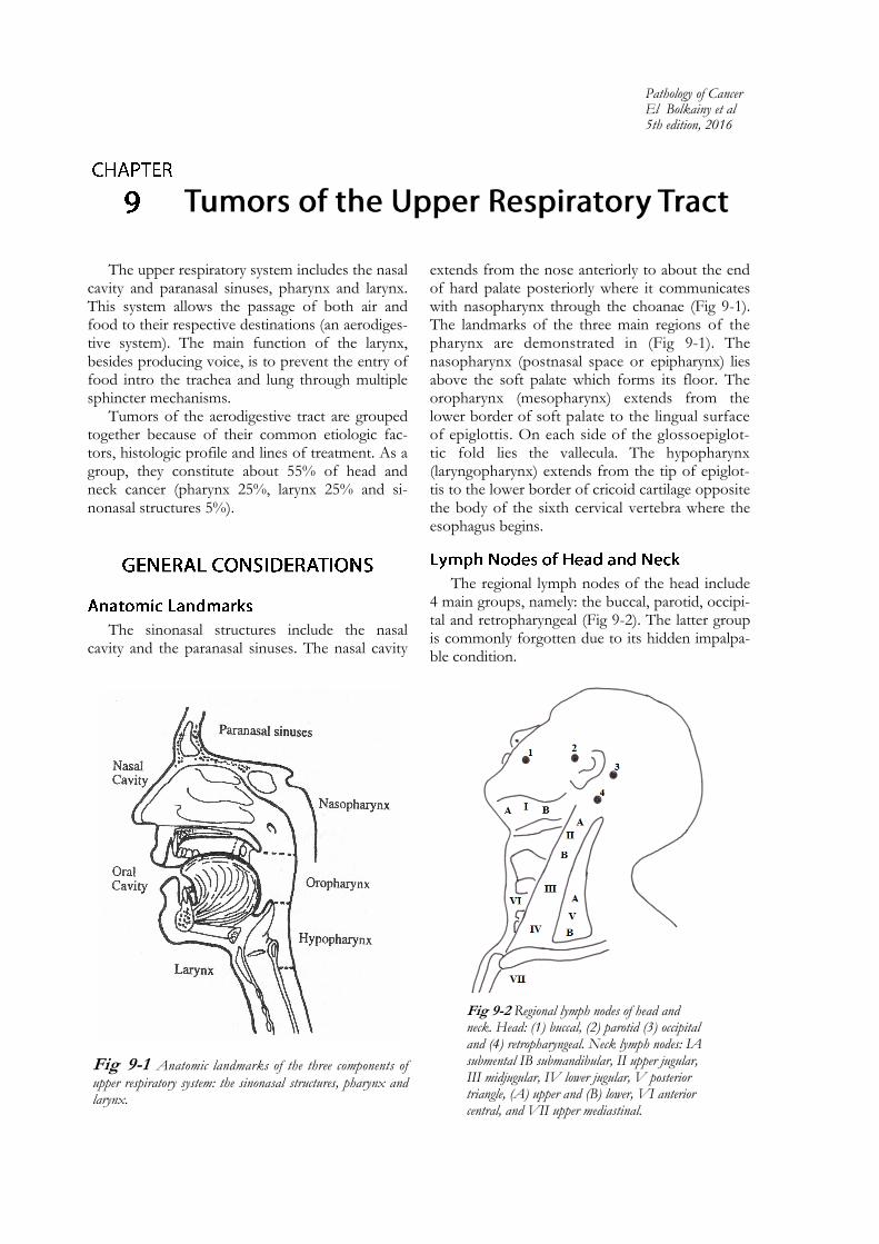
extends from the nose anteriorly to about the end of hard palate posteriorly where it communicates with nasopharynx through the choanae (Fig 9-1). The landmarks of the three main regions of the pharynx are demonstrated in (Fig 9-1). The nasopharynx (postnasal space or epipharynx) lies above the soft palate which forms its floor. The oropharynx (mesopharynx) extends from the lower border of soft palate to the lingual surface of epiglottis. On each side of the glossoepiglot-tic fold lies the vallecula. The hypopharynx (laryngopharynx) extends from the tip of epiglottis to the lower border of cricoid cartilage opposite the body of the sixth cervical vertebra where the esophagus begins.
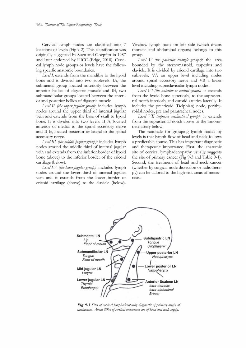
The regional lymph nodes of the head include 4 main groups, namely: the buccal, parotid, occipi-tal and retropharyngeal (Fig 9-2). The latter group is commonly forgotten due to its hidden impalpa-ble condition.
Fig 9-1 Anatomic landmarks of the three components of
upper respiratory system: the sinonasal structures, pharynx and larynx.
Fig 9-2 Regional lymph nodes of head and neck. Head: (1) buccal, (2) parotid (3) occipital and (4) retropharyngeal. Neck lymph nodes: IA submental IB submandibular, II upper jugular, III midjugular, IV lower jugular, V posterior triangle, (A) upper and (B) lower, VI anterior central, and VII upper mediastinal.
Pathology of Cancer El Bolkainy et al 5th edition, 2016
162 Tumors of The Upper Respiratory Tract
Cervical lymph nodes are classified into 7 locations or levels (Fig 9-2). This classification was originally suggested by Suen and Goepfert in 1987 and later endorsed by UICC (Edge, 2010). Cervi-cal lymph node groups or levels have the follow-ing specific anatomic boundaries:
Level I: extends from the mandible to the hyoid bone and is divided into two sublevels: IA, the submental group located anteriorly between the anterior bellies of digastric muscle and IB, two submandibular groups located between the anteri-or and posterior bellies of digastric muscle.
Level II (the upper jugular group): includes lymph nodes around the upper third of internal jugular vein and extends from the base of skull to hyoid bone. It is divided into two levels: II A, located anterior or medial to the spinal accessory nerve and II B, located posterior or lateral to the spinal accessory nerve.
Level III (the middle jugular group): includes lymph nodes around the middle third of internal jugular vein and extends from the inferior border of hyoid bone (above) to the inferior border of the cricoid cartilage (below).
Level IV (the lower jugular group): includes lymph nodes around the lower third of internal jugular vein and it extends from the lower border of cricoid cartilage (above) to the clavicle (below).
Virchow lymph node on left side (which drains thoracic and abdominal organs) belongs to this group.
Level V (the posterior triangle groups): the area bounded by the sternomastoid, trapezius and clavicle. It is divided by cricoid cartilage into two sublevels: VA an upper level including nodes around spinal accessory nerve and VB a lower level including supraclavicular lymph nodes.
Level VI (the anterior or central group): it extends from the hyoid bone superiorly, to the supraster-nal notch interiorly and carotid arteries laterally. It includes the precricoid (Delphian) node, perithy-roidal nodes, pre and paratracheal nodes.
Level VII (superior mediastinal group): it extends from the suprasternal notch above to the innomi-nate artery below.
The rationale for grouping lymph nodes by levels is that lymph flow of head and neck follows a predictable course. This has important diagnostic and therapeutic importance. First, the anatomic site of cervical lymphadenopathy usually suggests the site of primary cancer (Fig 9-3 and Table 9-1). Second, the treatment of head and neck cancer (whether by surgical node dissection or radiothera-py) can be tailored to the high-risk areas of metas- tasis.
Fig 9-3 Sites of cervical lymphadenopathy diagnostic of primary origin of carcinomas. About 80% of cervical metastases are of head and neck origin.
Pathology of Cancer 2016, El Bolkainy et al 163
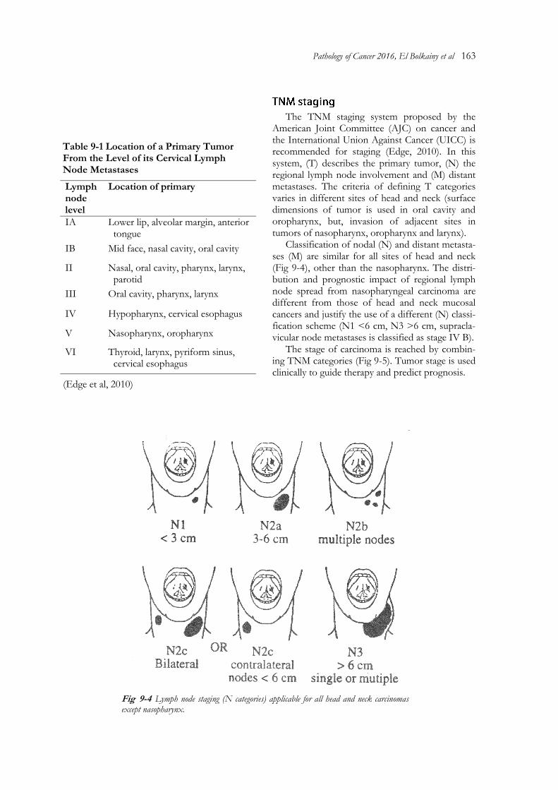
Fig 9-4 Lymph node staging (N categories) applicable for all head and neck carcinomas except nasopharynx.
The TNM staging system proposed by the American Joint Committee (AJC) on cancer and the International Union Against Cancer (UICC) is recommended for staging (Edge, 2010). In this system, (T) describes the primary tumor, (N) the regional lymph node involvement and (M) distant metastases. The criteria of defining T categories varies in different sites of head and neck (surface dimensions of tumor is used in oral cavity and oropharynx, but, invasion of adjacent sites in tumors of nasopharynx, oropharynx and larynx).
Classification of nodal (N) and distant metasta-ses (M) are similar for all sites of head and neck (Fig 9-4), other than the nasopharynx. The distri-bution and prognostic impact of regional lymph node spread from nasopharyngeal carcinoma are different from those of head and neck mucosal cancers and justify the use of a different (N) classi-fication scheme (N1 <6 cm, N3 >6 cm, supracla-vicular node metastases is classified as stage IV B).
The stage of carcinoma is reached by combin-ing TNM categories (Fig 9-5). Tumor stage is used clinically to guide therapy and predict prognosis.
Lymph node level
Location of primary
IA Lower lip, alveolar margin, anterior tongue
IB Mid face, nasal cavity, oral cavity
II Nasal, oral cavity, pharynx, larynx, parotid
III Oral cavity, pharynx, larynx
IV Hypopharynx, cervical esophagus
V Nasopharynx, oropharynx
VI Thyroid, larynx, pyriform sinus, cervical esophagus
Table 9-1 Location of a Primary Tumor From the Level of its Cervical Lymph Node Metastases
(Edge et al, 2010)
164 Tumors of The Upper Respiratory Tract
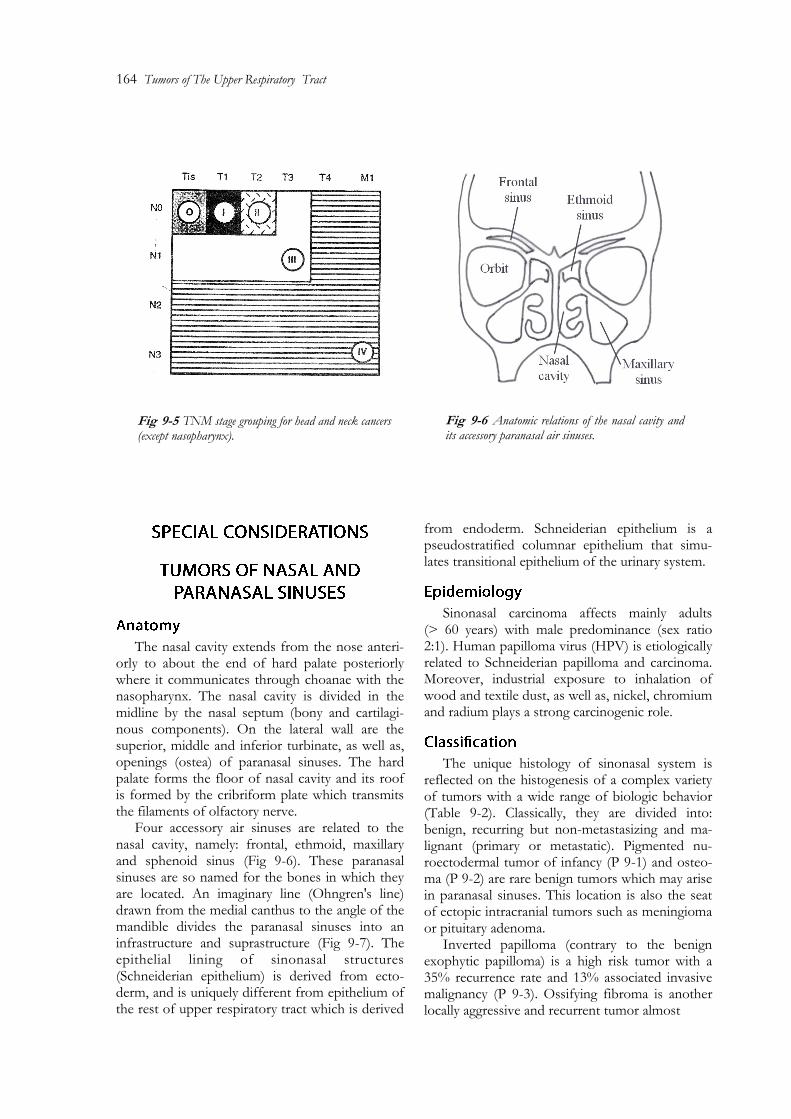
The nasal cavity extends from the nose anteri-orly to about the end of hard palate posteriorly where it communicates through choanae with the nasopharynx. The nasal cavity is divided in the midline by the nasal septum (bony and cartilagi-nous components). On the lateral wall are the superior, middle and inferior turbinate, as well as, openings (ostea) of paranasal sinuses. The hard palate forms the floor of nasal cavity and its roof is formed by the cribriform plate which transmits the filaments of olfactory nerve.
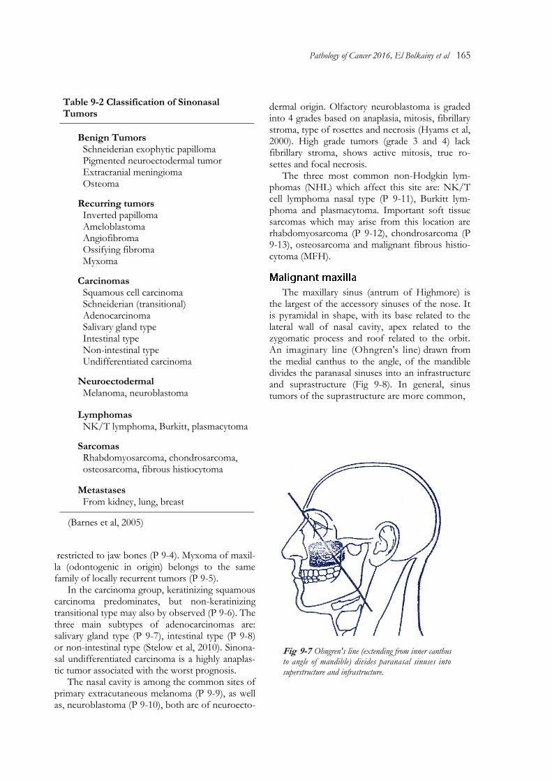
Four accessory air sinuses are related to the nasal cavity, namely: frontal, ethmoid, maxillary and sphenoid sinus (Fig 9-6). These paranasal sinuses are so named for the bones in which they are located. An imaginary line (Ohngren's line) drawn from the medial canthus to the angle of the mandible divides the paranasal sinuses into an infrastructure and suprastructure (Fig 9-7). The epithelial lining of sinonasal structures (Schneiderian epithelium) is derived from ecto-derm, and is uniquely different from epithelium of the rest of upper respiratory tract which is derived
from endoderm. Schneiderian epithelium is a pseudostratified columnar epithelium that simu-lates transitional epithelium of the urinary system.
Sinonasal carcinoma affects mainly adults (> 60 years) with male predominance (sex ratio 2:1). Human papilloma virus (HPV) is etiologically related to Schneiderian papilloma and carcinoma. Moreover, industrial exposure to inhalation of wood and textile dust, as well as, nickel, chromium and radium plays a strong carcinogenic role.
The unique histology of sinonasal system is reflected on the histogenesis of a complex variety of tumors with a wide range of biologic behavior (Table 9-2). Classically, they are divided into: benign, recurring but non-metastasizing and ma-lignant (primary or metastatic). Pigmented nu-roectodermal tumor of infancy (P 9-1) and osteo-ma (P 9-2) are rare benign tumors which may arise in paranasal sinuses. This location is also the seat of ectopic intracranial tumors such as meningioma or pituitary adenoma.
Inverted papilloma (contrary to the benign exophytic papilloma) is a high risk tumor with a 35% recurrence rate and 13% associated invasive malignancy (P 9-3). Ossifying fibroma is another locally aggressive and recurrent tumor almost
Fig 9-5 TNM stage grouping for head and neck cancers (except nasopharynx).
Fig 9-6 Anatomic relations of the nasal cavity and its accessory paranasal air sinuses.
Pathology of Cancer 2016, El Bolkainy et al 165
restricted to jaw bones (P 9-4). Myxoma of maxil-la (odontogenic in origin) belongs to the same family of locally recurrent tumors (P 9-5).
In the carcinoma group, keratinizing squamous carcinoma predominates, but non-keratinizing transitional type may also by observed (P 9-6). The three main subtypes of adenocarcinomas are: salivary gland type (P 9-7), intestinal type (P 9-8) or non-intestinal type (Stelow et al, 2010). Sinona-sal undifferentiated carcinoma is a highly anaplas-tic tumor associated with the worst prognosis.
The nasal cavity is among the common sites of primary extracutaneous melanoma (P 9-9), as well as, neuroblastoma (P 9-10), both are of neuroecto-
dermal origin. Olfactory neuroblastoma is graded into 4 grades based on anaplasia, mitosis, fibrillary stroma, type of rosettes and necrosis (Hyams et al, 2000). High grade tumors (grade 3 and 4) lack fibrillary stroma, shows active mitosis, true ro-settes and focal necrosis.
The three most common non-Hodgkin lym-phomas (NHL) which affect this site are: NK/T cell lymphoma nasal type (P 9-11), Burkitt lym-phoma and plasmacytoma. Important soft tissue sarcomas which may arise from this location are rhabdomyosarcoma (P 9-12), chondrosarcoma (P 9-13), osteosarcoma and malignant fibrous histio-cytoma (MFH).
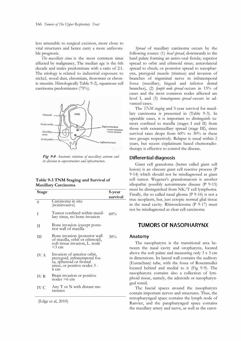
The maxillary sinus (antrum of Highmore) is the largest of the accessory sinuses of the nose. It is pyramidal in shape, with its base related to the lateral wall of nasal cavity, apex related to the zygomatic process and roof related to the orbit. An imaginary line (Ohngren's line) drawn from the medial canthus to the angle, of the mandible divides the paranasal sinuses into an infrastructure and suprastructure (Fig 9-8). In general, sinus tumors of the suprastructure are more common,
Fig 9-7 Ohngren's line (extending from inner canthus to angle of mandible) divides paranasal sinuses into superstructure and infrastructure.
(Barnes et al, 2005)
166 Tumors of The Upper Respiratory Tract
less amenable to surgical excision, more close to vital structures and hence carry a more unfavora-ble prognosis.
The maxillary sinus is the most common sinus affected by malignancy, The median age is the 6th decade and males predominate with a ratio of 2:1. The etiology is related to industrial exposure to nickel, wood dust, chromium, thorotrast or chron-ic sinusitis. Histologically Table 9-2), squamous cell carcinoma predominates (75%).
Spread of maxillary carcinoma occurs by the
following routes: (1) local spread, downwards to the hard palate forming an antro-oral fistula; superior spread to orbit and ethmoid sinus; anterolateral spread to cheek; or posterior spread to nasophar-ynx, pterygoid muscle (trismus) and invasion of branches of trigeminal nerve in infratemporal fossa (maxillary, lingual and inferior dental branches), (2) lymph node spread occurs in 15% of cases and the most common nodes affected are level I, and (3) hematogenous spread occurs in ad-vanced cases.
The TNM staging and 5-year survival for maxil-lary carcinoma is presented in (Table 9-3). In operable cases, it is important to distinguish tu-mors confined to maxilla (stages I and II) from those with extramaxillary spread (stage III), since survival rates drops from 60% to 30% in these two groups respectively. Relapse is usual within 2 years, but recent cisplatinum based chemoradio-therapy is effective to control the disease.
Giant cell granuloma (better called giant cell lesion) is an obscure giant cell reactive process (P 9-14) which should not be misdiagnosed as giant cell tumor. Wegener's granulomatosis is another idiopathic possibly autoimmune disease (P 9-15) must be distinguished from NK/T cell lymphoma. Finally, the so called nasal glioma (P 9-16) is not a true neoplasm, but, just ectopic normal glial tissue in the nasal cavity. Rhinoscleroma (P 9-17) must not be misdiagnosed as clear cell carcinoma
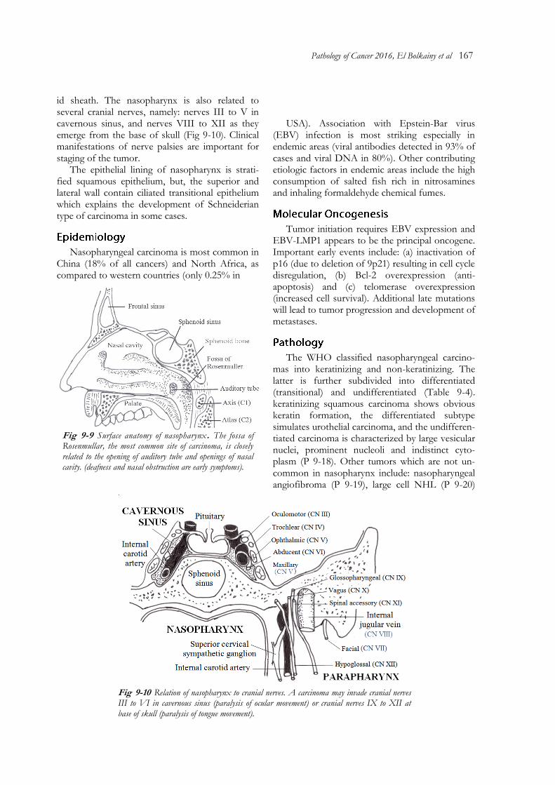
The nasopharynx is the transitional area be-tween the nasal cavity and oropharynx, located above the soft palate and measuring only 3 x 3 cm in dimensions. Its lateral wall contains the auditory (Eustachian) tube, with the fossa of Rosenmuller located behind and medial to it (Fig 9-9). The nasopharynx contains also a collection of lym-phoid tissue, namely, the adenoids or nasopharyn-geal tonsil.
The fascial spaces around the nasopharynx contain important nerves and structures. Thus, the retropharyngeal space contains the lymph node of Ranvier, and the parapharyngeal space contains the maxillary artery and nerve, as well as the carot-
Fig 9-8 Anatomic relations of maxillary antrum and its division to superstructure and infrastructure.
Stage
5-year survival
0 Carcinoma in situ (noninvasive)
I Tumor confined within maxil-lary sinus, no bone invasion
60%
II Bone invasion (except poste-rior wall of maxilla
III Bone invasion (posterior wall of maxilla, orbit or ethmoid), soft tissue invasion, L. node <3 cm
30%
IV A Invasion of anterior orbit, pterygoid, infratemporal fos-sa, sphenoid or frontal sinus, or positive nodes 3 -6 cm
IV B Brain invasion or positive nodes >6 cm
IV C Any T or N with distant me-tastases
Table 9-3 TNM Staging and Survival of Maxillary Carcinoma
(Edge et al, 2010)
Pathology of Cancer 2016, El Bolkainy et al 167
id sheath. The nasopharynx is also related to several cranial nerves, namely: nerves III to V in cavernous sinus, and nerves VIII to XII as they emerge from the base of skull (Fig 9-10). Clinical manifestations of nerve palsies are important for staging of the tumor.
The epithelial lining of nasopharynx is strati-fied squamous epithelium, but, the superior and lateral wall contain ciliated transitional epithelium which explains the development of Schneiderian type of carcinoma in some cases.
Nasopharyngeal carcinoma is most common in China (18% of all cancers) and North Africa, as compared to western countries (only 0.25% in
USA). Association with Epstein-Bar virus
(EBV) infection is most striking especially in endemic areas (viral antibodies detected in 93% of cases and viral DNA in 80%). Other contributing etiologic factors in endemic areas include the high consumption of salted fish rich in nitrosamines and inhaling formaldehyde chemical fumes.
Tumor initiation requires EBV expression and EBV-LMP1 appears to be the principal oncogene. Important early events include: (a) inactivation of p16 (due to deletion of 9p21) resulting in cell cycle disregulation, (b) Bcl-2 overexpression (anti-apoptosis) and (c) telomerase overexpression (increased cell survival). Additional late mutations will lead to tumor progression and development of metastases.
The WHO classified nasopharyngeal carcino-mas into keratinizing and non-keratinizing. The latter is further subdivided into differentiated (transitional) and undifferentiated (Table 9-4). keratinizing squamous carcinoma shows obvious keratin formation, the differentiated subtype simulates urothelial carcinoma, and the undifferen-tiated carcinoma is characterized by large vesicular nuclei, prominent nucleoli and indistinct cyto-plasm (P 9-18). Other tumors which are not un-common in nasopharynx include: nasopharyngeal angiofibroma (P 9-19), large cell NHL (P 9-20)
Fig 9-9 Surface anatomy of nasopharynx. The fossa of Rosenmullar, the most common site of carcinoma, is closely related to the opening of auditory tube and openings of nasal cavity. (deafness and nasal obstruction are early symptoms).
Fig 9-10 Relation of nasopharynx to cranial nerves. A carcinoma may invade cranial nerves III to VI in cavernous sinus (paralysis of ocular movement) or cranial nerves IX to XII at base of skull (paralysis of tongue movement).
168 Tumors of The Upper Respiratory Tract
and chordoma of basiocciput bone (P 9-21).
The spread of carcinoma is by the following three routes: (1) local spread: lateral to pterygoid muscle (trismus) or Eustachian tube; anterior spread to the nasal cavity, upward spread behind the sphenoid to the base of skull (paralysis of cranial nerves II to IV) or along the hypoglossal foramen (cranial nerves XI and XII), jugular and carotid vessel invasion, (2) lymph node metastases occur in 85 to 95% of cases, bilateral in 53%, with affection of all levels especially level V and the retropharyngeal node of Ranvier, and (3) hematogenous spread in 20% of cases to distant locations.
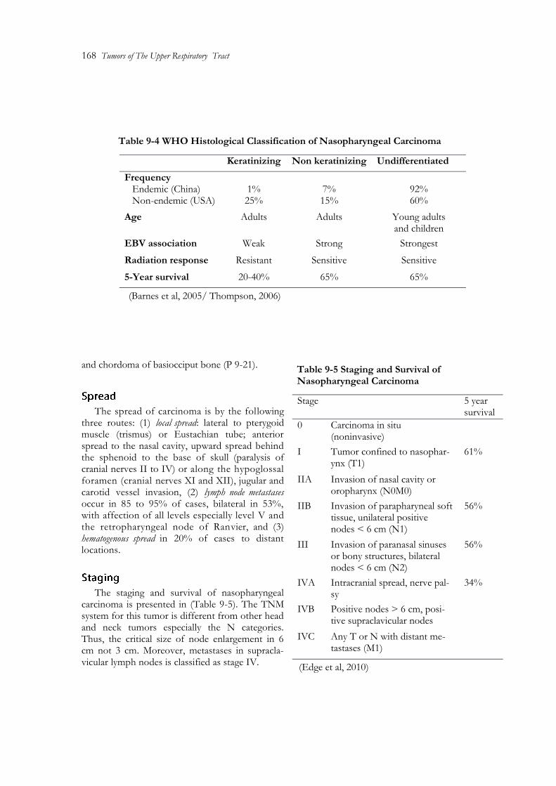
The staging and survival of nasopharyngeal carcinoma is presented in (Table 9-5). The TNM system for this tumor is different from other head and neck tumors especially the N categories. Thus, the critical size of node enlargement in 6 cm not 3 cm. Moreover, metastases in supracla-vicular lymph nodes is classified as stage IV.
Stage 5 year survival
0 Carcinoma in situ (noninvasive)
I Tumor confined to nasophar-ynx (T1)
61%
IIA Invasion of nasal cavity or oropharynx (N0M0)
IIB Invasion of parapharyneal soft tissue, unilateral positive nodes < 6 cm (N1)
56%
III Invasion of paranasal sinuses or bony structures, bilateral nodes < 6 cm (N2)
56%
IVA Intracranial spread, nerve pal-sy
34%
IVB Positive nodes > 6 cm, posi-tive supraclavicular nodes
IVC Any T or N with distant me-tastases (M1)
Table 9-5 Staging and Survival of Nasopharyngeal Carcinoma
(Edge et al, 2010)
Keratinizing Non keratinizing Undifferentiated
Frequency Endemic (China) Non-endemic (USA)
1% 25%
7% 15%
92% 60%
Age Adults Adults Young adults and children
EBV association Weak Strong Strongest
Radiation response Resistant Sensitive Sensitive
5-Year survival 20-40% 65% 65%
Table 9-4 WHO Histological Classification of Nasopharyngeal Carcinoma
(Barnes et al, 2005/ Thompson, 2006)
Pathology of Cancer 2016, El Bolkainy et al 169
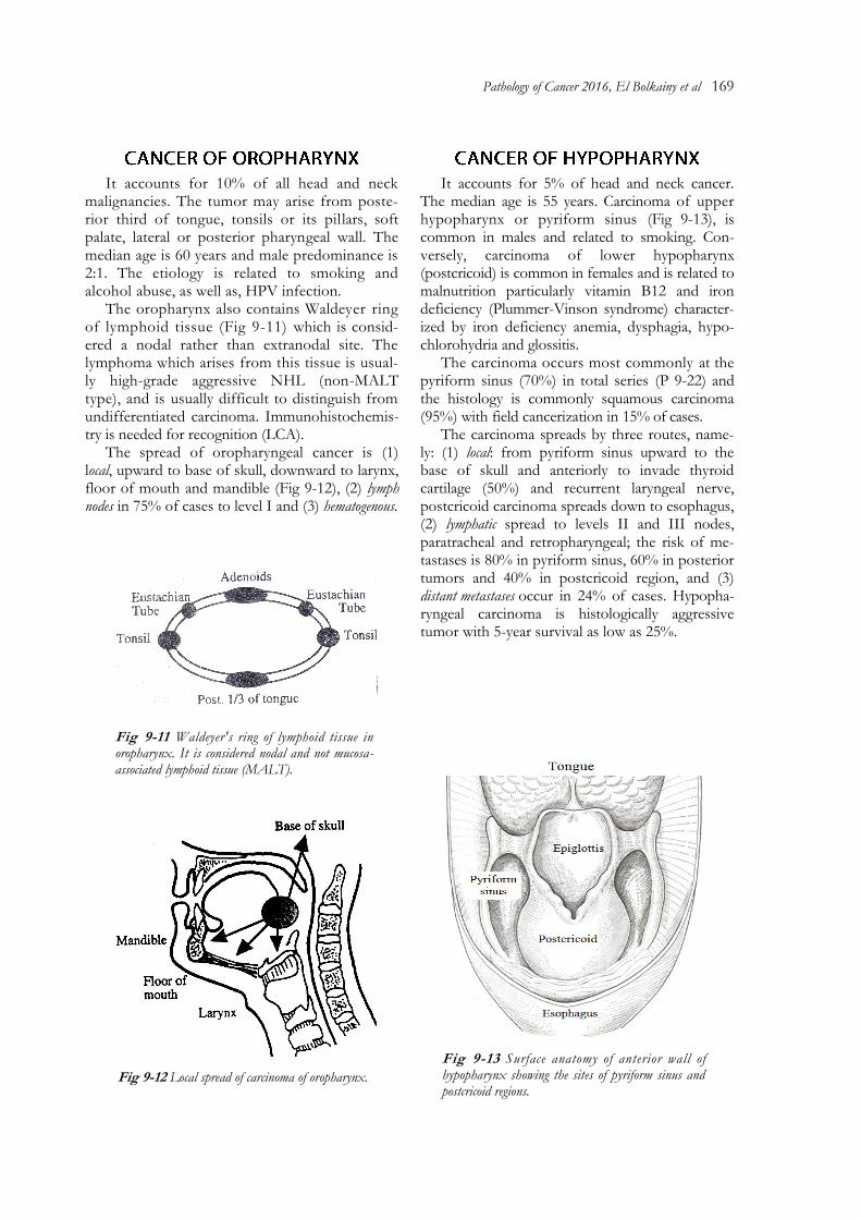
Fig 9-11 Waldeyer's ring of lymphoid tissue in oropharynx. It is considered nodal and not mucosa-associated lymphoid tissue (MALT).
It accounts for 10% of all head and neck malignancies. The tumor may arise from poste-rior third of tongue, tonsils or its pillars, soft palate, lateral or posterior pharyngeal wall. The median age is 60 years and male predominance is 2:1. The etiology is related to smoking and alcohol abuse, as well as, HPV infection.
The oropharynx also contains Waldeyer ring of lymphoid tissue (Fig 9-11) which is consid-ered a nodal rather than extranodal site. The lymphoma which arises from this tissue is usual-ly high-grade aggressive NHL (non-MALT type), and is usually difficult to distinguish from undifferentiated carcinoma. Immunohistochemis-try is needed for recognition (LCA).
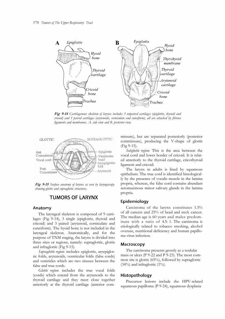
The spread of oropharyngeal cancer is (1) local, upward to base of skull, downward to larynx, floor of mouth and mandible (Fig 9-12), (2) lymph nodes in 75% of cases to level I and (3) hematogenous.
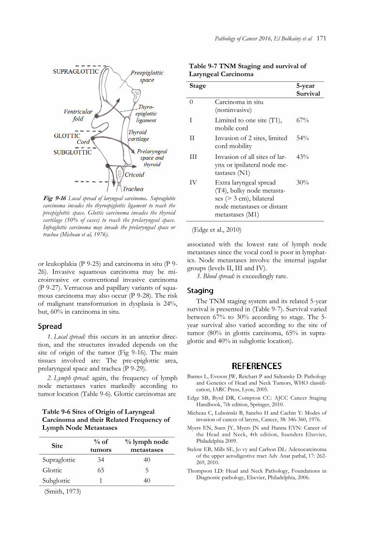
It accounts for 5% of head and neck cancer. The median age is 55 years. Carcinoma of upper hypopharynx or pyriform sinus (Fig 9-13), is common in males and related to smoking. Con-versely, carcinoma of lower hypopharynx (postcricoid) is common in females and is related to malnutrition particularly vitamin B12 and iron deficiency (Plummer-Vinson syndrome) character-ized by iron deficiency anemia, dysphagia, hypo-chlorohydria and glossitis.
The carcinoma occurs most commonly at the pyriform sinus (70%) in total series (P 9-22) and the histology is commonly squamous carcinoma (95%) with field cancerization in 15% of cases.
The carcinoma spreads by three routes, name-ly: (1) local: from pyriform sinus upward to the base of skull and anteriorly to invade thyroid cartilage (50%) and recurrent laryngeal nerve, postcricoid carcinoma spreads down to esophagus, (2) lymphatic spread to levels II and III nodes, paratracheal and retropharyngeal; the risk of me-tastases is 80% in pyriform sinus, 60% in posterior tumors and 40% in postcricoid region, and (3) distant metastases occur in 24% of cases. Hypopha-ryngeal carcinoma is histologically aggressive tumor with 5-year survival as low as 25%.
Fig 9-13 Surface anatomy of anterior wall of hypopharynx showing the sites of pyriform sinus and postcricoid regions.
Fig 9-12 Local spread of carcinoma of oropharynx.
170 Tumors of The Upper Respiratory Tract
Fig 9-14 Cartilagenous skeleton of larynx includes 3 unpaired cartilages (epiglottis, thyroid and cricoid) and 3 paired cartilages (arytenoids, corniculate and cuneiform), all are attached by fibrous ligaments and membranes. A. side view and B. posterior view.
The laryngeal skeleton is composed of 9 carti-lages (Fig 9-14), 3 single (epiglottis, thyroid and cricoid) and 3 paired (arytenoid, corniculate and cuneiform). The hyoid bone is not included in the laryngeal skeleton. Anatomically, and for the purpose of TNM staging, the larynx is divided into three sites or regions, namely: supraglottic, glottis and infraglottic (Fig 9-15).
Supraglottic region: includes epiglottis, aryepiglot-tic folds, arytenoids, ventricular folds (false cords) and ventricles which are two sinuses between the false and true cords.
Glottic region: includes the true vocal folds (cords) which extend from the arytenoids to the thyroid cartilage and they meet close together anteriorly at the thyroid cartilage (anterior com-
missure), but are separated posteriorly (posterior commissure), producing the V-shape of glottis (Fig 9-15).
Subglottic region: This is the area between the vocal cord and lower border of cricoid. It is relat-ed anteriorly to the thyroid cartilage, cricothyroid ligament and cricoid.
The larynx in adults is lined by squamous epithelium. The true cord is identified histological-ly by the presence of vocalis muscle in the lamina propria, whereas, the false cord contains abundant seromucinous minor salivary glands in the lamina propria.
Carcinoma of the larynx constitutes 1.5% of all cancers and 25% of head and neck cancer. The median age is 60 years and males predom-inate with a ratio of 4.5: 1. The carcinoma is etiologically related to tobacco smoking, alcohol overuse, nutritional deficiency and human papillo-ma virus infection.
The carcinoma presents grossly as a nodular mass or ulcer (P 9-22 and P 9-23). The most com-mon site is glottis (65%), followed by supraglottic (34%) and infraglottic (1%).
Precursor lesions include the HPV-related squamous papilloma (P 9-24), squamous dysplasia
Fig 9-15 Surface anatomy of larynx as seen by laryngoscopy showing glottis and supraglottic structures.
Pathology of Cancer 2016, El Bolkainy et al 171
or leukoplakia (P 9-25) and carcinoma in situ (P 9-26). Invasive squamous carcinoma may be mi-croinvasive or conventional invasive carcinoma (P 9-27). Verrucous and papillary variants of squa-mous carcinoma may also occur (P 9-28). The risk of malignant transformation in dysplasia is 24%, but, 60% in carcinoma in situ.
1. Local spread: this occurs in an anterior direc-tion, and the structures invaded depends on the site of origin of the tumor (Fig 9-16). The main tissues involved are: The pre-epiglottic area, prelaryngeal space and trachea (P 9-29).
2. Lymph spread: again, the frequency of lymph node metastases varies markedly according to tumor location (Table 9-6). Glottic carcinomas are
associated with the lowest rate of lymph node metastases since the vocal cord is poor in lymphat-ics. Node metastases involve the internal jugular groups (levels II, III and IV).
3. Blood spread: is exceedingly rare.
The TNM staging system and its related 5-year survival is presented in (Table 9-7). Survival varied between 67% to 30% according to stage. The 5-year survival also varied according to the site of tumor (80% in glottis carcinoma, 65% in supra-glottic and 40% in subglottic location).
Barnes L, Eveson JW, Reichart P and Sidransky D: Pathology and Genetics of Head and Neck Tumors, WHO classifi-cation, IARC Press, Lyon, 2005.
Micheau C, Luboinski B, Sancho H and Cachin Y: Modes of invasion of cancer of larynx, Cancer, 38: 346-360, 1976.
Myers EN, Suen JY, Myers JN and Hanna EYN: Cancer of the Head and Neck, 4th edition, Saunders Elsevier, Philadelphia 2009.
Stelow EB, Mills SE, Jo vy and Carlson DL: Adenocarcinoma of the upper aerodigestive tract Adv Anat pathal, 17: 262-269, 2010.
Thompson LD: Head and Neck Pathology, Foundations in Diagnostic pathology, Elsevier, Philadelphia, 2006.
Fig 9-16 Local spread of laryngeal carcinoma. Supraglottic carcinoma invades the thyroepiglottic ligament to reach the preepiglottic space. Glottic carcinoma invades the thyroid cartilage (50% of cases) to reach the prelaryngeal space. Infraglottic carcinoma may invade the prelaryngeal space or trachea (Micheau et al, 1976).
Table 9-7 TNM Staging and survival of Laryngeal Carcinoma
Stage 5-year Survival
0 Carcinoma in situ (noninvasive)
I Limited to one site (T1), mobile cord
67%
II Invasion of 2 sites, limited cord mobility
54%
III Invasion of all sites of lar-ynx or ipsilateral node me-tastases (N1)
43%
IV Extra laryngeal spread (T4), bulky node metasta-ses (> 3 cm), bilateral node metastases or distant metastases (M1)
30%
Site % of
tumors % lymph node
metastases
Supraglottic 34 40
Glottic 65 5
Subglottic 1 40
Table 9-6 Sites of Origin of Laryngeal Carcinoma and their Related Frequency of Lymph Node Metastases