Acute airway obstruction is one of the most common causes of acute respiratory failure in children. Left untreated, it can rapidly progress to cardiopulmonary arrest and death. There are several important anatomical differences between pediatric and adult patients that render children more sus-ceptible to acute airway obstruction. In addition, there are several diseases that can cause life-threatening acute airway compromise. An understanding of the developmental anat-omy and physiology, as well as the myriad diseases that can cause airway compromise in children is therefore essential for all healthcare personnel that provide care for critically ill children.
Developmental Anatomy
The airway is divided into the upper respiratory tract, which begins with the nose and lips and extends down to the glottis, and the lower respiratory tract which is the airway below the glottis. The upper respiratory tract begins with the nasal and oral cavities, which together comprise the pharynx. The pharynx is connected to the esophagus and the larynx. The larynx and its unique anatomy continue into the chest in the form of a cylindrical structure called the trachea. The larynx is a unique structure whose primary functions are in speech production and protection of the airway. It is formed by cartilaginous, bony, and connective tissue structures. The glottis is the area around the vocal cords. The subglottis is the area directly below the vocal cords leading into the tra-chea. The cords are closed during the end of the expiratory phase, and they open at the beginning of the inspiratory phase. The trachea is a cylindrical structure formed by 16–20 U-shaped cartilaginous rings and a muscular/cartilaginous part that completes the tube.
Though basic principles in the management of the airway in children are the same as in adults, there are important developmental characteristics that distinguish the pediatric
Abstract
Acute airway obstruction is one of the most common causes of acute respiratory failure in children. Left untreated, it can rapidly progress to cardiopulmonary arrest and death. There are several important anatomical differences between pediatric and adult patients that ren-der children more susceptible to acute airway obstruction. In addition, there are several diseases that can cause life- threatening acute airway compromise. An understanding of the developmental anatomy and physiology, as well as the myriad diseases that can cause airway compromise in children is therefore essential for all healthcare personnel that pro-vide care for critically ill children.
Life-Threatening Diseases of the Upper Respiratory Tract
Derek S. Wheeler
2
D.S. Wheeler, MD, MMMDivision of Critical Care Medicine, Cincinnati Children’s Hospital Medical Center, University of Cincinnati College of Medicine, Cincinnati, OH, USAe-mail: [email protected]
airway from the adult airway (Table 2.1). These affect both mask ventilation and tracheal intubation. In the neonate and infant, important anatomic differences include a proportion-ately larger head and tongue, narrower nasal passages, an anterior and cephalad larynx, long epiglottis, and a short tra-chea and neck. These factors contribute to making infants “obligate” nasal breathers. The cricoid cartilage is the nar-rowest point of the airway in children younger than 10 years of age as opposed to the glottis in the adult. Even minimal edema will have a proportionately greater effect in children because of their smaller tracheal diameters. In older children, prominent adenoidal and tonsillar tissue can obstruct visual-ization of the larynx. Also, there are specific congenital ana-tomic airway anomalies that occur in children which makes management of the airway even more complex. As the child becomes older the airway becomes more comparable to the adult anatomy, and by 8 or 9 years of age the airway is con-sidered similar to the adult airway, with the exception of the size of the airway itself.
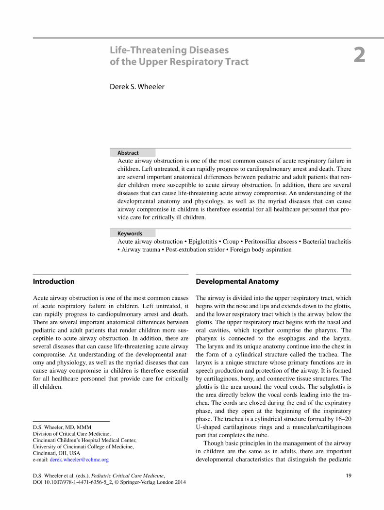
All parts of the pediatric airway are very small and fragile. Even trauma caused during tracheal intubation can cause significant edema and obstruction of the upper air-way. For example, 1 mm of circumferential edema can result in a 16-fold increase in resistance in a 4 mm infant airway (Fig. 2.1). The narrow caliber of the airway results in greater baseline resistance. Any process that narrows the airway fur-ther will cause an exponential rise in airway resistance and hence a significant increase in the work of breathing. When the child perceives distress, the resultant increase in respi-ratory effort will further augment turbulence and increase resistance.
Because the neonate is primarily a nasal breather, any degree of obstruction of the nasopharynx may result in a significant increase in the work of breathing and present clinically as nasal flaring, tachypnea, and retractions. The tongue of infants and small children dominates the over-all capacitance of the oropharynx, so any pediatric patient who presents with altered mental status will be at risk for the development of upper airway obstruction secondary to loss of muscle tone affecting the tongue. Occlusion of the
oropharynx by the tongue is not uncommon in this setting, but tilting of the head, lifting the chin, or insertion of an oral airway may correct this obstruction (the so-called triple air-way maneuver).
Older children have tonsillar and adenoidal tissues that are large in proportion to the rest of the upper airway. Although these rarely are the cause of an upper airway catas-trophe, they are vulnerable to traumatization and bleeding during clinical interventions such as insertion of an oral or a nasal airway. The pediatric trachea is easily distensible and compressible due to incomplete closure of semiformed carti-laginous rings. Any maneuver that overextends the neck will contribute to compression of this structure and secondary upper airway obstruction. As the cricoid ring represents the narrowest portion of the upper airway in children it is often the site of occlusion in tracheobronchial foreign body aspiration.
Acute Airway Obstruction
Children are at particular risk for acute airway obstruction (AAO) due to the anatomic differences between the pediatric and adult airway discussed above [1, 2]. In fact, children may appear surprisingly well from a clinical standpoint, despite being on the verge of cardiorespiratory collapse. Infants have a high oxygen demand due to a higher metabolic rate relative to body size and weight. Consequently, in the presence of apnea or inadequate ventilation, hypoxemia develops more rapidly in the child than adult, and acute decompensation of
Table 2.1 Major anatomic differences between the airway of infant vs. adult
Infant Adult
Head Large, prominent occiput Flat occiputTongue Relatively larger Relatively smallerLarynx Cephalad position Opposite to C4–C6
Opposite to C2–C3Epiglottis Omega-shaped & soft Flat and flexibleVocal cords Short & concave HorizontalNarrowest portion Cricoid ring, below cords Vocal cordsCartilage Soft FirmLower airways Smaller, less developed Larger, more cartilage
Normal
Infant
Adult
Edema ∆ diameter ∆ resistance
4 mm 2 mm
6 mm8 mm
↓ 50 %
↓ 25 %
↑ 16 ×
↑ 3 ×
Fig. 2.1 Age-dependent effects of a reduction in airway caliber on the airway resistance and airflow. Normal airways are represented on the left, edematous airways are represented on the right. According to Poiseuille’s law, airway resistance is inversely proportional to the radius of the airway to the fourth power when there is laminar flow and to the fifth power when there is turbulent flow. One mm of circumferential edema will reduce the diameter of the airway by 2 mm, resulting in a 16-fold increase in airway resistance (cross-sectional area reduced by 75 %). In contrast, an equivalent amount (1 mm) of circumferen-tial edema results in only a 3-fold increase in airway resistance (cross- sectional area reduced by 25 %) in an adult. Note that turbulent air flow (such as occurs during crying) in the infant would increase the resistance by 32-fold
D.S. Wheeler
21
cardiorespiratory status may be swift and often difficult to reverse [3, 4]. Upper airway obstruction (Table 2.2) often leads to acute respiratory failure and is an important cause of out-of-hospital cardiopulmonary arrest, in stark contrast to adults in which primary cardiac disease commonly precipi-tates cardiopulmonary arrest. Once respiratory arrest pro-gresses to cardiac arrest, outcome is dismal [5–7], and prompt recognition of AAO and appropriate, timely inter-vention is crucial to assure the best possible outcome.
Physiology of Airway Obstruction
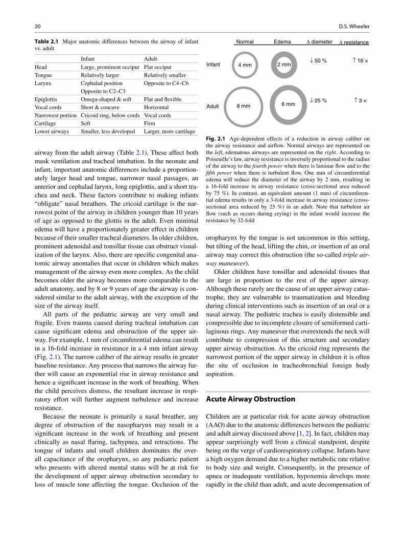
The pediatric airway is highly compliant and the cartilagi-nous support less well-developed compared to the adult air-way and is therefore more susceptible to dynamic airway collapse in the presence of airway obstruction. The normal respiratory dynamics change significantly in the presence of airway obstruction (Fig. 2.2). A forced inhalation that is required to generate airflow in the presence of a partial upper airway obstruction requires a stronger contraction of the dia-phragm and respiratory muscles, generating a greater decrease (i.e. more negative relative to atmospheric pressure) in intra-pleural and intra-luminal airway pressures. The larger gradient between atmospheric pressure and the airway
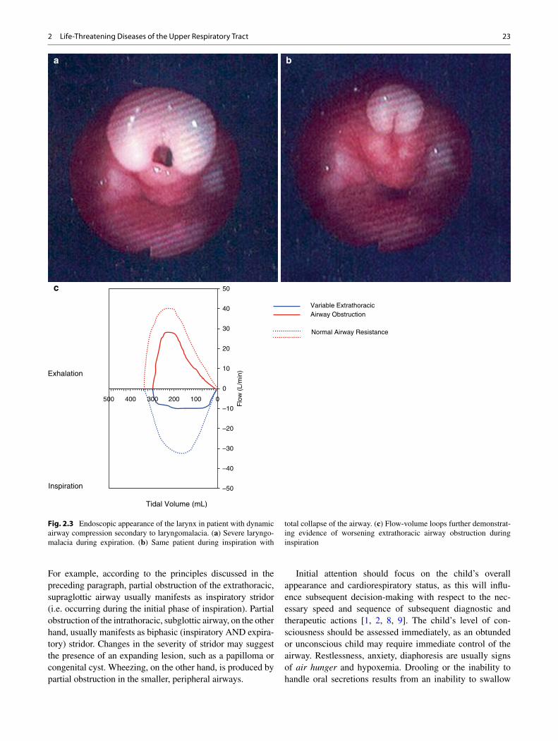
pressure leads to dynamic collapse of the extrathoracic tra-chea just beyond the level of obstruction. This explains why obstruction of the extrathoracic airway is worse during inspi-ration (Fig. 2.3). Conversely, lower airway or intrathoracic airway obstruction (e.g. aspirated foreign body, asthma, bronchiolitis, etc) results in a ball-valve effect and subse-quent air-trapping. Increased respiratory effort during exha-lation is required, generating an increase in intra-pleural pressures and leading to dynamic compression of the intra-thoracic airways. This explains why obstruction of the intra-thoracic airways is worse during expiration (Fig. 2.4).
The movement of a gas (i.e. air) through a partially closed, collapsible tube (i.e. airway) obeys the laws of physics. According to the Venturi effect, the pressure exerted by a gas (i.e. air) as it flows through a partially closed tube is equal in all directions except when there is linear movement, which creates additional pressure in the forward vector with a cor-responding fall in the lateral vectors. This decrease in lateral pressure (i.e. the distending pressure keeping the collapsible tube open) causes the tube to narrow, leading to partial obstruction. In addition, according to the Bernoulli principle, the velocity of a gas increases as it flows through a partially obstructed tube, creating an additional decrease in intralumi-nal pressure and further exacerbating the obstruction (Fig. 2.5). This pattern of intermittent flow produces audible sounds that are characterized (depending upon the level of partial obstruction) as stertor, gurgling, stridor, wheezes, rhonchi, and rales. For example, stretor is a snoring or snort-ing sound that is produced by turbulence within the naso-pharynx. Gurgling is produced by turbulence within the oropharynx due to the mixture of air and secretions. Stridor is the sound produced by turbulent airflow in a partially obstructed trachea, either due to intrinsic obstruction or extrinsic compression [1, 2, 8–14].
According to Hagen-Poiseuille’s law, the change in air flow resulting from a reduction in airway diameter is directly proportional to the airway radius elevated to the fourth power:
Q P r 8 L4= ( ) ( )∆ π / hh
where Q is flow, ∆P is the pressure gradient from one end of the airway to the other end, r is the radius of the airway, η is the viscosity of the air, and L is the length of the air-way. Therefore, increasing the length of the airway (L), increasing the viscosity of the air (η), or decreasing the radius of the airway will reduce laminar air flow. Changing the airway radius, however, has the greatest effect on flow. Hagen- Poiseuille’s law holds for conditions of laminar flow. Laminar flow is highly organized, streamlined, and efficient. Turbulent flow, on the other hand, is highly disorganized, chaotic, and inefficient (Fig. 2.6). Laminar flow is typically found in the peripheral airways, while turbulent flow is found
Table 2.2 Common causes of upper airway obstruction in children
2 Life-Threatening Diseases of the Upper Respiratory Tract
22
in the proximal, upper airways. Importantly, the Poiseuille’s law assumes conditions of laminar flow. When turbulent flow is present, as occurs during partial airway obstruction, air-way resistance is inversely proportional to the radius of the airway to the fifth power! The Reynolds number can be used to determine whether conditions favor laminar versus turbu-lent flow. It is calculated by the following equation:
Re 2Vr= rr hh/
Where Re is the Reynolds number, V is the velocity of flow of air, r is the radius of the airway, η is the viscosity of the air, and ρ is the density of the air. Laminar flow is favored when the Reynolds number is less than 2,000, while turbulent flow
is favored when the Reynolds number is greater than 4,000. In other words, the higher the Reynolds number, the more likely turbulent flow is present. As such, higher velocities (such as occurs during crying, coughing, agitation, or respi-ratory distress), larger caliber airways, lower air density, and lower air viscosity all favor turbulent flow [15, 16].
Clinical Manifestations of Airway Obstruction
Careful assessment of the time in the respiratory cycle in which stridor predominates may provide valuable diagnostic clues in determining the site of airway obstruction [2, 8–15].
– –
–
–
–
–
–
–
–
–
–––
–
–
–
–
––
A
A
A
A A
A
A
AA
A A
A
A
–
–
––
–
A A
–
–
Obstruction
Obstruction
+
+
+
++
vocal cords
extra-thoracictracheo
pleural space
lung
disphragam
Atmospheric pressure
Negative to A
Positive to A
=
=
=
++
++
++
+
+
+
+
+++++
+
+
a b
c d
Fig. 2.2 (a) Normal Inspiration. At end-expiration, intrapleural pres-sure is less than atmospheric pressure, so it should maintain airway patency. In infants the highly compliant chest wall does not provide the support required. Thus airway closure occurs with each breath. Descent of the diaphragm and contraction of the intercostals muscles develop a greater negative intrathoracic pressure relative to intraluminal and atmospheric pressure. The net result is a longitudinal stretching of the larynx and trachea, dilation of the intrathoracic trachea and bron-chi, movement of air into the lungs, and some dynamic collapse of the extrathoracic trachea due to the increased compliance of the trachea ad the negative intraluminal pressure in relation to atmospheric pressure. (b) Normal expiration. Intraluminal pressures are slightly positive in relation to atmospheric pressure, so air is forced out of the lungs.
(c) Extra-thoracic obstruction (obstructed inspiration). Respiratory dynamics occurring with upper airway obstruction; note the severe dynamic collapse of the extrathoracic trachea below the level of obstruc-tion. This collapse is greatest at the thoracic inlet, where the largest pressure gradient exists between negative intratracheal pressure and atmospheric pressure. (d) Intra-thoracic obstruction (obstructed expira-tion). Respiratory dynamics occurring with lower airway obstruction. Breathing through a partially obstructed lower airway (such as occurs in bronchiolitis or asthma) results in greater positive intrathoracic pres-sures, with dynamic collapse of the intrathoracic airways (prolonged expiration or wheezing) (Reprinted from Zalzal [11]. With permission from Elsevier)
D.S. Wheeler
23
For example, according to the principles discussed in the preceding paragraph, partial obstruction of the extrathoracic, supraglottic airway usually manifests as inspiratory stridor (i.e. occurring during the initial phase of inspiration). Partial obstruction of the intrathoracic, subglottic airway, on the other hand, usually manifests as biphasic (inspiratory AND expira-tory) stridor. Changes in the severity of stridor may suggest the presence of an expanding lesion, such as a papilloma or congenital cyst. Wheezing, on the other hand, is produced by partial obstruction in the smaller, peripheral airways.
Initial attention should focus on the child’s overall appearance and cardiorespiratory status, as this will influ-ence subsequent decision-making with respect to the nec-essary speed and sequence of subsequent diagnostic and therapeutic actions [1, 2, 8, 9]. The child’s level of con-sciousness should be assessed immediately, as an obtunded or unconscious child may require immediate control of the airway. Restlessness, anxiety, diaphoresis are usually signs of air hunger and hypoxemia. Drooling or the inability to handle oral secretions results from an inability to swallow
Exhalation
Inspiration
Tidal Volume (mL)
500 400 300 200 100 0
50
Variable ExtrathoracicAirway Obstruction
Normal Airway Resistance
40
30
20
10
0
–10
Flo
w (
L/m
in)
–20
–30
–40
–50
a
c
b
Fig. 2.3 Endoscopic appearance of the larynx in patient with dynamic airway compression secondary to laryngomalacia. (a) Severe laryngo-malacia during expiration. (b) Same patient during inspiration with
total collapse of the airway. (c) Flow-volume loops further demonstrat-ing evidence of worsening extrathoracic airway obstruction during inspiration
2 Life-Threatening Diseases of the Upper Respiratory Tract
24
secondary to pain or swelling of affected tissues and is typi-cally seen with supraglottic pathology (e.g. supraglottitis, retropharyngeal abscess). Accessory muscle use is an addi-tional sign of increased work of breathing and is indicative of compromised gas exchange.
During quiet breathing, airflow is laminar and resistance to airflow is inversely proportional to the fourth power of the airway radius as stipulated by Poiseuille’s law. When airflow is turbulent (e.g., during crying) resistance to airflow is inversely proportional to the fifth power of radius such that
even a minor reduction in the cross-sectional area of the air-way will result in a marked increase in airflow resistance and work of breathing. For these reasons, the infant or child with airway obstruction should be kept calm and as quiet as pos-sible to prevent generation of turbulent airflow, increased airway resistance, and worsening respiratory distress. In general, any child in severe respiratory distress will assume a position that maximizes oxygenation and ventilation and should be allowed to remain in this position of comfort. For example, the child with supraglottitis will sit erect with the
Exhalation
Inspiration
Tidal Volume (mL)
500 400 300 200 100 0
50
Variable Intrathoracic
Airway Obstruction
Normal Airway Resistance
40
30
20
10
0
–10
Flo
w (
L/m
in)
–20
–30
–40
–50
c
a b
Fig. 2.4 Endoscopic appearance of the trachea in a patient with dynamic airway compression secondary to tracheomalacia. (a) Normal trachea. (b) Child with severe tracheomalacia demonstrating total col-
lapse of the airway during inspiration. (c) Flow-volume loops further demonstrating evidence of worsening intrathoracic airway obstruction during expiration
D.S. Wheeler
25
head tilted forward in the sniffing position, whereas a child with a retropharyngeal abscess will assume a head tilt or opisthotonus posture because of spasm of the muscles sup-porting the cervical spine [1, 2, 8, 9].
Infectious Disorders of the Pediatric Airway
The clinical spectrum of infectious causes of upper airway obstruction has changed dramatically in the last few decades, especially following the introduction of vaccines against diph-theria and Haemophilus influenzae. Many of the infectious causes of upper airway obstruction pose less of a threat today as a result of advances in prevention, early diagnosis, and treatment. Nevertheless, infectious causes of upper airway obstruction remain a common cause of upper airway obstruc-tion as well as an important source of morbidity and potential mortality in the pediatric age group [17–19] (Table 2.3).
Viral Laryngotracheobronchitis (“Croup”)
Viral laryngotracheobronchitis, or croup, is the most common infectious cause of upper airway obstruction in children with an annual incidence of 18 per 1,000 children in the United States [20]. Croup primarily affects children between the ages of 6 months and 4 years with a peak incidence between 1 to 2 years of age. While sporadic cases may be seen throughout the year, the peak incidence occurs during early fall and win-ter. Males are affected slightly more commonly than females [17–21]. Croup is caused by an inflammation affecting the subglottic tissues, occasionally affecting the tracheobronchial tree as well, usually due to a viral infection. Croup is most commonly caused by parainfluenza virus type 1, though para-influenza virus types 2 and 3, influenza A and B, respiratory syncytial virus (RSV), human metapneumovirus, adenovirus, rhinovirus, coronavirus, enterovirus, and Mycoplasma pneu-moniae are commonly implicated as well [21–25].
IncreasedVelocity
DecreasedPressure
Fig. 2.5 Illustration of the Venturi effect and Bernoulli’s principle
Laminar Flow:
Turbulent Flow:
Fig. 2.6 Laminar versus turbulent air flow
Table 2.3 Infectious causes of upper airway obstruction
Onset Gradual Rapid onset Viral prodrome followed by rapid deterioration
Viral prodrome followed by rapid deteriorationViral prodrome 6–12 h
1–7 daysTypical age at onset 6 months–4 years 2–8 years 6 months–8 years < 5 yearsSeasonal occurrence Late fall to winter Throughout the year Fall to winter Throughout the yearCausative agents Parainfluenza, respiratory
syncytial virus, influenza AHaemophilus influenzae type b (classically), Streptococcus pneumoniae, GABHS
Fever Low-grade High fever High fever High feverCough “Barking” or “seal-like” None Usually absent Usually absentSore throat None Severe None SevereDrooling None Frequent None FrequentPosture Any position Sitting forward, mouth open,
neck extended (“tripod position”)
Any position Sitting forward, mouth open, neck extended (“tripod position”)
Voice Normal or hoarseness Muffled Normal or hoarseness MuffledAppearance Nontoxic Toxic Toxic Toxic
2 Life-Threatening Diseases of the Upper Respiratory Tract
26
Most children with croup can be managed in the outpa-tient setting, though between 1 % to 30 % of children require hospitalization and 2 % of hospitalized children require tra-cheal intubation and mechanical ventilatory support [26–28]. Children with croup typically present with several days of viral prodromal symptoms (cough, coryza, rhinorrhea, low- grade fever) with progressively worsening hoarseness, the classic “seal-like” or barky cough, and stridor (most com-monly inspiratory in nature, though biphasic stridor is indic-ative of more severe degree of airway obstruction). The absence of drooling (i.e. dysphagia and inability to handle oral secretions) can help differentiate children with croup from those with a more serious bacterial illness, such as supraglottitis. Conversely, children with high fevers, drool-ing, absence of a cough, and/or a toxic appearance are more likely to have a more serious infection such as bacterial tra-cheitis, retropharyngeal abscess, or supraglottitis [29]. The classic harsh cough or bark may progress to inspiratory stri-dor and frank dyspnea in severe cases, and various scales have been devised to quantify the severity of stridor and document the progression of the illness and subsequent response to therapy [30–33].
While radiographic examination is useful to rule out other important causes of airway obstruction (e.g. supraglottitis, foreign body, retropharyngeal abscess, etc), the classic stee-ple sign (Fig. 2.7) may be absent in as many as half of chil-dren with croup [21, 34–36]. When visible, the subglottic narrowing is dynamic and is more accentuated during inspi-ration, because of the more negative intraluminal airway pressure during inspiration [37]. Children with a longstand-ing history of stridor or those under 4 months of age should
be carefully evaluated for anatomical airway obstruction, such as laryngeal cyst or papillomatosis, vocal cord paresis, extrinsic airway compression (e.g. vascular ring), or laryngo-tracheal stenosis.
Croup is generally self-limited and frequently requires only supportive care. Humidification with continuous cool mist has been the standard accepted treatment for many years [34, 38–40]. The mechanism by which humidified cool mist improves symptomatology is not well understood, and probably reflects a placebo effect [38–40]. In addition, recent studies suggest that continuous cool mist therapy is not effective for croup [40, 41]. Nebulized racemic epi-nephrine rapidly reduces airway edema and improves symptoms, though the effect is transient and disappears within 2–3 h of administration [32, 42–48]. Both the race-mic and L-isomer form of epinephrine appear to be safe and effective, however racemic epinephrine has not been shown to decrease the need for either tracheal intubation or trache-otomy in children with croup [42, 44, 45]. Rebound or worsening of airway obstruction after the drug effect wears off may occur with the use of racemic epinephrine, and for this reason, treated patients should be observed for 4–6 h after administration [48].
Several studies have shown substantial improvement in symptoms following administration of corticosteroids. Administration of corticosteroids appears to improve symp-tomatology, shorten the duration of hospital stay, and reduce the need for racemic epinephrine [26, 46, 49–55]. A sin-gle dose of dexamethasone is usually adequate for mild to moderate croup. The dose of dexamethasone ranges from 0.15 mg/kg of oral preparation to 0.6 mg/kg of parenteral
Fig. 2.7 Typical radiographic appearance of croup demonstrating symmetric narrowing of the subglottic region (“steeple sign”) (a) Normal anatomy (left) (v vestibule, p pyriform sinuses), (b) Subglottic narrowing (arrow) consistent with croup (right)
D.S. Wheeler
27
preparation. Nebulized corticosteroids appear to be effec-tive as well, especially when administered in combination with racemic epinephrine [46, 55–62]. Children with severe croup who are managed in the PICU setting may require a more prolonged course of corticosteroids. Although few adequately controlled, randomized studies exist to suggest any benefit to prolonged administration of corticosteroids in critically ill children with acute respiratory failure secondary to croup, the wealth of anecdotal experience would suggest this to be a reasonable practice. Corticosteroids appear to be advantageous in relieving upper airway obstruction regard-less of the route of administration [63–66]. While the pre-cise mechanism of action of corticosteroids in croup is not readily known, the rapid response observed following cor-ticosteroid administration suggests that decreased capillary permeability and peripheral vasoconstriction plays an impor-tant role [63]. The anti-inflammatory effects of corticoste-roids (via inhibition of pro-inflammatory gene expression) require 6–12 h for maximal effect [63–66].
The use of helium-oxygen mixtures may be beneficial in some children with croup. Helium-oxygen mixtures create conditions that favor laminar flow as opposed to turbulent flow. Recall that airway resistance is inversely proportional to the airway radius to the fifth power (r5) under conditions of turbulent air flow (versus airway radius to the fourth power when laminar flow is present). Helium is a colorless, odor-less gas with the lowest density of any gas except hydro-gen. In addition, helium is more viscous than ambient air. As such, helium will reduce the Reynolds number and change a turbulent flow pattern to a laminar flow pattern, resulting in lower airway resistance and improved bulk flow [15, 16, 67]. Helium-oxygen gas (Heliox) mixtures have been shown to improve the work of breathing and gas exchange in chil-dren with croup [67–79]. Heliox has few side effects and is easy to administer by face mask, hood, high-flow nasal can-nula, or via a tracheal tube in children on mechanical ventila-tory support [74]. In order to minimize the risk of asphyxia secondary to administration of 100 % helium, helium-oxy-gen mixtures should only be administered from pre-mixed helium-oxygen cylinders. Currently, 80:20 helium-oxygen and 70:30 helium:oxygen mixtures are available. Therefore, children with a high oxygen requirement are unlikely to benefit (and may actually worsen) from heliox administra-tion. The beneficial effects of helium are reduced with lower ratios of helium-to-oxygen, though it appears likely that helium will have at least some therapeutic value even at low concentrations [16, 68, 75].
Until the airway inflammation resolves, severe upper air-way obstruction may develop and occasionally necessitate tracheal intubation. Prior to the introduction of corticosteroid therapy, tracheal intubation was required in approximately 2 % of children hospitalized with croup [8, 17–19]. Tracheal intubation is now commonly limited to those children who
either have pre-existing airway abnormalities or who have been tracheally intubated in an outside facility prior to trans-fer. Generally, tracheal intubation with a tube smaller than what would be normally predicted for age and weight should be used in the minority of children requiring tracheal intu-bation and mechanical ventilatory support. Extubation can usually be accomplished within 2–3 days once an air leak has developed around the tracheal tube. Bronchoscopy is reserved for children who fail to develop an air leak after 7 days, or in children less than 6 months of age, who have a high likelihood of congenital malformations of the airway [8, 17–19]. Although the etiologies of many infections of upper respiratory tract are viral, bacterial superinfection may occur. However, uncomplicated croup is viral in origin and should not be treated with antibiotics. Antibiotic therapy may be considered in those children who fail to improve or who require tracheal intubation.
Supraglottis (“Epiglottitis”)
Epiglottitis is a true emergency, though the term is some-what misleading as the supraglottic structures are most severely affected. Supraglottitis is perhaps more appropri-ate for this reason and classically affects children between the age of 2 and 8 years [8, 17–19]. Historically, supraglot-titis was most commonly caused by Haemophilus influenzae type B. Streptococcus pneumoniae, group A β-hemolytic Streptococcus (GABHS), and Staphylococcus aureus are reported more commonly in the post-Hib vaccination era [80–86]. Since the development and widespread use of the conjugated HIB vaccine, there has been a significant decrease in the incidence of supraglottitis. The incidence of supraglot-titis in children <5 years of age has decreased from 41 cases per 100,000 in 1987 to 1.3 cases per 100,000 in 1997 [87]. The incidence of supraglottitis appears to have stabilized at around 1.3 cases per 100,000 children, primarily due to low or incomplete vaccination coverage in localized populations, as well as cases of supraglottitis caused by microorganisms other than Haemophilus influenzae type b [88].
Supraglottitis requires a high index of suspicion, especially now in the post-Hib vaccination era when many physicians have never seen a child with true supraglottitis. In addition, especially now in the post-Hib vaccination era, epiglottitis may present with atypical features, especially in children under 2 years of age [81, 89, 90]. Children classically pres-ent with rapidly progressive signs and symptoms, including high fever, irritability, drooling, and respiratory distress (the “4 D’s” include drooling, dysphagia, dyspnea, and dyspho-nia). These children are toxic in appearance and prefer to rest in the tripod position. Stridor is relatively late and ominous. There is usually no viral prodrome. By the time that affected children present for medical attention, they are generally
2 Life-Threatening Diseases of the Upper Respiratory Tract
28
toxic and have inspiratory stridor [8, 17–19]. The voice tends to be muffled, rather than hoarse as in children with croup. Affected children assume a characteristic sniffing position in an attempt to maintain optimal airway patency. These chil-dren are usually anxious, which is a strong indication that their airway is significantly compromised.
When supraglottitis is suspected, intraoral examination and lateral neck radiograph should be deferred and only per-formed if equipment and personnel are available to secure the airway immediately. Excessive manipulation of the child and his airway should be avoided to minimize the risk of acute exacerbation of airway obstruction. The appearance on a lateral neck radiograph obtained with hyperextension of the neck is classic (Fig. 2.8), though diagnosis is usually confirmed by direct inspection of the airway in the operat-ing room. When a diagnosis of supraglottitis is entertained based upon initial history and physical examination, the child should be accompanied by a physician skilled in air-way management to the operating room. The child should be allowed to remain in his or her “position of comfort” and all anxiety-provoking procedures (e.g. phlebotomy, oral exami-nation, etc) should be deferred. Direct laryngoscopy under anesthesia should be performed while maintaining spontane-ous breathing [8, 17–19, 91–94]. Cultures of the supraglottic region should be obtained, and the trachea should be intu-bated. Treatment with broad-spectrum antibiotics effective against β-lactamase-producing microorganisms is initiated once cultures have been obtained (e.g. a second- or third- generation cephalosporin such as cefuroxime or ceftriaxone, or alternatively, ampicillin/sulbactam) [8, 17–19, 95, 96]. Antibiotic therapy should be tailored to the pathogenic organ-ism, and the duration of antibiotic therapy is determined by the clinical response. Generally, symptomatic improvement and the development of an audible air leak around the tra-cheal tube occurs within 24–48 h [97]. Despite the virtual elimination of invasive HIB infection, it is important for
pediatric intensivists to understand the management issues surrounding patients with supraglottitis to avoid disastrous outcomes.
Recent studies suggest that a less conservative approach than the one described above (i.e., direct visualization of the airway and tracheal intubation in the operative room set-ting) may be feasible in select patients [86, 98–101]. Whether this reflects a changing spectrum of disease is not known at this time. It should also be noted that most of the studies describing this less conservative approach to man-agement are retrospective in nature and limited to the adult population. It is likely that the management of this once common malady of childhood will continue to evolve in the years to come.
Bacterial Tracheitis
Bacterial tracheitis, also known as pseudomembranous croup, is a relatively uncommon, but potentially life- threatening cause of infectious upper airway obstruction in children [8, 17–19]. It is characterized by thick, muco-purulent, membranous tracheal secretions that do not clear with cough, which have the potential to occlude the airway. Bacterial tracheitis is more insidious compared to supraglot-titis and affects children between the ages of 6 months and 8 years with a peak incidence during fall and winter [8, 17–19, 102–104]. Children frequently present with a viral prodrome of several days duration, accompanied by low-grade fever, cough, and stridor (similar to croup). The viral prodrome is followed by rapid clinical deterioration characterized by a high fever and upper airway obstruction. Affected children are more toxic in appearance compared to children with croup. Radiographic examination often is indistinguishable from that of croup (steeple sign), and in fact, the noted similarities between these two disorders have led some authors to suggest
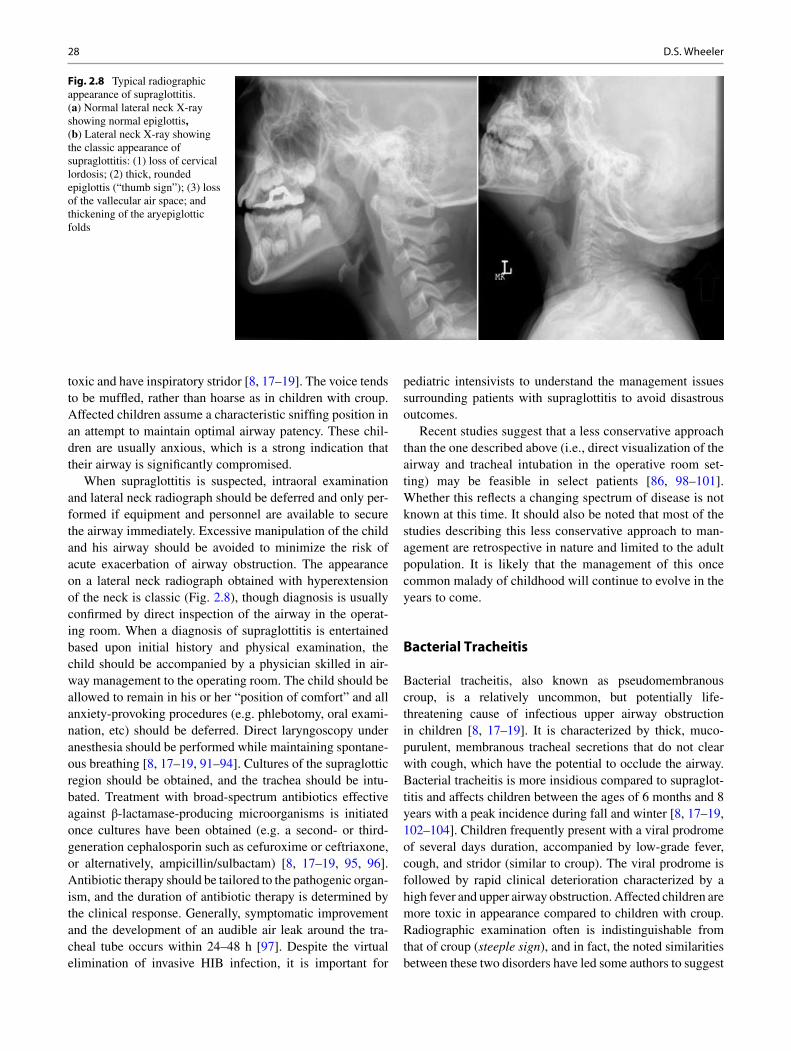
Fig. 2.8 Typical radiographic appearance of supraglottitis. (a) Normal lateral neck X-ray showing normal epiglottis, (b) Lateral neck X-ray showing the classic appearance of supraglottitis: (1) loss of cervical lordosis; (2) thick, rounded epiglottis (“thumb sign”); (3) loss of the vallecular air space; and thickening of the aryepiglottic folds
D.S. Wheeler
29
that bacterial tracheitis represents a bacterial superinfection of croup.
Historically, bacterial tracheitis is most commonly sec-ondary to Staphylococcus aureus, though Streptococcus pneumoniae, GABHS, Hemophilus influenzae, Moraxella catarrhalis, anaerobic bacteria, and viruses have been impli-cated as well [102–112]. Infection with the parainfluenza virus has been implicated as the prodromal infection in many cases, further lending credence to the suggestion that bacte-rial tracheitis represents a bacterial superinfection of croup. Children with bacterial tracheitis are managed using the epi-glottis management algorithm described above [107, 111]. Direct inspection of the airway under anesthesia should be performed in the operating room, and usually reveals subglot-tic edema with ulcerations, erythema, and pseudomembrane formation in the trachea. Removal of pseudomembranes and dead tissue from the airway at diagnosis, tracheal intubation, and administration of broad-spectrum antibiotics, directed against staphylococcal and streptococcal species are the cor-nerstone of treatment. Empiric therapy should be broadly directed against both gram-positive and gram- negative organ-isms until culture results are available. Staphylococcal cov-erage is of obvious importance. Anaerobic organisms may be treated with clindamycin. Extubation may be attempted following clinical improvement and the development of an air leak around the tracheal tube, usually within 3–5 days [8, 17–19, 107, 111].
Retropharyngeal Abscess
Retropharyngeal abscess has been called “the epiglottitis of the new millennium” [113]. The retropharyngeal space is comprised of a loose network of connective tissue and lymph nodes that drain the nasopharynx, paranasal sinuses, middle ear, teeth, and facial bones. Infection and abscess for-mation in this area generally result from lymphatic spread of infection or direct spread from the nasopharynx, parana-sal sinuses, or middle ear. These lymph nodes atrophy dur-ing early childhood, thereby decreasing the risk of disease in older children and adolescents [8, 17–19, 114–116]. For this reason, trauma (e.g. from placing a pencil or stick in the mouth) and foreign body ingestion account for the majority of cases in older children and adolescents. Most cases of ret-ropharyngeal abscess occur in children less than 5 years of age, so there is a significant overlap in the affected age range compared to supraglottitis and bacterial tracheitis [8, 17–19].
Children with retropharyngeal abscess present with a non-specific constellation of symptoms that progress to high fever, sore throat, and neck stiffness. Fever, sore throat, dysphagia, drooling, muffled voice, and limited neck movement or tor-ticollis are the most common presenting symptoms. Airway symptoms include stridor or stretor and difficulty in breath-ing. Symptoms often mimic those of supraglottitis. However,
in contrast to supraglottitis, children with retropharyngeal abscess normally have a sore throat and cough for several days before showing symptoms of fever and respiratory dis-tress. The neck stiffness may mimic that seen in children with meningitis, such that these children are often evaluated for meningitis. Physical examination may reveal the presence of a bulging unilateral neck mass. Additional physical findings commonly include diffuse erythema, tonsillar exudates, and swelling or bulging of the involved tonsillar region. Cervical adenopathy appears to be greatest on the side of the neck where deep infection is most involved [8, 17–19, 117–119].
The diagnosis of retropharyngeal abscess is confirmed by the presence of an abnormally increased pre-vertebral space on lateral neck radiographs (Fig. 2.9). Additional radio-graphic findings include the presence of gas or air fluid lev-els in the retropharyngeal space and the loss of the normal cervical lordosis. Computed tomography (CT) with contrast confirms the presence of abscess, determines its extent, and
Fig. 2.9 Typical radiographic appearance of a peritonsillar abscess. A retropharyngeal space measured from the most anterior aspect of C2 to the soft tissues of the posterior pharyngeal wall >7 mm (normal 3–6 mm) or a retrotracheal space >14 mm is suggestive of RTA. Normal prevertebral spaces are as follows: Anterior to C2: less than or equal to 7 mm; Anterior to C3–C4: less than 5 mm or less than 40 % of the AP diameter of the C3 and C4 vertebral bodies. A good rule of thumb to remember is that the upper pre-vertebral soft tissue should be no wider than one vertebral body width. NOTE: Adequate hyperextension of the head and neck is necessary in order to properly interpret the film. If the head and neck are not properly positioned, the pre-vertebral space will appear widened. In addition, crying can cause rapid changes in the size of the retropharyngeal space. If there is any doubt, repeat radiographic examination with either more hyperextension of the neck, fluoroscopy, or CT imaging is indicated
2 Life-Threatening Diseases of the Upper Respiratory Tract
30
identifies its relationship to the airway [119–123]. While blood cultures are generally negative, culture of the abscess often yields anaerobic microorganisms such as Prevotella, Porphyromonas, Fusobacterium and Peptostreptococcus spp, as well as Staphylococcus aureus, GABHS, and Haemophilus influenzae [124, 125].
Treatment with broad spectrum antibiotics and close observation is highly effective, with drainage of the abscess recommended in children refractory to antibiotic therapy. Complications are rare with early recognition and appropri-ate treatment, though complications include spontaneous rupture into the pharynx leading to aspiration or spread of the infection laterally to the side of the neck or dissection into the posterior mediastinum through the facial planes and the prevertebral space. While rare, death can occur from aspi-ration, upper airway obstruction, erosion into major blood vessels, or extension to the mediastinum with mediastinitis. Tracheal intubation is often necessary to protect the patient from aspiration of the purulent content [8, 17–19, 119].
Peritonsillar Abscess (“Quinsy” Tonsillitis)
Peritonsillar abscess (PTA) rarely requires admission to the PICU, but can lead to significant airway obstruction and respi-ratory compromise if not recognized and left untreated [126]. PTA, also known as “quinsy” tonsillitis is the most common deep space head and neck infection in children and is thought to result from the direct contiguous spread of infection from the tonsils. Older children and adolescents appear to be most commonly affected, with no seasonal predilection. Children with PTA present with sore throat, neck pain, odynophagia or dysphagia, and fever. Physical examination typically reveals enlargement of the cervical lymph nodes, uvular deviation, and a muffled voice. Treatment options include broad spec-trum antibiotics, needle aspiration of the abscess, incision and drainage, and tonsillectomy [127–129]. Complications include extension of the infection, acute upper airway obstruc-tion (rare), and rupture of the abscess with aspiration of puru-lent material and subsequent pneumonia [127–129].
Recurrent Respiratory Papillomatosis
Recurrent respiratory papillomatosis (RRP) is the most com-mon benign laryngeal neoplasm in children, and is usually caused by perinatal transmission of HPV-6 or HPV-11. RRP is characterized by the proliferation of squamous epithelial cells in the upper respiratory tract, which occasionally form lesions that cause severe to life-threatening airway obstruc-tion. Affected children are usually between the ages of 2 and 5 years and typically present with stridor and voice changes. The classic triad consists of a first-born child delivered vagi-
nally to an adolescent mother. The diagnosis of RRP is based upon endoscopic observation of characteristic lesions. Because any region of the upper aerodigestive tract is involved laryngoscopy, bronchoscopy, and careful inspection of oro-pharynx and nasopharynx should be performed [17–19, 130].
The primary goal of treatment is to prevent airway obstruc-tion while the lesions are in the proliferative phase and mini-mize any complications of therapy. The mainstay of treatment for RRP is surgical debulking of the lesions in the operating room by one of several methods, including physical debride-ment with forceps and/or CO2 laser vaporization, as often as weekly to assure a safe airway. Adjuvant medical therapies to control aggressive papillomatosis include topical chemother-apy, corticosteroids, podophyllin, tetracycline, autogenous vaccine, immune stimulators, acyclovir, isotretinoin, inter-feron, and cidofovir [131–133]. Tracheotomy should be avoided if at all possible, though tracheotomy is occasionally required due to acquired tracheal stenosis [17–19, 130–133].
Infectious Mononucleosis
Acute airway obstruction secondary to enlargement of the tonsils and adenoids is a well-recognized complication of infectious mononucleosis. Fortunately, this complication is exceedingly rare and appears to occur primarily in younger children. Parenteral corticosteroids are recommended, based primarily upon case reports and retrospective series [134–138]. Tracheal intubation may be required if airway obstruc-tion is severe, and in such cases tonsillectomy is generally recommended [139, 140].
Non-infectious Disorders of the Pediatric Airway
Obesity
The prevalence of childhood obesity has increased dramati-cally in recent years. Obesity is associated with decreased upper airway patency, principally related to increased fat deposition in the lateral walls of the pharynx [141] and is a common cause of obstructive sleep apnea syndrome (OSAS) in children [142, 143]. Adenotonsillectomy is a commonly performed surgical procedure in this population, and children with OSAS secondary to obesity frequently develop compli-cations that monitoring and care in the PICU [144–149].
Angioedema
Angioedema is an immunologically mediated, nonpitting edema that frequently results in acute airway obstruction. It
D.S. Wheeler
31
is caused by a kinin- and complement-mediated increase in capillary permeability that leads to edema, usually affecting the head and neck, face, lips, tongue, and larynx. Angioedema is most often due to ingestion (either food or medication), upper respiratory tract infection, and insect envenomation [18].
Angioedema represents a type 1 anaphylactic reaction and results from immunoglobulin E (IgE)-mediated acti-vation of mast cells leading to the release of histamine and other mediators. Signs and symptoms typically occur approximately 15 min after exposure to an allergen and may lead to respiratory and circulatory collapse. Early symptoms include itching of the eyes, nose, and throat associated with facial flushing and a tightening sensation in the throat. Tachycardia, bronchospasm, urticaria and a “feeling of impending doom” are other features sugges-tive of an anaphylactic reaction. Respiratory distress is secondary to edema of the larynx, trachea, and even hypopharynx. The type 1 reaction may also lead to a “late” allergic response causing airway obstruction that appears several hours after exposure to the allergen, such as food or medications. Children with angioedema rap-idly improve with intravenous corticosteroids, antihista-mines, and subcutaneous epinephrine, which are the mainstay of treatment. The airway should be secured in any child demonstrating signs and symptoms of acute air-way obstruction, such as stridor and respiratory distress [18].
Adenotonsillar Hypertrophy
Adenotonsillar hypertrophy is usually the result of infection. The majority of the children with adenotonsillar hypertro-phy present with symptoms of chronic airway obstruction, especially at night time; however a small group will pres-ent suddenly during an acute viral upper respiratory tract infection that causes additional swelling. Infectious mono-nucleosis commonly causes enlargement of lymphoid tis-sue and may precipitate acute obstruction in rare situations (see preceding paragraphs). Children with underlying cra-niofacial abnormalities such as Down’s syndrome [150, 151] and children with hypotonia are more susceptible to acute episodes of obstruction from adenotonsillar swelling. Careful questioning of caregivers will often elicit a history of preceding chronic airway obstruction, especially at night time with loud snoring, obstructed and irregular breathing, and even brief periods of apnea. During an acute infection, these symptoms become more severe. Severe, long-stand-ing airway obstruction may progress to cor pulmonale and right heart failure. Therefore, failure to respond to medi-cal therapy is usually an indication for tonsillectomy and adenoidectomy.
Acquired Subglottic Stenosis
A history of previous tracheal intubation dramatically increases the incidence of subglottic injury [152–155]. Subglottic stenosis may be congenital (see chapter on airway malformations) or acquired. Prior to 1965 and before the advent of neonatal intensive care, acquired subglottic stenosis more commonly affected older children and adults following either trauma or infection (particularly supraglottitis, diphthe-ria, and tuberculosis). Acquired subglottic stenosis in these cases was frequently observed as a complication following tracheotomy, and not prolonged tracheal intubation. Significant advances in pediatric critical care medicine, vaccination, and antimicrobial therapy has decreased the incidence of trache-otomy in this age group [152–155], and since 1965 the major-ity of cases of acquired subglottic stenosis involve children who develop subglottic stenosis following prolonged tracheal intubation due to preterm delivery. Early studies in this age group suggested that acquired subglottic stenosis occurs in 1.8 % of infants less than 1,500 g and one in 678 of infants greater than 1,500 g [153]. Given the significant advances in neonatal intensive care (especially with regards to the use of surfactant and non-invasive positive pressure ventilation), the incidence of acquired subglottic stenosis in this age group is likely lower. Regardless, even in the absence of newer data, acquired subglottic stenosis remains a significant complica-tion following prolonged tracheal intubation.
Affected children generally present with feeding diffi-culty, changes in voice, stridor (usually biphasic), and respi-ratory distress. The feeding problems are so severe that failure to thrive is common. Occasionally affected children will present with recurrent croup or asthma which is refrac-tory to medical therapy. The mainstay for diagnosis of sub-glottic stenosis is rigid bronchoscopy under general anesthesia. Flexible bronchoscopy may help to identify the level of airway collapse. CT or MRI scan may be necessary in order to rule out the possibility of extrinsic vascular compression. Finally, affected children should undergo a thorough evaluation for swallowing dysfunction, gastro-esophageal reflux, and pulmonary function prior to surgical correction. Surgical options include an anterior cricoid spilt, tracheotomy, and laryngotracheoplasty [152, 156–161].
Laryngeal Neoplasms and Mediastinal Masses
With the exception of laryngeal papillomatosis, laryngeal tumors are rare in children. Some of the rapidly develop-ing malignant mediastinal masses (e.g. lymphomas, certain type of acute leukemias) may impinge upon the intratho-racic trachea and lead to severe respiratory compromise. Aggressive medical therapy should be commenced immedi-ately to decrease the size of tumor mass. Tracheal intubation
2 Life-Threatening Diseases of the Upper Respiratory Tract
32
should only be considered for severe respiratory compro-mise as these masses may also impinge on the bronchial tree distal to the tip of the endotracheal tube and will not improve with positive-pressure ventilation. A stepwise and carefully planned, multi-disciplinary approach is clearly warranted here. In addition, less invasive diagnostic algo-rithms to avoid the need for general anesthesia, as well as the use of adjuncts such as non-invasive positive pressure ventilation and helium-oxygen mixtures should also be con-sidered [162–170].
Airway Trauma
Damage to the upper airway can occur from multiple causes, including foreign body aspiration, thermal or chemical injury, and direct trauma to the airway itself, either blunt or penetrating.
Post-extubation Stridor
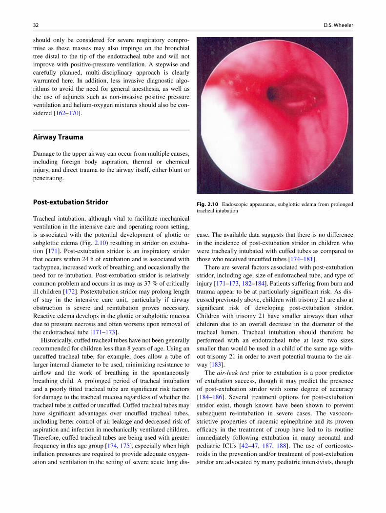
Tracheal intubation, although vital to facilitate mechanical ventilation in the intensive care and operating room setting, is associated with the potential development of glottic or subglottic edema (Fig. 2.10) resulting in stridor on extuba-tion [171]. Post-extubation stridor is an inspiratory stridor that occurs within 24 h of extubation and is associated with tachypnea, increased work of breathing, and occasionally the need for re-intubation. Post-extubation stridor is relatively common problem and occurs in as may as 37 % of critically ill children [172]. Postextubation stridor may prolong length of stay in the intensive care unit, particularly if airway obstruction is severe and reintubation proves necessary. Reactive edema develops in the glottic or subglottic mucosa due to pressure necrosis and often worsens upon removal of the endotracheal tube [171–173].
Historically, cuffed tracheal tubes have not been generally recommended for children less than 8 years of age. Using an uncuffed tracheal tube, for example, does allow a tube of larger internal diameter to be used, minimizing resistance to airflow and the work of breathing in the spontaneously breathing child. A prolonged period of tracheal intubation and a poorly fitted tracheal tube are significant risk factors for damage to the tracheal mucosa regardless of whether the tracheal tube is cuffed or uncuffed. Cuffed tracheal tubes may have significant advantages over uncuffed tracheal tubes, including better control of air leakage and decreased risk of aspiration and infection in mechanically ventilated children. Therefore, cuffed tracheal tubes are being used with greater frequency in this age group [174, 175], especially when high inflation pressures are required to provide adequate oxygen-ation and ventilation in the setting of severe acute lung dis-
ease. The available data suggests that there is no difference in the incidence of post-extubation stridor in children who were tracheally intubated with cuffed tubes as compared to those who received uncuffed tubes [174–181].
There are several factors associated with post-extubation stridor, including age, size of endotracheal tube, and type of injury [171–173, 182–184]. Patients suffering from burn and trauma appear to be at particularly significant risk. As dis-cussed previously above, children with trisomy 21 are also at significant risk of developing post-extubation stridor. Children with trisomy 21 have smaller airways than other children due to an overall decrease in the diameter of the tracheal lumen. Tracheal intubation should therefore be performed with an endotracheal tube at least two sizes smaller than would be used in a child of the same age with-out trisomy 21 in order to avert potential trauma to the air-way [183].
The air-leak test prior to extubation is a poor predictor of extubation success, though it may predict the presence of post-extubation stridor with some degree of accuracy [184–186]. Several treatment options for post-extubation stridor exist, though known have been shown to prevent subsequent re-intubation in severe cases. The vasocon-strictive properties of racemic epinephrine and its proven efficacy in the treatment of croup have led to its routine immediately following extubation in many neonatal and pediatric ICUs [42–47, 187, 188]. The use of corticoste-roids in the prevention and/or treatment of post-extubation stridor are advocated by many pediatric intensivists, though
Fig. 2.10 Endoscopic appearance, subglottic edema from prolonged tracheal intubation
D.S. Wheeler
33
there is very little evidence to support the universal use of corticosteroids at this time. Dexamethasone at a dose of 1–1.5 mg/kg/day divided every 6–8 h (maximum daily dose 40 mg/day) has also been administered in an attempt to interrupt the progressive cycle of inflammation that results in edema of injured tissue following extubation. Again, the evidence that this therapy prevents re-intubation is limited [189–196], though one meta-analysis suggested a possible benefit [193]. Regardless, in a national survey of pediatric critical care fellowship program directors, 66 % of those surveyed continue to rely on the air leak test and use corti-costeroids to prevent post-extubation stridor and extubation failure. Further, the majority stated that they would delay extubation and administer corticosteroids in the presence an air leak of ≥ 30 cm H2O [194]. Finally, helium- oxygen mixtures (see preceding discussion) have also been used in several studies [195–201]. Heliox should be viewed only as a temporizing measure until either the aforementioned therapies become effective or the disease process naturally resolves. In the majority of cases, post-extubation stridor is self limited, though re-intubation is occasionally required. Unfortunately, re-intubation further exacerbates the reac-tive airway edema. Ideally, a smaller tracheal tube (gen-erally one size smaller) than previously used should be placed with the hope of causing less airway injury. The new tracheal tube is generally left in place until air-leak
is observed 24–48 h later. Anatomic airway problems like subglottic stenosis, tracheal compression, etc should be considered if post-extubation stridor persists following the second attempt at extubation.
Foreign Body Aspiration
Foreign body aspiration is an important cause of accidental death in infants and young children, compounded by the fact that infants seem to place almost any object in their mouths [202]. While most foreign bodies pass through the vocal cords and lodge in the lower airways, laryngeal foreign bodies are not uncommon and are immediately life-threatening. The clinical presentation depends upon the location of the foreign body as well as the degree of obstruction. Importantly, the actual aspira-tion event is not always identified and a high index of suspicion is required. Foreign bodies that are lodged in the glottic or sub-glottic airway (extrathoracic obstruction) often produce symp-toms that mimic croup such as sudden onset of stridor and respiratory distress. In contrast, foreign bodies lodged in the distal trachea (intrathoracic obstruction) tend to produce coughing and wheezing, mimicking asthma or bronchiolitis.
The most commonly aspirated foreign bodies include veg-etable matter like peanuts, grapes, and popcorn [202]. Large objects that are lodged in the proximal esophagus and apply
a b
Fig. 2.11 Radiopaque foreign body (coin) in the esophagus. (a) AP Chest view, (b) Lateral Neck view
2 Life-Threatening Diseases of the Upper Respiratory Tract
34
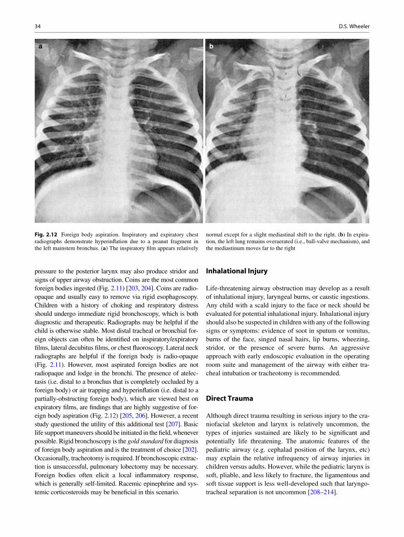
pressure to the posterior larynx may also produce stridor and signs of upper airway obstruction. Coins are the most common foreign bodies ingested (Fig. 2.11) [203, 204]. Coins are radio-opaque and usually easy to remove via rigid esophagoscopy. Children with a history of choking and respiratory distress should undergo immediate rigid bronchoscopy, which is both diagnostic and therapeutic. Radiographs may be helpful if the child is otherwise stable. Most distal tracheal or bronchial for-eign objects can often be identified on inspiratory/expiratory films, lateral decubitus films, or chest fluoroscopy. Lateral neck radiographs are helpful if the foreign body is radio-opaque (Fig. 2.11). However, most aspirated foreign bodies are not radiopaque and lodge in the bronchi. The presence of atelec-tasis (i.e. distal to a bronchus that is completely occluded by a foreign body) or air trapping and hyperinflation (i.e. distal to a partially- obstructing foreign body), which are viewed best on expiratory films, are findings that are highly suggestive of for-eign body aspiration (Fig. 2.12) [205, 206]. However, a recent study questioned the utility of this additional test [207]. Basic life support maneuvers should be initiated in the field, whenever possible. Rigid bronchoscopy is the gold standard for diagnosis of foreign body aspiration and is the treatment of choice [202]. Occasionally, tracheotomy is required. If bronchoscopic extrac-tion is unsuccessful, pulmonary lobectomy may be necessary. Foreign bodies often elicit a local inflammatory response, which is generally self- limited. Racemic epinephrine and sys-temic corticosteroids may be beneficial in this scenario.
Inhalational Injury
Life-threatening airway obstruction may develop as a result of inhalational injury, laryngeal burns, or caustic ingestions. Any child with a scald injury to the face or neck should be evaluated for potential inhalational injury. Inhalational injury should also be suspected in children with any of the following signs or symptoms: evidence of soot in sputum or vomitus, burns of the face, singed nasal hairs, lip burns, wheezing, stridor, or the presence of severe burns. An aggressive approach with early endoscopic evaluation in the operating room suite and management of the airway with either tra-cheal intubation or tracheotomy is recommended.
Direct Trauma
Although direct trauma resulting in serious injury to the cra-niofacial skeleton and larynx is relatively uncommon, the types of injuries sustained are likely to be significant and potentially life threatening. The anatomic features of the pediatric airway (e.g. cephalad position of the larynx, etc) may explain the relative infrequency of airway injuries in children versus adults. However, while the pediatric larynx is soft, pliable, and less likely to fracture, the ligamentous and soft tissue support is less well-developed such that laryngo-tracheal separation is not uncommon [208–214].
a b
Fig. 2.12 Foreign body aspiration. Inspiratory and expiratory chest radiographs demonstrate hyperinflation due to a peanut fragment in the left mainstem bronchus. (a) The inspiratory film appears relatively
normal except for a slight mediastinal shift to the right. (b) In expira-tion, the left lung remains overaerated (i.e., ball-valve mechanism), and the mediastinum moves far to the right
D.S. Wheeler
35
Blunt trauma to the airway appears to be more common in children compared to penetrating trauma and is more common in the adolescent age group [208–214]. The most frequent causes of injury are motor vehicle accidents or direct blows to the larynx. Edema and hematoma formation frequently lead to acute upper airway obstruction. Although less common compared to adults, laryngeal fractures occasionally occur. Laryngotracheal separation, while relatively uncommon, is potentially life-threatening. The severity of airway obstruc-tion dictates the extent of the initial evaluation and manage-ment. An unstable airway should be immediately secured using the flexible bronchoscope. Tracheal intubation without endoscopic evaluation is best avoided. If immediate surgical intervention is required, tracheotomy is preferable to cricothy-rotomy. If the airway is stable, radiographic evaluation should include chest radiograph (to look for associated injuries, such as pneumothorax, pneumomediastinum, or subcutaneous emphysema), lateral neck radiograph (to evaluate the cervical spine), and CT. A barium swallow may be helpful to rule out the possibility of esophageal tear or laceration [208–214].
References
1. Dickison AE. The normal and abnormal pediatric upper airway. Recognition and management of obstruction. Clin Chest Med. 1987;8:583–96.
2. Westmore RF. Management of acute airway obstruction. In: Westmore RF, Muntz HR, McGill TJI, editors. Pediatric otolaryn-gology. Principles and practice pathways. New York: Thieme Medical Publishers; 2000. p. 845–62.
3. Cross KW, Tizard JP, Trythall DA. The gaseous metabolism of the newborn infant. Acta Paediatr. 1957;46:379–84.
5. Meert KL, Donaldson A, Nadkarni V, Tieves KS, Schleien CL, Brilli RJ, Clark RS, Shaffner DH, Levy F, Statler K, Dalton HJ, van der Jagt EW, Hackbarth R, Pretzlaff R, Hernan L, Dean JM, Moler FW. Multicenter cohort study of in-hospital pediatric cardiac arrest. Pediatr Crit Care Med. 2009;10:544–53.
6. Nitta M, Iwami T, Kitamura T, Nadkarni VM, Berg RA, Shimizu N, Ohta K, Nishiuchi T, Hayashi Y, Hiraide A, Tamai H, Kobayashi M, Morita H. Age-specific differences in outcomes after out-of- hospital cardiac arrests. Pediatrics. 2011;128:e812–20.
7. Moler FW, Donaldson AE, Meert K, Brilli RJ, Nadkarni V, Shaffner DH, Schleien CL, Clark RS, Dalton HJ, Statler K, Tieves KS, Hackbarth R, Pretzlaff R, van der Jagt EW, Pineda J, Hernan L, Dean JM. Multicenter cohort study of out-of-hospital pediatric car-diac arrest. Crit Care Med. 2011;39:141–9.
8. Rotta AT, Wiryawan B. Respiratory emergencies in children. Respir Care. 2003;48:248–58.
9. McNiece WL, Dierdorf SF. The pediatric airway. Semin Pediatr Surg. 2004;13:152–65.
10. Zalzal GH. Stridor and airway compromise. Pediatr Clin N Am. 1989;36:1389–402.
11. Zalzal GH. Pediatric stridor and airway management. Int Congr Ser. 2003;1240:803–8.
12. Hollinger ID. Etiology of stridor in the neonate, infant, and child. Ann Otol Rhinol Laryngol. 1980;89:397–400.
13. Hirschberg J. Acoustic analysis of pathological cries, stridor, and coughing sounds in infancy. Int J Pediatr Otorhinolaryngol. 1980;2:287–300.
14. Snyder SR, Kivlehan SM, Collopy KT. What’s behind stridor? Case studies in diagnosis and care. Airway obstruction could be imminent with this alarming sound. EMS World. 2013;42:30–31, 33–34, 36.
15. Lusk RP, Khosla S. Principles of fluid dynamics. In: Holinger LD, Lusk RP, Green CG, editors. Pediatric laryngology and broncho-esophagology. Philadelphia: Lippincott-Raven; 1997. p. 381–91.
16. Hess DR, Fink JB, Venkataraman ST, Kim IK, Myers TR, Tano BD. The history and physics of heliox. Respir Care. 2006;51:608–12.
17. Sie KCY. Infectious and inflammatory disorders of the larynx and trachea. In: Westmore RF, Muntz HR, McGill TJI, editors. Pediatric otolaryngology. Principles and practice pathways. New York: Thieme Medical Publishers; 2000. p. 811–25.
19. Myer CM. Inflammatory diseases of the pediatric airway. In: Cotton RT, Myer CM, editors. Practical pediatric otolaryngology. Philadelphia: Lippincott-Raven; 1999. p. 547–59.
20. Leung AK, Kellner JD, Johnson DW. Viral croup: a current per-spective. J Pediatr Health Care. 2004;18:297–301.
21. Cunningham MJ. The old and new of acute laryngotracheal infec-tions. Clin Pediatr (Phila). 1992;31:56–64.
22. Denny FW, Murphy TF, Clyde Jr WA, Collier AM, Henderson FW. Croup: an 11-year study in a pediatric practice. Pediatrics. 1983;71:871–6.
23. Yang TY, Lu CY, Kao CL, Chen RT, Ho YH, Yang SC, Lee PI, Chen JM, Lee CY, Huang LM. Clinical manifestations of parainfluenza infection in children. J Microbiol Immunol Infect. 2003;36:270–4.
24. Crowe Jr JE. Human metapneumovirus as a major cause of human respiratory tract disease. Pediatr Infect Dis J. 2004;23:S215–21.
25. Rihkanen H, Ronkko E, Nieminen T, Komsi KL, Raty R, Saxen H, Ziegler T, Roivainen M, Soderlund-Venermo M, Beng AL, Hovi T, Pitkaranta A. Respiratory viruses in laryngeal croup of young chil-dren. J Pediatr. 2008;152:661–5.
26. Kairys SW, Olmstead EM, O’Connor GT. Steroid treatment of laryngotracheitis: a meta-analysis of the evidence from randomized trials. Pediatrics. 1989;83:683–93.
27. Durward AD, Nicoll SJ, Oliver J, Tibby SM, Murdoch IA. The outcome of patients with upper airway obstruction transported to a regional paediatric intensive care unit. Eur J Pediatr. 1998;157:907–11.
28. Dobrovoljac M, Geelhoed GC. 27 years of croup: an update high-lighting the effectiveness of 0.15 mg/kg dexamethasone. Emerg Med Australas. 2009;21:309–14.
29. Tibballs J, Watson T. Symptoms and signs differentiating croup and epiglottitis. J Paediatr Child Health. 2011;47:77–82.
30. Chin R, Browne GJ, Lam LT, McCaskill ME, Fasher B, Hort J. Effectiveness of a croup clinical pathway in the management of children with croup presenting to an emergency department. J Paediatr Child Health. 2002;38:382–7.
31. Fitzgerald DA, Mellis CM, Johnson M, Allen H, Cooper P, Van Asperen P. Nebulized budesonide is as effective as nebulized adren-aline in moderately severe croup. Pediatrics. 1996;97:722–5.
32. Westley CR, Cotton EK, Brooks JG. Nebulized racemic epineph-rine by IPPB for the treatment of croup: a double-blind study. Am J Dis Child. 1978;132:484–7.
33. Husby S, Agertoft L, Mortensen S, Pedersen S. Treatment of croup with nebulized steroid (budesonide): a double blind, placebo con-trolled study. Arch Dis Child. 1993;68:352–5.
34. Skolnik NS. Treatment of croup: a critical review. Am J Dis Child. 1989;36:1389–402.
35. Dawson KP, Steinberg A, Capaldi N. The lateral radiograph of neck in laryngo-tracheo-bronchitis (croup). J Qual Clin Pract. 1994;14:39–43.
36. Huang CC, Shih SL. Images in clinical medicine. Steeple sign of croup. N Engl J Med. 2012;367:66.
37. Swishchuk LE. Upper airway, nasal passages, sinuses, and mas-toids. In: Swishchuk LE, editor. Emergency radiology of the acutely ill and injured child. 2nd ed. Baltimore: Williams & Wilkins; 1986. p. 127–40.
2 Life-Threatening Diseases of the Upper Respiratory Tract
36
38. Bourchier D, Dawson KP, Fergusson DM. Humidification in viral croup: a controlled trial. Aust Paediatr J. 1984;20:289–91.
40. Neto GM, Kentab O, Klassen TP, Osmond MH. A randomized con-trolled trial of mist in the acute treatment of moderate croup. Acad Emerg Med. 2002;9:873–9.
41. Scolnik D, Coates AL, Stephens D, Da Silva Z, Lavine E, Schuh S. Controlled delivery of high vs low humidity vs mist therapy for croup in emergency departments: a randomized controlled trial. JAMA. 2006;295:1274–80.
42. Waisman Y, Klein BL, Boenning DA, et al. Prospective randomised double-blind study comparing L-epinephrine and racemic epi-nephrine aerosols in the treatment of laryngotracheitis (croup). Pediatrics. 1992;89:302–6.
43. Prendergast M, Jones JS, Hartman D. Racemic epinephrine in the treatment of laryngotracheitis: can we identify children for outpa-tient therapy? Am J Emerg Med. 1994;12:613–6.
44. Gardner HG, Powell KR, Roden VJ, Cherry JD. The evaluation of racemic epinephrine in the treatment of infectious croup. Pediatrics. 1973;52:52–5.
45. Kristjansson S, Berg-Kelly K, Winso E. Inhalation of racemic adrenaline in the treatment of mild and moderately severe croup: Clinical symptom score and oxygen saturation measurements for evaluation of treatment effects. Acta Paediatr. 1994;83:1156–60.
46. Duman M, Ozdemir D, Atasever S. Nebulised L-epinephrine and steroid combination in the treatment of moderate to severe croup. Clin Drug Investig. 2005;25:183–9.
47. Argent AC, Hatherill M, Newth CJ, Klein M. The effect of epinephrine by nebulization on measures of airway obstruc-tion in patients with acute severe croup. Intensive Care Med. 2008;34:138–47.
48. Rizos JD, DiGravio BE, Sehl MJ, Tallon JM. The disposition of children with croup treated with racemic epinephrine and dexameth-asone in the emergency department. J Emerg Med. 1998;16:535–9.
49. Ausejo M, Saenz A, Pham B, Kellner JD, Johnson DW, Moher D, Klassen TP. The effectiveness of glucocorticoids in treating croup: meta-analysis. BMJ. 1999;319:595–600.
50. Geelhoed GC, Macdonald WB. Oral and inhaled steroids in croup: a randomized, placebo-controlled trial. Pediatr Pulmonol. 1995;20:355–61.
51. Johnson DW, Jacobson S, Edney PC, Hadfield P, Mundy ME, Schuh S. A comparison of nebulized budesonide, intramuscular dexamethasone, and placebo for moderately severe croup. N Engl J Med. 1998;339:498–503.
52. Klassen TP, Craig WR, Moher D, Osmond MH, Pasterkamp H, Sutcliffe T, et al. Nebulized budesonide and oral dexamethasone for treatment of croup: a randomized, controlled trial. JAMA. 1998;279:1629–32.
53. Donaldson D, Poleski D, Knipple E, Filips K, Reetz L, Pascual RG, Jackson RE. Intramuscular versus oral dexamethasone for the treat-ment of moderate-to-severe croup: a randomized, double- blind trial. Acad Emerg Med. 2003;10:16–21.
54. Eboriadou M, Chryssanthopoulou D, Stamoulis P, Damianidou L, Haidopoulou K. The effectiveness of local corticosteroids therapy in the management of mild to moderate viral croup. Minerva Pediatr. 2010;62:23–8.
55. Dobrovoljac M, Geelhoed GC. How fast does oral dexamethasone work in mild to moderately severe croup? A randomized double- blinded clinical trial. Emerg Med Australas. 2012;24:79–85.
56. Cetinkaya F, Tufekci BS, Kutluk G. A comparison of nebulized budesonide, and intramuscular, and oral dexamethasone fo treat-ment of croup. Int J Pediatr Otorhinolaryngol. 2004;68:453–6.
57. Bjornson CL, Klassen TP, Williamson J, Brant R, Mitton C, Plint A, Bulloch B, Evered L, Johnson DW. A randomized trial of a sin-gle dose of oral dexamethasone for mild croup. N Engl J Med. 2004;351:1306–13.
58. Geelhoed GC. Budesonide offers no advantage when added to oral dexamethasone in the treatment of croup. Pediatr Emerg Care. 2005;21:359–62.
59. Amir L, Hubermann H, Halevi A, Mor M, Mimouni M, Waisman Y. Oral betamethasone versus intramuscular dexamethasone for the treatment of mild to moderate viral croup: a prospective, random-ized trial. Pediatr Emerg Care. 2006;22:541–4.
60. Sparrow A, Geelhoed G. Prednisolone versus dexametha-sone in croup: a randomized equivalence trial. Arch Dis Child. 2006;91:580–3.
61. Fifoot AA, Ting JY. Comparison between single-dose oral prednis-olone and oral dexamethasone in the treatment of croup: a random-ized, double-blinded trial. Emerg Med Australas. 2007;19:51–8.
62. Chub-Uppakarn S, Sangsupawanich P. A randomized compari-son of dexamethasone 0.15 mg/kg versus 0.6 mg/kg for the treat-ment of moderate to severe croup. Int J Pediatr Otorhinolaryngol. 2007;71:473–7.
63. Falkenstein E, Wehling M. Nongenomically initiated steroid actions. Eur J Clin Invest. 2000;30:51–4.
64. Klaustermeyer WB, Hale FC. The physiologic effect of an intravenous glucocorticoid in bronchial asthma. Ann Allergy. 1976;37:80–6.
65. Wolfson DH, Nypaver MM, Blaser M, Hogan A, Evans RI, Davis AT. A controlled trial of methylprednisolone in the early emergency department treatment of acute asthma in children. Pediatr Emerg Care. 1994;10:335–8.
66. Rodrigo C, Rodrigo G. Early administration of hydrocortisone in the emergency room treatment of acute asthma: a controlled clini-cal trial. Respir Med. 1994;88:755–61.
67. Gupta VK, Cheifetz IM. Heliox administration in the pediatric intensive care unit: an evidence-based review. Pediatr Crit Care Med. 2005;6:204–11.
68. Duncan PG. Efficacy of helium-oxygen mixtures in the manage-ment of severe viral and post-intubation croup. Can Anaesth Soc J. 1979;26:206–12.
69. Skrinskas GJ, Hyland RH, Hutcheon MA. Using helium-oxygen mixtures in the management of acute airway obstruction. Can Med Assoc J. 1983;128:555–8.
70. Mizrahi S, Yaari Y, Lugassy G, Cotev S. Major airway obstruc-tion relieved by helium/oxygen breathing. Crit Care Med. 1986;14:986–7.
71. Terregino CA, Nairn SJ, Chansky ME, Kass JE. The effect of heliox on croup: a pilot study. Acad Emerg Med. 1998;5:1130–3.
72. Grosz AH, Jacobs IN, Cho C, Schears GJ. Use of helium-oxygen mixtures to relieve upper airway obstruction in a pediatric popula-tion. Laryngoscope. 2001;111:1512–4.
73. Weber JE, Chudnofsky CR, Younger JG, Larkin GL, Boczar M, Wilkerson MD, Zuriekat GY, Nolan B, Eicke DM. A randomized comparison of helium-oxygen mixture (Heliox) and racemic epi-nephrine for the treatment of moderate to severe croup. Pediatrics. 2001;107:E96.
74. DiCecco RJ, Rega PP. The application of heliox in the management of croup by an air ambulance service. Air Med J. 2004;23:33–5.
75. Ho AM-H, Dion PW, Karmakar MK, Chung DC, Tay BA. Use of heliox in critical upper airway obstruction. Physical and physio-logic considerations in choosing the optimal helium: oxygen mix. Resuscitation. 2002;52:297–300.
76. Wigmore T, Stachowski E. A review of the use of heliox in the criti-cally ill. Crit Care Resusc. 2006;8:64–72.
77. Myers TR. Use of heliox in children. Respir Care. 2006;51:619–31. 78. Vorwerk C, Coats TJ. Use of helium-oxygen mixtures in the
treatment of croup: a systematic review. Emerg Med J. 2008;25: 547–50.
79. Kline-Krammes S, Reed C, Giuliano Jr JS, Schwartz HP, Forbes M, Pope J, Besunder J, Gothard MD, Russell K, Bigham MT. Heliox in children with croup: a strategy to hasten improvement. Air Med J. 2012;31:131–7.
D.S. Wheeler
37
80. Glenn GM, Schofield T, Krober M. Group A streptococcal supra-glottitis. Clin Pediatr (Phila). 1990;29:674–6.
81. Senior BA, Radkowski D, MacArthur C, Sprecher RC, Jones D. Changing patterns in pediatric supraglottitis: a multi-institutional review, 1980 to 1992. Laryngoscope. 1994;104:1314–22.
82. Gorelick MH, Baker MD. Epiglottitis in children, 1979 through 1992. Effects of Haemophilus influenzae type b immunization. Arch Pediatr Adolesc Med. 1994;148:47–50.
83. Gonzalez Valdepena H, Wald ER, Rose E, Ungkanont K, Casselbrant ML. Epiglottitis and Haemophilus influenzae immu-nization: the Pittsburgh experience-a five-year review. Pediatrics. 1995;96:424–7.
84. McEwab J, Giridharan W, Clarke RW, Shears P. Paediatric acute epiglottitis: not a disappearing entity. Int J Pediatr Otorhinolaryngol. 2003;67:317–21.
85. Shah RK, Roberson DW, Jones DT. Epiglottitis in the Hemophilus influenzae type B vaccine era: changing trends. Laryngoscope. 2004;114:557–60.
87. Talan DA, Moran GJ, Pinner RW. Progress toward eliminating Haemophilus influenzae type b disease among infants and children- United States, 1987–1997. Ann Emerg Med. 1999;34:109–11.
88. From the Centers for Disease Control and Prevention. Progress toward elimination of Haemophilus influenzae type b invasive dis-ease among infants and children– United States, 1998–2000. JAMA. 2002;287:2206–7.
89. Brilli RJ, Benzing 3rd G, Cotcamp DH. Epiglottitis in infants less than 2 years of age. Pediatr Emerg Care. 1989;5:16–21.
90. Losek JD, Dewitz-Zink BA, Melzer-Lange M, Havens PL. Epiglottitis: comparison of signs and symptoms in children less than 2 years of age and older. Ann Emerg Med. 1990;19:55–8.
91. Kimmons Jr HC, Peterson BM. Management of acute epiglottitis in pediatric patients. Crit Care Med. 1986;14:278–9.
92. Crockett DM, Healy GB, McGill TJ, Friedman EM. Airway man-agement of acute supraglottitis at the Children’s Hospital, Boston: 1980–1985. Ann Otol Rhinol Laryngol. 1988;97:114–9.
93. Crysdale WS, Sendi K. Evolution in the management of acute epi-glottitis: a 10-year experience with 242 children. Int Anesthesiol Clin. 1988;26:32–8.
94. Damm M, Eckel HE, Jungehulsing M, Roth B. Airway endoscopy in the interdisciplinary management of acute epiglottitis. Int J Pediatr Otorhinolaryngol. 1996;38:41–51.
95. Wald E, Reilly JS, Bluestone CD, Chiponis D. Sulbactam/ampi-cillin in the treatment of acute epiglottitis in children. Rev Infect Dis. 1986;5:S617–9.
96. Sawyer SM, Johnson PD, Hogg GG, Robertson CF, Oppedisano F, MacIness SJ, Gilbert GL. Successful treatment of epiglottitis with two doses of ceftriaxone. Arch Dis Child. 1994;70:129–32.
97. Gonzalez C, Reilly JS, Kenna MA, Thompson AE. Duration of intubation in children with acute epiglottitis. Otolaryngol Head Neck Surg. 1986;95:477–81.
98. Acevedo JL, Lander L, Choi S, Shah RK. Airway management in pediatric epiglottitis: a national perspective. Otolaryngol Head Neck Surg. 2009;140:548–51.
99. Guardiani E, Bliss M, Harley E. Supraglottitis in the era following widespread immunization against Haemophilus influenza Type B: evolving principles in diagnosis and management. Laryngoscope. 2010;120:2183–8.
100. Shah RK, Stocks C. Epiglottitis in the United States: national trends, variances, prognosis, and management. Laryngoscope. 2010;120:1256–62.
101. Bizaki AJ, Numminen J, Vasama JP, Laranee J, Rautiainen M. Acute supraglottitis in adults in Finland: review and analysis of 308 cases. Laryngoscope. 2011;121:2107–13.
102. Jones R, Santos JI, Overall Jr JC. Bacterial tracheitis. JAMA. 1979;242:721–6.
103. Liston SL, Gehrz RC, Siegel LG, Tilelli J. Bacterial tracheitis. Am J Dis Child. 1983;137:764–7.
104. Donaldson JD, Maltby CC. Bacterial tracheitis in children. J Otolaryngol. 1989;18:101–4.
105. Bernstein T, Brilli R, Jacobs B. Is bacterial tracheitis changing? A 14-month experience in a pediatric intensive care unit. Clin Infect Dis. 1998;27:458–62.
106. Salamone FN, Bobbitt DB, Myer CM, Rutter MJ, Greinwald Jr JH. Bacterial tracheitis reexamined: is there a less severe manifes-tation? Otolaryngol Head Neck Surg. 2004;131:871–6.
107. Stroud RH, Friedman NR. An update on inflammatory disorders of the pediatric airway: epiglottitis, croup, and tracheitis. Am J Otolaryngol. 2001;22:268–75.
108. Hopkins A, Lahiri T, Salerno R, Heath B. Changing epidemiology of life-threatening upper airway infections: the reemergence of bacterial tracheitis. Pediatrics. 2006;118:1418–21.
109. Huang YL, Peng CC, Chiu NC, Lee KS, Hung HY, Kao HA, Hsu CH, Chang JH, Huang FY. Bacterial tracheitis in pediatrics: 12 year experience at a medical center in Taiwan. Pediatr Int. 2009;51:110–3.
110. Tebruegge M, Pantazidou A, Thorburn K, Riordan A, Round J, De Munter C, Walters S, Curtis N. Bacterial tracheitis: a multi-centre perspective. Scand J Infect Dis. 2009;41:548–57.
111. Shargorodsky J, Whittemore KR, Lee GS. Bacterial tracheitis: a therapeutic approach. Laryngoscope. 2010;120:2498–501.
112. Miranda AD, Valdez TA, Pereira KD. Bacterial tracheitis: a varied entity. Pediatr Emerg Care. 2011;27:950–3.
113. Lee SS, Schwartz RH, Bahadori RS. Retropharyngeal abscess: epiglottitis of the new millennium. J Pediatr. 2001;138:435–7.
114. Yeoh LH, Singh SD, Rogers JH. Retropharyngeal abscess in a children’s hospital. J Laryngol Otol. 1985;99:555–66.
115. Coutlhard M, Isaacs D. Retropharyngeal abscess. Arch Dis Child. 1991;66:1227–30.
116. Broughton RA. Nonsurgical management of deep neck infections in children. Pediatr Infect Dis J. 1992;11:14–8.
117. Thompson JW, Cohen SR, Reddix P. Retropharyngeal abscess in children: a retrospective and historical analysis. Laryngoscope. 1988;98:589–92.
118. Morrison Jr JE, Pashley NR. Retropharyngeal abscesses in chil-dren: a 10-year review. Pediatr Emerg Care. 1988;4:9–11.
119. Craig FW, Schunk JE. Retropharyngeal abscess in children: clini-cal presentation, utility of imaging, and current management. Pediatrics. 2003;111:1394–8.
120. Coticchia JM, Getnick GS, Yun RD, Arnold JE. Age-, site-, and time-specific differences in pediatric deep neck abscesses. Arch Otolaryngol Head Neck Surg. 2004;130:201–7.
121. Vural C, Gungor A, Comerci S. Accuracy of computerized tomog-raphy in deep neck infections in the pediatric population. Am J Otolaryngol. 2003;24:143–8.
122. Hoffman C, Pierrot S, Contencin P, Morisseau-Durand MP, Manach Y, Cougloigner V. Retropharyngeal infections in children: treatment strategies and outcomes. Int J Pediatr Otorhinolaryngol. 2011;75:1099–103.
123. Grisaru-Soen G, Komisar O, Aizenstein O, Soudack M, Schwartz D, Paret G. Retropharyngeal and parapharyngeal abscess in chil-dren: epidemiology clinical features, and treatment. Int J Pediatr Otorhinolaryngol. 2010;74:1016–20.
124. Asmar BI. Bacteriology of retropharyngeal abscess in children. Pediatr Infect Dis J. 1990;9:595–7.
125. Brook I. Microbiology and management of peritonsillar, retropha-ryngeal, and parapharyngeal abscesses. J Oral Maxillofac Surg. 2004;62:1545–50.
126. Schraff S, McGinn JD, Derkay CS. Peritonsillar abscess in chil-dren: a 10-year review of diagnosis and management. Int J Pediatr Otorhinolaryngol. 2001;57:213–8.
2 Life-Threatening Diseases of the Upper Respiratory Tract
38
127. Millar KR, Johnson DW, Drummond D, Kellner JD. Suspected peritonsillar abscess in children. Pediatr Emerg Care. 2007; 23:431–8.
128. Powell J, Wilson JA. An evidence-based review of peritonsillar abscess. Clin Otolaryngol. 2012;37:136–45.
129. Baldassari C, Shah RK. Pediatric peritonsillar abscess: an over-view. Infect Disord Drug Targets. 2012;12:277–80.
130. Kashima HK, Mounts P, Shah K. Recurrent respiratory papilloma-tosis. Obstet Gynecol Clin North Am. 1996;23:699–706.
131. Peyton Shirley W, Wiatrak B. Is cidofovir a useful adjunctive therapy for recurrent respiratory papillomatosis in children? Int J Pediatr Otorhinolaryngol. 2004;64:413–8.
132. Gallagher TQ, Derkay CS. Pharmacotherapy of recurrent respira-tory papillomatosis: an expert opinion. Expert Opin Pharmacother. 2009;10:645–55.
133. Venkatesan NN, Pine HS, Underbrink MP. Recurrent respiratory papillomatosis. Otolaryngol Clin North Am. 2012;45:671–94.
134. Boglioli LR, Taff ML. Sudden asphyxial death complicating infec-tious mononucleosis. Am J Forensic Med Pathol. 1998;19:174–7.
135. Burstin PP, Marshall CL. Infectious mononucleosis and bilateral peritonsillar abscesses resulting in airway obstruction. J Laryngol Otol. 1998;112:1186–8.
136. Irving JA, Cameron BR, Ludemann JP, Taylor G. Florid infectious mononucleosis: clinicopathological correlation in acute tonsillec-tomy. Int J Pediatr Otorhinolaryngol. 2002;66:87–92.
137. Lobo S, Williams H, Singh V. Massive retropharyngeal lymphade-nopathy in an infant: an unusual presentation of infectious mono-nucleosis. J Laryngol Otol. 2004;118:983–4.
138. Ganzel TM, Goldman JL, Padhya TA. Otolaryngologic clinical patterns in pediatric infectious mononucleosis. Am J Otolaryngol. 1996;17:397–400.
139. Stevenson DS, Webster G, Stewart IA. Acute tonsillectomy in the management of infectious mononucleosis. J Laryngol Otol. 1992;106:989–91.
140. Wohl DL, Isaacson JE. Airway obstruction in children with infec-tious mononucleosis. Ear Nose Throat J. 1995;74:630–8.
141. Pierce RJ, Worsnop CJ. Upper airway function and dysfunction in respiration. Clin Exp Pharmacol Physiol. 1999;26:1–10.
142. Arens R, Marcus CL. Pathophysiology of upper airway obstruc-tion: a developmental perspective. Sleep. 2004;27:997–1019.
143. Erler T, Paditz E. Obstructive sleep apnea syndrome in children: a state-of-the-art review. Treat Respir Med. 2004;3:107–22.
144. Spector A, Scheid S, Hassink S, Deutsch ES, Reilly JS, Cook SP. Adenotonsillectomy in the morbidly obsess child. Int J Pediatr Otorhinolaryngol. 2003;67:359–64.
145. Mitchell RB, Kelly J. Adenotonsillectomy for obstructive sleep apnea in obese children. Otolaryngol Head Neck Surg. 2004;131:104–8.
146. Shine NP, Coates HL, Lannigan FJ. Obstructive sleep apnea, mor-bid obesity, and adenotonsillar surgery: a review of the literature. Int J Pediatr Otorhinolaryngol. 2005;69:1475–82.
147. Shine NP, Coates HL, Lannigan FJ, Duncan AW. Adenotonsillar surgery in morbidly obese children: routine elective admission of all patients to the intensive care unit is unnecessary. Anaesth Intensive Care. 2006;34:724–30.
148. Tweedie DJ, Bajaj Y, Ifeacho SN, Jonas NE, Jephson CG, Cochrane LA, Hartley BE, Albert DM, Wyatt ME. Peri-operative complications after adenotonsillectomy in a UK pediatric tertiary referral centre. Int J Pediatr Otorhinolaryngol. 2012;76:809–15.
149. Gleich SJ, Olson MD, Sprung J, Weingarten TN, Schroeder DR, Warner DO, Flick RP. Perioperative outcomes of severely obese children undergoing tonsillectomy. Paediatr Anaesth. 2012;22:1171–8.
150. Jacobs IN, Gray RF, Todd NW. Upper airway obstruction in chil-dren with down syndrome. Arch Otolaryngol Head Neck Surg. 1996;122:945–50.
151. de Jong AL, Sulek M, Nihill M, Duncan NO, Friedman EM. Tenuous airway in children with trisomy 21. Laryngoscope. 1997;107:345–50.
152. McMurray JS, Myer III CM. Management of chronic airway obstruction. In: Westmore RF, Muntz HR, McGill TJI, editors. Pediatric otolaryngology. Principles and practice pathways. New York: Thieme Medical Publishers; 2000. p. 863–81.
153. Ratner I, Whitfield J. Acquired subglottic stenosis in the very-low- birth-weight infant. Am J Dis Child. 1983;137:40–3.
154. da Silva OP. Factors influencing acquired upper airway obstruc-tion in newborn infants receiving assisted ventilation because of respiratory failure: an overview. J Perinatol. 1996;16:272–5.
155. Wiel E, Vilette B, Darras JA, Scherpereel P, Leclerc F. Laryngotracheal stenosis in children after intubation. Report of five cases. Paediatr Anaesth. 1997;7:415–9.
156. Cotton RT, Myer 3rd CM. Contemporary surgical management of laryngeal stenosis in children. Am J Otolaryngol. 1984;5:360–8.
158. Cotton RT, Myer 3rd CM, Bratcher GO, Fitton CM. Anterior cri-coid split, 1977–1987. Evolution of a technique. Arch Otolaryngol Head Neck Surg. 1988;114:1300–2.
159. Cotton RT. Management of subglottic stenosis. Otolaryngol Clin North Am. 2000;33:111–30.
160. Hartnick CJ, Hartley BE, Lacy PD, Liu J, Willging JP, Meyer 3rd CM, Cotton RT. Surgery for pediatric subglottic stenosis: disease- specific outcomes. Ann Otol Rhinol Laryngol. 2001; 110:1109–13.
161. White DR, Bravo M, Vijayasekaran S, Rutter MJ, Cotton RT, Elluru RG. Laryngotracheoplasty as an alternative to tracheotomy in infants younger than 6 months. Arch Otolaryngol Head Neck Surg. 2009;135:445–7.
162. Dilworth K, Thomas J. Anaesthetic consequences for a child with complex multilevel airway obstruction- recommendations for avoid-ing life-threatening sequelae. Paediatr Anaesth. 2003;13:620–3.
163. Lam JC, Chui CH, Jacobsen AS, Tan AM, Joseph VT. When is a mediastinal mass critical in a child? An analysis of 29 patients. Pediatr Surg Int. 2004;20:180–4.
164. Slinger P, Karsli C. Management of the patient with a large ante-rior mediastinal mass: recurring myths. Curr Opin Anaesthesiol. 2007;20:1–3.
165. Perger L, Lee EY, Shamberger RC. Management of children and adolescents with a critical airway due to compression by an ante-rior mediastinal mass. J Pediatr Surg. 2008;43:1990–7.
166. Bigham MT, Nowak JE, Wheeler DS. Therapeutic application of helium-oxygen and mechanical ventilation in a child with acute myelogenous leukemia and airway obstruction. Pediatr Emerg Care. 2009;25:469–72.
167. Stricker PA, Gurnaney HG, Litman RS. Anesthetic management of children with an anterior mediastinal mass. J Clin Anesth. 2010;22:159–63.
168. Garey CL, Laituri CA, Valusek PA, St. Peter SD, Snyder CL. Management of anterior mediastinal masses in children. Eur J Pediatr Surg. 2011;21:310–3.
169. Bassanezi BS, Oliveira-Filho AG, Miranda ML, Soares L, Aguiar SS. Use of BiPAP for safe anesthesia in a child with a large ante-rior mediastinal mass. Paediatr Anaesth. 2011;21:985–7.
170. Gun F, Erginel B, Unuvar A, Kebudi R, Salman T, Celik A. Mediastinal masses in children: experience with 120 cases. Pediatr Hematol Oncol. 2012;29:141–7.
171. Koka BV, Jeon IS, Andre JM, MacKay I, Smith RM. Postintubation croup in children. Anesth Analg. 1977;56:501–5.
172. Kemper KJ, Benson MS, Bishop MJ. Predictors of postextubation stridor in pediatric trauma patients. Crit Care Med. 1991;19:352–5.
173. Gomes Cordeiro AM, Fernandes JC, Troster EJ. Possible risk fac-tors associated with moderate or severe airway injuries in children
D.S. Wheeler
39
who underwent endotracheal intubation. Pediatr Crit Care Med. 2004;5:364–8.
174. Newth CJ, Rachman B, Patel N, Hammer J. The use of cuffed versus uncuffed endotracheal tubes in pediatric intensive care. J Pediatr. 2004;144:333–7.
175. Silver GM, Freiburg C, Halerz M, Tojong J, Supple K, Gamelli RL. A survey of airway and ventilator management strategies in North American pediatric burn units. J Burn Care Rehabil. 2004;25:435–40.
176. Deakers TW, Reynolds G, Stretton M, Newth CJ. Cuffed endotra-cheal tubes in pediatric intensive care. J Pediatr. 1994;125:57–62.
177. Khine HH, Corddry DH, Kettrick RG, Martin TM, McCloskey JJ, Rose JB, et al. Comparison of cuffed and uncuffed endotracheal tubes in young children during general anesthesia. Anesthesiology. 1997;86:627–31; discussion 27A.
178. Fine GF, Borland LM. The future of the cuffed endotracheal tube. Paediatr Anaesth. 2004;14:38–42.
179. Ashtekar CS, Wardhaugh A. Do cuffed endotracheal tubes increase the risk of airway mucosal injury and post-extubation stridor in children? Arch Dis Child. 2005;90:1198–9.
180. Weber T, Salvi N, Orliaquet G, Wolf A. Cuffed vs non-cuffed endotracheal tubes for pediatric anesthesia. Paediatr Anaesth. 2009;19 Suppl 1:46–54.
181. Dorsey DP, Bowman SM, Klein MB, Archer D, Sharar SR. Perioperative use of cuffed endotracheal tubes is advantageous in young pediatric burn patients. Burns. 2010;36:856–60.
182. Kurachek SC, Newth CJ, Quasney MW, Rice T, Sachdeva RC, Patel NR, Takano J, et al. Extubation failure in pediatric intensive care: a multiple-center study of risk factors and outcomes. Crit Care Med. 2003;31:2657–64.
183. Shott SR. Down syndrome: analysis of airway size and a guide for appropriate intubation. Laryngoscope. 2000;110:585–92.
184. Maury E, Guglielminotti J, Alzieu M, Qureshi T, Guidet B, Offenstadt G. How to identify patients with no risk for postextuba-tion stridor? Crit Care Med. 2004;19:23–8.
185. Mhanna MJ, Zamel YB, Tichy CM, Super DM. The “air leak” test around the endotracheal tube, as a predictor of postextubation stri-dor, is age dependent in children. Crit Care Med. 2002;30:2639–43.
186. Wratney AT, Benjamin Jr DK, Slonim AD, He J, Hamel DS, Cheifetz IM. The endotracheal tube air leak test does not predict extubation outcome in critically ill pediatric patients. Pediatr Crit Care Med. 2008;9:490–6.
187. Kelley PB, Simon JE. Racemic epinephrine use in croup and dis-position. Am J Emerg Med. 1992;10:181–3.
188. Nutman J, Brooks LJ, Deakins KM, Baldesare KK, Witte MK, Reed MD. Racemic versus l-epinephrine aerosol in the treat-ment of postextubation laryngeal edema: results from a prospec-tive, randomized, double-blind study. Crit Care Med. 1994;22: 1591–4.
189. Tellez DW, Galvis AG, Storgion SA, Amer HN, Hoseyni M, Deakers TW. Dexamethasone in the prevention of postextubation stridor in children. J Pediatr. 1991;118:289–94.
190. Couser RJ, Ferrara TB, Falde B, Johnson K, Schilling CG, Hoekstra RE. Effectiveness of dexamethasone in preventing extu-bation failure in preterm infants at increased risk for airway edema. J Pediatr. 1992;121:591–6.
191. Anene O, Meert KL, Uy H, Simpson P, Sarnaik AP. Dexamethasone for the prevention of postextubation airway obstruction: a prospective, randomized, double-blind, placebo-controlled trial. Crit Care Med. 1996;24:1666–9.
192. Meade MO, Guyatt GH, Cook DJ, Sinuff T, Butler R. Trials of corticosteroids to prevent postextubation airway complications. Chest. 2001;120(Suppl):464S–8.
193. Markovitz BP, Randolph AG. Corticosteroids for the prevention of reintubation and postextubation stridor in pediatric patients: a meta-analysis. Pediatr Crit Care Med. 2002;3:223–6.
194. Lukkassen IM, Hassing MB, Markhorst DG. Dexamethasone reduces reintubation rate due to postextubation stridor in a high- risk paediatric population. Acta Paediatr. 2006;95:74–6.
195. Malhotra D, Gurcoo S, Qazi S, Gupta S. Randomized comparative efficacy of dexamethasone to prevent postextubation upper airway complications in children and adults in ICU. Indian J Anaesth. 2009;53:442–9.
196. Saleem AF, Bano S, Haque A. Does prophylactic use of dexa-methasone have a role in reducing post extubation stridor and rein-tubation in children? Indian J Pediatr. 2009;76:555–7.
197. Foland JA, Super DM, Dahdah NS, Mhanna MJ. The use of the air leak test and corticosteroids in intubated children: a sur-vey of pediatric critical care fellowship directors. Respir Care. 2002;47:662–6.
198. Kemper KJ, Izenberg S, Marvin JA, Heimbach DM. Treatment of postextubation stridor in a pediatric patient with burns: the role of heliox. J Burn Care Rehabil. 1990;11:337–9.
199. Kemper KJ, Ritz RH, Benson MS, Bishop MS. Helium-oxygen mixture in the treatment of postextubation stridor in pediatric trauma patients. Crit Care Med. 1991;19:356–9.
200. Jaber S, Carlucci A, Boussarsar M, Fodil R, Pigeot J, Maggiore S, Harf A, Isabey D, Brochard L. Helium–oxygen in the postextuba-tion period decreases inspiratory effort. Am J Respir Crit Care Med. 2001;164:633–7.
201. Berkenbosch JW, Grueber RE, Graff GR, Tobias JD. Patterns of helium-oxygen (heliox) usage in the critical care environment. J Intensive Care Med. 2004;19:335–44.
202. Lima JA, Fischer GB. Foreign body aspiration in children. Paediatr Respir Rev. 2002;3:303–7.
203. Wai Pak M, Chung Lee W, Kwok Fung H, van Hasselt CA. A prospective study of foreign-body ingestion in 311 children. Int J Pediatr Otorhinolaryngol. 2001;58:37–45.
204. Arana A, Hauser B, Hachimi-Idrissi S, Vandeplas Y. Management of ingested foreign bodies in childhood and review of the litera-ture. Eur J Pediatr. 2001;160:468–72.
205. Donnelly LF, Frush DP, Bisset III GS. The multiple presentations of foreign bodies in children. AJR. 1998;170:471–7.
206. Girardi G, Contador AM, Castro-Rodriguez JA. Two new radiological findings to improve the diagnosis of bronchial foreign- body aspiration in children. Pediatr Pulmonol. 2004; 38:261–4.
207. Brown JC, Chapman T, Klein EJ, Chisholm SL, Phillips GS, Osincup D, Sakchalathorn P, Bittner R. The utility of adding expi-ratory or decubitus chest radiographs to the radiographic evalua-tion of suspected pediatric airway foreign bodies. Ann Emerg Med. 2013;61:19–26.
208. Ford HR, Gardner MJ, Lynch JM. Laryngotracheal disruption from blunt pediatric neck injuries: impact of early recognition and intervention on outcome. J Pediatr Surg. 1995;30:331–5.
209. Gold SM, Gerber ME, Shott SR, Myer 3rd CM. Blunt laryngotra-cheal trauma in children. Arch Otolaryngol Head Neck Surg. 1997;123:83–7.
210. Grant WJ, Meyers RL, Jaffe RL, Johnson DG. Tracheobronchial injuries after blunt chest trauma in children-hidden pathology. J Pediatr Surg. 1998;33:1707–11.
211. Cay A, Imamoglu M, Sarihan H, Kosucu P, Bektas D. Tracheobronchial rupture due to blunt trauma in children: report of two cases. Eur J Pediatr Surg. 2002;12:419–22.
212. Lichenstein R, Gillette DL. An unusual presentation of stri-dor: blunt pediatric laryngotracheal trauma. J Emerg Med. 2002;22:375–8.
213. Corsten G, Berkowitz RG. Membranous tracheal rupture in chil-dren following minor blunt cervical trauma. Ann Otol Rhinol Laryngol. 2002;111:197–9.
214. Myer 3rd CM. Trauma of the larynx and craniofacial structures: airway implications. Paediatr Anaesth. 2004;14:103–6.
2 Life-Threatening Diseases of the Upper Respiratory Tract