136
Ligand-Exchange and Metal Complexation studied by Mass Spectrometry Fundamentals and applications in analytical chemistry Hans Krabbe

Ligand-Exchange and Metal Complexation studied by Mass Spectrometry
Fundamentals and applications in analytical chemistry
Hans Krabbe

This thesis was financially supported by Shimadzu Benelux.
ISBN : 978907867528 0

VRIJE UNIVERSITEIT
Ligand-Exchange and Metal Complexation studied by Mass Spectrometry
Fundamentals and applications in analytical chemistry
ACADEMISCH PROEFSCHRIFT
ter verkrijging van de graad Doctor aande Vrije Universiteit Amsterdam,
op gezag van de rector magnificusprof.dr. L.M. Bouter,
in het openbaar te verdedigenten overstaan van de promotiecommissie
van de faculteit der Exacte Wetenschappenop vrijdag 14 december 2007 om 13.45 uur
in de aula van de universiteit,De Boelelaan 1105
door
Johannes Gerardus Krabbe
geboren te Leidschendam

promotoren: prof.dr. H. Irth
prof.dr. W.M.A. Niessen
copromotor: dr. H. Lingeman
Contents
7 Chapter 1. Introduction
21 Chapter 2. Metal complexation, coordination chemistry and ligand-exchange
reactions
37 Scope of thesis
41 Chapter 3. Metal-complex formation in continuous-flow ligand-exchange reactors
studied by electrospray mass spectrometry
59 Chapter 4. Screening for metal ligands by liquid chromatography –ligand-exchange
– electrospray mass spectrometry
81 Chapter 5a. Ligand-Exchange Detection of Phosphorylated Peptides Using Liquid
Chromatography Electrospray Mass Spectrometry
103 Chapter 5b. Selective detection and identification of phosphorylated proteins by
simultaneous ligand-exchange fluorescence detection and mass spectrometry
121 Summary
127 Samenvatting
131 List of publications
133 Dankwoord
136 Curriculum Vitae
5
Contents


7
Chapter 1
1
Chapter 1. Introduction
8
Chapter 1
1
1.1 Metal ions, metal complexes and ligand-exchange reac-tions
Metal ions, metal complexes and metal-ligand exchange reactions are essential in a large variety of natural and industrial processes. Metal ions can be incorporated in proteins, in metal complexes or they can exist as unbound species. These metal ions, either unbound or incorporated in larger molecules, possess a wide variety of functions and properties. For example, metal ions incorporated in proteins are responsible for protein folding like in metallothionein [1, 2] and protein kinase C [3] Metal ions in proteins are also involved in catalytic processes e.g., as nucleophilic catalysts like Zn(II) in carbonic anhydrase [4] or in electron transfer as, for instance, the Fe(III) in rubredoxin and ferredoxin [5]. Metal ions can also be incorporated in other organic as well as inorganic structures, fulfilling a variety of roles, such as for catalysis in (industrial) chemical reactions, e.g., in the Haber-Bosch process where iron(III) acts as a catalyst in the large scale production of NH3 [6], or for medicinal purposes e.g. cisplatin in platinum-based chemotherapy against cancer [7]. Cisplatin uses a series of ligand-exchange reactions in its function to change the structure of DNA, finally leading to death of (tumor) cells. A second example of a ligand-exchange reaction, which is more known and essential to life, is the uptake and delivery of oxygen and carbon dioxide by hemoglobin in the human body.
Due to the importance and the related interest of researchers in metal complexes and metal-ligand interactions in several fields, e.g., drug research [8] and catalyst design [9], novel detection methods are necessary for better under-standing these compounds and their interactions as well as for obtaining quantita-tive information.
1.2 Conventional techniques for studying metal ions and complexes
Conventional techniques to study metal complexes and interactions are spectrophotometry, potentiometry, [10, 11] X-ray diffraction [12] and nuclear magnetic resonance spectroscopy (NMR) [13]. Despite their specific advantages each of these techniques also has significant drawbacks. Spectrophotometry and potentiometry are accurate techniques but only provide indirect information about a specific metal complex. Moreover, the data obtained will be more difficult or even impossible to interpret, when dealing with more than one metal complex present in solution [14]. Occasionally, X-ray diffraction can be applied to study the stoichiometry of the metal complex, although a pure sample in the form of crystal powder should be available. Moreover, studying metal complexes by X-ray diffraction as well as NMR require relatively large amounts of pure compounds

9
Chapter 1
1
[15].
1.3 Mass spectrometry
In the last two decades, mass spectrometry has become increasingly popular for a large variety of applications. More recently, the popularity of using MS for studying metal complexes and metal interactions has increased. The potential of mass spectrometry (MS) for studying metal ions, metal-ligand interactions and metal complexes is readily recognized [16, 17], since direct information on the nature and quantitiy of metal complexes can be obtained with relatively small amounts of complexes, even when present in mixtures. Some of the benefits and advantages of the use of MS in the study of metal ions and their complexes are outlined below.
The specific m/z obtained in MS provides information about the specific metal ion, e.g., which metal ion and/or oxidation state as well as information about the particular form in which the metal ion is present, e.g., complexated with ligand(s). Additionally, MS provides the possibility to analyze complex samples and to monitor complexes in concentrations lower than 1 x 10⁻⁵ M, which is more representative for environmental and biological samples [18]. Moreover, MS enables to simultaneously monitor the response of ligand-exchange reactions and additionally obtain chemical information about the specific compound in one single run [19, 20].
In contrast to, for instance, NMR MS is able to analyze ionic compounds irrespective of the nuclear spin of atoms, and to observe individual ions from a solution in which the species are in equilibrium. Moreover, MS provides the possibility to identify paramagnetic as well as diamagnetic species [21].
Additionally, MS may serve as tool for the direct study of metal complex intermediates which generally cannot be studied with other techniques. [22]. Bortolini et al. [23] demonstrated the use of ESI-MS and NMR in combination with ab initio calculations to identify the structures of highly reactive intermediates of vanadium in the synthesis of vanadium haloperoxidases.
Since a large number of metals possess specific isotopic patterns, and mass spectrometry can distinguish between these different isotopes, nature provides researchers a useful tool for identifying metal species in mass spectra. Moreover, the isotope ratio studied by MS can be used for the direct investigation of different processes, as is for instance demonstrated by Terada et al in the study of the Europium isotopic composition in the investigation of meteorites of stars and supernovae [24].
Several mass spectrometric methods can be applied in studying metal species and interactions, e.g., Inductive Coupled Plasma-MS (ICP-MS), Fast Atom Bombardment-MS (FAB-MS), Atmospheric Pressure Chemical Ionization-MS

10
Chapter 1
1
(APCI-MS), Matrix Assisted Laser Desorption Ionization-MS (MALDI-MS) and Elec-trospray Ionization-MS (ESI-MS). Due to the difference in the generation of ions, the various combinations of ionization techniques and mass spectrometry provide specific and often complementary information. The two currently most common ionization techniques for the study of metal ions and metal-ligand interactions (ICP-MS and ESI-MS) are described in somewhat more detail below.
1.3.1 Inductively coupled plasma mass spectrometry
Conventionally, ICP-MS is used for elemental (trace)-analysis or (metal) speciation analysis. Metal speciation is concerned with the quantitation and iden-tification of metals present in samples. In ICP-MS, the samples are nebulized and the analytes are introduced to an inductively coupled plasma, which atomizes and ionizes the analytes. ICP-MS provides atomic or elemental information, in contrast to, e.g., ESI-MS and MALDI-MS, which provides molecular information [25]. ICP-MS is able to detect monoisotopic positive ions for most elements. The sensitivity depends on the ionization energy of the elements. Due to the generally low ionization energy of metal ions, they are well suited to ICP-MS detection and, therefore, show a high sensitivity in contrast to, for instance, halogens which are less easily ionized and are generally more difficult to detect by ICP-MS [26]. ICP-MS is known for its tolerance to matrix compounds and its large linear range independent of the chemical environment of the analyte. Therefore, it is suitable in the detection of analytes present in difficult matrices. An example of the power of ICP-MS is demonstrated by Palmer et al. in the ecotoxicological biomonitoring of trace amounts of rare earth metals, such as lanthanide, cesium and europium in Paramoera walkeri bacteria in the Antarctica region [27].
The most important disadvantage of ICP-MS is that it only provides information at the atomic level, not about the molecular state of the metal ion. Therefore, it can play a complementary role to other MS techniques, which provide information at the molecular level, especially ESI-MS and MALDI-MS. For example, metallic species of mercury and tin are less toxic than the methylmercury or organotin species [28]. Using ICP-MS no distinction can be made between the metallic and organometallic species, unless a separation is performed prior to detection. In that case distinction can only be made by retention time. ESI-MS and APCI-MS on the other hand can detect intact molecular species and thereby discriminate between different organometallic species in the sample.
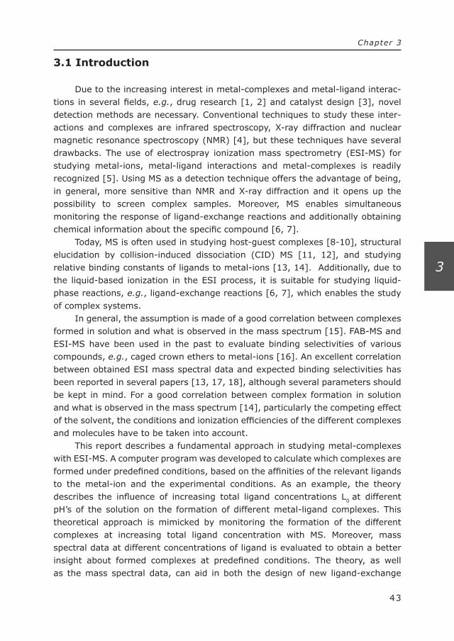
Sacks et al [29] combined the power of ICP-MS and APCI-MS in a parallel approach to obtain elemental and molecular information, respectively, in the analysis of Selenium-amino acid complexes. Because of its high sensitivity for Se, ICP-MS was used to detect low concentration of Se-species in a yeast matrix. In fact, LC–ICP-MS was used to provide a peak recognitioning marker (peak width

Figure 1.1: Combination of ICP-MS and API-MS in one single run for the determination of Se-
species. [29]
11
Chapter 1
1
and retention time) to locate Se-related compounds in the LC–APCI-MS trace. By correlating ICP-MS traces and APCI-MS traces (see Figure 1.1), molecular information of the Se-species was obtained which would be less easily identified or even lost if only the APCI-MS approach was used.
1.3.2 Electrospray ionization mass spectrometry
The benefits of using electrospray ionization mass spectrometry to study metal complexes and metal ligand interactions are easily recognized. ESI-MS has the ability to preserve metal complexes and transfer them from solution phase to gas phase [30]. ESI is a liquid based ionization technique, where the ions are essentially generated already in solution [31]. ESI-MS is generally assumed to provide a good correlation between metal complexes present in solution and those observed in the mass spectrum [21]. Additionally, due to the dynamic character of the liquid-based ionization in the ESI process, it is possible to monitor liquid-phase reactions of metal ions in solution, e.g., ligand-exchange reactions [19, 20], and consequently, to study dynamic complex systems [32]. Unfortunately, the presumed correlation between liquid state and gas phase is not always true since solution phase, electrochemical and gas-phase processes may interfere in the transfer of the complexes from the liquid phase to the gas phase [33-36].
12
Chapter 1
1
Di Marco and Bombi [21] reviewed the application of ESI-MS in studying metal-ligand solution equilibria. They outlined the possibilities, drawbacks and aspects which should be kept in mind when using this technique for studying metal-ligand complexes and metal-ligand solution equilibria. They reported that although ESI-MS in metal-ligand solution equilibria is still not fully understood, ESI-MS provides in many cases very useful and representative results in the characterization and quantitation of metal complexes. It should, however, be noted that that there is not always a good correlation between solution phase binding constants and the metal complex composition observed in the mass spectrum [37]. Shen and Brodbelt studied the complexes of two ligands with large differences in affinity to a metal ion by ESI-MS. Despite the large differences in affinity, unexpected mixed complexes are observed in the mass spectra, demonstrating that the ESI process has a preference which not necessarily represents the affinity in solution.
Although ESI-MS is frequently used to analyze highly polar compounds such as metal complexes, APCI-MS can also be applied for this purpose. In contrast to ESI, the ionization in APCI is performed in the gas phase. The choice between APCI-MS or ESI-MS can be made based on the polarity of the metal complex. Less polar complexes are generally better suited for analysis with APCI-MS. Pereira et al. demonstrated the use of APCI-MS for the structural elucidation of several labile iridium complexes. [38]
1.4 Applications of MS in studying metal-ligand complexes and interactions
Mass spectrometry has been used for several applications ranging from metal speciation as described in the ICP-MS section, structural elucidation of metal-ligand complexes and organometallic compounds [39, 40], studying relative binding constants [41] and studying gas-phase reactions [42, 43].
1.4.1 Structural elucidation of metal complexes using mass spec-trometry
In the structural elucidation of metal complexes, mass spectrometry can be a useful tool [44]. As an example, Hansen et al. [39] studied and characterized complexes of several nucleotides and related compounds (adenosine, cytidine, guanosine, uridine, adenosine-5′-monophosphate, adenosine-3′,5′-cyclic mono-phosphate, ribose, or 2′-deoxyadenosine) with antimony (Sb(V)) using collision-induced dissociation (CID). Vachet et al. [45] studied the influence of the ligand donor group on dissociation of Cu(II) complexes. They observed a trend between the intrinsic binding strength of the functional group (ligand) either to Cu(II)
13
Chapter 1
1
and Cu(I) and the ligand remaining coordinated to the Cu(II) and Cu(I) upon competition. This trend can also be related to the flexibility of the functional group and, therefore, is related to the ability to direct the dipole moment towards the metal ion. Jellen et al [46] reported that the stability of a complex in CID conditions is affected by the size of the complex, meaning the overall number of vibrational degrees of freedom. They studied metal complexes with similar binding energies and varying number of degrees of freedom reporting a linear effect on its stability related to the number of degrees freedom.
Next to the study of metal complexes in solution phase, metal complexes can also be studied in the gas phase, thereby obtaining intrinsic (or solvent free) metal-ligand interactions. One approach is to introduce a metal complex, or several metal complexes into an ion-trap MS, followed by isolation of the specific metal complex. After isolation, the selected complex is subjected to gas phase ligands introduced in the ion-trap. Vachet et al. [47] subjected 2nd row transition metals including manganese and zinc to gas phase reagents such as ammonia, water and methanol. Depending on the initial coordination of the metal ion, the gas-phase complexation reaction is studied as a function of the electrondonating properties of the reagent ligands, the type of metal ion and its electronic structure. Depending on the coordination number, the different initial complexes reacted differently with the reagent gasses.
In some cases, structure elucidation of metal complexes by CID-MS can result in ambiguous information, for instance if internal fragmentation of a ligand is occurring instead of the dissociation of the coordination bond [15].
1.4.2 Studying relative binding constants by ESI-MS and host-guest complex formation
FAB-MS and ESI-MS have been used to evaluate binding selectivities of various compounds [48]. Brodbelt et al. [49-51] for example describe the use of ESI-MS in the study of relative binding constants of specific ligands, e.g., crown ethers and lariat ethers, with alkali metal ions.
A powerful approach in the study of relative binding constants or relative binding selectivities is the host-guest approach. In host-guest interactions usually only one known complex with a known stoichiometry is formed. The host-guest approach is used to quantitatively determine either the selectivity factors of the ligand for a series of metal ions, the stability constant of the complex or the selectivity factor of a series of ligands for a certain metal ion. In these type of studies the response factor of the different complexes formed is an important parameter, which may obscure the interpretation of metal binding selectivities. Complexes may show different ionization efficiencies, resulting in an indirect rela-tionship between response and relative binding selectivities. With respect to this,
14
Chapter 1
1
Leize et al. reported that the ESI response factor of an ion is related to its solvation energy [52]. They showed that under given experimental conditions host-guest complexes of, e.g., crown ethers, with metal ions of the same charge have similar response factors. Therefore, relative intensities in ESI-MS closely correspond to the relative concentrations of the ions in the solution being analyzed. Additionally, Gabelica et al. [53] reported that the influence of the response factor on equilibra-tion association constants for complexes does not comply with the requirements stated by Leize. The response function derived provided a tool for the correction for differences in response factors of the free ligand and the metal complex, mass discrimination of the mass spectrometer and/or moderate in-source dissociation.
Literature usually describes the study of relative binding constants, e.g., using the host-guest approach, for metal-ligand complexes with a 1:1 ratio [51, 54]. A higher ratio of metal:ligand ratio makes the interpretation of the mass spectral data and the correlation to the theoretical affinity of ligands towards a metal ion more complicated if not impossible The different metal:ligand complexes may show different ionization efficiencies and thereby respond differently to the addition of an additional ligand or metal ion. Often, the ionization efficiencies of the 1:1 complexes are believed to be similar, although this is not necessarily true. Additionally, the ionization efficiencies of higher ratio metal:ligand complexes are usually different. Therefore, the interpretation of the intensities in the mass spectrum must be done with great care. In such studies, the use of continuous-flow ligand-exchange reactors can play a useful role [19, 20].
1.4.3 Coordination Electrospray Mass Spectrometry
A different application of metal ions is in coordination electrospray mass spectrometry [55]. In coordination electrospray mass spectrometry a metal ion is pre- or post-column mixed with the analytes of interest. In the post-column approach, the metal ion is mixed after the separation of the analytes. Analytes with affinity for the metal ion will be complexated, and the metal complex formed is introduced into the MS. In the pre-column approach, the metal ion is already premixed with the analyte, prior to separation and MS-analysis. Depending on the stability of metal complex and the influence of the metal complexation on the separation a pre- or post-column approach is chosen [56] although one should keep in mind that metal complexes may dissociate during the separation process. The aim of coordination electrospray mass spectrometry and the metal complex-ation can be twofold. Firstly, it may provide an improved ionization efficiency as for instance demonstrated for Ag+ complexation of (nonpolar) compounds such as fatty acids, flavanoids, amino acids, glycosides and aromatic compounds. The second goal is to influence the fragmentation behavior in CID. Both aims are combined in the Ag+-complexation of flavanoids as demonstrated by Zhang and
15
Chapter 1
1
Brodbelt [57]. Enhanced ionization was achieved for some flavanoids that are not readily protonated in ESI. In the structural characterization of isomeric flavanoid diglycosides, it was found that [Ag – flavanoids]+ complexes displayed unique fragments allowing to distinguish flavanoids diglycosides in each isomeric series.
Leary et al. [58] demonstrated that coordination electrospray-MS can aid to elucidate the linkage position of the sialic acid moiety in complex oligosaccharides derived from glycoproteins. Due to the unstable glycosidic bond between sialic acid and the oligosaccharide, conventional CID-MS can not be applied. By metal complexation, the glycosidic bond to the sialic acid is stabilized.
1.5 Considerations for studying metal-ligand complexes in solution by ESI-MS
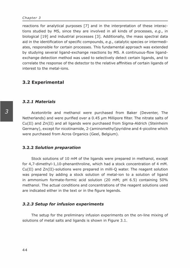
The ESI mass spectra are assumed to reflect the constituents present in solution. However, various processes may be responsible for deviations of the alleged assumption. The changing solution conditions during droplet evaporation, e.g., changes in pH [59] and/or ionic strength or an unequal evaporation of constituents [21], may alter the metal complex or the equilibrium conditions. A second aspect is that the gas phase ions generated may undergo gas phase reactions, such as CID and/or the formation of gas phase adducts. Moreover, ion adducts, e.g., with NH4
+ or Na+ in positive ion mode and HCOO– and CH3COO– in negative ion mode can be formed either in the liquid phase or in the gas phase. Moreover, species which are stable in solution may not be stable in the gas phase due to the absence of solvent [18]. All the processes may have an impact on the appearance of the mass spectra observed.
Another problem in the study of metal complexes and their interactions is the occurrence of charge reduction [21, 60]. Charge reduction is caused by evaporation of solvent from the droplets, produced in the ESI process. While the solvent stabilizes the charge on the metal ion in solution, it may induce reduction of the metal ion in the evaporating droplet, e.g., by removing H+ from water;
[M(H2O)n]3+ + H2O [M(OH)n-1]
2+ + [H3O]+
The charge reduction results in a change in the oxidation state of the metal ion and the accompanied chemical properties. This charge reduction occurs especially when the ionization potential of the metal ion is large in comparison with the ionization energy of the solvent. In contrast to protic solvents, non-protic solvents like acetonitrile and dimethyl sulfoxide will stabilize the charge on the metal ion in the gas phase [21, 61]. This explains the preference of using non-protic solvents in the study of multiple charge metal ions and complexes. These effects are not only exerted by solvent molecules, but also by ligands. Fortunately, due to the
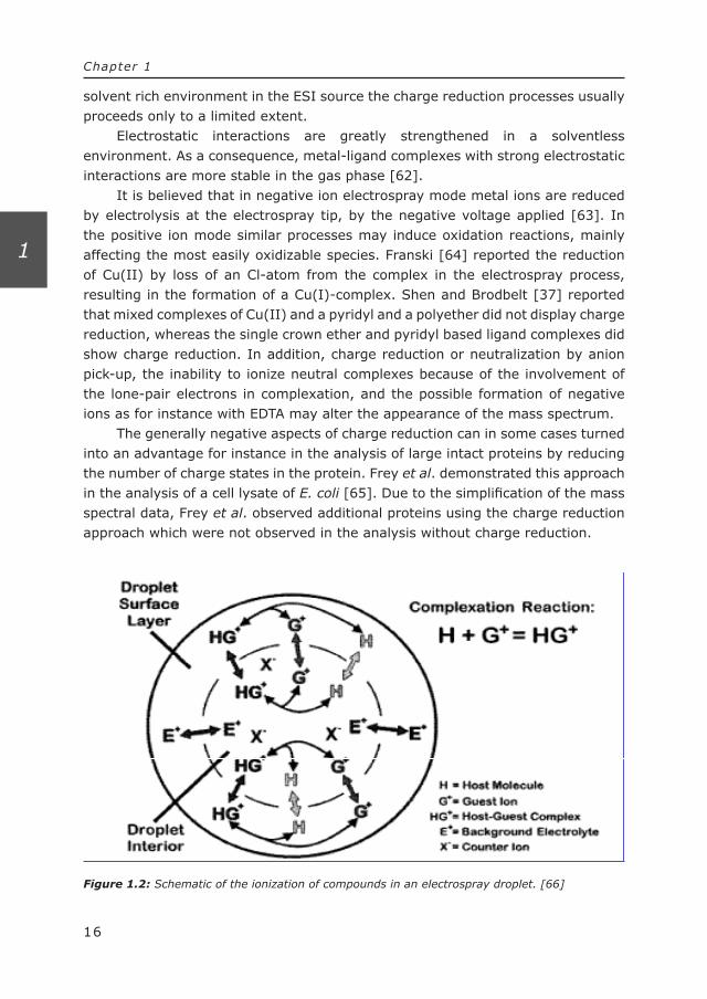
Figure 1.2: Schematic of the ionization of compounds in an electrospray droplet. [66]
16
Chapter 1
1
solvent rich environment in the ESI source the charge reduction processes usually proceeds only to a limited extent.
Electrostatic interactions are greatly strengthened in a solventless environment. As a consequence, metal-ligand complexes with strong electrostatic interactions are more stable in the gas phase [62].
It is believed that in negative ion electrospray mode metal ions are reduced by electrolysis at the electrospray tip, by the negative voltage applied [63]. In the positive ion mode similar processes may induce oxidation reactions, mainly affecting the most easily oxidizable species. Franski [64] reported the reduction of Cu(II) by loss of an Cl-atom from the complex in the electrospray process, resulting in the formation of a Cu(I)-complex. Shen and Brodbelt [37] reported that mixed complexes of Cu(II) and a pyridyl and a polyether did not display charge reduction, whereas the single crown ether and pyridyl based ligand complexes did show charge reduction. In addition, charge reduction or neutralization by anion pick-up, the inability to ionize neutral complexes because of the involvement of the lone-pair electrons in complexation, and the possible formation of negative ions as for instance with EDTA may alter the appearance of the mass spectrum.
The generally negative aspects of charge reduction can in some cases turned into an advantage for instance in the analysis of large intact proteins by reducing the number of charge states in the protein. Frey et al. demonstrated this approach in the analysis of a cell lysate of E. coli [65]. Due to the simplification of the mass spectral data, Frey et al. observed additional proteins using the charge reduction approach which were not observed in the analysis without charge reduction.
17
Chapter 1
1
Finally, another important aspect in the ESI-MS analysis of metal complexes, especially in complex mixtures is ionization suppression. Metal ions and other sample constituents with less efficient solvation properties accumulate at the surface of the droplet and may prevent highly solvated metal ion or metal complexes to be ionized less efficiently. See Fig 1.2 [66].
In conclusion, mass spectrometry and especially ESI-MS is a useful tool in the analysis and characterization of metal-ligand complexes. Despite the many potential problems in ESI-MS of metal-ligand complexes, it turns out that with adequate precautions and control of experimental parameters reliable data can be obtained on solution-phase metal-ligand complexes [21]. The dynamic nature of the ESI-MS process actually enables monitoring of liquid-phase reactions involving metal ligand interactions. This is discussed in more detail in Chapter 2.
1.6 Reference List
1. Kaltashov, I. A.; Zhang, M. X.; Eyles, S. J.; Abzalimov, R. R. Analytical and Bioanalytical Chemistry2006, 386, 472-481.
2. Leszczyszyn, O. I.; Evans, C. D.; Keiper, S. E.; Warren, G. Z. L.; Blindauer, C. A. Inorganica Chimica Acta2007, 360, 3-13.
3. Shindo, M.; Irie, K.; Fukuda, H.; Ohigashi, H. Bioorganic & Medicinal Chemistry 2003, 11, 5075-5082.
4. Peschke, M.; Blades, A. T.; Kebarle, P. Journal of the American Chemical Society 2000, 122, 1492-1505.
5. Petros, A. K.; Reddi, A. R.; Kennedy, M. L.; Hyslop, A. G.; Gibney, B. R. Inorganic Chemistry2006, 45, 9941-9958.
6. Anon Catalysis Letters2000, 67, 5-13. 7. Huang, H. F.; Zhu, L. M.; Reid, B. R.; Drobny, G. P.; Hopkins, P. B. Science1995,
270, 1842-1845. 8. Gu, X. F.; Wang, Y. H.; Kumar, A.; Ye, G. F.; Parang, K.; Sun, G. Q. Journal of
Medicinal Chemistry2006, 49, 7532-7539. 9. Di Nicola, C.; Karabach, Y. Y.; Kirillov, A. M.; Monari, M.; Pandolfo, L.; Pettinari,9. Di Nicola, C.; Karabach, Y. Y.; Kirillov, A. M.; Monari, M.; Pandolfo, L.; Pettinari,
C.; Pombeiro, A. J. L. Inorganic Chemistry2007, 46, 221-230. 10. Garcia-Mina, J. M. Organic Geochemistry2006, 37, 1960-1972. 11. Patel, R. N.; Singh, N.; Shrivastava, R. P.; Shukla, K. K.; Singh, P. K.11. Patel, R. N.; Singh, N.; Shrivastava, R. P.; Shukla, K. K.; Singh, P. K. Proceedings
of the Indian Academy of Sciences-Chemical Sciences2002, 114, 115-124. 12. Biswas, M.; Rosair, G. M.; Pilet, G.; El Fallah, M. S.; Ribas, J.; Mitra, S.
Polyhedron2007, 26, 123-132. 13. Ashok, M.; Ravinder, V.; Prasad, A. V. S. S. Transition Metal Chemistry2007, 32,
23-30. 14. Wang, K. S.; Gokel, G. W. Journal of Organic Chemistry1996, 61, 4693-4697. 15. Combariza, M. Y.; Vachet, R. W.15. Combariza, M. Y.; Vachet, R. W. Analytica Chimica Acta2003, 496, 233-248. 16. Benson, L. M.; Kumar, R.; Cavanagh, J.; Naylor, S.16. Benson, L. M.; Kumar, R.; Cavanagh, J.; Naylor, S. Rapid Communications in
Mass Spectrometry2003, 17, 267-271. 17. Beck, J. L.; Colgrave, M. L.; Ralph, S. F.; Sheil, M. M. Mass Spectrometry Reviews
2001, 20, 61-87. 18. Combariza, M. Y.; Fahey, A. M.; Milshteyn, A.; Vachet, R. W. International Journal
of Mass Spectrometry2005, 244, 109-124.
18
Chapter 1
1
19. Krabbe, J. G.; Lingeman, H.; Niessen, W. M. A.; Irth, H. Journal of Chromatog raphy A2005, 1093, 36-46.
20. Krabbe, J. G.; Lingeman, H.; Niessen, W. M. A.; Irth, H. Analytical Chemistry 2003, 75, 6853-6860.
21. Di Marco, V. B.; Bombi, G. G. Mass Spectrometry Reviews2006, 25, 347-379. 22. Bortolini, O.; Conte, V. Mass Spectrometry Reviews2006, 25, 724-740. 23. Bortolini, O.; Carraro, M.; Conte, V.; Moro, S. European Journal of Inorganic
Chemistry2003, 42-46. 24. Terada, K.; Itoh, K.; Hidaka, H.; Yoshida, T.; Iwamoto, N.; Aoki, W.; Williams, I.
S. New Astronomy Reviews2006, 50, 582-586. 25. Xuan, Y.; Scheuermann, E. B.; Meda, A. R.; Hayen, H.; von Wiren, N.; Weber, G.25. Xuan, Y.; Scheuermann, E. B.; Meda, A. R.; Hayen, H.; von Wiren, N.; Weber, G.
Journal of Chromatography A2006, 1136, 73-81. 26. Lobinski, R.; Schaumloffel, D.; Szpunar, J. Mass Spectrometry ReviewsMass Spectrometry Reviews2006,
25, 255-289. 27. Palmer, A. S.; Snape, I.; Stark, J. S.; Johnstone, G. J.; Townsend, A. T. Marine
Pollution Bulletin 2006, 52, 1441-1449. 28. Fernandez, R. G.; Bayon, M. M.; Alonso, J. I. G.; Sanz-Medel, A. Journal of Mass
Spectrometry2000, 35, 639-646. 29. Sacks, G. L.; Derry, L. A.; Brenna, J. T. Analytical Chemistry2006, 78, 8445-
8455. 30. Stewart, I. I. Spectrochimica Acta Part B-Atomic Spectroscopy1999, 54, 1649-
1695. 31. Cech, N. B.; Enke, C. G. Mass Spectrometry Reviews2001, 20, 362-387. 32. Ross, A. R. S.; Ikonomou, M. G.; Thompson, J. A. J.; Orians, K. J. Analytical
Chemistry1998, 70, 2225-2235. 33. Suckau, D.; Shi, Y.; Beu, S. C.; Senko, M. W.; Quinn, J. P.; Wampler, F. M.;
McLafferty, F. W. Proceedings of the National Academy of Sciences of the United States of America1993, 90, 790-793.
34. Jarrold, M. F. Accounts of Chemical Research1999, 32, 360-367. 35. Hoaglund-Hyzer, C. S.; Counterman, A. E.; Clemmer, D. E. Chemical Reviews
1999, 99, 3037-3079. 36. Gross, D. S.; Schnier, P. D.; RodriguezCruz, S. E.; Fagerquist, C. K.; Williams, E. R. Proceedings of the National Academy of Sciences of the United States of
America1996, 93, 3143-3148. 37. Shen, J.; Brodbelt, J. Journal of Mass Spectrometry1999, 34, 137-146. 38. Pereira, R. M. S.; Paula, V. I.; Buffon, R.; Tomazela, D. M.; Eberlin, M. N.38. Pereira, R. M. S.; Paula, V. I.; Buffon, R.; Tomazela, D. M.; Eberlin, M. N.
Inorganica Chimica Acta2004, 357, 2100-2106. 39. Hansen, H. R.; Pergantis, S. A.39. Hansen, H. R.; Pergantis, S. A. Analytical and Bioanalytical Chemistry2006, 385,
821-833. 40. Rehulka, P.; Popkov, A.; Nadvornik, M.; Planeta, J.; Mazanec, K.; Chmelik, J.
Journal of Mass Spectrometry2006, 41, 448-453. 41. Williams, S. M.; Brodbelt, J. S.; Huang, Z. L.; Lai, H. G.; Marchand, A. P. Analyst
2003, 128, 1352-1359. 42. Combariza, M. Y.; Vachet, R. W. Journal of the American Society for Mass Spectrometry2004, 15, 1128-1135. 43. Combariza, M. Y.; Vachet, R. W. Journal of the American Society for Mass Spectrometry2002, 13, 813-825. 44. Budimir, N.; Fournier, F.; Bailly, T.; Burgada, R.; Tabet, J. C. Rapid Communica-
tions in Mass Spectrometry2005, 19, 1822-1828. 45. Chaparro, A. L.; Vachet, R. W. Journal of Mass Spectrometry2003, 38, 333-342. 46. Jellen, E. E.; Chappell, A. M.; Ryzhov, V. Rapid Communications in Mass
19
Chapter 1
1
Spectrometry2002, 16, 1799-1804. 47. Vachet, R. W.; Hartman, J. A. R.; Callahan, J. H. Journal of Mass Spectrometry
1998, 33, 1209-1225. 48. Kempen, E. C.; Brodbelt, J. S. Analytical Chemistry2000, 72, 5411-5416. 49. Williams, S. M.; Brodbelt, J. S.; Bartsch, R. A. Journal of the American Society for
Mass Spectrometry2003, 14, 1215-1228. 50. Reyzer, M. L.; Brodbelt, J. S.; Marchand, A. P.; Chen, Z. B.; Huang, Z. L.; Namboothiri, I. N. N. International Journal of Mass Spectrometry2001, 204, 133-142. 51. Kempen, E. C.; Brodbelt, J. S. Analytical Chemistry1999, 71, 5493-5500. 52. Leize, E.; Jaffrezic, A.; VanDorsselaer, A. Journal of Mass Spectrometry1996,
31, 537-544. 53. Gabelica, V.; Galic, N.; Rosu, F.; Houssier, C.; De Pauw, E. Journal of Mass Spec-
trometry2003, 38, 491-501. 54. Kempen, R. C.; Brodbelt, J. S. Analytical Chemistry2001, 73, 384-390. 55. Satterfield, M.; Brodbelt, J. S. Analytical Chemistry2000, 72, 5898-5906. 56. Collins, R. N. Journal of Chromatography A2004, 1059, 1-12. 57. Zhang, J. M.; Brodbelt, J. S. Analytical Chemistry2005, 77, 1761-1770. 58. Leavell, M. D.; Leary, J. A. Journal of the American Society for Mass Spectrometry
2001, 12, 528-536. 59. Hao, C. Y.; March, R. E.; Croley, T. R.; Smith, J. C.; Rafferty, S. P. Journal of Mass
Spectrometry 2001, 36, 79-96. 60. Operti, L.; Rabezzana, R. Mass Spectrometry Reviews2006, 25, 483-513. 61. Shvartsburg, A. A.; Wilkes, J. G.; Lay, J. O.; Siu, K. W. M. Chemical Physics
Letters2001, 350, 216-224. 62. Loo, J. A. International Journal of Mass Spectrometry2000, 200, 175-186. 63. Mollah, S.; Pris, A. D.; Johnson, S. K.; Gwizdala, A. B.; Houk, R. S. Analytical
Chemistry2000, 72, 985-991. 64. Franski, R. Journal of Mass Spectrometry2004, 39, 272-276. 65. Frey, B. L.; Lin, Y.; Westphall, M. S.; Smith, L. M. Journal of the American Society
for Mass Spectrometry2005, 16, 1876-1887. 66. Sherman, C. L.; Brodbelt, J. S. Analytical Chemistry2003, 75, 1828-1836.

21
Chapter 2
2
Chapter 2. Metal complexation, coordination
chemistry and ligand-exchange reactions

Me + L MeL ]][[
][1 LMe
MeLK =K
22
Chapter 2
2
Metal complexation can be seen as a selective process. A metal ion can attract or interact with a ligand (an anion or a neutral compound with coordina-tion properties) in two ways. Firstly, the metal ion forms a coordination bond with the specific ligand, resulting in the formation of a metal complex. In a coordina-tion bond, the d-shell electrons and unoccupied d-orbitals present in transition metals are involved in the interactions with lone pair electrons of the ligand. This is called an inner-sphere complex since the ligands are attached directly to the central metal atom or ion. On the other hand, the metal ion can form an (electro-static) bond with a negatively charged ion or via other weak interactions to form an outer-sphere complex, as for instance in NaCl. The anions and also solvent molecules will not compete with the coordinated ligands for bonding to the metal ion. Coordination and electrostatic interactions can exist simultaneously. In many cases, the electrostatic interactions between an anionic coordination ligand and the metal ion plays also a role [1] in metal complexes.
The formation of a coordination bond is much more selective than the elec-trostatic interactions. As will be discussed in Ch.2, nature uses this selectivity in a large number of biological processes.
2.1 Theory of metal complexation
In metal complexation, a metal (M) is complexated by one or several ligand(s) (L) via a coordination bond. Metal complexation results in an equilibrium between the free form of the complex constituents and the complex. Depending on the equilibration constant (K) and the concentration of metal ions and ligands under the given conditions, the metal complex or the unbound constituents are predominantly present.
The equilibration constant represents a value describing the equilibrium between the concentration of metal complex and the free constituents. A high value of the equilibrium constant favors the presence of the metal complex, whereas a low value favors the presence of unbound constituents.
2.1.1 Theoretical calculations
Calculations performed in this thesis are based on normal equilibration calcu-lations which were incorporated in a computer program, that allows the prediction of the type and concentrations of metal complexes formed as a function of

3
3212
211 ][][][1(][
LKKKLKKLKMe
Me tot
+++=
Me + L MeL
MeL + L MeL2
MeL2 + L MeL3
]][[][
1 LMeMeLK =
]][[][ 2
2 LMeLMeLK =
]][[][
2
33 LMeL
MeLK =
Equation 1
Equation 2
Equation 3
H + L HL ]][[][LH
HLKa = Equation 4
23
Chapter 2
2
experimental conditions. The equilibration constants of the different metal-ligand complexes are obtained from the NIST Database 46 [2]. Most transition metal ions have either 4 or 6 coordination sites. This means that with typical bidentate ligands MeL, MeL2 and/or MeL3 may be formed. The complexation reactions involved and corresponding equilibration constants can be written as follows;
In most cases, the acid-base equilibrium of the ligand (Equation 4) also plays a role as it determines the fraction of the ligand concentration present in the correct state to form the complexes. Therefore, the influence of the pH of the solution is also incorporated in the computer program.
Under equilibrium conditions, both the metal ion and the ligand concentra-tion is distributed over various species in solution. The total ligand concentration (Ltot) and the total metal concentration (Metot) can be written as follows:
Ltot = [L] + [MeL] + 2[MeL2] + 3[MeL3] + [HL]
Metot = [Me] + [MeL] + [MeL2] + [MeL3]
By rearranging and implementing equations 1 to 3 in equations 5 and 6 and solving equation 6 for [Me] leads to Equation 7. The free [Me] in solution can be written as:
When equation 7 is now incorporated in equation 5, the concentrations of the various species in solution can be calculated using Brent’s method [3] for finding the roots of the following function (function 1). This algorithm was incorporated in a Delphi-program, where the unbound ligand concentration (L) is calculated at a specific total L concentration (Ltot) and a predefined total metal concentration
Equation 5Equation 5
Equation 6Equation 6
Equation 7Equation 7
totatottot
atottot
atottot
a
LxHKMeKLKxHKKMeKKKLKK
xHKKKMeKKKKKLKKKxHKKKKKKK
−++−+
+++−+
++++−+
+
+
+
+
])[((
])[2((
])[3((
])[(
11
2121121
32132121321
4321321
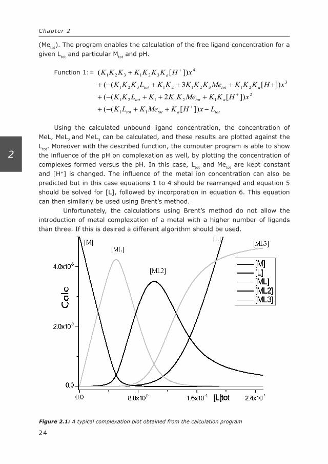
Figure 2.1: A typical complexation plot obtained from the calculation program
24
Chapter 2
2
(Metot). The program enables the calculation of the free ligand concentration for a given Ltot and particular Mtot and pH.
Function 1:=
Using the calculated unbound ligand concentration, the concentration of MeL, MeL2 and MeL3 can be calculated, and these results are plotted against the Ltot. Moreover with the described function, the computer program is able to show the influence of the pH on complexation as well, by plotting the concentration of complexes formed versus the pH. In this case, Ltot and Metot are kept constant and [H+] is changed. The influence of the metal ion concentration can also be predicted but in this case equations 1 to 4 should be rearranged and equation 5 should be solved for [L], followed by incorporation in equation 6. This equation can then similarly be used using Brent’s method.
Unfortunately, the calculations using Brent’s method do not allow the introduction of metal complexation of a metal with a higher number of ligands than three. If this is desired a different algorithm should be used.

25
Chapter 2
2
A typical complexation plot obtained using the calculation procedure of the Delphi-program is shown in Figure 2.1. In this specific example, one metal ion is complexated with a maximum number of three ligands, depending on the concen-tration of the ligand. The concentrations of calculated species (plotted on the y-axis), either the metal concentration [Me], the unbound ligand concentration [L] and/or the different complexes are plotted against the total concentration of ligand.
2.2 Ligand-exchange principles
A ligand-exchange reaction is a solution-phase reaction in which a specific ligand complexated by a metal ion is exchange by another ligand which is introduced into the initial solution. This results in the formation of a new metal-ligand complex and the increase in concentration of the free ligand which was initially complexated by the metal ion.
Ligand-exchange reactions play an important role in all kind of processes. The folding and unfolding of cytchrome c, for example, is controlled by a relatively rapid ligand-exchange reaction in the heme group [4]. Or the uptake of heavy metal ions by hyper accumulator plants such as Berkheya coddii capable to purify polluted soil e.g., from Ni is also based on ligand-exchange reactions [5].
In the treatment of several forms of cancer, cisplatin or derivatives of this platinum based complex are used as chemotherapeutic agents. Before the platinum complex can bind to cell DNA, it is “activated” by a ligand-exchange reaction, where at first one of the Cl-atoms is replaced by a water molecule. As a result, the platinum complex can bind to DNA, followed by a second exchange of the other Cl atom by water. The bound platinum complex changes the structure of the DNA, eventually leading to cell death. [6] This latter example demonstrates the use of ligand-exchange reactions for medical purposes in the treatment for specific cancers.
Ligand-exchange reactions can also be used in the synthesis of for instance organometallic dendrimers [7] or gold nanoparticles [8]. In this thesis, the use of ligand-exchange reactions as an analytical tool is explored in a variety of applica-tions.
2.3 Application of ligand-exchange principles in analytical chemistry
Ligand-exchange principles have widely been used in analytical chemistry. Ligand-exchange chromatography has been used as a method to isolate or separate compounds with complexing properties, e.g., metal ions [9], amines,
26
Chapter 2
2
carboxylic acids [10], phosphates and phosphorylated proteins [11]. In ligand-exchange chromatography, separation of ligands or metal ions is achieved as the result of differences in coordination (inner-sphere) interactions between the ligands towards an immobilized metal ion or between metal ions towards an immobilized ligand. Often, additional electrostatic interactions play a role as well. This coordination or inner-sphere interaction differentiates ligand-exchange chro-matography from ion exchange chromatography, which is based on outer-sphere electrostatic interactions of the ions with groups of opposite charge immobilized at the stationary phase. Thus, ligand-exchange chromatography is more selective than ion exchange chromatography. Since in a coordination bond between a ligand and a metal ion the interaction occurs in the inner-sphere, steric hindrance plays an important role in ligand-exchange principles.
In the 1960s and 1970s, Davankov reported a method for the separation of enantiomers based on ligand-exchange principles. In this approach a metal ion was complexated with an amino acid. The complex then functioned as a chiral selector. The enationselectivity can be applied in different ways, e.g., by immobi-lizing the chiral selector to the HPLC stationary phase [12].
One widely-used form of ligand-exchange chromatography is immobilized metal-ion affinity chromatography (IMAC). In IMAC, a metal ion is immobilized on a stationary support by relatively strong coordination interaction. Analytes with a specific affinity for the metal ion are retained in the IMAC column.
IMAC is predominantly applied in the separation and isolation of proteins and peptides [13, 14]. The interaction primarily involves binding of the side chains of specific amino acids to the immobilized metal ion. Tishechenko et al. demon-strated the use of IMAC by addressing the specific interactions of Cu(II) and Ni(II) in the isolation of exochitinases and specific immunoglobulins [15]. Ni(II) is widely used in IMAC for isolating histidine-tagged proteins. Proteins can be easily tagged with histidine, simplifying the purification of the specific his-tagged protein by introducing the sample to a Ni(II) immobilized IMAC column. Moreover, the interest in phosphorylation of proteins has increased in recent years. Phosphor-ylation plays an important role in signal transduction and regulatory functions inside a cell [16]. Fe(III) or Ga(III) loaded IMAC columns can be applied in the isolation of phosphorylated proteins and peptides [17].
2.4 Requirements of continuous-flow ligand-exchange reac-tions
Next to the use of ligand-exchange principles in chromatographic separations, it can also be applied in a continuous-flow approach, where the metal ion and ligands of interest are present in the same (solution) phase. There are several ways to study and monitor ligand-exchange reactions. The concept of ligand-

27
Chapter 2
2
exchange reactions in a continuous-flow system, has been reported previously, e.g., in the detection of inositol triphosphates [18] and organosulphur compounds [19]. In a continuous-flow ligand-exchange reaction detection system, a metal complex is mixed with the ligand of interest (Li), in a continuous flow. The metal complex consists of a metal ion (Me) and one or several reporter ligands (Lr). Depending on the concentration of the metal complex, the concentration of the ligand of interest and the affinity of both the reporter ligand and the ligand of interest, the reporter ligand is exchanged by the ligand of interest.
The ligand-exchange reaction can be monitored in several ways, e.g., by monitoring the decrease of the metal-reporter ligand complex, by detecting the metal-ligand of interest complex, but most commonly the reporter ligand is monitored, by fluorescence [18] or mass spectrometric detection [20]. By monitoring the change in the reporter ligand concentration, the ability to monitor the ligand-exchange reaction is not dependent on the properties of the ligand of interest.
Several factors are important in ligand-exchange reactions and in using ligand-exchange reactions as an analytical detection tool.
2.4.1 Metal ion selection
Since coordination chemistry is a rather selective process, the choice of the metal ion is an important aspect when implementing a ligand-exchange reaction as an analytical tool. The choice of the metal ion depends primarily on the ligand of interest. Based on the affinity to specific hetero atoms, metal ions can be classified in three main groups: hard, borderline and soft Lewis acids. Hard Lewis acids, e.g., Fe(III), Ga(III) and Ca(II) associate with hard Lewis bases such as phosphate and sulfate. Borderline Lewis acids, e.g., Fe(II), Cu(II) and Zn(II) prefer interactions with pyridines, sulfite and nitrite, whereas soft Lewis acids like Ag(I), Pd(I) and Cu(I) preferably interact with soft Lewis bases like thiols and cyanides. The affinity of metal ions for a specific ligand also varies within each class. One should keep in mind that, despite their preferences, Lewis acids can still interact with Lewis bases of the other classes. Moreover, when studying metal-ligand interactions in general the kinetics of complex formation and dissociation should be considered. One can think, for instance, of a ligand with a high affinity for the metal ion but slow formation kinetics. Despite the high affinity of the ligand to the metal ion, complex formation will not be observed when using a continuous-flow set-up, since the available time for complex formation is too short.
28
Chapter 2
2
2.4.2 Selection of the reporter ligand
The selection of the reporter ligand is another important aspect when designing a ligand-exchange reaction [20-22]. It is determined by two types of properties of the ligand: the affinity to the metal ion and the detection properties in the detection system used.
The choice depends on the affinity of the reporter ligand to the metal ion. In general, under similar concentrations, the affinity of the ligand of interest towards the metal ion should be higher than the affinity of the reporter ligand. Next to the selectivity provided by the metal ion, the affinity of the reporter ligand can also influence additional selectivity and/or sensitivity. A low affinity reporter ligand provides a more efficient exchange reaction when a ligand of interest with a high affinity is introduced. Since complex formation and ligand exchange are equilibrium processes, the exchange also depends on the concentration of the metal-reporter ligand complex and the concentration of introduced ligand of interest. A low affinity reporter ligand exchanges with the ligand of interest at lower concentrations than a reporter ligand with a high affinity. Theoretically, a relatively low affinity reporter ligand provides a higher sensitivity, since the ligand of interest exchanges the reporter ligand at lower concentrations. However, a possible disadvantage of the use of a low affinity reporter ligand is that interferences with a higher affinity for the metal ion also result in a ligand-exchange reaction and thereby provide a false positive response. This problem can be overcome by increasing the selectivity of the ligand-exchange reaction by increasing the affinity of the reporter ligand. In this way, depending on the requirements and on the presence of interferences, the selection of the reporter ligand can be optimized.
Next to these affinity considerations, the detection properties of the reported ligand should also be kept in mind. In fluorescence detection, the exchange of the reporter ligand by the ligand of interest can only be monitored, if the free reporter ligand possesses fluorescent properties while its fluorescence is quenched when it is complexated with the metal ion [18]. Again, interfering compounds may interfere in the fluorescence yield as well.
Fluorescence detection, frequently applied to monitor ligand-exchange reactions, is a rather selective detection method. However, this advantage can turn into a disadvantage because it restricts the choice of reporter ligands to fluorescent ligands. In this respect, mass spectrometry as a detection tool to monitor ligand-exchange reactions is superior to fluorescence detection: the detection is based on detecting ions with a specific m/z, e.g., of the reporter ligand. The choice of the reporter ligand now primarily depends on the required affinity of the reporter ligand. Most ligands show good detection performance in (electrospray) mass spectrometry, because the ionization is based on protonation or deprotonation of hetero atoms like N or O, also involved in complexation. The
29
Chapter 2
2
hyphenation of mass spectrometry with a ligand-exchange reaction/detection will be explained in more detail in section 2.4.7.
2.4.3 Metal:reporter ligand ratio
The ratio between the metal ion and the reporter ligand is another important aspect in designing the ligand-exchange reaction/detection system. Depending on the coordination number (the number of possible coordination sites) of the metal ion, a number of reporter ligand can be coordinated by the metal ion. Moreover, the ligand can possess several binding sites and thereby form a chelate complex with the metal ion. For instance, a metal ion with a coordination number of six, can bind to a maximum of six monodentate (one binding possibility) ligands or three bidentate (two binding possibilities) ligands.
In a metal-reporter ligand complex, where coordination sites are unoccupied, the ligand of interest might not exchange the reporter ligand but forms a coordi-nation bond at the available coordination site instead. In that case, no response is observed in the reporter ligand trace. In this particular case, monitoring the metal-reporter ligand complex might be a good alternative since a decrease in response of the metal-reporter ligand complex is expected. However, when the metal-reporter complex is monitored as a marker for complex formation, one needs to have knowledge about the formed complex and the complex must possess characteristic detection properties, in order to be able to distinguish between the different complexes before and after the addition of the ligand of interest. In contrast, when the metal-reporter ligand ratio is higher than the available coordi-nation sites, unbound reporter ligand is present in solution, resulting in a higher background.
With respect to sensitivity, a monodentate reporter ligand with similar affinity is preferred over a multidentate reporter ligand, since a bidentate ligand of interest exchanges only one bidentate reporter ligand, but in theory it exchanges two monodentate reporter ligands.
2.4.4 pH and temperature
The pH value is an important parameter in ligand-exchange reactions. The pH has a strong influence on the degree of ionization of the reporter ligand and the ligand of interest. At a pH below the pKa of the ligands, the ionization of the ligands might influence the affinity of the ligands to the metal ion. Due to the competition of H3O
+ with either the metal ion and the reporter ligand or with the metal ion and the ligand of interest, it might interfere in complex formation since it can compete with the metal ion for the binding site of the ligand. At lower pH,

30
Chapter 2
2
the ionic interactions between the ligand and the metal ion decreases, resulting in a more selective method. This is nicely demonstrated by the interaction of phosphates with Fe(III) [23]. At higher pH, the complex between the metal ion and the phosphate is stronger due to the additional electrostatic interactions. At pH below 3, these electrostatic interactions are reduced and the main interaction between the Fe(III) and phosphates are of coordination nature. Many interfer-ences, e.g., acidic compounds [24], show a much lower affinity for Fe(III) at such a low pH.
Next to pH, the temperature plays a role in ligand-exchange reactions and complex formation. In general, the equilibrium constant of a metal complex-ation reaction is lower at higher temperature. Besides the complex formation, the temperature also influences the mixing of the metal-reporter ligand complex flow stream and the flow introducing the ligands of interest in continuous-flow systems.
2.4.5. Coupling of a separation prior to the ligand-exchange reac-tion/detection
By hyphenation of a (bio)chemical assay and a separation method, e.g., LC prior to the (bio)chemical assay, the analysis can be extended from pure compounds to complex samples, such as biological samples and natural extracts. In contrast to biochemical assays, where the LC solvent conditions can exert a negative influence on the biochemical reaction, the requirements for LC in combination with a ligand-exchange reaction as a chemical assay is less strict with respect to modifier content, salt concentration and temperature. Reversed-phase liquid chromatography (RPLC) is currently the predominant HPLC separation method. The use of organic modifier in the mobile phase is an important aspect to consider when applying a biochemical assay after the RPLC separation. Many biochemical assays can not withstand (high) percentages of organic modifier in the mobile phase [25]. Moreover, a gradient of organic modifier may alter the response between the beginning and the end of the gradient. Metal based ligand-exchange reactions are generally less influenced by the presence or variation of organic modifier content. Although salt concentrations and temperature are aspects to be considered, the influence on the ligand-exchange reaction is generally much less than in biochemical assays.
2.4.6. Detection in ligand-exchange reactions
With respect to the choice of detection technique, the detection method should be able to selectively determine the response of the ligand of interest in

31
Chapter 2
2
the chemical assay. In a ligand-exchange reaction, the reporter ligand free in solution should display different properties, as compared to when the reporter ligand is complexated by the metal ion. Irth et al. [18] demonstrated a fluo-rescence based ligand-exchange detection method for the analysis of inositol phosphates. The phosphates showed a higher affinity for the Fe(III) than Methyl-calcein Blue (MCB) which acted as a fluorescent reporter ligand. The fluorescent properties of MCB are quenched when complexated with Fe(III). Thereby, the presence of inositol phosphates was detected in the ligand-exchange method as a fluorescence response generated by the release/exchange of MCB.
Next to fluorescence detection as a final detection step for the ligand-exchange detection, mass spectrometry can also be used and actually has several advantages over fluorescence detection. First, the use of MS opens the possibility of having a wide variety of potential reporter ligands. In fluorescence based assays the reporter ligand should possess native fluorescence or should be fluo-rescently labeled without changing the (bio)chemical properties of the reporter ligand. Moreover, as reported previously, the reporter ligand should possess different fluorescence properties free in solution and when it is complexated by the metal ion. In MS, the reporter ligand can primarily be chosen as a function of its affinity to the metal ion of interest, of course keeping the ionization efficiency in mind. The m/z is specific for the reporter ligand and therefore an increase in the extracted ion trace will indicate the presence of a ligand of interest with a higher affinity for the metal ion compared to the reporter ligand. Moreover, with MS it is possible to monitor multiple reporter molecules, e.g., also the metal-reporter ligand can be monitored, resulting in an additional marker for the ligand-exchange reaction. Besides obtaining information from the chemical assay, MS is, next to monitoring the response of the chemical assay, also able to simultane-ously provide information about the ligand of interest, e.g., the metal-ligand of interest complex and, if MS-MS is available, structural information of the ligand of interest.
Next to using either fluorescence or MS detection in (bio)chemical assays, a combination of both techniques in a so-called parallel setup can also be applied [22,26]. In such an approach, the sensitivity of fluorescence detection is combined with the specificity and identification power of MS. Van Elswijk et al. [26] developed a parallel assay for the monitoring of estrogen activity in plant extracts by combining a fluorescence based biochemical assay and mass spec-trometry for the identification of active substances.
Figure 2.2 shows a schematic diagram of (A) the direct approach and (B) the parallel approach. In a parallel approach, the flow is split after the separation, where one part is directed to the fluorescence-based (bio)chemical assay for (bio)chemical information and the other part of the flow is directed towards the MS for structural information.

(B)
Figure 2.2.B: The parallel approach
Figure 2.2.A: Approach of a ligand-exchange reactor, where MS is used to monitor the response
of the ligand-exchange reactor as well as to obtain information about the compound of interest
(A)
32
Chapter 2
2
2.4.7 Applications of hyphenation LC with continuous-flow ligand-exchange reactions and mass spectrometric detection
A continuous-flow ligand-exchange reaction can function as a specific chemical assay to obtain information about the compound(s) of interest. A ligand-exchange reaction provides the affinity of the ligand(s) of interest to the metal ion relative to the reporter ligand. As such, this approach is very similar to a continuous-flow biochemical assay, as for instance reported for the screening of enzymatic inhibitors for specific enzymatic conversions like in the screening of

33
Chapter 2
2
inhibitors in natural extracts for acetylcholine esterase [27] or for the screening of enzymatic inhibitors for the enzyme cathepsin B [25,28]. In general, a continuous (bio)chemical assays can provide highly specific information of the ligands studied.
The combination of a ligand-exchange reaction and mass spectrometry provides an analytical tool to study metal complexation and monitor metal affinity of ligands. Moreover, it provides the possibility to screen selectively for specific compounds in a wide variety of applications e.g., in biochemistry, medicine and industry. Fundamentals and application of continuous-flow ligand-exchange systems combined with mass spectrometric detection are discussed in this thesis. The build-up of the thesis is reported in the next section.
2.5 Reference List
[1] K.Jitsukawa, T.Mabuchi, H.Einaga, and H.Masuda, European Journal of Inorganic Chemistry, 2006, 4254-4263.
[2] R.M.Smith and A.E.Martell, NIST database 46: Critically selected Stability Constants of Metal Complexes, version 8, (1975).
[3] Press, W. H. Flannery, B. P.; Teulkolsky, S. A.; Vetterling, W. T. Numerical Recipes in Pascal, 2nd ed.; Cambridge University Press: 1992; p 285.
[4] S.R.Yeh, S.Takahashi, B.Fan, and D.L.Rousseau, Nat.Struct.Biol., 1997, 4, 51-56. [5] B.Robinson, R.R.Brooks, A.W.Howes, J.H.Kirkman, and P.E.H.Gregg, J.Geochem.
Explor., 1997,60 115-126. [6] H.F.Huang, L.M.Zhu, B.R.Reid, G.P.Drobny, and P.B.Hopkins, Science, 1995, 270,
1842-1845. [7] K.Onitsuka, A.Iuchi, M.Fujimoto, and S.Takahashi, Chemical Communications,
2001, 741-742. [8] L.O.Brown and J.E.Hutchison, Journal of the American Chemical Society, 1997 ,
119, 12384-12385. [9] A.Blazewicz, R.Swieboda, and R.Kocjan, Chemia Analityczna, 2006, 51, 307-318. [10] I.Paunovic, R.Schulin, and B.Nowack, Journal of Chromatography A, 2005,
1100, 176-184. [11] E.Kinoshita-Kikuta, E.Kinoshita, A.Yamada, M.Endo, and T.Koike, Proteomics,
2006, 6, 5088-5095. [12]C.Cheng and F.Y.Lin, Chromatographia, 1994, 39, 15-22. [13]G.Tishchenko, B.Hodrova, J.Simunek, and M.Bleha, Journal of Chromatography A,
2003, 983, 125-132. [14]G.S.Chaga, Journal of Biochemical and Biophysical Methods, 2001, 49, 313-334. [15]G.Tishchenko, B.Hodrova, J.Simunek, and M.Bleha, Journal of Chromatography A,
2003, 983, 125-132. [16]A.Pandey and M.Mann, Nature, 2000, 405, 837-846. [17]M.C.Posewitz and P.Tempst, Analytical Chemistry, 1999, 71, 2883-2892. [18]H.Irth, M.Lamoree, G.J.De Jong, U.A.T.Brinkman, R.W.Frei, R.A.Kornfeldt, and
L.Persson, Journal of Chromatography, 1990, 499, 617-625. [19]C.E.Werkhoven-Goewie, W.M.A.Niessen, U.A.T.Brinkman, and R.W.Frei, Journal of
Chromatography, 1981, 203, 165-172. [20]J.G.Krabbe, H.Lingeman, W.M.A.Niessen, and H.Irth, Analytical Chemistry, 2003,
34
Chapter 2
2
75, 6853-6860. [21]J.G.Krabbe, H.Lingeman, W.M.A.Niessen, and H.Irth, Journal of Chromatography
A, 2005, 1093, 36-46. [22]J.G.Krabbe, F.Gao, J.Li, J.E.Ahlskog, H.Lingeman, W.M.A.Niessen, and H.Irth,
Journal of Chromatography A, 2006, 1130, 287-295. [23]L.D.Holmes and M.R.Schiller, Journal of Liquid Chromatography & Related Tech-
nologies, 1997, 20, 123-142. [24] J.Stupak, H.Z.Liu, Z.P.Wang, B.J.Brix, L.Fliegel, and L.Li, Journal of Proteome
Research, 2005, 4, 515-522. [25]A.R.de Boer, J.M.Alcaide-Hidalgo, J.G.Krabbe, J.Kolkman, C.N.V.Boas, W.M.A.Niessen, H.Lingeman, and H.Irth, Analytical Chemistry, 2005, 77, 7894-
7900. [26]D.A.van Elswijk, U.P.Schobel, E.P.Lansky, H.Irth, and J.van der Greef,
Phytochemistry, 2004, 65, 233-241. [27]C.F.de Jong, R.J.E.Derks, B.Bruyneel, W.Niessen, and H.Irth, Journal of Chroma-
tography A, 2006, 1112, 303-310. [28]A.R.de Boer, B.Bruyneel, J.G.Krabbe, H.Lingeman, W.M.A.Niessen, and H.Irth,
Lab on A Chip, 2005, 5, 1286-1292.


37
Scope of thesis
Scope of thesis

Metal ions and their interactions with all kinds of ligands have raised increasing interest in recent years. Mass spectrometry has taken a prominent role in the study of metal complexes and their interactions. This is due to several advantages of mass spectrometry such as better sensitivity and direct information about the formed complex as compared to conventional detection methods. The goal of this thesis is to investigate metal-ligand exchange reactions by mass spectrometry and implement ligand-exchange reactions as selective analytical tools, e.g., to obtain specific information about metal ions and ligands of interest or to develop specific and selective analyte detection strategies. In this context, mass spec-trometry not only provides chemical information, but also information about the compound which is responsible for a change in the metal-ion reporter complex.
Chapter 1 provides a brief introduction about metal ions, metal complexes and ligand-exchange reactions. The main focus of the chapter is to give the reader insight about the detection of metal species and reactions by mass spectrometry, mainly focusing on the different mass spectrometric techniques which can be used and what their strengths and weaknesses are. Moreover, the behavior of metal species in the mass spectrometer is discussed.
Chapter 2 focuses on the metal-ligand interactions, and how these interac-tions can be monitored or detected. Moreover, this chapter provides a guideline how to develop continuous-flow ligand-exchange reactions as analytical tools for all kind of applications, thereby focusing on mass spectrometric detection of these reactions.
Chapter 3 deals with the fundamental study of metal complexes and metal-ligand reactions by electrospray ionization mass spectrometry. Electro-spray ionization mass spectrometry was used to investigate complex formation of different metal-complexes in a continuous-flow ligand-exchange reactor. Normal equilibration calculations which are incorporated in a computer program, provide the prediction of the type and concentrations of metal complexes formed as a function of experimental conditions. These theoretical calculations were compared with mass spectral data using an approach which mimics the calcula-tions. Moreover, this chapter discusses the influence of the pH on the complex-ation of the metal ion with the ligand. The usefulness of mass spectrometry is demonstrated by monitoring a ligand-exchange reaction by mass spectrometry, obtaining information about affinity properties of the introduced ligand to the metal ion as well as structural information about the ligand itself. The detection of this ligand-exchange reaction is based on the specific release of a reporter ligand from a metal-reporter ligand complex by a high affinity ligand in a continuous-flow system.
38
Scope of thesis
Chapter 4 describes the utilization of electrospray ionization mass spectrom-etry for the selective detection of metal ligands after a post-column continuous-flow ligand-exchange reaction. By applying a chromatographic separation prior to the ligand-exchange reactor it is demonstrated that (complex) mixtures of ligands can be analyzed in one single run. Moreover, this chapter discusses the relationship between the affinity of the reporter ligand to the metal ion and the selectivity of the ligand-exchange reactor, thereby providing a tool to tune the ligand-exchange reaction to either sensitivity or selectivity.
Chapter 5 is divided in two parts, demonstrating two possible strategies. Chapter 5a demonstrates application of a mass spectrometry based ligand-exchange reactor in the screening of phosphorylated peptides, separated by a reversed-phase liquid chromatography. The ligand-exchange reaction is directly monitored by mass spectrometry. A specific reporter trace indicates the presence of a phosphorylated compound, whereas in mass spectral information can be obtained about the compound itself. Chapter 5b uses the same ligand-exchange principle in a slightly different approach. After the chromatographic separation of the peptide mixture, the effluent is split is two parts. One part is directed to a fluo-rescence based ligand-exchange reactor, whereas the other part is directed to a mass spectrometer. By correlating the response in the fluorescence based ligand-exchange reactor, which selectively detects phosphorylated peptides, with the mass spectrometry chromatograms, phosphorylated peptides can be selectively subjected to tandem mass spectrometric analysis, for structural elucidation of the phosphorylated fragments of the protein. A common problem in metal affinity studies of phosphorylated compounds is the interferences of other compounds which possess high affinity to the metal ion. To address this problem, the sample will be subjected to alkaline phosphatase to remove phospho-groups and rerun in the system, expecting no response anymore in the fluorescence trace.
39
Scope of thesis

41
Chapter 3
3
Chapter 3. Metal-complex formation in
continuous-flow ligand-exchange reactors
studied by electrospray mass spectrometry
Journal of the American Society for Mass Spectrometry, 2007, 18(4), 707-713
42
Chapter 3
3
Abstract
Electrospray ionization mass spectrometry was used to investigate complex formation of different metal-complexes in a continuous-flow ligand-exchange reactor. A computer program was developed based on normal equilibrium calcula-tions to predict the formation of various metal-ligand complexes. Corresponding to these calculations,infusion electrospray mass spectrometric experiments were performed to investigate the actual complex formation in solution. The data clearly show good correlation between the theoretically calculated formation of metal – ligand complexes and the experimental mass spectrometric data. Moreover, the approach demonstrates that the influence of the pH can be investigated using a similar approach. Indirectly, these infusion experiments provide information on relative binding constants of different ligands towards a metal-ion. To demonstrate this, a continuous-flow ligand-exchange detection system with mass spectro-metric detection was developed. Injection of ligands, with different affinity for the metal-ion, into the reactor shows good correlation between binding constants and the response in the ligand-exchange detection system. Additional information on the introduced ligand, and the complexes formed after introduction of the ligand, can be obtained from interpretation of the mass spectra.
43
Chapter 3
3
3.1 Introduction
Due to the increasing interest in metal-complexes and metal-ligand interac-tions in several fields, e.g., drug research [1, 2] and catalyst design [3], novel detection methods are necessary. Conventional techniques to study these inter-actions and complexes are infrared spectroscopy, X-ray diffraction and nuclear magnetic resonance spectroscopy (NMR) [4], but these techniques have several drawbacks. The use of electrospray ionization mass spectrometry (ESI-MS) for studying metal-ions, metal-ligand interactions and metal-complexes is readily recognized [5]. Using MS as a detection technique offers the advantage of being, in general, more sensitive than NMR and X-ray diffraction and it opens up the possibility to screen complex samples. Moreover, MS enables simultaneous monitoring the response of ligand-exchange reactions and additionally obtaining chemical information about the specific compound [6, 7].
Today, MS is often used in studying host-guest complexes [8-10], structural elucidation by collision-induced dissociation (CID) MS [11, 12], and studying relative binding constants of ligands to metal-ions [13, 14]. Additionally, due to the liquid-based ionization in the ESI process, it is suitable for studying liquid-phase reactions, e.g., ligand-exchange reactions [6, 7], which enables the study of complex systems.
In general, the assumption is made of a good correlation between complexes formed in solution and what is observed in the mass spectrum [15]. FAB-MS and ESI-MS have been used in the past to evaluate binding selectivities of various compounds, e.g., caged crown ethers to metal-ions [16]. An excellent correlation between obtained ESI mass spectral data and expected binding selectivities has been reported in several papers [13, 17, 18], although several parameters should be kept in mind. For a good correlation between complex formation in solution and what is observed in the mass spectrum [14], particularly the competing effect of the solvent, the conditions and ionization efficiencies of the different complexes and molecules have to be taken into account.
This report describes a fundamental approach in studying metal-complexes with ESI-MS. A computer program was developed to calculate which complexes are formed under predefined conditions, based on the affinities of the relevant ligands to the metal-ion and the experimental conditions. As an example, the theory describes the influence of increasing total ligand concentrations L0 at different pH’s of the solution on the formation of different metal-ligand complexes. This theoretical approach is mimicked by monitoring the formation of the different complexes at increasing total ligand concentration with MS. Moreover, mass spectral data at different concentrations of ligand is evaluated to obtain a better insight about formed complexes at predefined conditions. The theory, as well as the mass spectral data, can aid in both the design of new ligand-exchange
44
Chapter 3
3
reactions for analytical purposes [7] and in the interpretation of these interac-tions studied by MS, since they are involved in all kinds of processes, e.g., in biological [19] and industrial processes [3]. Additionally, the mass spectral data aid in the identification of specific compounds, e.g., catalytic species or intermedi-ates, responsible for certain processes. This fundamental approach was extended by studying several ligand-exchange reactions by MS. A continuous-flow ligand-exchange detection method was used to selectively detect certain ligands, and to correlate the response of the detector to the relative affinities of certain ligands of interest to the metal-ions.
3.2 Experimental
3.2.1 Materials
Acetonitrile and methanol were purchased from Baker (Deventer, The Netherlands) and were purified over a 0.45 μm Millipore filter. The nitrate salts of Cu(II) and Zn(II) and all ligands were purchased from Sigma-Aldrich (Steinheim Germany), except for nicotinamide, 2-(aminomethyl)pyridine and 4-picoline which were purchased from Acros Organics (Geel, Belgium).
3.2.2 Solution preparation
Stock solutions of 10 mM of the ligands were prepared in methanol, except for 4,7-dimethyl-1,10-phenanthroline, which had a stock concentration of 4 mM. Cu(II) and Zn(II)-solutions were prepared in milli-Q water. The reagent solution was prepared by adding a stock solution of metal-ion to a solution of ligand in ammonium formate-formic acid solution (20 mM; pH 6.5) containing 50% methanol. The actual conditions and concentrations of the reagent solutions used are indicated either in the text or in the figure legends.
3.2.3 Setup for infusion experiments
The setup for the preliminary infusion experiments on the on-line mixing of solutions of metal salts and ligands is shown in Figure 3.1.
Figure 3.1: Schematic drawing of the set-up used in infusion and direct-injection experiments.
For the direct infusion experiments, pump 1a and pump 1b generate a concentration gradient of
either metal-ions or a ligand at a flow-rate of 50 µL/min (unless otherwise reported), the second
pump (3) continuously delivers the ligand or metal-ion of interest at 50 µL/min (unless otherwise
reported), 4 a 10 µL thermostated reaction coil, and 5 the ESI-MS instrument. For the direct
injection experiments additionally an autosampler (2) is added to the system. In these cases
pumps 1a and 1b generate a sample carrier-flow. (50 µL/min running buffer unless otherwise
reported); and 2, the reagent pump provides 50 µL/min metal-ion-reporter-ligand solution (unless
otherwise reported)).
45
Chapter 3
3
The system consisted of a Shimadzu (‘s Hertogenbosch, The Netherlands) LCMS-2010A, and a separate Shimadzu LC-10Ai pump (pump 2). The LCMS-2010A consisted of two LC-10ADvp pumps (pump 1a and Pump 1b), a SCL-10ADvp system controller, a SIL-10ADvp autosampler, a CTO-ACvp oven and a single quadruple mass spectrometer equipped with an ESI probe. The binary pumps 1a and 1b were used to generate a concentration gradient of either a metal-ion or a ligand at 50 µL/min. Thus, pump 1a was pumping the running buffer consisting of methanol/5 mM ammonium formate (50/50 v/v) and pump 1b was pumping a certain concentration of either metal-ions or ligand in running buffer. The LC-10Ai pump (pump 3) delivered, at 50 µL/min, either the ligand or the metal-ion solution at a fixed concentration. The total flow into the home-made 40 µL, 0.18 mm I.D. knitted PEEK reaction coil and the ESI-MS was 100 µL/min. The reaction coil was thermostatted at 60 °C by means of a water bath.

46
Chapter 3
3
3.2.4 Setup for direct-injection – ligand-exchange – ESI-MS
The general set-up applied in experiments involving the direct-injection combination with the continuous-flow ligand-exchange reaction coupled to ESI-MS was very similar to the direct infusion system. An autosampler was placed between the binary gradient pump 1 and the Y-piece for the mixing of the solution from pump 2 (see Figure 3.1).
In the continuous-flow detection system, the metal/reporter-ligand solution (50 µL/min) was continuously mixed with a sample carrier flow (50 µL/min), into which the pure compounds were directly injected.
3.2.5 Mass spectrometry settings
MS detection was performed in positive-ion ESI. The probe voltage was 4 kV. Nitrogen (99.999% purity, Praxiar, Oevel, Belgium) gas flow was set at 1.5 L/min. A nitrogen counter gas flow was set at 0.06 MPa, which was applied to obtain better solvent evaporation. The curved desolvation line (CDL) temperature and the block temperature were set at 200 °C. ESI-MS data were acquired by switching between full-spectrum mode and selected-ion monitoring (SIM), detecting a number of specific m/z-values related to the reporter-ligand, the metal-reporter-ligand complex, the ligand of interest and/or complexes of the metal-ion and the ligand of interest. In addition to the ligand and complex m/z-values, a system monitoring compound (SMC) was continuously detected in SIM during the direct injection in order to monitor the overall stability of the system and possible ion suppression effects.
3.2.6 MS/MS settings
For structural elucidation and complex identification, a Micromass Q-TOF2 mass spectrometer equipped with a Z-spray ESI source was used. The ESI source conditions were as follows: source temperature 80 °C, desolvation temperature 100 °C, capillary voltage 3 kV. The cone voltage was set at 30 V. Nitrogen (99.999% purity; Praxair) was used with flow rates of 20 L/h for nebulization, 50 L/h for cone gas, and 350 L/h for desolvation. Argon (99.9995% purity; Praxair) was used as collision gas in MS/MS experiments.
47
Chapter 3
3
3.3 Results and Discussion
3.3.1 General setup
The set-up used for both infusion experiments and flow-injection analysis is shown in Figure 3.1. Pump 3 continuously introduces a constant concentration of metal-ion. The binary gradient pump (pumps 1a and 1b) delivers the ligand to be studied at the concentration range defined by the gradient settings. In typical experiments, the binary pump was set to run a 0-100% gradient of the ligand of interest in 20 mM ammonium formate-formic acid buffer/methanol (50/50 v/v) at a total flow rate of 100 µL/min. ESI-MS was used to monitor both the free ligand and metal-ligand complex concentrations in the full scan mode. For flow-injection experiments, an autoinjector was placed between pump 1 and the mixing union Y, using pump 1 as a sample carrier flow and pump 3 to deliver the premixed metal-reporter-ligand solution.
3.3.2 Infusion experiments (Zn(II) and 1,10-phenanthroline)
In many papers describing ESI-MS detection of metal ligand complexes, the assumption is made that ESI mass spectra reflect, to a large extent, the composition of the metal-complexes formed in solution [9, 10, 15, 18], although in-source fragmentation should be kept in mind [21, 22]. In order to evaluate the correlation between solution complex formation and the observed mass spectrum, a computer program was developed to calculate the type and concentration of metal-complexes formed under predefined conditions as a function of experi-mental parameters. Since the presence of methanol alters the pH, the apparent pH*, reflecting the actual pH after the addition of methanol (pH* is measured with a pH-meter), is used in the calculations of the different complexes throughout this paper.
Figure 3.2a displays the theoretical plot of the concentration of the different metal-complexes as a function of the concentrations of Zn(II), indicated as Me, and 1,10-phenanthroline (C12H8N2, 180.1 Da), indicated as L.
The Zn(II) concentration of 25 µM and the pH (pH* 6.5) are kept constant, while the ligand concentration is increased in time from 0 to 125 µM. Zn(II) can bind three bidentate ligands such as 1,10-phenanthroline, resulting in the formation of 1:1 (MeL), 1:2 (MeL2) and 1:3 (MeL3) metal-ligand complexes. From these calculations, the ligand concentrations can be derived, for which maxima of the individual metal-ligand complexes can be observed.

Figure 3.2: a) Theoretical plot of the complex formation of Zn(II) with 1,10-phenanthroline at
different concentrations of 1,10-phenanthroline at a pH* 6.5. b) Obtained mass chromatogram for
experimental approach at a pH* 6.5. A continuous flow of 50 µM Zn(II) was mixed with a gradient
of the concentration of 1,10-phenanthroline in time. The gradient consisted of 0 µM to 250 µM in
60 min. The maximum concentration of 1,10-phenanthroline was 250 µM. The total concentration
of Zn(II) and 1,10-phenanthroline which reached the MS was, 25 µM and 125 µM respectively.
48
Chapter 3
3

Figure 3.3: Average mass spectrum when the maximum concentration of 1:1 Zn:1,10-phenan-
throline complex is formed (pH* 6.5). Where M = Zn(II); L = 1,10-phenanthroline; X1 = formate
and X2 = nitrate.
49
Chapter 3
3
The m/z traces shown in Figure 3.2b, reflect the experimentally measured responses for some of the observed metal-complex species during an infusion experiment. For clarification, the traces are normalized.
Good correlation between the theoretical plot and the experimental data is achieved. Although a close relationship between the observed intensity of the mass spectrum and the calculated concentration of the formed complex was reported for 1:1 complexes of crown ethers with Na+ and K+ [18], in the present case it is more difficult to correlate intensities to concentrations of metal-complexes, because various complexes are formed (see below) and these complexes appear to have different ionization efficiencies. Despite this limitation, the measured optimum total ligand concentration for the maximum concentration of MeL (≈ 23 µM) and MeL2 (≈ 50 µM) are in good agreement with the values predicted from the theoretical plot, 25 µM and 53 µM, respectively.
Since complexes of Zn(II) show specific isotope patterns (64Zn:66Zn:67Zn:68Zn; 49:28:4:19), they can easily be recognized in the mass spectrum. Figure 3.3 shows an averaged mass spectrum measured at pH* 6.5 at a ligand concentration of ~23 µM, at which the concentration of MeL is at a maximum.
The spectrum shows the protonated 1,10-phenanthroline at m/z 181, various ions related to MeL as well as ions related to MeL2 and MeL3. Interestingly, the abundances of the peaks at m/z 289 and 469, corresponding to [MeL+formate]+

50
Chapter 3
3
and [MeL2+formate]+, respectively, differ from what is predicted by the theoretical model. Under the conditions chosen, the MeL related signal is predicted to be similar to the MeL2 signal whereas in the experimental data, the ratio of the MeL:MeL2 signals is 1:4. This indicates that MeL2 shows significantly higher ionization efficiency than MeL. Despite the differences in ionization efficiency, the normalized patterns observed for MeL and MeL2 closely coincide with those predicted by the theoretical model, moreover all observed MeLx complexes show a similar pattern.
Next to [MeL+formate]+, a number of other MeL-related ions are observed, e.g., peaks at m/z 245 and 321. While the latter can be explained as a methanol adduct of [MeL+formate]+, as frequently observed in spectra from this type of ESI source, the peak at m/z 245 is more difficult to explain. The isotope pattern reveals that one Zn metal-ion is present and that the molecular species is singly charged. When the intensity of the peak at m/z is plotted against the total concentration of ligand, it resembles the profile of [MeL2+formate]+. In-source CID and MS–MS experiments, however, demonstrate that the ion with m/z 245 may be a fragment of the ion with m/z 289, which could be formed by the loss of CO2 from the [MeL+formate]+. Further fragmentation of the ion with m/z 245 in MS–MS results in protonated 1,10-phenanthroline (m/z 181), which in turn can be fragmented by two subsequent losses of HCN, similarly to protonated 1,10-phenanthroline itself. The fragmentation of the ion at m/z 289 is not yet fully understood: the loss of CO2 implies that the singly-charged ion with m/z 245 would be a complex of Zn(II), 1,10-phenanthroline and a hydride anion, while further fragmentation to the ion with m/z 181 should involve the loss of neutral Zn.
Similar to MeL, a number of different ion species are observed for MeL2, e.g., a doubly-charged [MeL2]
2+ complex with m/z 212 and a [MeL2+nitrate]+
complex with m/z 486.0. The latter complex is known from the literature [23], but under the present conditions the formate concentration from the ammonium formate buffer (5 mM) exceeds the nitrate concentration (50 µM), resulting from the zinc nitrate used. The formation of doubly-charged ions implies that possibly a number of neutral complexes, e.g., [MeL+ (formate)2] or [MeL+formate+nitrate], are formed as well. These are obviously not observed under positive-ion ESI conditions, but to some extent they may obscure the actual agreement between the experimental and the theoretical plot (Figure 3.2).
A small peak due to [MeL3]2+ is observed in Figure 3.3 as well. This peak
becomes obviously far more abundant in spectra acquired at higher ligand concen-trations (data not shown). No [MeL3+formate]+ ions were observed at m/z 649. This suggests that in the MeL and MeL2 complexes, formate acts as inner-sphere ligands and not as outer-sphere anionic ligands.

51
Chapter 3
3
3.3.3 pH* variation
In order to study the influence of the pH on metal-complex formation, similar continuous-flow experiments were performed at a pH* below the pKa of the ligand. At pH* of 3.4, the increased competition between the H+ and Mn+ results in a shift of the total concentration of ligand at which the maximum concentration of the relevant complex is observed. Again, there is good agreement between the experimental plot at a pH* of 3.4 and the theoretical plot (data not shown).
The averaged mass spectrum at the total ligand concentration for a maximum MeL complex at a pH* of 3.4 is similar to Figure 3.3, except for some changes in relative abundances of various ions, e.g., the [MeL2]
2+ (m/z 212) is more abundant than [MeL2+formate]+ (m/z 469).
3.3.4 Infusion experiments (Cu(II) and 2,2’-bipyridyl)
Similar infusion experiments and correlation with theoretical plots were carried out for other combinations of a metal-ion and a bidentate ligand. The results obtained with Cu(II) and 2,2’-bipyridyl (C10H8N2, 156.1 Da) are briefly discussed here, especially with respect to the mass spectral interpretation. (see Table 3.1)
Table 3.1: Complexes of Cu(II) and 2,2’-bipyridyl formed at a 1:1 metal to ligand ratio
m/z % Relative abundance Interpretation187.6 9 [MeL2]
2+
219.0* [MeL]+
219.9 6 [MeL+H]+
236.9* 14 [MeL + H2O]+
251.0* 15 [MeL+MeOH]+
264.0 100 [MeL+formate]+
280.9 6 [MeL+nitrate]+
375.1* 70 [MeL2]+
420.1 45 [MeL2+formate]+
437.2 5 [MeL2++nitrate]+
* Indicates complexes of Cu(I).
Good correlation between the theoretical plot and the experimental results at various pH* was achieved (data not shown). Table 3.1 shows the most abundant ions of averaged mass spectra observed at ~45 µM of 2,2’-bipyridyl (pH* 6.5), that is the ligand concentration were the MeL complex concentration is at a maximum.
Reduction of Cu(II) to Cu(I) can occur upon dissociation of its complex

52
Chapter 3
3
with certain ligands such as bipyridyl [24] or upon reduction of Cu(II) complexes in the reaction with solvents, e.g. acetonitrile [25, 26]. In the ESI process, the reduction of Cu(II) to Cu(I) is much faster than the process in solution. Cu(I) is a closed-shell ion [4], which may explain the ease of this reaction. Keeping this in mind, several of the observed ions, e.g., m/z 263.9, 251.0, and 375.0 are explained as [MeL+H2O]+, and [MeL+CH3OH]+ after reduction of Cu(II) to Cu(I). Again, a similar 1:1 complex (m/z 219.9) as in the experiments with Zn(II) and 1,10-phenanthroline (m/z 245.0) was observed. MeL2 complexes show similar behavior where, next to [MeL2+formate]+, [MeL2+nitrate]+ and [MeL2]
2+, the most abundant complex is m/z 375 which can be interpreted as [MeL2]
+ after reduction of Cu(II) to Cu(I). The MeL3 complexes expected are [MeL3]
+withm/z 531 with Cu(I) (not observed) or [MeL3]
2+withm/z 265.7 with Cu(II).
3.3.5 Direct-injection ligand-exchange detection
Next to the characterization of different metal-ligand species, the ability to readily detect metal-ligand complexes by ESI-MS also offers the possibility to study ligand-exchange reactions. We have demonstrated that ligand-exchange reactions can successfully be used as analytical tool [6, 7].
The principle of the ligand-exchange detection method relies on the exchange of a reporter-ligand with a competing ligand of interest. The extent of the exchange reaction depends on the concentration and the affinity of the ligand L for the metal-ion relative to the reporter-ligand R. Indirectly, such infusion experiments can provide information on relative binding constants of ligands towards a metal-ion. Since the concentration of R is continuously monitored by ESI–MS, a positive response in the MS-trace can be related a ligand with a high affinity (relative to the reporter-ligand). By changing the nature of R, and therefore the binding strength of MeR, the method can be tuned to achieve either higher or lower selectivity [7]. Furthermore, using MS as detection method, both R and MeR can be monitored in order to measure the extent of the ligand-exchange reaction. Typically, the species with the highest ionization efficiency are finally selected as reporter molecule.
Next to the ionization efficiency, the binding constant of MeR and the number of available binding sites have to be considered. Weak MeR complexes result in an easy dissociation of the complex and, therefore, a decrease of the MeR signal. Moreover, when all available coordination sites are occupied, a decrease of the reporter complex relies only on the exchange itself.
Figure 3.4 displays the ligand-exchange detection method for different concentrations of ligands with a known affinity for Cu(II) using 2,2’-bipyridyl as reporter-ligand R.

Figure 3.4: Ligand-exchange detection of different ligands. Reagent conditions: Cu(II)-(2,2’-
bipyridyl)2 in 5 mM NH4HCO2/methanol (50/50 v/v) pH* 6.5. The reporter-ligand trace (R) is 2,2’-
bipyridyl ([M+H]+; m/z 157) and the complex related trace (C) is [Cu(II)+2,2’-bipyridyl+formate]+
(m/z 264). 10, 50, 100 and 200 µM injections (10 µL) are performed of the ligands of interest.
1, glycinamide (m/z 75); 2, 2-aminomethyl-pyridine (m/z 109); 4, benzimidazole (m/z 119);
5, nicotinamide (m/z 123); 6, 1,10-phenanthroline (m/z 181); 7, 5-methyl-1,10-phenanthroline
(m/z 195); 8, 2,9-dimethyl-1,10-phenanthroline (m/z 209); 9 4,7-dimethyl-1,10-phenanthroline
(m/z 209). * 10, 50, 100 and 200 µM injections (10 µL) of EDTA, which is not observed as a ligand
in positive electrospray mass spectrometry, since it has an overall negative charge.
53
Chapter 3
3
The upper trace corresponds to the protonated molecule of the free 2,2’-bipyridyl. A peak in the 2,2’-bipyridyl trace indicates that ligand-exchange has occurred upon injection of a ligand of interest. Additional proof that ligand exchange occurred is obtained by analyzing the MS trace corresponding to one or

54
Chapter 3
3
more copper-2,2’-bipyridyl complex species. In the present example, m/z 264.0, corresponding to [MeR+formate]+ was used and a ligand-exchange reaction results in a negative peak. The ESI-MS spectrum provides additional information on identity of the analyte ligand and the metal-analyte complexes formed. This is especially interesting when dealing with unknown ligands or when information about specific metal-complexes is required.
The response in the free 2,2’-bipyridyl trace is related to the relative binding constants of the Cu(II)-analyte complexes. EDTA, which forms strong complexes with Cu(II), shows the highest response. Increasing EDTA concentra-tions result in increasing positive peak heights in the free 2,2’-bipyridyl trace and increasing negative peak heights in the Cu(II)-2,2’-bipyridyl trace. The molecular structure of the injected ligand also influences the response of the ligand-exchange detection. EDTA is a hexadentate ligand, which, in principle, can displace or exchange three bidentate reporter-ligands from MeR. Whereas upon injection of a bidentate ligand, e.g. 1,10-phenanthroline, only one reporter-ligand is exchanged. This effect is substantiated by the results (Figure 3.4) where EDTA gives a higher response in the reporter-ligand trace than 1,10-phenan-throline. At the same time, the decrease in the metal-reporter-ligand complex trace is similar for the injection of EDTA or 1,10-phenanthroline. One molecule of EDTA exchanges three reporter-ligand molecules from one metal-reporter-ligand complex molecule, but affects only one metal-reporter-ligand complex, whereas a 1,10-phenanthroline molecule only exchanges one reporter-ligand molecule from that complex.
In some applications, it may be of interest to study the complexes formed with the analyte ligands in detail. Next to homogeneous complexes, the formation of heterogeneous, mixed-ligand complexes has to be considered. By injecting 5-methyl-1,10-phenanthroline (C13H10N2, 194.1 Da) in the Cu(II)-2,2’-bipyridyl ligand-exchange detection system, all three types of metal-complexes that may be formed, i.e., complexes of the analyte ligand (MeL), mixed complexes with both the analyte ligand and reporter-ligand (MeLR) and complexes with the reporter-ligand (MeR) (data not shown).
3.3.6 Correlation of response in ligand-exchange detection and af-finity of the injected ligands
As expected, an increase in the concentration of the injected ligand results in an increase in the response in the reporter-ligand trace. For EDTA, 1,10-phen-anthroline, 5-methyl-1,10-phenanthroline, and 4,7-dimethyl-1,10-phenanthro-line, the increase of the response shows a linear relationship with the increase in concentration (correlation coefficient of > 0.99), while such a linear correlation is not observed for 2,9-dimethyl-1,10-phenanthroline. The latter may be caused

55
Chapter 3
3
by steric hindrance (which is also related to the equilibration constant) and/or ion suppression.
As expected, the type of MeL complex formed depends on the concentra-tion of the ligand of interest. At low concentrations, the MeL complex species are relatively more abundant, while at higher injected concentrations a shift to the MeL2 and MeL3 complexes is observed. At the same time, data show that the response due to MeL3 complexes starts decreasing at lower ligand concentration, while the decrease of MeL2 and MeL starts at higher ligand concentrations.
3.4 Conclusions
Continuous-flow ligand-exchange detection systems coupled to ESI-MS are useful tools in studying interactions between metal-ions and a variety of ligands. In principle, monitoring the dissociation and formation of metal-ligand complexes by means of ESI-MS provides a wealth of information. However, a proper picture on the liquid-phase complexation processes can only be obtained if the mass spectral data adequately reflect the metal-ligand complexes formed in solution. This study demonstrates that for several model systems good correlation is found between experimental data obtained by ESI-MS and theoretically predicted metal-ligand concentrations, even when experimental parameters like solution pH vary. Infusion experiments were also found to be a fast method to obtain a proper insight in the complexes formed, prior to the development of an actual ligand-exchange detection system. The various complexes formed may well differ in ionization efficiency. In addition, these experiments show the complexity of the liquid phase constituents during complex formation and ligand-exchange reactions.
Injection of several ligands with different affinity for the metal-ion in a continuous-flow ligand-exchange reaction clearly shows that there is a good correlation between binding constants and the ligand-exchange detection response. Additional information of the introduced ligand and the complexes formed after introduction of the ligand can be obtained from interpretation of the mass spectrum.
This fundamental approach may aid in the future design of a customized ligand-exchange reaction, e.g., as a selective detection method, to study relative binding constants or study the ligand-metal interactions in general. The applica-bility of these ligand-exchange reactions is not restricted to the metal-ions Cu(II) and Zn(II), but can be expanded to other metal-ions of interest or ligands of interest.

56
Chapter 3
3
3.5 Acknowledgements
Shimadzu Benelux is acknowledged for the loan of equipment.
3.6 References
1. Farrell, N. Biomedical uses and applications of inorganic chemistry. An overview. Coord. Chem. Rev. 2002, 232, 1-4.
2. Molenveld, P.; Engbersen, J. F. J.; Kooijman, H.; Spek, A. L.; Reinhoudt, D. N. Efficient catalytic phosphate diester cleavage by the synergetic action of two Cu(II) centers in a dinuclear cis-diaqua Cu(II) calix[4]arene enzyme model. J. Am. Chem. Soc. 1998, 120, 6726-6737. 3. Chen, P. Electrospray ionization tandem mass spectrometry in high-throughput screening of homogeneous catalysts. Angew. Chem. Int. Ed. 2003, 42, 2832- 2847. 4. Chaparro, A. L.; Vachet, R. W. Tandem mass spectrometry of Cu(II) complexes: the effects of ligand donor group on dissociation. J. Mass Spectrom. 2003, 38, 333-342. 5. Traeger, J. C. Electrospray mass spectrometry of organometallic compounds. Int. J. Mass Spectrom. 2000, 200, 387-401. 6. Krabbe, J. G.; Lingeman, H.; Niessen, W. M. A.; Irth, H. Ligand-exchange detection of phosphorylated peptides using liquid chromatography electrospray mass spectrometry. Anal. Chem. 2003, 75, 6853-6860. 7. Krabbe, J. G.; Lingeman, H.; Niessen, W. M. A.; Irth, H. Screening for metal ligands by liquid chromatography-ligand-exchange-electrospray mass spectrom- etry. J. Chromatogr. A 2005, 1093, 36-46. 8. Satterfield, M.; Brodbelt, J. S. Relative binding energies of gas-phase pyridyl ligand/metal-complexes by energy-variable collisionally activated dissociation in a quadrupole ion trap. Inorg. Chem. 2001, 40, 5393-5400. 9. Tsierkezos, N. G.; Schroder, D.; Schwarz, H. Complexation of nickel(II) by ethyl enediamine investigated by means of electrospray ionization mass spectrometry. Int. J. Mass Spectrom. 2004, 235, 33-42. 10. Vincenti, M. Host-guest chemistry in the mass-spectrometer. J. Mass Spectrom. 1995, 30, 925-939. 11. Jellen, E. E.; Chappell, A. M.; Ryzhov, V. Effects of size of noncovalent complexes on their stability during collision-induced dissociation. Rapid Commun.Mass Spectrom. 2002, 16, 1799-1804. 12. Kumar, M. R.; Prabhakar, S.; Kumar, M. K.; Reddy, T. J.; Vairamani, M. Dissocia tion of gas-phase dimeric complexes of lactic acid and transition metal-ions formed under electrospray ionization conditions; the role of reduction of the metal-ion. Rapid Commun. Mass Spectrom. 2005, 19, 113-120. 13. Blair, S. M.; Brodbelt, J. S.; Marchand, A. P.; Kumar, K. A.; Chong, H. S. Evaluation of binding selectivities of caged crown ligands toward heavy metals by electrospray ionization/quadrupole ion trap mass spectrometry. Anal. Chem. 2000, 72, 2433-2445. 14. Young, D. S.; Hung, H. Y.; Liu, L. K. An easy and rapid method for determination of stability constants by electrospray ionization mass spectrometry. Rapid Commun. Mass Spectrom. 1997, 11, 769-773. 15. Di Marco, V. B.; Bombi, G. G. Electrospray mass spectrometry (ESI-MS) in the
57
Chapter 3
3
study of metal-ligand solution equilibria. Mass Spectrom. Rev. 2006, 25, 347- 379. 16. Young, D. S.; Hung, H. Y.; Liu, L. K. Estimation of selectivities and relative cation ization efficiencies of different [crown plus M](+) by electrospray mass spectrom- etry. J. Mass Spectrom. 1997, 32, 432-437. 17. Goolsby, B. J.; Brodbelt, J. S.; Adou, E.; Blanda, M. Determination of alkali metal-
ion binding selectivities of calixarenes by matrix-assisted laser desorption ionization and electrospray ionization in a quadrupole ion trap. Int. J. Mass Spectrom. 1999, 193, 197-204. 18. Wang, K. S.; Gokel, G. W. Correlation of solution and gas phase complexation assessed by electrospray ionization mass spectrometry: Application to one-, two-, and three-ring macrocycles. J. Org. Chem. 1996, 61, 4693-4697. 19. Gomez-Ariza, J. L.; Garcia-Barrera, T.; Lorenzo, F.; Bernal, V.; Villegas, M. J.; Oliveira, V. Use of mass spectrometry techniques for the characterization of metal bound to proteins (metallomics) in biological systems. Anal. Chim. Acta 2004, 524 15-22. 20. Press, W. H. Flannery, B. P.; Teulkolsky, S. A.; Vetterling, W. T. Numerical Recipes in Pascal, 2nd ed.; Cambridge University Press: 1992; p 285. 21. Bure, C.; Lange, C. Comparison of dissociation of ions in an electrospray source, or a collision cell in tandem mass spectrometry. Curr. Org. Chem. 2003, 7, 1613- 1624. 22. Kubwabo, C.; Vais, N.; Benoit, F. M. Characterization of microcystins using in- source collision-induced dissociation. Rapid Commun. Mass Spectrom. 2005, 19 597-604. 23. Vachet, R. W.; Callahan, J. H. Quadrupole ion trap studies of the structure and reactivity of transition metal-ion pair complexes. J. Mass Spectrom. 2000, 35 311-320. 24. Shen, J.; Brodbelt, J. Formation of doubly charged transition metal-polyether- pyridyl mixed-ligand complexes by electrospray ionization. J. Mass Spectrom. 1999, 34 137-146. 25. Shvartsburg, A. A.; Wilkes, J. G.; Lay, J. O.; Siu, K. W. M. Fragmentation and charge transfer in gas-phase complexes of divalent metal-ions with acetonitrile. Chem. Phys. Lett. 2001, 350, 216-224. 26. Seto, C.; Stone, J. A. The reactions of Cu2+(CH3CN)(n) (n = 2-4) and Cu2+(CH3CN)(3)(H2O) at low collision energy with neutral molecules in a triple sector quadrupole instrument. Int. J. Mass Spectrom. 1998, 175, 263-276.

59
Chapter 4
4
Chapter 4. Screening for metal ligands by
liquid chromatography –ligand-exchange –
electrospray mass spectrometry
Journal of Chromatography A, 2005, 1093, 36-46

60
Chapter 4
4
Abstract
Electrospray ionization mass spectrometry is applied for the selective detection of metal ligands after a post-column continuous-flow ligand-exchange reaction. The detection is based on the specific release of a reporter ligand from a metal-reporter ligand complex by a high affinity ligand. Constant infusion and direct-injection experiments are performed to optimize the method. The on-line coupling of a liquid chromatographic separation prior to the continuous flow ligand-exchange reaction enables the screening for high affinity ligands in complex samples. The feasibility of the method is demonstrated by using several ligands with a different affinity for either Cu(II) or Zn(II) ions. The selectivity of the ligand-exchange detection method can be tuned by the choice of the reporter ligand. This is demonstrated by using either 2,2’-bipyridyl or 5-methyl-1,10-phen-anthroline as reporter ligands.

61
Chapter 4
4
4.1 Introduction
The importance of electrospray ionization mass spectrometry (ESI-MS) and tandem mass spectrometry in the characterization of metal complexes and organo-metallic compounds is readily recognized [1]. The adduct formation of analytes with various metal ions is also studied in order to either influence the sensitivity, as in coordination ESI [2,3], or to change the fragmentation reactions of the analytes involved [4,5]. ESI-MS also plays an important role in the study of host-guest complexes, relevant for instance in understanding molecular recognition and in the field of supramolecular chemistry [6-8].
However, since ESI is a liquid-based ionization technique, it can also be used for the continuous monitoring of liquid-phase reactions, e.g., ligand-exchange reactions with metal ions, e.g., see [9]. In principle, this enables the study of ligand-metal interactions in complex (biological) systems. A large variety of biological processes rely on the (non-)reversible binding and dissociation of specific ligands to metal ions, i.e., on liquid-exchange reactions. An example, crucial to life, is the uptake, transport and delivery of oxygen and carbon dioxide by hemoglobin, containing an iron-porphyrin active site.
In fact, the study of ligand-exchange reactions by ESI-MS opens a number of research opportunities. A continuous-flow ligand-exchange reaction can be applied to assess (relative) affinities of various ligands to a certain metal ion, or alternatively the (relative) affinities of various metal ions for a particular ligand [10]. While this may be useful for fundamental purposes, it also gives way to various more practical applications. For instance, it allows the development of screening and analysis methods based on ligand-exchange reactions. Recently, we demonstrated the selective detection of phosphorylated peptides in peptide mixtures separated by liquid chromatography, based on the high affinity of a phospho-group to Fe(III) metal ions [11]. Upon mixing the column effluent in a continuous-flow reactor with an Fe(III)-reporter ligand complex, the presence of a high-affinity phosphorylated peptide in the effluent will release the reporter ligand from its complex with Fe(III). Using ESI-MS, not only the change in the free reporter ligand concentration can be measured and quantitated, but also the identity of the phosphorylated peptide involved may be established [11].
The ability to monitor (relative) affinities of ligands to metal ions may also be applied in the discovery of new drugs targeting metalloproteins [12,13] as well as in the optimization of other systems of homogeneous catalysis [14]. The biological activity of a variety of enzymes is based on catalytic metal-ligand complexes, for instance the cytochrome P450 enzyme complex [15], in which the iron-porphyrin active site catalyses the oxidation of endogenous and exogenous molecules in the cell, and is thus heavily involved in phase I drug metabolism.
A more common approach to analyze ligands with a certain affinity for
62
Chapter 4
4
metal ions is immobilized metal affinity chromatography (IMAC) [16,17]. A major drawback of this approach is that all ligands are exposed to the immobilized metal ion prior to separation, resulting in the possibility that a high concentration of an high affinity ligand will exclude the simultaneous enrichment of low affinity ligands by occupying all available coordination sites.
In the present study, the potential of ESI-MS in monitoring ligand-exchange reactions is explored from a more general point of view. Competition between various ligands for the metal ions Cu(II) and Zn(II) is investigated in a continuous-flow ligand-exchange reactor coupled to ESI-MS. The ligands can be introduced into the reactor by means of either flow injection or after separation by liquid chromatography. The latter opens the possibility to screen mixtures of potential ligands for their (relative) affinity to a particular metal-ligand complex. Since the reporter ligand can be chosen more freely in ESI-MS, in contrast to for instance in fluorescence based methods, this method opens a way to tune for selectivity of the method. Additionally, upon introduction of ligands MS provides a possibility to monitor not only the reporter ligand, but also dissociation of metal-reporter ligand complexes and formation of metal-ligand of interest complexes. The approach further provides insight in the identity of the complexes formed under different experimental conditions, e.g. with respect to solution pH, organic modifier content, and mixing ratios between metal-ions, reporter ligand, and the ligand investigated.
4.2 Experimental
4.2.1 Materials
Acetonitrile and methanol were purchased from Baker (Deventer, The Netherlands) and were purified over a 0.45-μm Millipore filter. The nitrate salts of Cu(II) and Zn(II) and all ligands were purchased from Sigma-Aldrich (Steinheim Germany), except for nicotinamide, 2-(aminomethyl)pyridine and 4-picoline which were purchased from Acros Organics (Geel, Belgium).
4.2.2 Solution preparation
Stock solutions of 10 mM of the ligands were prepared in methanol, except for 4,7-dimethyl-1,10-phenanthroline which had a stock concentration of 4 mM. Cu(II) and Zn(II)-solutions were prepared in milli-Q water. The reagent solution was prepared by adding a stock solution of metal ion to a solution of ligand in ammonium formate solution (5mM; pH 6.3) containing 50% methanol. The actual

Figure 4.1: Schematic drawing of the set-up used in infusion, direct-injection and LC experiments.
For the direct infusion experiments, pump 1a and pump 1b generate a concentration gradient of
either metal ions or a ligand at a flow-rate of 100 µL/min (unless otherwise reported), pump 2
continuously delivers the ligand or metal ion of interest at 100 µL/min (unless otherwise reported),
4 a 40 µL thermostated reaction coil, and 5 the ESI-MS instrument. For the direct infusion and
LC-experiment additionally 3 an autosampler and column 6 are added to the system. In these
cases pumps 1a and 1b generate a sample carrier-flow or a gradient. (100 µL/min LC-gradient or
running buffer unless otherwise reported); and 2, the reagent pump provides 100 µL/min metal
ion-reporter ligand solution (unless otherwise reported)).
63
Chapter 4
4
concentrations of the reagent solutions used are indicated either in the text or in the legend of the figure.
4.2.3 Setup for infusion experiments
The setup for the preliminary infusion experiments on the on-line mixing of solutions of metal salts and ligands is shown in figure 4.1.
The system consists of a Shimadzu (‘s Hertogenbosch, The Netherlands) LCMS-2010A, and a separate Shimadzu LC-10Ai pump (pump 2). The LCMS-2010A consisted of two LC-10ADvp pumps (pump 1a and Pump 1b), a SCL-10ADvp system controller, a SIL-10ADvp autosampler, a CTO-ACvp column oven and a single quadruple mass spectrometer equipped with an ESI-probe. The binary pumps 1a and 1b are used to generate either a metal ion or a ligand concentration gradient at 100 µL/min. Thus, pump 1a is pumping the running buffer consisting of methanol/5 mM ammonium formate (50/50 v/v) and pump 1b is pumping a
64
Chapter 4
4
certain concentration of either metal ions or ligand in running buffer. The LC-10Ai pump (pump 2) delivers at 100 µL/min either the ligand or the metal ion solution at a fixed concentration. The total flow into the home-made 40 µL, 0.18 mm I.D. knitted PEEK reaction coil and the ESI-MS was 200 µL/min. The reaction coil is thermostated at 60 °C by means of a water bath.
4.2.4 Setup for direct-injection and LC – ligand-exchange – ESI-MS
The general set-up applied in experiments involving direct-injection and LC in combination with the continuous-flow post-column ligand-exchange reaction coupled to ESI-MS is very similar to the direct infusion system. An autosampler and an LC column are placed between the binary gradient pump 1 and the T-piece for the mixing of the solution from pump 2 (see Figure 4.1). For direct-injection experiments, the LC column is replaced by a short PEEK tubing.
In the continuous-flow post-column detection system, the metal/reporter ligand solution (100 µL/min) is continuously mixed with a sample carrier flow (100 µL/min), in which either pure compounds are directly injected, or the column effluent from the LC. In the LC-experiments with Cu(II), a 100 x 2.1 mm I.D. Luna Phenomenex C8(2) reversed-phase LC column (3 µm particles) is applied, and in the LC-experiments with Zn(II), a 50 x 1 mm I.D. Luna Phenomenex C18(2) reversed-phase LC column (3 µm particles). The gradients used in the LC-experiments are reported in text and in the legend of the figure. The mixing of the two flows is improved by a home-made 40 µL, 0.18 mm I.D. knitted PEEK reaction coil placed in a thermostated bath of 60 °C.
4.2.5 Mass spectrometry settings
MS detection was performed in positive-ion ESI. The probe voltage was 4 kV. Nitrogen (99.999% purity, Praxiar, Oevel, Belgium) gas flow was set at 1.5 L/min. A nitrogen counter gas flow was set at 0.06 MPa. It was applied for better solvent evaporation. The curved desolvation line (CDL) temperature and the block temperature were set at 200 °C. ESI-MS data were acquired by switching between full-spectrum mode and selected-ion monitoring (SIM), detecting a number of specific m/z-values related to the reporter ligand, the metal-reporter ligand complex, the ligand of interest and/or complexes of the metal-ion and the ligand of interest. In addition to the ligand and complex m/z-values, a system monitoring compound (SMC) was continuously detected in SIM during the direct injection and LC-experiments.
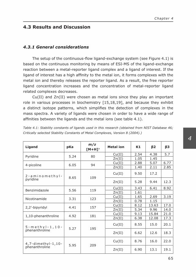
65
Chapter 4
4
4.3 Results and Discussion
4.3.1 General considerations
The setup of the continuous-flow ligand-exchange system (see Figure 4.1) is based on the continuous monitoring by means of ESI-MS of the ligand-exchange reaction between a metal-reporter ligand complex and a ligand of interest. If the ligand of interest has a high affinity to the metal ion, it forms complexes with the metal ion and thereby releases the reporter ligand. As a result, the free reporter ligand concentration increases and the concentration of metal-reporter ligand related complexes decreases.
Cu(II) and Zn(II) were chosen as metal ions since they play an important role in various processes in biochemistry [15,18,19], and because they exhibit a distinct isotope patterns, which simplifies the detection of complexes in the mass spectra. A variety of ligands were chosen in order to have a wide range of affinities between the ligands and the metal ions (see table 4.1).
Table 4.1: Stability constants of ligands used in this research (obtained from NIST Database 46;
Critically selected Stability Constants of Metal Complexes, Version 8 (2004).)
Ligand pKa m/z [M+H]+
Metal ion K1 β2 β3
Pyridine 5.24 80Cu(II) 2.54 4.38 5.7Zn(II) 1.05 1.45
4-picoline 6.05 94Cu(II) 2.88 5.07 6.77Zn(II) 1.40 2.11 2.85
2 - a m i n o m e t h y l -pyridine 8.65 109
Cu(II) 9.50 17.2
Zn(II) 5.28 9.44 12.3
Benzimidazole 5.56 119Cu(II) 3.43 6.41 8.92Zn(II) 1.61
Nicotinamide 3.31 123Cu(II) 1.65 2.69 3.19Zn(II) 0.78 1.15
2,2’-bipyridyl 4.41 157Cu(II) 8.12 13.63 17.0Zn(II) 5.34 9.96 14.0
1,10-phenanthroline 4.92 181Cu(II) 9.13 15.84 21.0Zn(II) 6.38 12.08 17.3
5 - m e t h y l - 1 , 1 0 -phenanthroline 5.27 195
Cu(II) 8.55 15.0 20.1
Zn(II) 6.62 12.6 18.3
4,7-dimethyl-1,10-phenanthroline 5.95 209
Cu(II) 8.76 16.0 22.0
Zn(II) 6.90 13.1 19.1
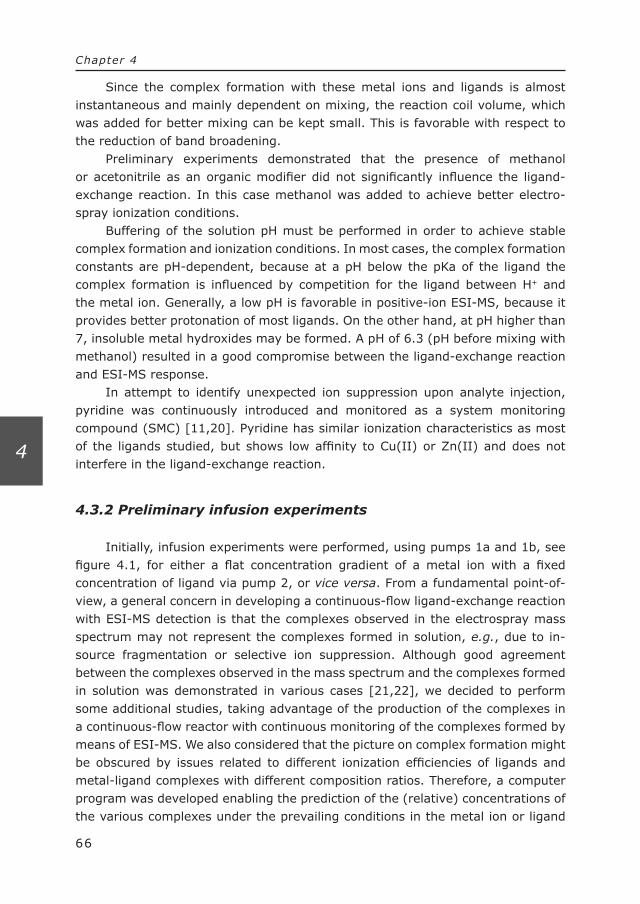
66
Chapter 4
4
Since the complex formation with these metal ions and ligands is almost instantaneous and mainly dependent on mixing, the reaction coil volume, which was added for better mixing can be kept small. This is favorable with respect to the reduction of band broadening.
Preliminary experiments demonstrated that the presence of methanol or acetonitrile as an organic modifier did not significantly influence the ligand-exchange reaction. In this case methanol was added to achieve better electro-spray ionization conditions.
Buffering of the solution pH must be performed in order to achieve stable complex formation and ionization conditions. In most cases, the complex formation constants are pH-dependent, because at a pH below the pKa of the ligand the complex formation is influenced by competition for the ligand between H+ and the metal ion. Generally, a low pH is favorable in positive-ion ESI-MS, because it provides better protonation of most ligands. On the other hand, at pH higher than 7, insoluble metal hydroxides may be formed. A pH of 6.3 (pH before mixing with methanol) resulted in a good compromise between the ligand-exchange reaction and ESI-MS response.
In attempt to identify unexpected ion suppression upon analyte injection, pyridine was continuously introduced and monitored as a system monitoring compound (SMC) [11,20]. Pyridine has similar ionization characteristics as most of the ligands studied, but shows low affinity to Cu(II) or Zn(II) and does not interfere in the ligand-exchange reaction.
4.3.2 Preliminary infusion experiments
Initially, infusion experiments were performed, using pumps 1a and 1b, see figure 4.1, for either a flat concentration gradient of a metal ion with a fixed concentration of ligand via pump 2, or vice versa. From a fundamental point-of-view, a general concern in developing a continuous-flow ligand-exchange reaction with ESI-MS detection is that the complexes observed in the electrospray mass spectrum may not represent the complexes formed in solution, e.g., due to in-source fragmentation or selective ion suppression. Although good agreement between the complexes observed in the mass spectrum and the complexes formed in solution was demonstrated in various cases [21,22], we decided to perform some additional studies, taking advantage of the production of the complexes in a continuous-flow reactor with continuous monitoring of the complexes formed by means of ESI-MS. We also considered that the picture on complex formation might be obscured by issues related to different ionization efficiencies of ligands and metal-ligand complexes with different composition ratios. Therefore, a computer program was developed enabling the prediction of the (relative) concentrations of the various complexes under the prevailing conditions in the metal ion or ligand
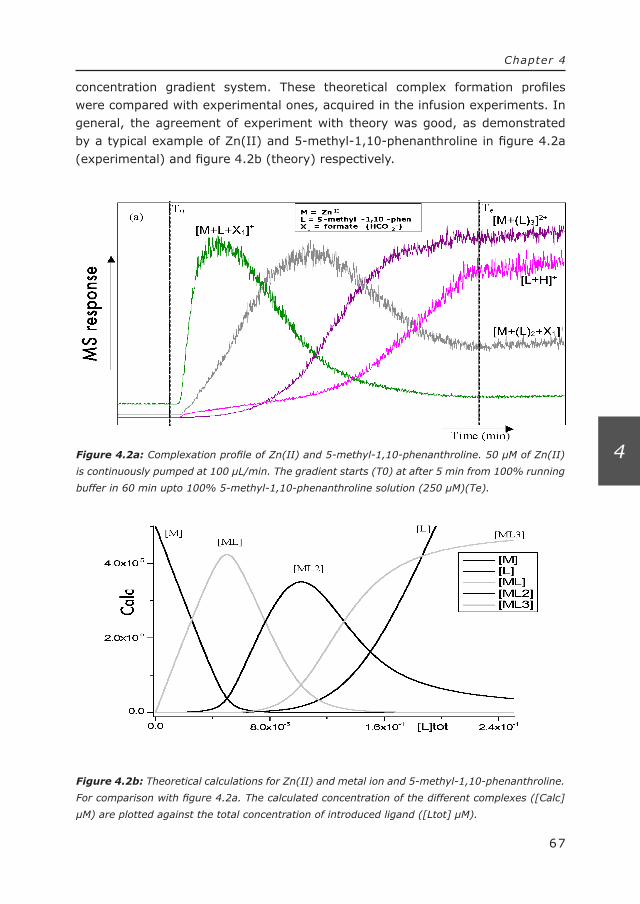
Figure 4.2a: Complexation profile of Zn(II) and 5-methyl-1,10-phenanthroline. 50 µM of Zn(II)
is continuously pumped at 100 µL/min. The gradient starts (T0) at after 5 min from 100% running
buffer in 60 min upto 100% 5-methyl-1,10-phenanthroline solution (250 µM)(Te).
Figure 4.2b: Theoretical calculations for Zn(II) and metal ion and 5-methyl-1,10-phenanthroline.
For comparison with figure 4.2a. The calculated concentration of the different complexes ([Calc]
µM) are plotted against the total concentration of introduced ligand ([Ltot] µM).
67
Chapter 4
4
concentration gradient system. These theoretical complex formation profiles were compared with experimental ones, acquired in the infusion experiments. In general, the agreement of experiment with theory was good, as demonstrated by a typical example of Zn(II) and 5-methyl-1,10-phenanthroline in figure 4.2a (experimental) and figure 4.2b (theory) respectively.

Figure 4.2c: Reversed complexation profile of Cu(II) and 2,2’-bipyridyl. 100 µM 2,2’-bipyridyl
in running buffer (methanol/ 5mM ammonium formate (50/50 v/v)) is pumped continuously
at 100 µL/min (pump 2). The gradient starts at 48.5 min from 100% running buffer in 8 min
upto 100% Cu(II) solution (50 µM) followed by a continuous flow of 100% Cu(II) solution for 8
min and than decreasing again to 100% running buffer in 8 min. 1, [Cu(II)(bipy)(HCOO-)]+; 2,
[Cu(II)(bipy)2(HCOO-)]+; 3, Cu(II)(bipy)2(NO3-)]+; 4, [Cu(I)(bipy)2]+.
68
Chapter 4
4
These experiments also enabled us to have a detailed look at the various complexes formed under these conditions. Detailed results are reported elsewhere [10]. Figure 4.2c shows the result of the reversed experiment where the concen-tration of ligand is kept constant and a metal concentration gradient is applied in time.
One of the interesting issues revealed in such experiments between Cu(II) and 2,2’-bipyridyl is the observation of a Cu(I)–2,2’-bipyridyl complex, see trace 4 in figure 4.2c. It is known [23], that in the presence of methanol Cu(II)-ions are reduced to Cu(I)-ions. The reduction is slow in solution, but quite fast in electrospray ionization. The infusion experiments provide information about the possibility to use the specific ligand as a reporter ligand. Furthermore, also certain m/z-traces can be obtained, which can function as additional reporter traces, and can be correlated to metal-ligand complexes.
When studying metal-ligand interactions in general the kinetics of complex formation and dissociation should be kept in mind. One can think for instance of a ligand with a high affinity for the metal ion but slow formation kinetics. Despite the high affinity of the ligand to the metal ion, complex formation will not be observed when using a continuous flow set-up described in this paper.

69
Chapter 4
4
4.3.3 Direct-injection – ligand-exchange – ESI-MS system
The direct injection of ligands of interest in a continuous flow ligand-exchange – ESI-MS detection provides the possibility to monitor specific m/z-values in SIM mode, enabling the simultaneous detection of the reporter ligand, the metal-reporter ligand complex, the ligand of interest and/or complexes of the metal-ion and the ligand of interest. In analyzing mixtures of unknown compounds for their affinity to a metal ion, the ESI-MS system may provide additional structural information about the unknown ligands as well.
Low-affinity ligands show low or no complex formation, whereas high-affinity ligands form complexes with the metal ion. Therefore, the relative affinity of a ligand for the metal ion can be derived from the changes in the ESI-MS trace of the free reporter ligand concentration or the dissociation of the reporter-ligand-complex traces. It is a relative affinity, as it relates to the affinity of the reported ligand for the metal ion. This means that one can actually tune the ligand-exchange reaction, depending on the application (see below). Several parameters influence the response of the system [11], e.g., the solvent pH, as discussed above, and the ratio between the metal ion and reporter ligand concentrations. A significant excess of reporter ligand increases the background signal resulting in a higher noise. An excess of metal ion results in unoccupied ligand binding sites leading to a reduced release of reporter ligand in the ligand-exchange reaction. For optimum response, bidentate reporter ligands were mixed with metal ions in a concentra-tion ratio of two to one.
4.3.4 Direct-injection – ligand-exchange – ESI-MS with Cu(II)
As a first test of the system, direct injection of some blank solvent, of EDTA, which is a known strong complexing agent for Cu(II), and some other ligands of interest were performed. The ligand 2,2’-bipyridyl was used as reporter ligand. Some results are summarized in Figure 4.3.
Upon injection of EDTA, a positive response is observed in the reporter ligand trace (at m/z 157), indicating that a ligand-exchange reaction occurred. At the same time, a loss of response was observed in the trace related to the Cu(II)-2,2’-bipyridyl complex trace (at m/z 264). No response change was observed upon injection of the blank.

Figure 4.3: Direct-injection – Ligand exchange – MS experiments with specific ligands. Numbers
indicate the traces of protonated ligands injected. 1: EDTA*; 2: 4-picoline; 3: Benzimidazole;
4: nicotinamide; 5: 1,10-phenanthroline; 6: 5-methyl-1,10-phenanthroline; 7: 4,7-dimehtyl-
1,10-phenanthroline. R, indicates the reporter ligand trace (2,2’-bipyridyl, [M+H]+; m/z 157)
and C, indicates the complex related trace ([Cu(II)(2,2’-bipyridyl)(HCO2-)]+; m/z 264). Reagent
conditions; 50 µM Cu(II)-(2,2’-bipyridyl)2 in methanol/5 mM ammonium formate (50/50 v/v);
flow-rate 200 µL/min. Mass spectrometric conditions; SIM and scan in the positive mode. All
ligands had a concentration of 100μM (absolute introduced amount 1 nmol). *Since EDTA has an
overall negative charged under these conditions, it is not observed in positive electrospray mass
spectrometry.
70
Chapter 4
4
Another important issue, demonstrated in Figure 4.3, is that not all of the injected ligand forms complexes with the metal ions. A fraction of the ligand continues to exist as a free ligand in solution, and can thus be monitored by ESI-MS. Some relevant traces of the protonated molecules of injected ligand are also shown in Figure 4.3. When dealing with unknown ligands, this feature can be used for identification purposes.
In general, injection of ligands with an affinity to the metal ion comparable to or higher than the reporter ligand, e.g., 1,10-phenanthroline, 5-methyl-1,10-phenanthroline, and 4,7-dimethyl-1,10-phenanthroline, resulted in a positive response in the reporter trace and a negative response in the complex related trace, whereas the low affinity ligand, e.g., 4-picoline, and nicotinamide, did not show any response in either the reporter ligand trace or in the complex related
71
Chapter 4
4
trace (cf. Table 4.1). Benzimidazole does show a negative response, however, the negative signal is related to ion suppression, since a negative response is also observed in the SMC-trace. Both low and high affinity ligands are observed at their specific m/z-value as protonated molecules. Additional increasing responses could be observed at m/z-values not related to the Cu(II)-2,2’-bipyridyl complex, but in fact related to the formation of a complex between the injected ligand and Cu(II) (or Cu(I), see above). Interpretation of some of the mass spectra acquired is reported elsewhere [10]. It must be mentioned, that some complexes formed, e.g., the Cu(II)-EDTA complex, are not observed in positive-ion ESI-MS, because they carry an overall neutral or negative charge.
Compared to other high-affinity ligands tested, EDTA gives a higher reporter ligand response but a lower response change of the metal-reporter ligand complexes. A possible explanation is that the hexadentate EDTA upon complex formation with a metal ion releases one, two or possibly even three bidentate reporter ligands. On the other hand, the bidentate 1,10-phenanthroline ligand, for instance, displaces only one reporter ligand. This implies that the choice of the reporter ligand can also have a distinct influence on the sensitivity of the system as a detector for the injected ligands. In this respect, the number of coor-dination sites of the metal ion and especially the nature of the reporter ligand as a monodentate or polydentate ligand is very important. With a monodentate reporter ligand, a bidentate ligand of interest may (depending on kinetics and steric hindrance) in the ligand-exchange reaction release two reporter ligands and therefore enhance the response of the ligand-exchange detection, measured from the change in the reporter ligand concentration, by a factor of two.
4.3.5 Tuning selectivity
In the past, ligand-exchange reaction detection systems based on fluo-rescence detection were described [24,25]. Compared to such a system, the proposed method based on ESI-MS has major advantages in a greater freedom in choosing the reporter ligand. The ability to protonate or deprotonate a ligand for ESI-MS detection is more common than the presence of a fluorophore in the ligand. In applications of the ligand-exchange reactor for the monitoring of biological reactions, this advantage will be even more pronounced, because in such cases often derivatization had to be applied to enable fluorescence detection of the relevant reporter ligands [20].
The greater flexibility in the choice of the reported ligand can actually be exploited to tune the selectivity of the ligand-exchange detector to include or exclude ligands with relatively low affinities for the metal ion. For instance, when a reporter ligand with a moderate affinity to the metal ion is chosen, all injected ligands with an affinity higher than the reporter ligand (at a certain concentration)
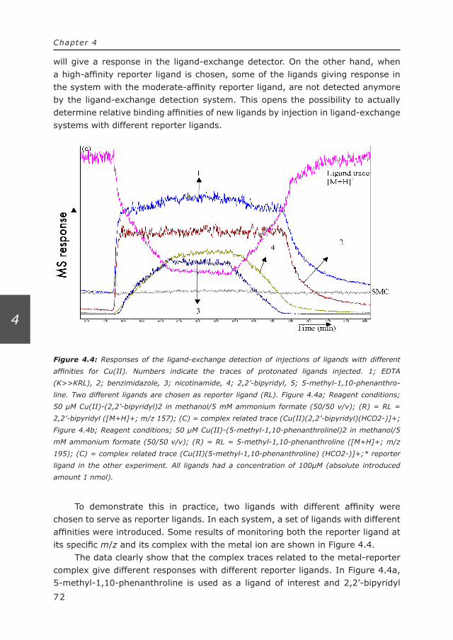
Figure 4.4: Responses of the ligand-exchange detection of injections of ligands with different
affinities for Cu(II). Numbers indicate the traces of protonated ligands injected. 1; EDTA
(K>>KRL), 2; benzimidazole, 3; nicotinamide, 4; 2,2’-bipyridyl, 5; 5-methyl-1,10-phenanthro-
line. Two different ligands are chosen as reporter ligand (RL). Figure 4.4a; Reagent conditions;
50 µM Cu(II)-(2,2’-bipyridyl)2 in methanol/5 mM ammonium formate (50/50 v/v); (R) = RL =
2,2’-bipyridyl ([M+H]+; m/z 157); (C) = complex related trace (Cu(II)(2,2’-bipyridyl)(HCO2-)]+;
Figure 4.4b; Reagent conditions; 50 µM Cu(II)-(5-methyl-1,10-phenanthrolinel)2 in methanol/5
mM ammonium formate (50/50 v/v); (R) = RL = 5-methyl-1,10-phenanthroline ([M+H]+; m/z
195); (C) = complex related trace (Cu(II)(5-methyl-1,10-phenanthroline) (HCO2-)]+;* reporter
ligand in the other experiment. All ligands had a concentration of 100μM (absolute introduced
amount 1 nmol).
72
Chapter 4
4
will give a response in the ligand-exchange detector. On the other hand, when a high-affinity reporter ligand is chosen, some of the ligands giving response in the system with the moderate-affinity reporter ligand, are not detected anymore by the ligand-exchange detection system. This opens the possibility to actually determine relative binding affinities of new ligands by injection in ligand-exchange systems with different reporter ligands.
To demonstrate this in practice, two ligands with different affinity were chosen to serve as reporter ligands. In each system, a set of ligands with different affinities were introduced. Some results of monitoring both the reporter ligand at its specific m/z and its complex with the metal ion are shown in Figure 4.4.
The data clearly show that the complex traces related to the metal-reporter complex give different responses with different reporter ligands. In Figure 4.4a, 5-methyl-1,10-phenanthroline is used as a ligand of interest and 2,2’-bipyridyl

73
Chapter 4
4
as reported ligand. Since 5-methyl-1,10-phenanthroline has a higher affinity to Cu(II) than 2,2’-bipyridyl, a positive response is seen in the reporter ligand trace. On the other hand, when 2,2’-bipyridyl is used as a ligand of interest, no response is observed in the 5-methyl-1,10-phenanthroline (reporter ligand) trace (see 4.4b).
Upon injection of benzimidazole as a ligand, a negative response was observed in the metal-reporter trace, as indicated by the arrows in Figure 4.4a and 4.4b. This decrease in response is not corresponding with ligand-exchange, but is due to ion suppression. A similar decrease was observed in the trace of the system monitoring compound (data not shown).
It is tempting to qualitatively or even quantitatively correlate the relative intensity of the response with the relative stability constants, i.e., to investigate whether a ligand with a higher affinity results in a higher release of reporter ligand. Table 4.2 displays the limit of detection (LOD) for the different injected ligands with both ligand-exchange reactions.
Table 4.2: Estimated limit of detection (LOD) (signal to noise ratio (S/N) =3) for ligand-exchange
detection of the different ligands using two different reporter ligands with corresponding different
affinities. (R.L. = reporter ligand; N.D. = not detectable). The LODs are based on the response of
the reporter ligand.
R.L.: 2,2’-bipyridylR.L.: 5-methyl-1,10-
phenanthroline
Introduced ligandsS/N
(100 µM)Estimated LOD
(µM)S/N
(100 µM)Estimated LOD
(µM)4-picoline N.D. - N.D. -
benzimidazole N.D. - N.D. -
nicotinamide N.D. - N.D. -
2,2’-bipyridyl R.L. - N.D. -
1,10-phenanthroline 5.17 57 4.57 665-methyl-1,10-phenanthroline
4.62 65 R.L. -
4,7-dimethyl-1,10-phenanthroline
4.13 73 3.68 81
EDTA 13.2 20 19.8 15
It is clear that under the given conditions similar concentrations of low affinity ligands will not give a response in the ligand-exchange detection method, whereas the high affinity ligands do. While a qualitative correlation appears to be observed, drawing conclusions must be done with great care, because the actual response is the result of various interrelated parameters. As indicated above, the relative release of reporter ligands is determined by the character of the injected ligand (mono- or polydentate) and of the reporter ligand. In addition, the ESI-MS response of a certain concentration of the reporter ligand might be influenced by

74
Chapter 4
4
other components in the mixture, i.e., a certain injected ligand might result in a relative suppression of the reporter ligand response without acting as a clear ion suppression agent. In addition, some of the complexes formed will have no net positive charge and are not detected by positive-ion ESI-MS at all. A complete quantitative assessment of the responses obtained in the ligand-exchange reactor is beyond the scope of the present study.
4.3.6 LC – ligand-exchange – ESI-MS with Cu(II)
A possible application area of the ligand-exchange ESI-MS detection system is in the screening of (complex) mixtures, e.g., combinatorial libraries, biological samples, or natural extracts, for ligands with particular affinities to certain metal ions. This might for instance be useful to screen for inhibitors of metal-containing enzymes or other metal-based homogeneous catalytic systems. Screening of complex mixtures can be performed via an on-line coupling with LC in order to separate the mixture prior to introduction into the ligand-exchange reactor. Because a fraction of the ligand introduced in the reactor is not involved in the ligand-exchange reactor, on-line ESI-MS and MS/MS detection enables the iden-tification of the unknown compounds in a mixture with affinity to the metal ion. This can be performed in the same run.
To test the efficacy of on-line LC – ligand-exchange – ESI-MS detection, various experiments were performed. Gradient elution, with either methanol or acetonitrile as organic modifier, was used to obtain an acceptable separation of a test mixture of ligands. Although both acetonitrile and methanol can be used as organic modifiers, methanol is used in further on-line LC experiments.
Figure 4.5 displays the chromatogram obtained when a test mixture of ligands with different affinities to Cu(II) is analyzed by means of the LC – ligand-exchange – ESI-MS detection method. In this experiment, 2,2’-bipyridyl was chosen as the reporter ligand.
In the top trace in Figure 4.5, the reporter ligand trace is given, showing response changes upon elution of ligands with higher affinity to Cu(II). In the middle, a trace related to the Cu(II)-reporter ligand complexes, also showing response changes upon elution of ligands with higher affinity to Cu(II). The lower traces show a ‘conventional’ LC–ESI-MS chromatogram of the separated ligands in the mixture. Peaks that show up in the lower trace, but do not appear in the upper two traces, correspond to compounds with a low affinity to Cu(II), e.g., nicotinamide and 4-picoline (cf. Table 4.1).

Figure 4.5: LC – Ligand-exchange – ESI-MS chromatogram for ligands with an affinity for Cu(II).
(R), reporter ligand trace ([M+H]+; m/z 157); (C1), [Cu(II)(2,2’-bipyridyl)(HCO2-)]+ and (C2),
[Cu(II)(2,2’-bipyridyl)2(HCO2-)]+. LC conditions; 100 × 2 mm ID C8 (3 µm particles). A gradient
ran from 100% A to 100% B in 2 min and remained stable at 100% B for 10 min. at a total flow of
100 µL/min. A, Methanol/5 mM NH4HCO2 (pH 6.3) (35/65 v/v); B, Methanol/5 mM NH4HCO2 (pH
6.3) (50/50 v/v). Reagent conditions; 50 µM Cu(II)(2,2’-bipyridyl)2 in B. The injected mixture
contained; 1, 2-(aminomethyl)-pyridine; 2, nicotinamide; 3, benzimidazole; 4, 4-picoline; 5,
1,10-phenanthroline; 6, 5-methyl-1,10-phenanthroline; 7, 2,9-dimethyl-1,10-phenanthroline. All
ligands had a concentration of 100 μM (absolute introduced amount 1 nmol).
75
Chapter 4
4
4.3.7 LC – ligand-exchange – ESI-MS with Zn(II)
Most of the results discussed in this paper were obtained using Cu(II) as metal ion. Obviously, the ligand-exchange detection principle is not limited to the use of Cu(II) alone. LC – Ligand-exchange – ESI-MS detection based on reactions of phosphopeptides with Fe(III)-MCB (methylcalcein blue) complexes was reported earlier [11]. Some of our results with Zn(II) are discussed in this section. Zn(II) is a common Lewis acid in biological systems and is involved in all kind of biochemical reactions [18]. Compared to Cu(II)-complexes, Zn(II)-complexes are generally weaker, which might pose a challenge in the ligand-exchange detection.

Figure 4.6: LC – Ligand-exchange – ESI-MS chromatogram for ligands with an affinity for
Zn(II). (R), reporter ligand trace ([M+H]+; m/z 157); (C), complex related trace [Zn(II)-(2,2’-
bipyridyl)(HCO2-)]+. LC conditions; 5 × 1 mm ID Phenomenex Luna C18(2) (3 µm particles).
A gradient was operated at 50 μL/min between pump 1a and pump 1b. The gradient ran from
20% B upto 85% B in 6 min where A was, Methanol/5 mM NH4HCOO (pH 6.3) (20/80 v/v);
B, Methanol/5 mM NH4HCOO (pH 6.3) (80/20 v/v). Reagent conditions; 50µM Zn(II)-(2,2’-
bipyridyl)2 in methanol/5 mM NH4HCOO (pH 6.3) (50/50 v/v). The injected mixture contained; 1,
2-(aminomethyl)-pyridine; 2, nicotinamide; 3, pyridine (also SMC); 4, 4-picoline; 5, 1,10-phen-
anthroline; 6, 5-methyl-1,10-phenanthroline; 7, 4,7-dimethyl-1,10-phenanthroline. The concen-
tration of the ligands was 100 μM (absolute introduced amount 1 nmol).
76
Chapter 4
4
The setup used in monitoring Zn(II)-related ligand exchange is identical to the one used in the detection for Cu(II)-related ligand exchange (see Figure 4.1). Like with Cu(II), also Zn(II) complexes can be easily recognized by means of their specific isotope pattern. Preliminary experiments, such as a study of the complex formation profiles and direct injections of ligands with different affinities for Zn(II), were performed (data not shown). This demonstrated that a system based on Zn(II) can be developed with the same ease as a system based on Cu(II).
Figure 4.6 shows a chromatogram obtained after LC separation of a test mixture of ligands and a Zn(II) ligand-exchange – ESI-MS detection. The same type of traces are shown as in Figure 4.5 for the Cu(II) system.
Again, we only detect a positive increase in the reporter ligand trace and an

77
Chapter 4
4
decrease in signal in the metal-reporter ligand complex trace when a ligand of interest was present which had a higher affinity to Zn(II). Under those conditions a ligand-exchange reaction occurred.
In general, the LC mobile-phase composition might interfere with the ligand-exchange detection. For instance, LC performed under acidic conditions can interfere since a ligand may be protonated under these conditions. Buffer comaptibility issues may be overcome by choosing a suitable buffer in the ligand-exchange reaction to control the pH of the ligand-exchange reaction. Of course, the current methodology requires compatibility of the buffer used with electro-spray MS detection.
4.4 Conclusions
In the present paper, we have demonstrated that by on-line coupling of the ligand-exchange – ESI-MS detection with an LC separation, it is possible to screen mixtures of ligands for their relative affinity to a particular metal-reporter ligand complex. Although the interactions of selected ligands with Cu(II) and Zn(II) was investigated, the approach can be considered as a template to study selective interactions between other ligands and other metal ions in a similar way.
The use of ESI-MS for detection allows a free choice of reporter ligand, and therefore the affinity of the reporter ligand to the metal ion, compared to other approaches, e.g., based on fluorescence detection. Additionally, MS provides the possibility to monitor not only the reporter ligand, but also dissociation of metal-reporter ligand complexes and formation of metal-ligand of interest complexes. Moreover information about the ligand of interest can be obtained This is especially interesting when dealing with unknown ligands.
During method development, direct-infusion experiments provide insight in the identity of the complexes formed under different experimental conditions, e.g. with respect to solution pH, organic modifier content, and mixing ratios between metal-ions, reporter ligand, and the ligand investigated. The selectivity of the ligand-exchange detection method can be tuned by the choice of the reporter ligand. This is demonstrated by using either 2,2 ‘-bipyridyl or 5-methyl-1,10-phenanthroline as reporter ligands. In case that the system should be used for the screening of low affinity analytes it is preferable to use a low affinity reporter ligand for detection. The same strategy should be followed when screening of low concentrations of high affinity ligands is considered.
The most significant improvement with respect to the more commonly used method IMAC lies in the possibility to screen a mixture of metal ligands with a large range of affinities. In IMAC, where all ligands are exposed to the immobilized metal ion prior to separation, a large concentration of a high affinity ligand would exclude the simultaneous enrichment of low affinity ligands by occupying all
78
Chapter 4
4
available metal coordination sites. In the current method, ligands are separated prior to the interaction with the metal ion.
Applications of this approach in the screening of (complex) mixtures, e.g., combinatorial libraries, biological samples, or natural extracts, for possible new drugs, catalysts or other ligands with particular affinities to certain metal ions are currently under development.
4.5 Acknowledgements
The late Dr. M.W.G. de Bolster from the section of organic chemistry and inorganic chemistry of the Vrije Universiteit Amsterdam is acknowledged for his helpful discussion. Shimadzu Benelux (‘s Hertogenbosch, The Netherlands) is acknowledged for the loan of equipment.
4.6 Reference List
[1] J.C. Traeger, Int. J. Mass Spectrom. 200 (2000) 387.[2] J.R. Seal, C.M. Havrilla, N.A. Porter, D.L. Hachey, J. Am. Soc. Mass Spectrom. 14
(2003) 872.[3] M. Gaub, A. von Brocke, G. Roos, K.A. Kovar, Phytochem. Anal. 15 (2004) 300.Anal. 15 (2004) 300.[4] M. van Stipdonk, V. Anbalagan, W. Chien, G. Gresham, G. Groenewold, D. Hanna, J.
Am. Soc. Mass Spectrom. 14 (2003) 1205.[5] C.P.G. Butcher, B.F.G. Johnson, J.S. McIndoe, X. Yang, X.B. Wang, L.S. Wang, J. Chem
Phys. 116 (2002) 6560.[6] M. Vincenti, J. Mass Spectrom. 30 (1995) 925.[7] N.G. Tsierkezos, D. Schroder, H. Schwarz, Int. J. Mass Spectrom. 235 (2004) 33.235 (2004) 33.[8] M. Satterfield, J.S. Brodbelt, Inorg. Chem. 40 (2001) 5393.[9] M.L. Reyzer, J.S. Brodbelt, A.P. Marchand, Z.B. Chen, Z.L. Huang, I.N.N. Namboothiri,
Int. J. Mass Spectrom. 204 (2001) 133.[10] J.G. Krabbe, H. Lingeman, W.M.A. Niessen, H. Irth, in preparation 2005.[11] J.G. Krabbe, H. Lingeman, W.M.A. Niessen, H. Irth, Anal. Chem. 75 (2003) 6853.[12] C. Matsumoto, O. Ohmori, T. Shinkai, H. Hori, J. Nakamura, Psychiatric Genetics 14
(2004) 209.[13] N. Farrell, Coord. Chem. Rev. 232 (2002) 1.[14] P. Chen, Angew. Chem. Int. Ed. 42 (2003) 2832.[15] C. de Graaf, N.P.E. Vermeulen, K.A. Feenstra, J. Med. Chem. 48 (2005) 2308.Chem. 48 (2005) 2308.[16] J.W. Wong, R.L. Albright, N.H.L. Wang, Sep. Purif. Methods 20 (1991) 49.[17] L.D. Holmes, M.R. Schiller, J. Liq. Chromatogr. Related Technol. 20 (1997) 123.[18] T. Dudev, C. Lim, J. Am. Chem. Soc. 122 (2000) 11146.[19] V.E. Vandell, P.A. Limbach, J. Mass Spectrom. 33 (1998) 212.[20] A.R. de Boer, T. Letzel, D.A. van Elswijk, H. Lingeman, W.M.A. Niessen, H. Irth, Anal.
Chem. 76 (2004) 3155.[21] M.J. Greig, H. Gaus, L.L. Cummins, H. Sasmor, R.H. Griffey, J. Am. Chem. Soc. 117
(1995) 10765.[22] E.C. Kempen, J.S. Bordbelt, Anal. Chem. 72 (2000) 5411.[23] A.L. Chaparro, R.W. Vachet, J. Mass Spectrom. 38 (2003) 333
79
Chapter 4
4
[24] H. Irth, M. Lamoree, G.J. de Jong, U.A.T. Brinkman, R.W. Frei, R.A. Kornfeldt, L. Persson, J. Chromatogr. 499 (1990) 617.
[25] C.E. Werkhoven-Goewie, W.M.A. Niessen, U.A.T. Brinkman, R.W. Frei, J. Chromatogr. 203 (1981) 165.

81
Chapter 5a
5a
Chapter 5a. Ligand-Exchange Detection
of Phosphorylated Peptides Using Liquid
Chromatography Electrospray Mass
Spectrometry
Analytical Chemistry, 2003, 75, 6853-6860
82
Chapter 5a
5a
Abstract
Electrospray ionization mass spectrometry (ESI-MS) is used to selectively detect analytes with a high affinity for metal ions. The detection method is based on the selective monitoring of a competing ligand at its specific m/z value that is released during the ligand-exchange reaction of a metal-ligand complex with analyte(s) eluting from a reversed-phase liquid chromatography column. The ligand-exchange reaction proceeds in a postcolumn reaction detection system placed prior to the inlet of the electrospray MS interface. The feasibility of metal affinity detection by ESI-MS is demonstrated using phosphorylated peptides and iron(III)methylcalcein blue as reactant, as a model system. Methylcalcein blue (MCB) released upon interaction with phosphorylated peptides is detected at m/z 278. The ligand-exchange detection is coupled to a C8 reversed-phase column to separate several nonphosphorylated enkephalins and the phosphorylated peptides pp60 c-src (P) and M2170. Detection limits of 2 µM were obtained for pp60 c-src (P) and M2170. The linearity of the detection method is tested in the range of 2-80 µmol/L phosphorylated compounds (r2 = 0.9996), and a relative standard deviation of less than 8% (n= 3) for all MCB responses of the different concentra-tions of phosphorylated compounds was obtained. The presented method showed specificity for phosphorylated peptides and may prove a useful tool for studying other ligand-exchange reactions and metal-protein interactions.

83
Chapter 5a
5a
5a.1. Introduction
The importance and applicability of mass spectrometry (MS) in (bio)chemistry is now widely recognized [1-9]. MS is a key technology for protein characteriza-tion, for example, for protein profiling and the measurement of protein-ligand interactions [10-14].
One area less frequently studied by MS is protein-metal interactions, although metal complexes of proteins play a key role in several biological processes [15-17]. For example, metal ions are important for the structure and folding of several proteins, where structural changes or unfolding due to the removal of the metals might lead to denaturation and inactivation of the proteins. In enzymatic reactions, the metal center in the proteins can act as a catalyst [18,19]. Other groups of metal-binding proteins play an important role in the metabolism and detoxifica-tion of certain metals in biological systems [20,21]. Moreover, several metal-protein complexes are known to act as transporters for all kinds of ligands, e.g., hemoglobin which is responsible for the oxygen transport from lungs to peripheral tissue in living organisms.
Conventional methods for studying metal-protein interactions involve, among others, infrared spectroscopy, [22] nuclear magnetic resonance spec-troscopy [22], UV detection [19] and more recently mass spectrometry [23]. Mass spectrometric studies of metal-ligand and metal-protein interactions were performed for stoichiometric determinations of complexes [24,25] and fragmen-tation of the metal complexes by collision-induced dissociation (CID) [26-29] for structural studies or identifications of unknown complexes.[30,31] Little work has been done to use MS for the direct detection of ligand-exchange reactions. Metal-based ligand-exchange reactions, in batch, were monitored either by infusion-MS, [32-34] or by liquid chromatography coupled to MS [35].
Ligand-exchange reactions play a key role in several (bio)-chemical processes, for instance, as catalysts in production processes [36] or in several enzymatic reactions as a coenzyme or cofactor [15,16,37]. Several postcolumn ligand-exchange detection methods, based on the same principle, are known. A continuous-flow ligand-exchange method based on a palladium-calcein complex was developed for the detection of various organosulfur containing compounds in wash water, vegetables, urine, and serum [38]. Irth et al. [39] developed a selective fluorescence detection method for inositol phosphates based on a postcolumn ligand-exchange reaction with a weakly fluorescent Fe(III)-methylcal-cein blue (MCB) complex. The MCB released in this reaction is highly fluorescent and allows the indirect detection of the nonfluorescent inositol phosphates. MCB has the advantage that it forms a relatively weak complex with Fe(III) making an exchange of a ligand with a higher affinity for the metal, e.g., phosphory-lated compounds, possible. A major disadvantage of using fluorescence is that

84
Chapter 5a
5a
the workable pH range is between [7] and [10,39] with a fluorescence maximum of MCB a pH of 8.5 [40]. In contrast, the highest selectivity of Fe(III) toward a phosphate group is between pH 3 and pH 5. Above pH 5, the selective affinity decreases due to competitive binding of hydroxyl ions to Fe(III) [41]. Moreover, the choice of fluorescent complexing agents is limited, making it rather difficult to apply this detection method to other biologically relevant analytes.
In the present paper, the use of electrospray ionization-mass spectrometry (ESI-MS) as a detection method is reported for the monitoring of ligand-exchange reactions and is applied for the selective detection of phosphorylated peptides in the presence of nonphosphorylated peptides. The interaction of the binding of metal ions and phosphorylated compounds is now a popular field of interest in biochemistry and medicinal chemistry [42-44]. A reversed-phase liquid chro-matographic separation is combined with the ligand-exchange reaction between Fe(III)-MCB and phospho groups, where MCB acts as a reporter molecule that is monitored by MS. Monitoring the exchanged MCB, at its specific m/z, makes it possible to identify and possibly quantify phosphorylated compounds in the presence of nonphosphorylated compounds. MS detection and identification is not limited to a relative high pH and can, if necessary, be performed in the optimum pH range to obtain the highest selectivity. More importantly, the reporter ligands, i.e., complexing agents used to indicate the presence of analyte with metal-binding properties, can be chosen more freely than in fluorescence detection [41].
5a.2 Experimental
5a.2.1 Materials
Acetonitrile and methanol were purchased from Baker (Deventer, The Netherlands) and were filtered over a 0.45-µm Millipore filter. Iron(III) nitrate was purchased from Acros Organics (Geel, Belgium), and methylcalcein blue (4-meth-ylumbelliferone-8-methylenesarcosine) (MCB) (M = 277.28) was obtained from Alfa Aesar (Karlsruhe, Germany). Adenosine monophosphate (AMP), adenosine diphosphate (ADP), and adenosine triphosphate (ATP) were obtained from Sigma-Aldrich (Zwijndrecht, The Netherlands). Ala-Ala-Ala-Ala (M = 302.33), which was used to continuously monitor the performance of the MS, M2170 (M =748.73), enkephalins, the phosphorylated peptide pp60 c-src (P) (M = 1543.50), and its nonphosphorylated analogue pp60 c-src (M = 1463.25) were purchased from Bachem (Budendorf, Switserland). Delta (Phospho) sleep induced peptide (DSIP(P)) was obtained from American Peptide Co. (Sunnyvale,CA).

85
Chapter 5a
5a
5a.2.2 Reagent Preparation
Stock solutions (1 mmol/L) of iron(III) nitrate and MCB were prepared in methanol/water (50/50,v/v) containing 0.1% formic acid. The reagent solution was prepared by adding a stock solution of iron(III) nitrate to a solution of MCB in ammonium formate buffer (5 mmol/L, pH 3.25) containing 50% methanol (MeOH). Unless otherwise reported the final concentration of Fe(III) and MCB was 40 and 80 µmol/L, respectively.
5a.2.3 Flow Injection Ligand-Exchange Analysis
The flow injection experiments were performed with a Shimadzu (’s Herto-genbosch, The Netherlands) single-quadruple LCMS 2010 system. The system consisted of two LC-10ADvp pumps, a SCL-10ADvp system controller, a SIL-10ADvp autoinjector, a CTO-ACvp column oven, and a single-quadruple mass spectrometer with an ESI.
5a.2.4 Liquid Chromatography Ligand-Exchange Analysis
The LC experiments were performed on a 100 x 2.00 mm i.d. Phenomenex Luna C8(2) column packed with 3-µm particles. A gradient was operated from 100% A up to 100% B in 20 min, where A consisted of acetonitrile/ammonium formate (5 mmol/L; pH 3.25; 10/90 v/v) and B consisted of acetonitrile/ammonium formate (5 mmol/L; pH 3.25; 40/60 v/v). The total flow was maintained at 50 µL/min. The LC column was placed in a column oven that was set at 40 °C. The ligand-exchange setup was the same as that used for the flow injection experiments, with the only difference that sample carrier flow was replaced by the LC eluent.
5a.2.5 Mass Spectrometry Settings
Mass spectrometric detection was performed in positive-ion electrospray ionization in selected-ion monitoring (SIM) mode. The total flow rate directed toward the MS detector was 100 µL/min. The probe voltage was 3.5 kV, and nitrogen (99.999% purity, Praxair, Oevel, Belgium) gas flow was set at 3.5 L/min. For good solvent evaporation, CDL temperature and block temperature were maintained at 200 and 95 °C, respectively, and for the extraction of ions, a CDL voltage of -10 V was applied. Infusion tandem MS for the identification of the complex-related trace was performed on an Agilent (Agilent Technologies, Palo Alto, CA) 1100 series LC/MSD trap SL mass spectrometer. Nitrogen (99.999% purity, Praxair) was used as nebulizer gas and dry gas, which was 16 psi and 5 L/
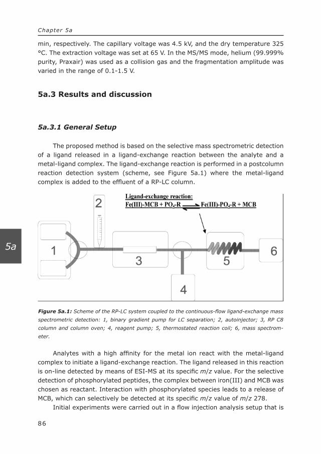
Figure 5a.1: Scheme of the RP-LC system coupled to the continuous-flow ligand-exchange mass
spectrometric detection: 1, binary gradient pump for LC separation; 2, autoinjector; 3, RP C8
column and column oven; 4, reagent pump; 5, thermostated reaction coil; 6, mass spectrom-
eter.
86
Chapter 5a
5a
min, respectively. The capillary voltage was 4.5 kV, and the dry temperature 325 °C. The extraction voltage was set at 65 V. In the MS/MS mode, helium (99.999% purity, Praxair) was used as a collision gas and the fragmentation amplitude was varied in the range of 0.1-1.5 V.
5a.3 Results and discussion
5a.3.1 General Setup
The proposed method is based on the selective mass spectrometric detection of a ligand released in a ligand-exchange reaction between the analyte and a metal-ligand complex. The ligand-exchange reaction is performed in a postcolumn reaction detection system (scheme, see Figure 5a.1) where the metal-ligand complex is added to the effluent of a RP-LC column.
Analytes with a high affinity for the metal ion react with the metal-ligand complex to initiate a ligand-exchange reaction. The ligand released in this reaction is on-line detected by means of ESI-MS at its specific m/z value. For the selective detection of phosphorylated peptides, the complex between iron(III) and MCB was chosen as reactant. Interaction with phosphorylated species leads to a release of MCB, which can selectively be detected at its specific m/z value of m/z 278.
Initial experiments were carried out in a flow injection analysis setup that is

Figure 5a.2: Complex formation with 7-fold amplification of complex trace (Fe-MCB trace).
87
Chapter 5a
5a
comparable to the setup in Figure 5a.1, with the exception that the binary gradient pump was replaced by an isocratic pump and no column and column oven were used. To prove that the complex formation between Fe(III) and MCB takes place, first a solution of free MCB was pumped continuously toward the MS, and after a certain period of time, Fe(III) was added. The addition of Fe(III) results in a large decrease in MCB trace (m/z 278), whereas the complex-related trace (m/z 395) exhibited an increase (Figure 5a.2).
From these experiments, indirect conclusions can be drawn that complex formation occurs and that the Fe(III)-MCB complex is stable under ESI ionization conditions. Mass spectrometric data obtained by infusion MS, revealed no significant responses for m/z 395 or other Fe(III)-MCB-related traces in either blank solution of running buffer, blank solution of free MCB, or blank solution of Fe(NO3)3.
It is difficult to develop a theoretical model to predict the amount of complex formed. Several parameters, such as ratio, other ligands present, and concentra-tion of Fe(III) and MCB, make the calculations rather complex. Most probably a
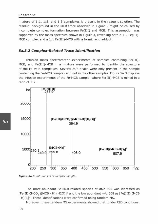
Figure 5a.3: Infusion MS of complex sample.
88
Chapter 5a
5a
mixture of 1:1, 1:2, and 1:3 complexes is present in the reagent solution. The residual background in the MCB trace observed in Figure 2 might be caused by incomplete complex formation between Fe(III) and MCB. This assumption was supported by the mass spectrum shown in Figure 3, revealing both a 1:2 Fe(III)-MCB complex and a 1:1 Fe(III)-MCB with a formic acid adduct.
5a.3.2 Complex-Related Trace Identification
Infusion mass spectrometric experiments of samples containing Fe(III), MCB, and Fe(III)-MCB in a mixture were performed to identify the structure of the Fe-MCB complexes. Several m/z-peaks were only present in the sample containing the Fe-MCB complex and not in the other samples. Figure 5a.3 displays the infusion experiments of the Fe-MCB sample, where Fe(III)-MCB is mixed in a ratio of 1:2.
The most abundant Fe-MCB-related species at m/z 395 was identified as [Fe(III)(HCO2
-)(MCB - H)-(H2O)]+ and the low abundant m/z 608 as [Fe(III)((MCB - H)-)2]
+. These identifications were confirmed using tandem MS. Moreover, these tandem MS experiments showed that, under CID conditions,
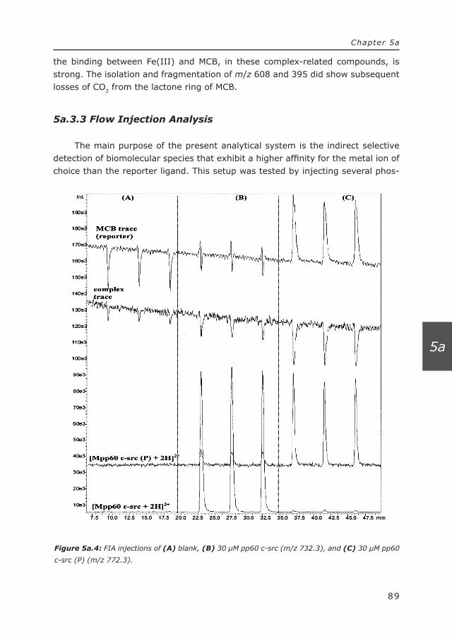
Figure 5a.4: FIA injections of (A) blank, (B) 30 µM pp60 c-src (m/z 732.3), and (C) 30 µM pp60
c-src (P) (m/z 772.3).
89
Chapter 5a
5a
the binding between Fe(III) and MCB, in these complex-related compounds, is strong. The isolation and fragmentation of m/z 608 and 395 did show subsequent losses of CO2 from the lactone ring of MCB.
5a.3.3 Flow Injection Analysis
The main purpose of the present analytical system is the indirect selective detection of biomolecular species that exhibit a higher affinity for the metal ion of choice than the reporter ligand. This setup was tested by injecting several phos-

90
Chapter 5a
5a
phorylated and nonphosphorylated compounds, see Figure 5a.4.SIM of MCB, released during the ligand-exchange reaction between phos-
phorylated peptides and Fe-MCB, was set at m/z 278. As can be clearly seen in Figure 5a.4, the injection of 30 µM phosphorylated pp60 c-src (P) (Figure 5a.4C) showed a significant response at the MCB trace, whereas the Fe-MCB trace showed, as expected, negative peaks, indicating a decrease of the complex concentra-tion due to the interaction with the phosphorylated peptide. The injection of the nonphosphorylated analogue (30 µM) (Figure 5a.4B) resulted in a response that was comparable to blank injections in Figure 5a.4A. A small increase of response was seen at the injections of nonphosphorylated pp60 c-src (Figure 5a.4B) in the phosphorylated pp60 c-src (P) trace, which could be attributed to an isotope of the dipotassium adduct of the nonphosphorylated pp60 c-src ([M + 2K]2+).
Several parameters influence the ligand-exchange reaction and mass spec-trometric detection sensitivity such as pH, concentration of reagent, and MS settings. These parameters were optimized by flow injection analysis using ATP and a compound, Ala-Ala-Ala-Ala, to monitor the mass spectrometric response. ATP, which is a model compound for phosphorylated analytes, showed similar results compared to other phosphorylated compounds.
5a.3.4 Optimization of pH
The pH is one of the most important parameters that influences both the ligand-exchange reaction and mass spectrometric detection. With respect to the ligand-exchange reaction, the pH influences the degree of ionization of MCB and of the phospho group and possible formation of other Fe(III) complexes such as hydroxy complexes (above pH 5). Also at 3 < pH < 5, optimal selectivity of Fe(III) is achieved for phospho groups.[41] In positive-ion electrospray mass spectrometry, generally a low pH is favorable because of a better protonation of the analytes. An optimal signal-to-noise ratio was found between 3 < pH < 3.5 (Figure 5a.5A).
A possible reason for the decrease in sensitivity above pH 3.5, is a decrease in ionization efficiency of MCB. The decrease of background MCB trace as well as the response of Ala-Ala-Ala-Ala supports this assumption.
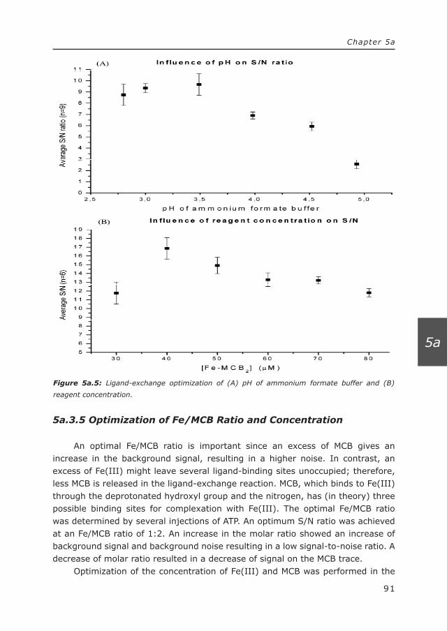
Figure 5a.5: Ligand-exchange optimization of (A) pH of ammonium formate buffer and (B)
reagent concentration.
91
Chapter 5a
5a
5a.3.5 Optimization of Fe/MCB Ratio and Concentration
An optimal Fe/MCB ratio is important since an excess of MCB gives an increase in the background signal, resulting in a higher noise. In contrast, an excess of Fe(III) might leave several ligand-binding sites unoccupied; therefore, less MCB is released in the ligand-exchange reaction. MCB, which binds to Fe(III) through the deprotonated hydroxyl group and the nitrogen, has (in theory) three possible binding sites for complexation with Fe(III). The optimal Fe/MCB ratio was determined by several injections of ATP. An optimum S/N ratio was achieved at an Fe/MCB ratio of 1:2. An increase in the molar ratio showed an increase of background signal and background noise resulting in a low signal-to-noise ratio. A decrease of molar ratio resulted in a decrease of signal on the MCB trace.
Optimization of the concentration of Fe(III) and MCB was performed in the

Figure 5a.6: Influence of reactor volumes at different temperatures on S/N ratio of MCB.
92
Chapter 5a
5a
range of 10-100 µmol/L Fe(III) in steps of 10 µmol/L using a 1:2 Fe(III)/MCB ratio. Since unbound MCB causes a background response, high reagent concen-trations might decrease the S/N ratio. On the other hand, low reagent concentra-tions causes a decreased response of phosphorylated compounds. Optimal S/N ratio was obtained at a 40 µM Fe(III) concentration, which is shown in Figure 5a.5B.
5a.3.6 Optimization of Reaction Coil and Reaction Temperature
Several open-tubular reactors were investigated with different internal diameters and reaction volumes. Since the reaction is almost simultaneous, only a small reaction volume is necessary. From the two investigated reactors with an internal diameter of 0.3 and 0.18 mm, respectively, the 0.18-mm-i.d. reactor gave the best results in terms of band broadening (peak shape and height). Several reactor volumes were investigated ranging from 2, 5, 10, to 20 µL resulting in a reaction time of 1.2, 3, 6, and 12 s, respectively.
Figure 5a.6 displays the average S/N ratio of the 2-µL reactor and 5-µL reactor at different reaction temperatures.
The better response of the 2-µL reactor compared to the 5-µL reactor can be
93
Chapter 5a
5a
attributed to lower band broadening. This supports the assumption that mixing is the most important parameter for a fast reaction. Furthermore, increasing the temperature up to 50 °C clearly improves the S/N ratio in both the 2- and 5-µL reactor. At higher temperatures, however, a decrease in S/N ratio is observed, which is possibly caused by air bubble formation. It should be noted that no disso-ciation of the Fe(III)-MCB complex is observed at higher temperatures, since no additional free MCB is detected.
Experiments show similar results when methanol is replaced by acetoni-trile. Acetonitrile does not seem to interfere with either complex formation or the ligand-exchange reaction. This is interesting for the combination of a RP-LC separation with the ligand-exchange reaction in the analysis of phosphorylated and nonphosphorylated peptides.
5a.3.7 Optimization of MS Conditions
MS parameters often are highly interrelated. Probe voltage, nitrogen flow, and source temperature strongly influence solvent evaporation and ionization of the analytes [45]. In contrast to biochemical mass spectrometric systems, reported, for example, by Hogenboom et al.,[46] where a high organic modifier content interferes with the complex formation especially for a model system consisting of anti-digoxigenin, a relatively high percentage of organic modifier does not influence the metal complex-based ligand-exchange reaction, making it even more amenable for the coupling with RP-LC as well as MS.
Increase of the probe high voltage shows an increase of the S/N ratio up to 4 kV, after which a steep decrease is caused by the increase of background noise. An optimum S/N ratio was seen at a nitrogen gas flow of 3.5 L/min. A lower gas flow resulted in a decrease of the MCB response, which is probably caused by less efficient droplet desolvation. Since the curved desolvation line (CDL), which is the entrance of the Shimadzu MS, is under an angle, increasing gas flow rates above 3.5 L/min may cause less sample to enter the CDL. This may be the reason for the decrease in response of unbound MCB at higher gas flow rates. The CDL is used for the electrostatic extraction of ions from the charged spray as well as for better desolvation of the charged droplet by heating the capillary. An increase of CDL temperature provides a better desolvation with an optimum at 200 °C. The change in CDL temperature, CDL voltage, and block temperature did not have a great influence on the S/N ratio, and the variables were set at the optimum values.
It should be noted that the optimization of the current method for the detection of MCB released in the ligand-exchange reaction resulted in a decrease of the Fe-MCB response. Therefore, this mass trace is not used for further data evaluation.

94
Chapter 5a
5a
5a.3.8 RP-LC Coupled to Ligand-Exchange Mass Spectrometric De-tection
The goal of this study was to develop a new fast screening method for phos-phorylated compounds using ligand-exchange reactions in combination with MS. To separate known or unknown phosphorylated and nonphosphorylated peptides, a reversed-phase C8-column was placed in front of the ligand-exchange mass spectrometric detection unit. In such a setup, complex samples can be separated and screened for active compounds and additionally, structural information on the active compound can also be acquired based on MS data. During the entire procedure, a compound (Ala-Ala-Ala-Ala; m/z 303.0) was present to monitor the MS response. The blank injection showed an acceptable minor decrease in reporter trace when running the acetonitrile gradient from 10 to 40%. Previous flow injection experiments showed only minor influences of the type and content of organic modifier on both complex equilibrium and the ligand-exchange reaction. Figure 7 shows the analysis of two phosphorylated compounds in the presence of four closely related nonphosphorylated peptides.
Only phosphorylated compounds show an increase in response of unbound MCB whereas for nonphosphorylated compounds such an increase is not observed. The peak present at t = 7.5 min was also present in the blank injection. An interesting feature is that apparently a fraction (15%, by estimation) of the phos-phorylated peptides still is unbound, enabling identification.

Figure 5a.7: LC-LE-MS experiments of a 6 µM mixture of the phosphorylated pp60 c-src (P) and
M2170(P) and several nonphosphorylated enkephalins.
95
Chapter 5a
5a
A calibration curve showed linearity for the reporter trace of MCB for pp60 c-src (P) and M2170(P) ranging from 2 to 80 µmol/L (R2 = 0.9996, n = 3). Although 2 µM (LOD; S/N = 3) concentrations of both phosphorylated peptides can be detected in the reporter trace, it should be emphasized that it may be difficult to distinguish between a high-abundant nonphosphorylated compound and a low-abundant phosphorylated compound. Since extremely high concentrations of other ligands may interfere with the ligand-exchange reaction between Fe(III) and the phospho groups, a false positive assignment might be given to these

Figure 5a.8: Calibration curve of Ala-methionine enkephalin (AME), pp60 c-src (P), and the area
of the MCB peak related to AME and pp60 c-src (P)
96
Chapter 5a
5a
nonphosphorylated ligands. Figure 8 again illustrates that the ligand-exchange reaction is specific for
phosphorylated compounds.
Even at higher concentrations of nonphosphorylated compounds, no increase is seen in peak area for the related MCB peak. It displays a calibration curve for the nonphosphorylated Ala-methionine enkephalin (AME) and phosphorylated pp60 c-src (P) and the area of the MCB peak related to these two compounds.
5a.4 Conclusions
A new method for monitoring ligand-exchange reactions by mass spectrom-etry is demonstrated for phosphorylated peptides in the presence of nonphos-phorylated peptides, by a gradient RPLC separation followed by a continuous-flow ligand-exchange mass spectrometric detection.
Principally, for the development of the method, known phosphorylated and nonphosphorylated peptides are analyzed, but in general, unknown phosphory-lated compounds can also be identified based on their phospho group(s). The mass/charge ratio of the unbound fraction of these compounds might provide structural information about the specific compound.

97
Chapter 5a
5a
The gradient of the LC separation hardly influenced the complex stability or ligand-exchange reaction. MCB, which acted as the reporter trace, showed linearity in the range from 2 to 80 µmol/L of phosphorylated compounds. A LOD in the low-micromolar level was achieved for the phosphorylated peptides. Nonphos-phorylated compounds did not show a significant response on the reporter trace, which proved this method to be selective toward phosphorylated compounds. A relative standard deviation of less than 8% (n = 3) for all MCB responses of the different concentrations of phosphorylated compounds was obtained.
Although in this research an Fe(III)-MCB complex was used for the analysis of phosphorylated compounds, other ligand-exchange reactions can also be applied for other metals and ligands [47]. For instance, the analysis of histidine-containing compounds by using of Ni2+, which is known to bind to histidine, [48,49] or for the analysis of certain metal-binding proteins[25] may be considered. In general, a suitable metal-ligand complex involves a metal ion with strong affinity for the analyte(s) and a reporter ligand with strong affinity for the metal, but which is still able to exchange with the desired “active” ligand. One important parameter is the reaction rate, which should be rapid to maintain fast analysis times and acceptable band broadening. Also, both the complex between the metal and the reporter ligand and the complex with the ligand of interest should be stable in mass spectrometry. In particular, dissociation of the ligand complex during the ionization phase may cause a significant decrease of detection sensitivity.
In comparison to fluorescence-based techniques, the advantage of the proposed approach can be found in the versatility of mass spectrometry, particu-larly when the choice of a suitable ligand is concerned. Suitable candidate ligands can be selected based on their binding constants and detection sensitivities in MS. In contrast to immobilized metal-affinity chromatography approaches, the current method offers detection sensitivities that principally only depend on the ionization of the known reporter ligand and the binding constant of the analyte. This should be beneficial particularly in the discovery of novel unknown metal-binding species. The method might be a useful tool for studying protein-metal interactions.
5a.5 Acknowledgements
Shimadzu (’s Hertogenbosch, The Netherlands) and Agilent Technologies (Amstelveen, The Netherlands) are acknowledged for the loan of equipment.
5a.6 References
(1) Gerber, S. A.; Scott, C. R.; Turecek, F.; Gelb, M. H. Anal. Chem. 2001, 73, 1651-1657.
(2) Griffin, T. J.; Aebersold, R. J. Biol. Chem. 2001, 276, 45497-45500.

98
Chapter 5a
5a
(3) Griffiths, W. J.; Jonsson, A. P.; Liu, S.; Rai, D. K.; Wang, Y. Biochem. J. 2001, 355, 545-561.
(4) Gygi, S. P.; Aebersold, R. Curr. Opin. Chem. Biol. 2000, 4, 489-494.
(5) Li, Y.-T.; Hsieh, Y.-L.; Henion, J. D.; Ganem, B. J. Am. Soc. Mass Spectrom.Mass Spectrom. 1993, 4, 631-637.
(6) Loo, J. A. J. Mass Spectrom. 1995, 30, 180-183.
(7) Loo, J. A. Mass Spectrom. Rev. 1997, 16, 1-23.
(8) Nguyen, D. N.; Becker, G. W.; Riggin, R. M. J. Chromatogr., A 1995, 705, 21-45.
(9) Winston, R. L.; Fitzgerald, M. C. Mass Spectrom. Rev. 1997, 16, 165-179.
(10) Adam, B. L.; Davis, J. W.; Ward, M. D.; Clements, M. A.; Cazares, L. H.; Semmes, O. J.; Schellhammer, P. F.; Yasui, Y.; Feng, Z. D.; Wright, G. L. Cancer Res. 2002, 62, 3609-3614.
(11) Bane, T. K.; LeBlanc, J. F.; Lee, T. D.; Riggs, A. D. Nucleic Acids Res. 2002, 30, e69.
(12) Chaurand, P.; DaGue, B. B.; Pearsall, R. S.; Threadgill, D. W.; Caprioli, R.W. Proteomics 2001, 1, 1320-1326.
(13) Chaurand, R.; Caprioli, R. M. Electrophoresis 2002, 23, 3125-3135.
(14) Molloy, M. P.; Phadke, N. D.; Chen, H.; Tyldesley, R.; Garfin, D. E.; Maddock, J. R.; Andrews, P. C. Proteomics 2002, 2, 899-910.
(15) Kendrick, M. J.; May, M. T.; Plishka, M. J.; Robinson, K. D. Metals in Biological Systems, 1st ed.; Ellis Horwood: New York, 1992.
(16) Lehninger, A. L.; Nelson, D. L.; Cox, M. M. Principles of Biochemistry, 2nd ed.; Worth Publishers: New York, 1993.
(17) Fedurco, M. Coord. Chem. Rev. 2000, 209, 263-331.
(18) Betrand, M.; Weber, G.; Schoefs, B. Trends Anal. Chem. 2003, 22, 254-262.
(19) Matzapetakis, M.; Farrer, B. T.; Weng, T. C.; Hemmingsen, L.; Penner-Hahn, J. E.; Pecoraro, V. L. J. Am. Chem. Soc. 2001, 124, 8042-8054.
(20) Masuoka, J.; Hegenauer, J.; Van Dyke, B. R.; Saltman, P. J. Biol. Chem. 1993, 268, 21533-21537.
(21) Bebianno, M. J.; Cravo, A.; Miguel, C.; Morias, S. Sci. Total Environ. 2003, 301, 151-161.
(22) Nath, M.; Pokharia, S.; Song, X.; Eng, G.; Gielen, M.; Kemmer, M.; Biesemans, M.; Willem, R.; de Vos, D. Appl. Organomet. Chem.Organomet. Chem. 2003, 17, 305-314.
(23) Loo, J. A. Int. J. Mass Spectrom. 2001, 204, 113-123.
(24) Johnson, K. A.; Verhagen, M. F. J. M.; Brereton, P. S.; Adams, M. W. W.; Amster, I. J. Anal. Chem. 2000, 72, 1410-1418.
(25) Benson, L. M.; Kumar, R.; Cavanagh, J.; Naylor, S. Rapid Commun. Mass Spectrom.

99
Chapter 5a
5a
2003, 17, 267-271.
(26) Jellen, E. E.; Chappell, A. M.; Ryzhov, V. Rapid Commun. Mass Spectrom. 2002, 16, 1799-1804.
(27) Schoener, D. F.; Olsen, M. A.; Cummings, P. G.; Basic, C. J. Mass Spectrom. 1993, 34, 1069-1078.
(28) Chaparro, A. L.; Vachet, R. W. J. Mass Spectrom. 2003, 38, 333-342.
(29) Ho, Y.-P.; Huang, P.-C.; Deng, K.-H. Rapid Commun. Mass Spectrom. 2003, 17, 114-121.
(30) Mollah, S.; Pris, D. P.; Johnson, S. K.; Gwizdala, A. B.; Houk, R. S. Anal. Chem. 2000, 72, 985-991.
(31) Moreau, S.; Awade, A. C.; Molle, D.; Le Graet, Y.; Brule, G. J. Agric. Food Chem. 1995, 43, 883-889.
(32) Bond, A. M.; Colton, R.; Traeger, J. C.; Harvey, J. Inorg. Chim. Acta 1993, 212, 233-239.
(33) Canty, A. J.; Colton, R. Inorg. Chim. Acta 1994, 215, 179-184.
(34) Bond, A. M.; Colton, R.; Mah, Y. A.; Traeger, J. C. Inorg. Chem. 1994, 33, 2548-2554.
(35) Lemr, K.; Holcapek, M.; Jandera, P.; Lycka, A. Rapid Commun. Mass Spectrom. 2000, 14, 1881-1888.
(36) Perry, M. C.; Burgess, J. Tetrahedron: Asymmetry 2003, 14, 951-961.
(37) Allwood, E. G.; Davies, D. R.; Gerrish, C.; Ellis, B. E.; Bolwell, G. P. FEBS Lett. 1999, 457, 47-52.
(38) Werkhoven-Goewie, C. E.; Niessen, W. M. A.; Brinkman, U. A. Th.; Frei, R.W. J. Chromatogr. 1981, 203, 165-172.
(39) Irth, H.; Lamoree, M.; De Jong, G. J.; Brinkman, U. A. T.; Frei, R. W.; Kornfeldt, R. A.; Persson, L. J. Chromatogr. 1990, 499, 617-625.
(40) Huitink, G. M.; Diehl, H. Talanta 1974, 21, 1193-1202.
(41) Holmes, D.; Schiller, M. R. J. Chromatogr. Relat. Technol. 1997, 20, 123-142.
(42) Liu, H.; Zheng, J.; Stupak, J.; Keller, B.; Booth, B.; Fliegel, L.; Li, L. 51st Annual ASMS Conference, Montreal, Canada, 2003.
(43) Hettich, R.; Schnier, P.; Buchanan, M. 47th ASMS Conference, Dallas TX, 1999.
(44) Oda, Y.; Nagasu, T.; Chait, B. T. Nat. Biotechnol. 2001, 19, 379-382.
(45) Niessen, W. M. A. Liquid Chromatography-Mass Spectrometry, 2nd ed.; Marcel Dekker: New York, 1999.
(46) Hogenboom, A. C.; de Boer, A. R.; Derks, R. J. E.; Irth, H. Anal. Chem.Chem. 2001, 73, 3816-3823.
(47) Hancock, R. D.; Martell, A. E. Chem. Rev. 1989, 89, 1875-1914.
(48) Gaberc-Porekar, V.; Menart, V. J. Biochem. Biophys. MethodsBiophys. Methods 2001, 49, 335-360.
(49) Muller, K. M.; Arndt, K. M.; Bauer, K.; Pluckthun, A. Anal. Biochem.Biochem. 1998, 259, 54-61.
100
Chapter 5a
5a


103
Chapter 5b
5b
Chapter 5b. Selective detection and
identification of phosphorylated proteins by
simultaneous ligand-exchange fluorescence
detection and mass spectrometry
Journal of Chromatography A, 2006, 1130, 287-295

104
Chapter 5b
5b
Abstract
A ligand-exchange method for the detection and identification of phosphory-lated peptides in complex mixtures is presented that is based on the character-ization of phosphorylated species by solution-phase interactions with Fe(III) ions and subsequent fluorescence readout. After the separation of the peptides and digest products on a reversed-phase LC column, the flow is split between the two detection systems. One part is directed towards an electrospray mass spectrom-eter for direct detection and identification of all the peptides present in the sample. The other part of the flow is directed towards a ligand-exchange detection system. This system relies on the specific release of a fluorescent reporter ligand from a Fe(III)-complex in the presence of phosphorylated peptides. To recognize false positive signals due to high-affinity non-phosphorylated high-acidic peptides and other compounds which are known to be a problem in for instance immobilized metal affinity chromatography (IMAC), a second run is performed after incubation of the sample with alkaline phosphatase. A positive signal in this second run indicates a high-affinity non-phosphorylated compound. The method is illustrated using digest from a phosphorylated α-casein. Automated switching between MS and MS-MS was performed to obtain additional information about the compounds present in the sample.
The linearity of the method was tested in the range of 0.5 µM to 80 µM of phosphorylated peptides. A limit of detection (LOD) of 0.5 µM was obtained for a mono-phosphorylated peptide. The interday (n=4) and intraday precision (n=3) expressed as relative standard deviation was better than 10%.
105
Chapter 5b
5b
5b.1 Introduction
Reversible phosphorylation of proteins plays a key role in all kinds of biological processes, e.g., signal transduction [1], regulation of cellular life, and controlling enzyme activity [2]. Therefore, analytical methods for identifying and quantifying these compounds are important to obtain knowledge about these processes, e.g., in clinical diagnostics or target discovery [3]. Moreover, phosphorylated peptides and proteins are used as biomarkers, for instance, for certain forms of cancer [4] or Alzheimer disease [5].
In general, the analysis of phosphoproteins is not straightforward due to their relatively low abundance, variable phosphorylation sites and the possible presence of phosphatases [1]. Techniques for studying protein and peptide (de)phosphorylation involve specific antibody probes (primarily for tyrosine phos-phorylated peptides), radioactive labeling or mass spectrometry (MS) [6]. MS detection and identification of phosphorylated compounds is usually based on detecting the loss of a mass 98 (H3PO4) and/or 80 (HPO3) in the positive-ion mode or using the phosphopeptide specific marker-fragments m/z 97 (H2PO4
-) and m/z 79 (PO3
-) in the negative-ion mode [7] followed by MS-MS analysis of the peptides found.
Another selective way for the separation of phosphorylated proteins and peptides from non-phosphorylated species is the use of immobilized metal affinity chromatography (IMAC), where the retention mechanism is based on the selective interaction of phosphorylated compounds with Fe(III) and Ga(III) via the formation of reversible metal-ligand complexes [8]. This approach is primarily used as a sample clean-up step, removing all non-phosphorylated compounds, although actual separation of different phosphorylated species is also reported [9]. A disadvantage of using IMAC is the limited loadability of the column, which may result in loss of retention of low abundant analytes in the presence of high abundant analytes and interferences and the possibility of non-specific binding of non-phosphorylated species to the support backbone.
To overcome these problems we have recently developed a continuous-flow post-column ligand-exchange detection method, in which the different analytes and interferences are separated prior to the interaction with the metal ion [10]. The application of MS detection in combination with such a system has the advantage that the reporter molecule used to detect the interaction of phos-phorylated species with Fe(III) can be chosen more freely compared to radio-labeling or fluorescence detection [11]. In MS, the choice of a reporter ligand is primarily based on its affinity towards the receptor molecule, e.g., proteins or metal ions, and on its ionization efficiency. For phosphorylated peptides, we have demonstrated that the interaction with Fe(III) ions can be monitored by electro-spray ioniazation (ESI)-MS in the presence of non-phosphorylated peptides and

106
Chapter 5b
5b
other interferences [12]. The method is based on the specific mass spectrometric detection of a reporter ligand which was released during the ligand-exchange reaction with phosphorylated compounds.
In this paper, we report the development and application of an alternative parallel approach for the detection and identification of phosphorylated peptides, similarly to the methodology developed by van Elswijk et al. to profile the estrogenic activity in a plant extract [13]. Samples containing both phosphory-lated and non-phosphorylated peptides are separated by reversed-phase LC, and subsequently, the HPLC effluent flow is split into two lines. One line is directed to a ligand-exchange reactor with fluorescence detection to selectively detect phos-phorylated species. The second line is directed towards MS or MS-MS detection where only those peaks are processed that correlate with signals in the ligand-exchange detection system. The potential of the method is demonstrated for the analysis of a mixture of peptides, and the phosphorylated protein α-casein. One major problem encountered when using a metal interaction approach is that non-phosphorylated compounds with a high affinity for the metal ion will give false positive results. This problem is addressed by applying a dephosphorylation step for the phosphorylated peptides and subsequent comparison of the two chro-matograms.
5b.2 Experimetal
5b.2.1 Materials
Methanol and acetonitrile were purchased from Baker (Deventer, The Netherlands) and were filtered over a 0.45 µm Millipore filter. Iron(III) nitrate was purchased from Alfa Aesar (Kalsruhe, Germany) and methylcalcein blue (MCB) was obtained from Acros Organics (Geel, Belgium). Kinase domain of insulin receptor-1, kinase domain of insulin receptor-2 and kinase domain of insulin receptor-5 were purchased from AnaSpec (San Jose, USA). The enkephalins, pp60 c-src (P) and its non-phosphorylated analog pp60 c-src were obtained from Bachem (Budendorf, Switzerland). Bovine serum albumin (BSA), myoglobin and α-casein were purchased from Sigma (St. Louis, USA). Tetrabutylammonium bromide (TBABr) was obtained from Sigma-Aldrich (Steinheim, Germany).
5b.2.2 Reagent preparation
Stock solutions of 10 mM iron(III) nitrate and MCB were prepared in methanol/water (50/50 v/v) containing 0.1% formic acid (FA) (Riedel-de Haën

Figure 5b.1: Schematic drawing of the general LC-UV-LE-FD set-up: 1, binary gradient pump LC-
gradient or running buffer; 2, autosampler; 3, UV-detection; 4, reagent pump (metal ion-reporter
ligand solution); 5, thermostated knitted reaction coil (0.18 I.D., PEEK-tubing); 6, fluorescence
detector; 7, C18-reversed phase LC-column (3µm particles, 100 × 2.1 mm I.D.)
107
Chapter 5b
5b
Sigma-Aldrich Laborchemikalien GmbH, Seelze, Germany) and methanol, respec-tively. The reagent solution for the LC-(UV)-LE-FD was prepared by adding a stock solution of iron(III) nitrate to a solution of MCB in methanol/Tris-HCl (20 mM; pH 7; 95/5 v/v). For the preparation of the reagent solution for the parallel assay a stock solution of iron(III) nitrate was added to a solution of MCB in methanol/Tris-HCl (30 mM; pH 7; 70/30 v/v).
5b.2.3 Liquid chromatography ligand-exchange analysis
The set-up of the LC-(UV)-LE-FD system is displayed in Figure 5b.1.
The LC experiments were performed with a Shimadzu (‘s Hertogenbosch, The Netherlands) system consisting of three LC-10ADvp pumps (two for the gradient and one for the delivery of reagent solution), a SCL-10ADvp system controller, a SIL-10ADvp autoinjector and a CTO-ACvp column oven. The samples were separated on a 100 x 2.0 mm i.d. Phenomenex Luna C18(2) column packed with 3-µm particles. Gradient elution was performed from 100% A to 100% B in 35 or 45 min. Solvent A consisted of acetonitrile/Tris-HCl (5 mM; pH 7; 2/98 or 15/85 v/v) and solvent B of acetonitrile/Tris-HCl (5 mM; pH 7; 40/60 v/v). In both solvents, 0.5 mM tetrabutylammonium bromide (TBABr) was added as an ion pair agent. The actual gradient program is specified in the legends of the figures. In order to monitor all compounds present in the sample, a UV-detector microUVIS 20 of Carlo Erba Instruments (Milan, Italy) is placed between the LC-column and the LE-FD system. The reaction coil consisted of knitted polyetheretherketone
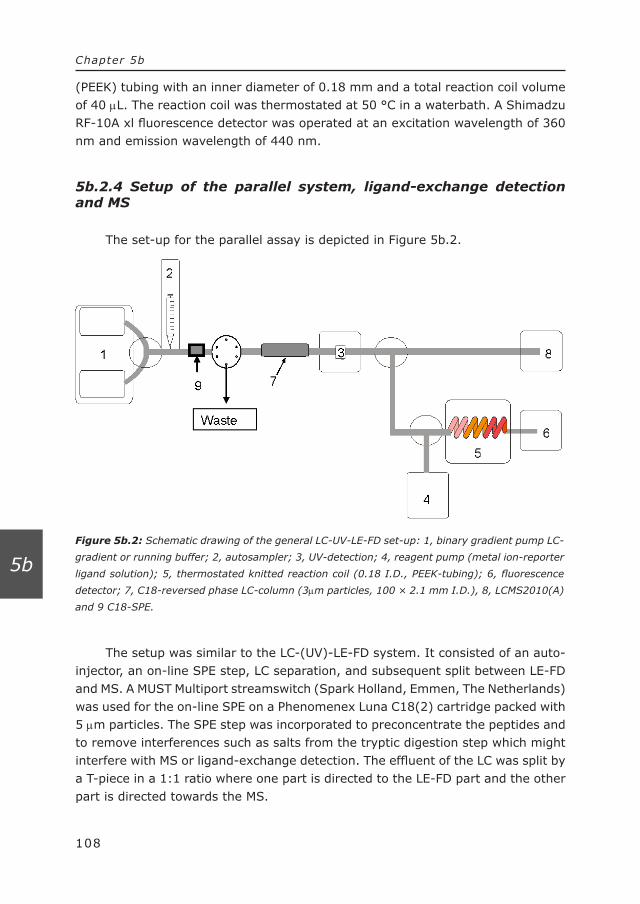
Figure 5b.2: Schematic drawing of the general LC-UV-LE-FD set-up: 1, binary gradient pump LC-
gradient or running buffer; 2, autosampler; 3, UV-detection; 4, reagent pump (metal ion-reporter
ligand solution); 5, thermostated knitted reaction coil (0.18 I.D., PEEK-tubing); 6, fluorescence
detector; 7, C18-reversed phase LC-column (3µm particles, 100 × 2.1 mm I.D.), 8, LCMS2010(A)
and 9 C18-SPE.
108
Chapter 5b
5b
(PEEK) tubing with an inner diameter of 0.18 mm and a total reaction coil volume of 40 µL. The reaction coil was thermostated at 50 °C in a waterbath. A Shimadzu RF-10A xl fluorescence detector was operated at an excitation wavelength of 360 nm and emission wavelength of 440 nm.
5b.2.4 Setup of the parallel system, ligand-exchange detection and MS
The set-up for the parallel assay is depicted in Figure 5b.2.
The setup was similar to the LC-(UV)-LE-FD system. It consisted of an auto-injector, an on-line SPE step, LC separation, and subsequent split between LE-FD and MS. A MUST Multiport streamswitch (Spark Holland, Emmen, The Netherlands) was used for the on-line SPE on a Phenomenex Luna C18(2) cartridge packed with 5 µm particles. The SPE step was incorporated to preconcentrate the peptides and to remove interferences such as salts from the tryptic digestion step which might interfere with MS or ligand-exchange detection. The effluent of the LC was split by a T-piece in a 1:1 ratio where one part is directed to the LE-FD part and the other part is directed towards the MS.

109
Chapter 5b
5b
5b.2.5 Mass Spectrometry settings
A Micromass (Wythenshawe, Manchester, UK) Q-TOF2 mass spectrometer equipped with a Z-spray electrospray (ESI) source was used for mass spectro-metric detection. Masslynx software (version 3.5) running under Windows NT was used for control of the system, data acquisition and data processing. The ESI source conditions were as follows: source temperature 150 °C, desolvation temperature 350 °C, capillary voltage 3 kV. The cone voltage was set at 30 V. Nitrogen (99.999% purity; Praxair, Oevel, Belgium) was used with flow rates of 20 L/h for nebulization, 50 L/h for cone gas, and 350 L/h for desolvation. Argon (99.9995% purity; Praxair) was used as collision gas in MS-MS experiments. Data-dependent automated switching between MS (survey scan) and MS-MS experiments was used, with an intensity threshold (set at 5) for changing from survey scan to MS-MS.
5b.3 Results and discussion
5b.3.1 General set-up
The current method is based on the specific detection of phosphopeptides by means of a ligand-exchange reaction of phosphorylated peptides with a Fe(III)-MCB complex. An LC separation is performed prior to the ligand-exchange detection method. Phosphorylated and non-phosphorylated peptides elute from the column and are on-line mixed with a continuous flow of the reagent solution (Fe(III)-MCB). MCB acts as a reporter molecule, which is displaced from the Fe(III)-MCB complex by the high-affinity phosphate group. MCB is highly fluorescent when it is free in solution, but when it is complexated with Fe(III) its fluorescence is quenched. Therefore, an increase in fluorescence response indicates the presence of phosphorylated compounds, although the possible presence of other interfer-ences with a high affinity for Fe(III) should be kept in mind, as is a common problem in metal-affinity based methods.
5b.3.2 Optimization of ligand-exchange detection
Several parameters play a role in this ligand-exchange detection method. The pH of the solution is important not only for the ligand-exchange reaction but also for the detection. The pH influences the degree of ionization of the ligands in the ligand-exchange detection. Fe(III) shows the highest selectivity for phos-phorylated compounds at a pH around 3 [9] while on the other hand the optimum

110
Chapter 5b
5b
fluorescence response of MCB is achieved at a pH around 7.5. At a pH above 8, undesired Fe(III)–hydroxy complexes will be formed. Therefore, for the ligand-exchange fluorescence detection the optimal pH is around 7. Although these ligand exchange reactions are known to be almost instantaneous, a reaction coil of 40 µL was implemented to allow the ligand-exchange reaction to take place. The temperature in the reaction coil was maintained at an optimal temperature (50 °C), to improve mixing and thereby increase the rate of the ligand-exchange reaction. The reagent concentration is an important parameter since the ligand-exchange responses are concentration dependent. Moreover, the ratio of metal to reporter ligand is also important since an excess of metal ions results in a large number of unoccupied coordination sites, which will result in a decrease in ligand-exchange response, whereas an excess of reporter ligand might increase the background noise.
5b.3.3 Fluorescence-based ligand-exchange detection
The selectivity of the liquid chromatography ligand-exchange detection method for phosphorylated compounds was tested with a mixture of two phos-phorylated and three non-phosphorylated peptides. Gradient LC was performed to separate the peptides. Figure 5b.3 demonstrates the selectivity of the method for phosphorylated peptides, since the two phosphorylated peptides show an increase in the fluorescence signal whereas two of the non-phosphorylated peptides, i.e., the non-phosphorylated analog of the two phosphorylated peptides and leucine enkephalin, did not give any response. However, the highly-acidic non-phosphory-lated peptide (peak 3) also showed a response in the LC-LE-FD detection method. This false positive problem is addressed in the next section. A UV-detector was placed on-line prior to the ligand-exchange detection method to detect all the peptides present. In the UV-chromatogram all five peptides were observed (Figure 5b.3).
The significant increase of organic modifier during the gradient elution influenced both the background fluorescence signal and the ligand-exchange response [14]. Figure 5b.3 also indicates that the number of phospho-groups on a peptide strongly influences the response factors in the ligand-exchange detection system. The tri-phospho peptide insulin receptor 5 (peak 5) showed a higher response than the mono-phospho peptide insulin receptor 2 (peak 2), while the non-phosphorylated peptide insulin receptor (peak 1) did not show a positive fluorescence response at all.
The method was tested for linearity in the range of 0.5 µM up to 80 µM. The limit of detection was 0.5 µM (S/N = 3), with an absolute detection limit of 5 pmol. The interday (n=4) and intraday (n=3) precision, expressed as relative standard deviation for a mixture of three phosphorylated peptides was better than 10 %.
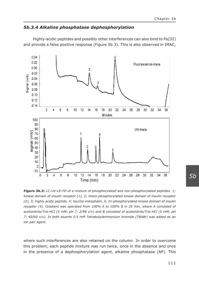
Figure 5b.3: LC-UV-LE-FD of a mixture of phosphorylated and non-phosphorylated peptides. 1;
kinase domain of insulin receptor (1), 2; mono-phosphorylated kinase domain of insulin receptor
(2), 3; highly acidic peptide, 4; leucine enkephalin, 5; tri-phosphorylated kinase domain of insulin
receptor (5). Gradient was operated from 100% A to 100% B in 35 min, where A consisted of
acetonitrile/Tris-HCl (5 mM; pH 7; 2/98 v/v) and B consisted of acetonitrile/Tris-HCl (5 mM; pH
7; 40/60 v/v). In both eluents 0.5 mM Tetrabutylammonium bromide (TBABr) was added as an
ion pair agent.
111
Chapter 5b
5b
5b.3.4 Alkaline phosphatase dephosphorylation
Highly-acidic peptides and possibly other interferences can also bind to Fe(III) and provide a false positive response (Figure 5b.3). This is also observed in IMAC,
where such interferences are also retained on the column. In order to overcome this problem, each peptide mixture was run twice, once in the absence and once in the presence of a dephosphorylation agent, alkaline phosphatase (AP). This

Figure 5b.4: LC-UV-LE-FD of a mixture of phosphorylated and non-phosphorylated peptides after
dephosphorylation by alkaline phosphatase. 1*; kinase domain of insulin receptor (1) and dephos-
phorylated mono-phosphorylated kinase domain of insuline receptor (2) and tri-phosphorylated
kinase domain of insulin receptor (5), 3; highly acidic peptide, 4; leucine enkephalin. For gradient
see legend Figure 5b.3.
112
Chapter 5b
5b
technique was previously used to distinguish between phosphorylated and non-phosphorylated peptides, based on comparing mass spectra and screening for Δ m/z 80 (loss of HPO3). [2,15] The idea behind this approach is that in the run of the sample without AP both interferences and the phosphorylated peptides will give a positive response in the fluorescent trace, whereas in the run of the sample with AP the dephosphorylated peptides will no longer give a positive response in the fluorescent trace. In this way, phosphorylated peptides can be distinguished from the other, non-phosphorylated interferences that show a positive response. Since the dephosphorylation is performed at an alkaline pH (pH 9.8) and in the presence of MgCl2, the untreated sample is also prepared under these conditions, with the only difference that no AP was added.
Figure 5b.4 shows the LC-LE-FD chromatogram of the same mixture as in Figure 5b.3, but after addition of alkaline phosphatase.
In the dephosphorylated sample, the only positive response is due to the acidic peptide (peak 3). In this way, a distinction can be made between phosphor-ylated peptides and interferences present in the sample. There is a possibility that

113
Chapter 5b
5b
the dephosphorylated species still has a significant affinity for Fe(III) and therefore will give a positive response in the fluorescence trace. Although first of all with the removal of the phosphate group, the affinity for Fe(III) will decrease signifi-cantly and therefore the response will decrease as well. Moreover, the removal of the phosphate group will most likely decrease the polarity of the species, thereby usually increasing the retention on the RP-LC column.
5b.3.5 Digest-LC-LE-FD and dephosphorylation
Large proteins will possibly interfere with the ligand-exchange detection method, due to e.g. electrostatic interactions, and are more difficult to detect by ESI-MS. The general method to analyze proteins by ESI-MS involves the use of proteases such as trypsin to obtain a mixture of peptides possibly including phos-phorylated peptides. Therefore, protein samples were enzymatically digested by trypsin prior to injection in the LC-LE-FD system.
Three model proteins were used to test the proposed method, the phos-phorylated protein α-casein and the non-phosphorylated proteins myoglobin and bovine serum albumin (BSA). Digest from both BSA and myoglobin did not show any response in LC-LE-FD system, whereas the on-line UV-detection showed a typical chromatogram for these protein-digest samples (data not shown). When α-casein was digested and injected, positive responses in the fluorescence trace were observed, indicating that phosphorylated peptides may be present although the positive peaks could also be due to other interferences with a high affinity to Fe(III). To distinguish between phosphorylated peptides and interferences, dephosphorylation of the sample with AP was performed. The gradient program applied allowed to focus on the relevant part of the chromatogram, showing the peaks of the phosphorylated peptides in the LE-FD detection. The results are shown in Figure 5b.5a (UV-trace) and in 5b.5b (LE-FD). Figure 5b.5c displays the fluorescence trace of the sample after dephosphorylation with AP.
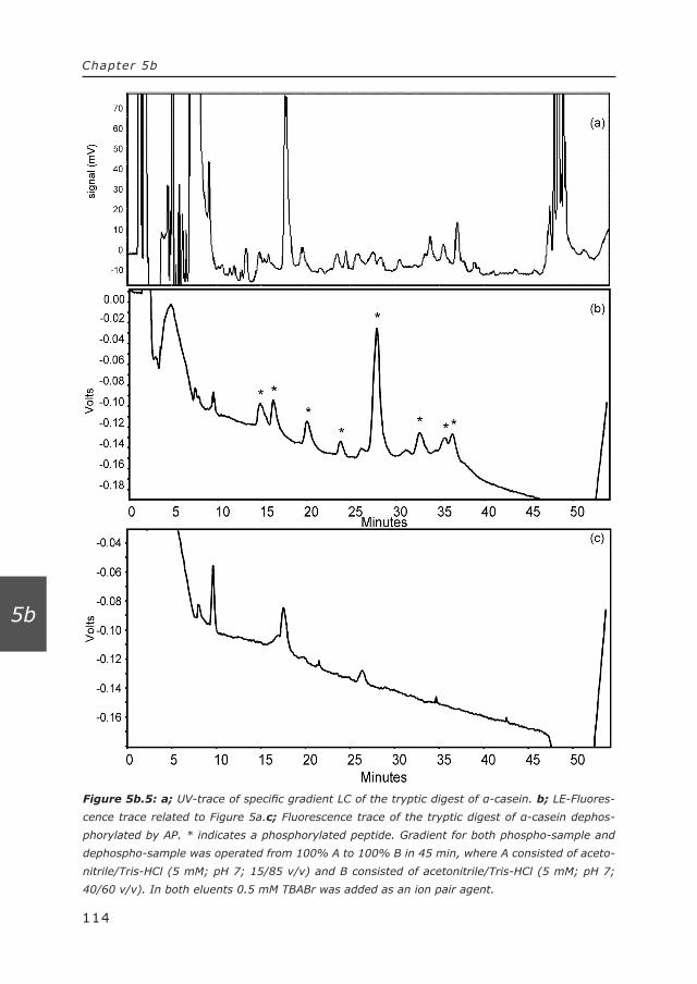
Figure 5b.5: a; UV-trace of specific gradient LC of the tryptic digest of α-casein. b; LE-Fluores-
cence trace related to Figure 5a.c; Fluorescence trace of the tryptic digest of α-casein dephos-
phorylated by AP. * indicates a phosphorylated peptide. Gradient for both phospho-sample and
dephospho-sample was operated from 100% A to 100% B in 45 min, where A consisted of aceto-
nitrile/Tris-HCl (5 mM; pH 7; 15/85 v/v) and B consisted of acetonitrile/Tris-HCl (5 mM; pH 7;
40/60 v/v). In both eluents 0.5 mM TBABr was added as an ion pair agent.
114
Chapter 5b
5b

115
Chapter 5b
5b
Taking the delay between the UV-trace and the LE-FD into account the peaks in the fluorescence trace can easily be correlated to peaks in the UV-trace. By comparing the fluorescence trace of the phosphorylated and dephosphorylated sample, the positive responses can be assigned to either phosphorylated peptides or non-phosphorylated interferences.
5b.3.4 Parallel setup for detection and identification of the phos-phorylated peptides
The HPLC-effluent initially used in the LE-FD is not compatible with the MS-detection. Therefore, the running buffer of the LC for the parallel setup was changed to a volatile ammonium-acetate buffer with pH 4. Additionally, the amount of the reagent buffer (4, see Figure 5b.2) added to the LE-FD part after the split was increased to neutralize the low pH, which was favorable in the LE-FD system. Secondly, a desalting step by means of solid-phase extraction column (9, see Figure 5b.2) is performed in between the injection and the LC-column, to remove high concentrations of non-volatile salts from the enzymatic digestion and/or dephosphorylation step. After the injection of the sample, the SPE traps the peptides, whereas the salts are flushed to waste. After flushing for 5 min, the valve is switched and the sample is directed to the LC-column.
To test the parallel setup, a mixture of the phosphorylated peptides pp60 c-src (P) and insulin receptor 2 and 5 and the non-phosphorylated peptides insulin receptor and leucine enkephalin was introduced in the parallel system. Again, the fluorescence trace (Figure 5b.6a) only showed a positive increase, when a phosphorylated peptide was present while the MS chromatogram and MS spectra provided additional information about the specific compounds (Figure 5b.6b).
The Q-TOF-MS has the possibility to perform automatic switching between the MS survey scan and MS-MS mode, when the ion intensity is above a certain predefined threshold. The MS-MS spectra provide extra information about the identity of the peptide which is giving a positive response in the fluorescence trace. This is especially interesting when dealing with unknown compounds.
When the same peptide mixture is subjected to the dephosphorylation procedure, it is clearly seen (Figure 5b.6c) that peaks due to both insulin receptors 2 and 5 are missing, because they are dephosphorylated by AP, whereas the intensity of their non-phosphorylated analog, insulin receptor 1, shows a large increase. This increase in insulin receptor response was also observed when UV-detection was used instead of MS-detection.

Figure 5b.6: LC-(MS)/(LE-FD) of a mixture of phosphorylated and non-phosphorylated peptides.
1; tri-phosphorylated kinase domain of insulin receptor (5), 2; mono-phosphorylated kinase
domain of insuline receptor (2), 3; phosphorylated pp60 c-src (P), 4; kinase domain of insulin
receptor (1) 5; leucine enkephalin. Gradient was operated from 100% A to 100% B in 40 min,
where A consisted of ammonium formate (10 mM; pH 4) containing 2% acetonitrile and B
consisted of acetonitrile/ammonium formate (10 mmol/L; pH 4; 40/60 v/v). a and b the fluores-
cence-chromatogram and MS-chromatogram respectively of the phosporylated sample. c; MS-
chromatogram of the dephosphorylated sample by AP. * indicates dephosphorylated peptide.
116
Chapter 5b
5b
117
Chapter 5b
5b
5b.3.5 Parallel setup for the detection of α-casein
Similar experiments with the parallel setup were performed for the α-casein digest. The digest was split in two parts, one of which was dephosphorylated by AP. The different m/z of the digest products obtained from the MS-spectra were searched against a protein database by means of MASCOT (developed by Matrix Science; http://www.matrixscience.com/) to identify the corresponding protein. MASCOT search resulted in a rank 1 match forrank 1 match for α-casein with a score of with a score of 80 (protein scores greater than 76 are significant) and a 37% sequence coverage. The fluorescence trace and corresponding digest fragment obtained from MS and MS-MS data provide information on the position of the phosphorylation in the protein. MS-MS may provide additional information when dealing with unknown phosphorylated peptides. For example, peak 1 in the fluorescence trace (Figure 5b.7a) corresponds to peak 1 in the MS-chromatogram (Figure 5b.7b). The mass spectrum (Figure 5b.7c) provides information about the compound responsible for the positive response in the fluorescence trace.
In this case, the m/z relates to the doubly charged T15-fragment of α-casein. The MS-MS-spectrum (Figure 5b.7d) and the corresponding database search confirm that the peak is fragment T15 of α-casein and that this fragment is phosphorylated. Since primarily the amino acid residues serine, threonine and tyrosine can bind a phosphate group, we can conclude that in the T15-fragment of α-casein phosphorylation must be at the serine. Dephosphorylation of the sample (data not shown) confirms that the T15-fragment is phosphorylated since its peak disappears in the fluorescence trace. In the same way, peak 2 was identified as the phosphorylated fragment T14-15.
Depending on the sample preparation and the chromatographic system (since highly phosphorylated peptides are usually more polar), the mass range of the mass spectrometer as well as the possibility to do negative electrospray ionization, the entire range of possible phosphorylated peptides can be monitored by this approach. In this particular case three phosphorylated peptides were identified, whereas one of these peptides (assumed to be the doubly phosphory-lated T8-fragment) was only (because of the threshold to switch between survey MS and MS-MS mode) based on the molecular mass and tryptic digest library.

Figure 5b.7: Detection and identification of phosphorylated tryptic peptides of a-casein where a;
fluorescent trace, b; MS-chromatogram, c; MS-spectrum of peak 1, d; MS-MS-spectrum of m/z
831.19 of peak 1. The MS-chromatogram displayed in Figure 5b.7b is not corresponding with the
MS-MS-experiments, since automated MS-MS was performed by the Q-TOF2 showing bad peak
shape which was caused by the switching between survey scan and the MS-MS mode. Therefore
for clarification the MS-chromatogram of a previous run (without automated MS-MS), with the
same sample and similar retention was chosen to function as MS-chromatogram in this Figure.
Gradient was operated from 100% A to 100% B in 40 min, where A consisted of acetonitrile/
ammonium formate (10 mM; pH 4; 12/88 v/v) and B consisted of acetonitrile/ammonium formate
(10 mmol/L; pH 4; 30/70 v/v)
118
Chapter 5b
5b
119
Chapter 5b
5b
5b.4 Conclusions
We demonstrated a methodology for the simultaneous and selective detection and identification of phosphorylated peptides and proteins in the presence of non-phosphorylated species. The current technique is particularly useful when dealing with unknown phosphorylated peptides or proteins, because automated MS-MS can be performed to obtain a mass fingerprint. In comparison to the splitless ligand-exchange detection method reported previously [12] the current method is better suited for the detection of low-abundant analytes, since the analyte concentration available for detection and quantitation is independent of the ligand-exchange reaction.
The advantage of the proposed method over other phospho-detection methods is that samples can selectively be screened for phosphorylated species and these species can simultaneously be identified. Moreover, the use of a continuous-flow ligand-exchange detection and the combination with an LC-separation prior to the detection method, makes it possible to detect low abundant phosphory-lated peptides in the presence of high abundant phosphorylated peptides and high affinity interferences, which is a common problem in, for instance, IMAC. False positives can be addressed by implementing a dephosphorylation step based on alkaline phosphatase.
5b.5 References
[1] M. Mann, S.E. Ong, M. Gronborg, H. Steen, O.N. Jensen, A. Pandey, Trends in Biotechnol. 20 (2002) 261.20 (2002) 261.
[2] A. Stensballe, S. Andersen, O.N. Jensen, Proteomics 1 (2001) 207. [3] M. Wind, H. Wesch, W.D. Lehmann, Anal. Chem. 73 (2001) 3006.Chem. 73 (2001) 3006. [4] Z. Kucerova, P. Majercakova, J. Biochem. Biophy. Methods 49 (2001) 523.Biophy. Methods 49 (2001) 523. [5] M. Bennecib, C.X. Gong, I. Grundke-Iqbal, K. Iqbal, Febs Lett. 485 (2000) 87. [6] H.L. Zhou, J.D. Watts, R. Aebersold,. Nat. Biotechnol. 19 (2001) 375.19 (2001) 375. [7] R.S. Annan, M.J. Huddleston, R. Verma, R.J. Deshaies, S.A. Carr, Anal. Chem. 73
(2001) 393. [8] M.C. Posewitz, P. Tempst, Anal. Chem. 71 (1999) 2883.[8] M.C. Posewitz, P. Tempst, Anal. Chem. 71 (1999) 2883.Chem. 71 (1999) 2883. [9] L.D. Holmes, M.R. Schiller, J. Liq. Chromatogr. Related Technol. 20 (1997) 123.Chromatogr. Related Technol. 20 (1997) 123. [10] J.G. Krabbe, H. Lingeman, W.M.A. Niessen, H. Irth, J. Chromatogr. A 1093A 1093
(2005) 36. [11]A.R. de Boer, T. Letzel, H. Lingeman, H. Irth, Anal. Bioanal. Chem. 381Bioanal. Chem. 381
(2005) 647. [12]J.G. Krabbe, H. Lingeman, W.M.A. Niessen, H. Irth, Anal. Chem. 75
(2003) 6853. [13]D.A. van Elswijk, U.P. Schobel, E.P. Lansky, H. Irth, J. van der Greef, Phytochem.
65 (2004) 233. [14]H. Irth, M. Lamoree, G.J. De Jong, U.A.T. Brinkman, R.W. Frei, R.A.
Kornfeldt, L.J. Persson, Chromatogr. 499 (1990) 617.499 (1990) 617. [15]P.C. Liao, J. Leykam, P.C. Andrews, D.A. Gage, J. Allison, Anal. Biochem. 219
(1994) 9.


121
Summary
Summary

122
Summary
Metal ions, metal complexes and metal-ligand exchange reactions are essential in a large variety of natural and industrial processes. Due to this importance and the related interest of researchers in metal complexes and metal-ligand interactions in several fields, e.g., drug research and catalyst design, novel detection methods are necessary for better understanding these compounds and their interactions as well as for obtaining quantitative information.
Mass spectrometry has taken a prominent role in the study of metal complexes and their interactions. This is due to several advantages of mass spectrometry such as better sensitivity and direct information about the formed complex as compared to conventional detection methods. This thesis describes the use of mass spectrometry in the study of complex formation as well as the application of ligand-exchange reactions coupled to mass spectrometry as a analytical tool, e.g., to obtain specific information on metal ions and ligands of interest or to develop specific and selective analyte detection strategies. In this context, mass spec-trometry not only provides chemical information, but also information about the compound that is responsible for a change in the metal-ion reporter complex.
Chapter 1 provides a brief introduction about metal ions, metal complexes and ligand-exchange reactions. The chapter gives the reader insight in the detection of metal species and reactions by mass spectrometry, mainly focusing on the different applicable mass spectrometric techniques and strengths and weaknesses. Moreover, the behavior of metal species in the mass spectrometer is discussed.
Chapter 2 focuses on the metal-ligand interactions, and how these interac-tions can be monitored or detected. Moreover, this chapter provides a guideline how to develop continuous-flow ligand-exchange reactions as analytical tools for all kind of applications, thereby focusing on mass spectrometric detection of these reactions.
Chapter 3 deals with the fundamental study of metal complexes and metal-ligand reactions by electrospray ionization mass spectrometry. Electrospray ionization mass spectrometry was used to investigate complex formation of different metal-complexes in a continuous-flow ligand-exchange reactor. Normal equilibration calculations which are incorporated in a computer program, provide the prediction of the type and concentrations of metal complexes formed as a function of experimental conditions. These theoretical calculations were compared with mass spectral data using an approach which mimics the calculations, showing good correlation between the experimental data obtained by MS and the theoretical calculations, even when experimental parameters such as pH of the solution vary. The usefulness of mass spectrometry is demonstrated by monitoring a ligand-exchange reaction by mass spectrometry, obtaining information about

123
Summary
affinity properties of the introduced ligand to the metal ion as well as structural information about the ligand itself. The detection of this ligand-exchange reaction is based on the specific release of a reporter ligand from a metal-reporter ligand complex by a high affinity ligand in a continuous-flow system. The choice of reporter ligand mainly depends on the affinity of the ligand toward the metal ion and the ionization efficiency. Therefore, it is possible to obtain a highly selective and highly sensitive ligand-exchange detection method.
Chapter 4 describes the utilization of electrospray ionization mass spectrom-etry for the selective detection of metal ligands after a post-column continuous-flow ligand-exchange reaction. By applying a chromatographic separation prior to the ligand-exchange reactor, it is demonstrated that (complex) mixtures of ligands can be analyzed in one single run. By choosing the reporter ligand based on its affinity towards the metal ion, it was possible to tune the ligand-exchange reaction to either sensitivity or selectivity, thereby keeping the affinity of the ligand of interest in mind. A reporter ligand with a higher affinity provides better selectivity whereas a reporter ligand with a lower affinity may provide higher sensitivity.
Chapter 5 is divided in two parts, demonstrating two possible strategies for the application of ligand-exchange reactors as analytical tools for screening for specific ligands. Chapter 5a demonstrates application of a mass spectrom-etry based ligand-exchange reactor in the screening of phosphorylated peptides, separated by a reversed-phase liquid chromatography. The ligand-exchange reaction is directly monitored by mass spectrometry. A specific reporter trace indicates the presence of a phosphorylated compound, whereas mass spectral information can be obtained about the compound itself. In this way, it was possible to monitor specifically phosphorylated peptides as well as to obtain information on the phosphorylated ligands. The method proved to be linear in the tested range of 2 to 80 μmol/L with a limit of detection (S/N = 3) of 2 μmol/L. Chapter 5b uses the same ligand-exchange principle in a slightly different approach. After the chro-matographic separation of the peptide mixture, the effluent is split is two parts. One part is directed to a fluorescence based ligand-exchange reactor, whereas the other part is directed to a mass spectrometer. By correlating the response in the fluorescence based ligand-exchange reactor, which selectively detects phosphor-ylated peptides, with the mass spectrometry chromatograms, phosphorylated peptides can be selectively subjected to tandem mass spectrometric analysis, for structural elucidation and identification of the phosphorylated fragments of the protein. Moreover, using fluorescence a LOD of 0.5 μmol/L was obtained for mono-phophorylated peptides. The number of phospho-groups influences the response in the ligand-exchange, which was demonstrated by the response of the mono-phosphorylated peptide which displayed a more or less three times lower
124
Summary
response in the fluorescence trace than the tri-phosphorylated peptide. A common problem in metal affinity studies of phosphorylated compounds is that some non-phosphorylated compounds, especially highly acidic peptides, also possess high affinity to the metal ion and may show complexation. To overcome this problem, the sample was introduced twice into the LC-ligand-exchange detection system, once as it is and once after treatment with alkaline phosphatase to remove phosphate-groups. Comparison of the fluorescence chromatogram of the two runs demonstrated the selective removal of phospho-groups (by disappearance of the fluorescence response), thereby enabling the discrimination between phosphory-lated compounds and non-phosphorylated interferences.
Summarizing, MS provides a tool to study metal-ligand interactions and metal complexes. The studied complexes showed a good correlation between theoretical calculations and mass spectral data. In theory, MS provides the possibility to study for instance (intermediate) (metal)-catalyst formation or protein-metal interactions.
Ligand-exchange reactions coupled to mass spectrometry provides a selective analytical tool to obtain information (e.g., affinity and structure) about the ligand of interest, where the choice of reporter ligand provides either sensitivity or selectivity or even both.


127
Samenvatting
Samenvatting
128
Samenvatting
Metaal ionen, metaal complexen and metal-ligand exchange reacties zijn essentieel in verschillende natuurlijke, medische en industriële processen. Vanwege deze belangrijke rol en de daaraan gerelateerde interesse van onder-zoekers, zijn nieuwe detectie-methodes onontbeerlijk voor de opheldering van en begrip over deze moleculen, de interacties en tevens voor het verkrijgen van kwantitatieve informatie.
Tegenwoordig heeft massa spectrometrie een prominente rol in het bestuderen van deze metaal complexen en de interacties. Deze rol komt onder andere door de vele voordelen die deze techniek biedt ten opzichte van andere technieken. Hierbij moet gedacht worden aan verbeterde gevoeligheid en het verkrijgen van directe informatie over de gevormde complexen. Dit proefschrift beschrijft het gebruik van massa spectrometrie bij het bestuderen van de vorming van metaal complexen en tevens de toepassing van ligand-exchange reacties als een analytisch gereedschap; bijvoorbeeld, voor het verkrijgen van informatie over het metaal ion en de liganden waar we in geïnteresseerd zijn of deze ligand-exchange reactie te gebruiken als een specifieke en selectieve detectie strategie. Hierbij geeft massa spectrometrie niet alleen chemische informatie, maar daarnaast ook informatie over het molecuul dat verantwoordelijk is voor de verandering in de metaal ion-reporter ligand interactie.
Hoofdstuk 1 bevat een korte introductie over metaal ionen, metaal complexen en ligand-exchange reacties. Dit hoofdstuk geeft de lezer inzicht in de detectie van metaal ionen, metaal complexen en de reacties met behulp van massa spectrom-etrie, daarbij voornamelijk kijkend naar de mogelijke massa spectrometrische technieken en hun voor- en nadelen. Daarnaast wordt het gedrag van metaal ionen en metaal complexen in de massa spectrometer besproken.
Hoofdstuk 2 beschrijft de metaal-ligand interacties en hoe deze interacties bestudeerd of gedetecteerd kunnen worden. Daarnaast geeft dit hoofdstuk een leidraad voor de ontwikkeling en gebruik van continuous-flow ligand-exchange reacties als analytische toepassingen voor allerlei doeleinden, waarbij de nadruk wordt gelegd op massa spectrometrische detectie van deze reacties.
Hoofdstuk 3 beschrijft het fundamenteel bestuderen van metaal complexen en metaal ion-ligand interacties met behulp van electrospray massa spectrome-trie (ESI-MS). ESI-MS is toegepast voor het bestuderen van complex formatie van verschillende complexen door middel van een continuous-flow ligand-exchange reactor. Een computer programma is ontwikkeld, dat gebruik maakt van standaard evenwichtsreactie-calculaties. Het programma geeft een voorspellende waarde voor het type en de concentratie van metaal complexen, waarbij de invloed van de experimentele condities meegewogen kan worden. Deze theoretische berek-eningen werden vergeleken met de massa spectra data. Deze vergelijking gaf een

129
Samenvatting
goede overeenkomst, tussen de experimentele data en de theoretische berek-eningen zelfs wanneer experimentele parameters zoals de pH gevarieerd werden. Het nut van het gebruik van massa spectrometrie is gedemonstreerd door ligand exchange-reacties te vervolgen, waarbij informatie over de affiniteit van de gein-troduceerde ligand voor het metaal ion en tevens structurele informatie over de ligand zelf. De detectie van deze ligand-exchange reactie is gebaseerd op het specifieke vrijkomen van een reporter ligand uit een metaal-ion reporter ligand complex, bij aanwezigheid van een ligand met een hogere affiniteit van een ander ligand. De keuze van reporter ligand hangt voornamelijk af van de affiniteit van de ligand voor het metaal ion en de mate waarin het ligand wordt geïoniseerd in ESI-MS. Op deze manier kan dus een hele selectieve en gevoelige ligand-exchange reactie methode ontwikkeld worden.
Hoofdstuk 4 beschrijft het gebruik van ESI-MS voor de post-kolom selectieve ligand-exchange detectie van metaal liganden. Door gebruik te maken van een chromatografische scheiding voorafgaand aan de ligand-exchange reactor, kunnen (complexe) mengsels van liganden in een enkele meting gedaan worden. Een geschikte keuze van de reporter-ligand, kijkend naar de affiniteit van de ligand voor het metaal ion, maakt het mogelijk om de selectiviteit en gevoeligheid te “tunen”, waarbij de liganden waarin we geïnteresseerd zijn in het achterhoofd worden gehouden. Een reporter ligand met een hoge affiniteit geeft een hogere selectiviteit en een ligand met een lagere affiniteit kan een betere gevoeligheid geven.
Hoofdstuk 5 is onderverdeeld in twee delen, waarin twee verschillende strategieën voor het gebruik van ligand-exchange reactoren als analytische toepassing voor de screening van specifieke liganden. Hoofdstuk 5a demonstreert de toepassing van een MS-gebaseerde ligand-exchange reactor in de screening van gefosforyleerde peptiden, die gescheiden worden met behulp van reversed-phase vloeistof chromatografie. De ligand-exchange wordt direct bestudeerd met MS. Een specifieke reporter trace geeft de aanwezigheid van een gefosforyleerde peptide aan, maar tevens kan met MS informatie worden verkregen over het gefosforyleerde peptide. Op deze manier is het mogelijk om specifiek gefosfor-yleerde peptiden te bestuderen. De methode was lineair in het gebied van 2 tot 80 µmol/L en een detectie limiet (S/N = 3) van 2 µmol/L. In hoofdstuk 5b wordt gebruik gemaakt van eenzelfde ligand-exchange principe in een andere opzet. Na de chromatografische scheiding van het peptide mengsel wordt het effluent in twee delen gesplitst. Een gedeelte wordt richting een op fluorescentie gebaseerde ligand-exchange reactor gedirigeerd en het andere gedeelte naar de MS. Door de uitslag van de fluorescentie gebaseerde ligand-exchange reactor, die selectief gefosforyleerde peptiden detecteert, te correleren aan de MS-chromatogram kunnen gefosforyleerde peptiden selectief worden gedetecteerd en met tandem-

130
Samenvatting
MS analyse worden bestudeerd. De tandem-MS analyse geeft informatie over de structuur en kan de gefosforyleerde peptiden identificeren. Met behulp van fluorescentie als detectie, werd een detectie limiet van 0.5 µmol/L verkregen. Het aantal fosfaat-groepen beïnvloedt de uitslag van de ligand-exchange reactie. Dit werd gedemonstreerd door een mono-gefosforyleerd peptide en een tri-gefosforyleerd peptide te introduceren. Het mono-gefosforyleerd peptide gaf een flourescentie uitslag die ongeveer drie keer minder intensief was dan de tri-gefos-foryleerde peptide. Een bekend probleem binnen metaal-affiniteits studies van gefosforyleerde verbindingen is de aanwezigheid van liganden, die ook een hoge affiniteit (bijvoorbeeld zure peptiden) hebben voor het metaal ion en daarbij dus een uitslag geven in de ligand-exchange detectie. Om een vals positieve uitslag te voorkomen wordt het monster twee keer geïntroduceerd. Een keer zonder en een keer na behandeling met alkaline phosphatase. De alkaline phosphatase verwijdert onder de juiste condities de fosfaat-groepen, waardoor deze gedefos-foryleerde peptiden geen positieve uitslag meer zullen geven in het chromato-gram, in tegenstelling tot het onbehandelde monster. De niet-gefosforyleerde peptiden daarentegen zullen nog steeds een positieve uitslag geven.
MS is een geschikte techniek voor de bestudering van metaal ion-ligand interacties en metaal complexen. De bestudeerde complexen gaven een duidelijk overeenkomst tussen theoretische berekeningen en MS-data. In theorie, biedt MS de mogelijkheid om bijvoorbeeld (intermediate) (metal)-catalyst formaties of eiwit-metaal ion interacties te bestuderen. Daarnaast biedt de koppeling van ligand-exchange reacties en massa spectrometrie een selectieve analytische toepassing voor het verkrijgen van informatie over de liganden die bestudeerd worden (bijvoorbeeld affiniteit en structuur), waarbij de keuze van de reporter-ligand een mogelijkheid biedt om de methode specifieker of gevoeliger te maken.

List of publications
Included in this thesis
Krabbe JG, Lingeman H, Niessen WM, Irth H.
Ligand-exchange detection of phosphorylated peptides using liquid chromatography
electrospray mass spectrometry.
Anal Chem. 2003 75 (24) 6853-6860
Krabbe JG, Lingeman H, Niessen WM, Irth H.
Screening for metal ligands by liquid chromatography-ligand-exchange-electrospray mass
spectrometry.
J Chromatogr A. 2005 (1093) 36-46
Krabbe JG, Gao F, Li J, Ahlskog JE, Lingeman H, Niessen WM, Irth H.
Selective detection and identification of phosphorylated proteins by simultaneous ligand-
exchange fluorescence detection and mass spectrometry.
J Chromatogr A. 2006 1130 (2) 287-295
Krabbe JG, de Boer AR, van der Zwan G, Lingeman H, Niessen WM, Irth H.
Metal-complex formation in continuous-flow ligand-exchange reactors studied by electrospray
mass spectrometry.
J Am Soc Mass Spectrom. 2007 (18) 707-13
Not included in this thesis
Waterval JC, Krabbe H, Teeuwsen J, Bult A, Lingeman H, Underberg WJ.
Comparison between transient isotachophoretic capillary zone electrophoresis and reversed-
phase liquid chromatography for the determination of peptides in plasma.
Electrophoresis. 1999 (20) 2909-16
de Boer AR, Bruyneel B, Krabbe JG, Lingeman H, Niessen WM, Irth H.
A microfluidic-based enzymatic assay for bioactivity screening combined with capillary liquid
chromatography and mass spectrometry.
Lab Chip. 2005 (11) 1286-1292
de Boer AR, Alcaide-Hidalgo JM, Krabbe JG, Kolkman J, van Emde Boas CN,Niessen WM,
Lingeman H, Irth H.
High-temperature liquid chromatography coupled on-line to a continuous-flow biochemical
screening assay with electrospray ionization mass spectrometric detection.
Anal Chem. 2005 77 (24) 7894-7900
Huertas-Perez JF, Heger M, Dekker H, Krabbe H, Lankelma J, Ariese F.
Simple, rapid, and sensitive liquid chromatography-fluorescence method for the quantification
of tranexamic acid in blood.
J Chromatogr A. 2007 (1157) 142-50
Martha CT, Elders N, Krabbe JG, Kool J, Niessen WMA, Orru RVA, Irth H
On-line Approach for Rapid Screening of Homogeneous Catalyst Performance with Mass
Spectrometric Detection
Submitted
List of publications

133
Dankwoord
Dankwoord

134
Dankwoord
Het gedeelte, dat tot aan een zekerheidgrenzende waarschijnlijkheid het meest gelezen wordt van de proefschriften, is het dankwoord en daar zal dit proefschrift waarschijnlijk geen uitzondering op zijn. Terugkijkend op mijn vier jaar durend promotie-onderzoek kan ik alleen maar zeggen dat ik veel geluk heb gehad met het onderzoek en met het team, dat ik om me heen heb (gehad) en heb mogen leren kennen.
Allereerst ben ik grootse dank verschuldigd aan mijn promotoren en copromotor. Hubi, enorm bedankt voor je inzet, enthousiasme en je begeleiding. Ik heb veel geleerd van je manier van denken en de snelle wijze waarop je problemen kunt analyseren en oplossen.
Wilfried, jij begon met je leerstoel ongeveer een half jaar nadat ik begonnen was met mijn vier jaar zwoegen. Ik heb veel geleerd van de vele discussies, die wij hadden over de wetenschap en de dingen die daarbuiten gebeuren. Je expertise op het gebied van Massa Spectrometrie was en is nog steeds een waardevolle bron voor ACAS.
En dan Henk, waar moet ik beginnen? Ik zal proberen om het netjes te houden. Grotendeels ben ik door jou op de VU begonnen aan de master Analytische Chemie. Ondanks het feit dat je het waarschijnlijk niet wilt horen, konden wij het direct goed met elkaar vinden. Ik heb je manier van werken, begeleiden en je “briljante” kijk op de wereld altijd enorm gewaardeerd en bewonderd. Daarnaast was je altijd een geweldig slachtoffer voor practical jokes. Wat zul jij nu een rust hebben.
Bij jullie alle drie, hoop ik dat de afronding van dit proefschrift niet het einde van ons contact zal zijn.
Naast de supervisors wil ik ook al mijn collega’s en stafleden bedanken bij ACAS. Allereerst mijn paranimfen, Arjen en Rico. Arjen, hartelijk dank voor de geweldige collega/vriend die je bent geweest de afgelopen jaren. Dit zelfde geldt voor Rico. De vele grappen die we hebben uitgehaald en de zogenaamde `relax’-momenten zal ik enorm missen. Ook wil ik de vele andere collega’s bedanken die mij plezier in mijn werk hebben bezorgd. In willekeurige volgorde, Jeroen, Natasja, Gisèle, Johannes, Mark, Big Niels, Niels M, Ansgar, Jon, Ben, Marek, Rene, Camiel, Daniel, Reggy Zaagmans, Lawrence, Mohamed, Pim en Dik. Daarnaast wil ik de andere helft van ACAS, de spectroscopie groep, ook bedanken voor de goede samenwerking. In het bijzonder Cees, Freek en Gert.
I would like to thank my students for their help in the research. Again in random order and hopefully not forgetting someone, Sevda, Lai-Ping, Li Jia and Gao Grace, Johanna, Hans S. and Arvid, Jeffrey, Sebastiaan, Ana, Herald, Dikke Paling, and Hans K.

135
Dankwoord
Dit onderzoek zou niet mogelijk zijn geweest zonder hulp van de fijnmecha-nische werkplaats. DJ Dick, Klaas en Roald, ontzettend bedankt voor jullie snelle en vakkundige hulp.
Naast werk is er natuurlijk ook nog een sociaal leven. Ik wil mijn familie, en in het bijzonder mijn ouders, mijn broer Pim (en Danielle) en zus Marieke (en Ralph) bedanken; allereerst voor hun steun maar ook voor het begrip en hun onvermoeide interesse in wat ik deed. Ik had vaak geen tijd en was altijd wel druk met iets. Desondanks heb ik altijd het gevoel gehad dat jullie voor de volle 100% en onvoorwaardelijk achter me hebben gestaan, wat ik ook deed. Jullie hebben het voor mij mogelijk gemaakt, om zover te komen. Tevens wil ik mijn vrienden, voornamelijk Jeroen, Maarten en Tom, danken voor de steun en de vriendschap. Ik wil ook graag de familie Slaghekke, en in het bijzonder Simone, Ben en Henk Zwijnenberg, bedanken voor hun steun.
And last but not least I want to thank a very special person. Mi, thank you so much for your support and help and for everything you taught me in such a short period of time.
-Hans -

136
Curriculum Vitae
Curriculum Vitae
Hans Krabbe werd geboren op 28 mei 1977 te Leidschendam. In 1994 behaalde hij zijn HAVO diploma om vervolgens in datzelfde jaar de studie Hoger Laboratorium Onderwijs in Enschede te beginnen. Hij verrichtte zijn afstudeerop-dracht in de richting Analytische Chemie aan de Universiteit Utrecht, bij de faculteit Farmacie en de vakgroep Analytische farmacie en toxicologie onder leiding van Joop Waterval en Willy Underberg. Het onderzoek was gericht op de analyse van peptiden in plasma met behulp van vloeistof chromatografie en capillaire electroforese. Na het behalen van zijn diploma in 1998, is Hans scheikunde gaan studeren aan de Vrije Universiteit Amsterdam, met als afstudeerrichting Analytische Chemie. In 1999 tot 2000 heeft hij de studie onderbroken voor een wereldreis, om vervolgens in 2001 te beginnen aan zijn afstudeerstage onder leiding van Maarten van Bommel en Prof. Dr. Irth, bij de vakgroep Analytische Chemie en Toegepaste Spectroscopie (ACAS). De opdracht was om gebiotinyl-eerde peptiden te bepalen met behulp van een enzyme-amplified biochemical detection. Ter afronding van de opleiding heeft hij een literatuurstudie gedaan over multidimensionale scheidingen. In maart 2002 behaalde Hans zijn doctoraal diploma Scheikunde. In ditzelfde jaar is hij begonnen aan zijn promotie-onderzoek bij de Faculteit Exacte Wetenschappen in de vakgroep ACAS. Onder leiding van zijn promotoren Prof. Dr. Hubertus Irth en Prof. Dr. Wilfried Niessen en co-promotor Dr. Henk Lingeman, heeft hij onderzoek gedaan naar de interacties van metaal ionen en liganden met behulp van massa spectrometrie en het gebruik van deze interacties voor analytische toepassingen. Momenteel is Hans bezig met de opleiding tot Klinisch Chemicus bij de Isala Klinieken te Zwolle.


