1 Preparation of Tissues for Study ■ Chemical fixatives such as formalin are used to preserve tissue structure by cross-linking and denaturing proteins, inactivating enzymes, and preventing cell autolysis or self-digestion. ■ Dehydration of the fixed tissue in alcohol and clearing in organic solvents prepare it for embedding and sectioning. ■ Embedding in paraffin wax or epoxy resin allows the tissue to be cut into very thin sections (slices) with a microtome. ■ Sections are mounted on glass slides for staining ,which is required to reveal specific cellular and tissue components with the microscope. ■ The most commonly used staining method is a combination of the stains hematoxylin and eosin (H&E), which act as basic and acidic dyes, respectively. ■ Cell substances with a net negative (anionic) charge, such as DNA and RNA, react strongly with hematoxylin and basic stains; such material is said to be “basophilic.” *Cationic substances, such as collagen and many cytoplasmic proteins react with eosin and other acidic stains and are said to be ….acidophilic . Light Microscopy ■ Bright-field microscopy, the method most commonly used by both students and pathologists, uses ordinary light and the colors are imparted by tissue staining. ■ Fluorescence microscopy uses ultraviolet light, under which only fluorescent molecules are visible, allowing localization of fluorescent probes which can be much more specific than routine stains. ■ Phase-contrast microscopy uses the differences in refractive index of various natural cell and tissue components to produce an image without staining, allowing observation of living cells. ■ Confocal microscopy involves scanning the specimen at successive focal planes with a focused light beam, often from a laser, and produces a 3D reconstruction from the images. Autoradiography ■ This process localizes cell components synthesized using radioactive precursors by detecting silver grains produced by weakly emitted radiation in a photographic emulsion coating the tissue section or cells. ■ With either light microscopy or TEM, autoradiography permits unique studies of processes such as tissue growth (using radioactive DNA precursors) or cellular pathways of macromolecular synthesis. Cell & Tissue Culture ■ Cells can be grown in vitro from newly explanted tissues (primary cultures) or as long-established cell lines and can be examined in the living state by phase-contrast light microscopy Enzyme Histochemistry ■ Histochemical (or cytochemical) techniques use specific enzymatic activities in lightly fixed or unfixed tissue sections to produce visible products in the specific enzyme locations. ■ Fixation and paraffin embedding denatures most enzymes, so histochemistry usually uses frozen tissue sectioned with a cryostat. ■ Enzyme classes for which histochemical study is useful include phosphatases, dehydrogenases, and peroxidases, with peroxidase often conjugated to antibodies used in immunohistochemistry.
Transcript
1
Preparation of Tissues for Study ■ Chemical fixatives such as formalin are used to preserve tissue structure by cross-linking and denaturing proteins, inactivating enzymes, and preventing cell autolysis or self-digestion. ■ Dehydration of the fixed tissue in alcohol and clearing in organic solvents prepare it for embedding and sectioning. ■ Embedding in paraffin wax or epoxy resin allows the tissue to be cut into very thin sections (slices) with a microtome. ■ Sections are mounted on glass slides for staining ,which is required to reveal specific cellular and tissue components with the microscope. ■ The most commonly used staining method is a combination of the stains hematoxylin and eosin (H&E), which act as basic and acidic dyes, respectively. ■ Cell substances with a net negative (anionic) charge, such as DNA and RNA, react strongly with hematoxylin and basic stains; such material is said to be “basophilic.” *Cationic substances, such as collagen and many cytoplasmic proteins react with eosin and other acidic stains and are said to be ….acidophilic .
Light Microscopy
■ Bright-field microscopy, the method most commonly used by both students and pathologists, uses ordinary light and the colors are imparted by tissue staining. ■ Fluorescence microscopy uses ultraviolet light, under which only fluorescent molecules are visible, allowing localization of fluorescent probes which can be much more specific than routine stains. ■ Phase-contrast microscopy uses the differences in refractive index of various natural cell and tissue components to produce an image without staining, allowing observation of living cells. ■ Confocal microscopy involves scanning the specimen at successive focal planes with a focused light
beam, often from a laser, and produces a 3D reconstruction from the images.
Autoradiography
■ This process localizes cell components synthesized using radioactive precursors by detecting silver grains produced by weakly emitted radiation in a photographic emulsion coating the tissue section or cells. ■ With either light microscopy or TEM, autoradiography permits unique studies of processes such as tissue growth (using radioactive DNA precursors) or cellular pathways of macromolecular synthesis.
Cell & Tissue Culture ■ Cells can be grown in vitro from newly explanted tissues (primary cultures) or as long-established cell lines and can be examined in the living state by phase-contrast light microscopy
Enzyme Histochemistry ■ Histochemical (or cytochemical) techniques use specific enzymatic activities in lightly fixed or unfixed tissue sections to produce visible products in the specific enzyme locations. ■ Fixation and paraffin embedding denatures most enzymes, so histochemistry usually uses frozen tissue sectioned with a cryostat. ■ Enzyme classes for which histochemical study is useful include phosphatases, dehydrogenases, and peroxidases, with peroxidase often conjugated to antibodies used in immunohistochemistry.
2
Visualizing Specific Molecules
■ Some substances specifically bind certain targets in cells. ■ Immunohistochemistry is based on specific reactions between an antigen and antibodies labeled with visible markers, often fluorescent compounds or peroxidase for light microscopy and gold particles for TEM. ■ If the cell or tissue antigen of interest is detected by directly binding a labeled primary antibody specific for that antigen, the process is considered direct immunohistochemistry. *Indirect immunohistochemistry uses an unlabeled primary anti- body that is detected bound to its antigen with labeled secondary antibodies. *The indirect immunohistochemical method is more commonly used because the added level of antibody binding amplifies the signal detected and provides greater technical flexibility. Specific gene sequences or mRNAs of cells can be detected microscopically using labeled complementary DNA (cDNA) probes in a technique called in situ hybridization (ISH). *Indirect immunohistochemistry uses an unlabeled primary anti- body that is detected bound to its antigen with labeled secondary antibodies. ■The indirect immunohistochemical method is more commonly used because the added level of antibody binding amplifies the signal detected and provides greater technical flexibility. ■Specific gene sequences or mRNAs of cells can be detected microscopically using labeled complementary DNA (cDNA) probes in a technique called in situ hybridization (ISH).
Interpretation of Structures in Tissue Sections Many steps in tissue processing, slide preparation and staining can introduce minor artifacts such as spaces and precipitates that are not normally present in the living tissue and must be recognized. ■ Sections of cells or tissues are essentially 2D planes through 3D structures, and understanding this fact is important for their correct interpretation and study
3
The Cell
Many cells show polarity, meaning different areas of the cell have different structures The most-studied polarity is in epithelial cells, they have 1-Apical domain 2-Basal (basolateral) domain
The Plasma Membrane
The plasma membrane (cell membrane or plasmalemma) that envelops every eukaryotic cell. It functions as a selective barrier regulating the passage of materials into and out of the cell and facilitating the transport of specific molecules.
IMPORTANT
Membranes range from 7.5 to 10 nm in thickness and consequently are visible only in the electron microscope (can NOT be resolved by the light microscope). They appear as a trilaminar unit in TEM. The line between adjacent cells sometimes seen faintly with the light microscope consists of plasma membrane proteins plus extracellular material, which together can reach a dimension visible by light microscopy.
1-Phospholipids
2- Cholesterol
3- Proteins:
A) Integral: incorporated directly within the lipid bilayer
B) Peripheral: bound to one of the two membrane surfaces, particularly on the cytoplasmic side
4-Oligosaccharide (carbohydrate) chains linked to many of the phospholipid (to form glycolipids)
and protein (to form glycoproteins) molecules.
Functions of Plasma Membrane
1. Physical barrier: Establishes a flexible boundary, protects cellular contents, and supports cell structure.
Phospholipid bilayer separates substances inside and outside the cell.
2. Selective permeability: Regulates entry and exit of ions, nutrients, and waste molecules through the membrane
3. Electrochemical gradients: Establishes and maintains an electrical charge difference across the plasma
membrane.
4. Communication: Contains receptors that recognize and respond to molecular signals.
4
The Cytoplasm
Inside the cell membrane, the fluid cytoplasm (or cytosol) bathes metabolically active structures called organelles,
which may be membranous (such as mitochondria) or nonmembranous protein complexes (such as
ribosomes).
Most organelles are positioned in the cytoplasm by movements along the polymers of the cytoskeleton, which also
determines a cell’s shape and motility.
Ribosomes
Ribosomes are macromolecular machines, about 20 × 30 nm in size, which assemble polypeptides (proteins) from
amino acids in a sequence specified by mRNA.
They can be free in the cytosol or bound to the rough endoplasmic reticulum. During protein synthesis many ribosomes typically bind the same strand of mRNA to form larger complexes called
polyribosomes, or polysomes
Differences between free and bound ribosomes
Free ribosomes synthesize cytosolic and cytoskeletal proteins and proteins for import into the nucleus,
mitochondria, and peroxisomes.
Bound ribosomes synthesize proteins that are to be incorporated into membranes, stored in lysosomes, or
eventually secreted from the cell.
Note that: The proteins produced by these ribosomes are segregated during translation into
the interior of the ER’s membrane cisternae.
Endoplasmic Reticulum (ER) The endoplasmic reticulum is an anastomosing network of intercommunicating channels or cisternae formed by a
continuous membrane network.
two types:
Rough endoplasmic reticulum (rER)
Smooth endoplasmic reticulum (sER)
Functions of ER
1-Synthesis: Provides a place for chemical reactions.
sER is the site of lipid synthesis and carbohydrate metabolism
rER synthesizes proteins for secretion, incorporation into the plasma, membrane, and as enzymes within lysosomes.
5
2-Transport: Moves molecules through cisternal space from one part of the cell to another, sequestered
away from the cytoplasm
3-Storage: Stores newly synthesized molecules, sER stores C
4-Detoxification: sER detoxifies both drugs and alcohol.
Histological appearance
rER: Light microscopy: Intense basophilia (blue to purple on H &E)
Electron microscopy: Appears as interconnected flat cisternae and tubules associated with ribosomes
sER:
Light microscopy: can not be seen under LM
Electron microscopy: Appears as interconnected tubules with various shapes and sizes and not stack of
flattened cisternae not associated with ribosomes
Golgi Apparatus Golgi apparatus, or Golgi complex, completes posttranslational modifications of proteins produced in the rER
and then packages and addresses these proteins to their proper destinations.
Material moves from the rER cisternae to the Golgi apparatus in small, membrane-enclosed carriers called transport
vesicles.
Has two sides (ends): Receiving end (cis): receives transport vesicles
Shipping end (tran): ships secretory vesicles
Histological appearance
Golgi cannot be seen in H & E staining.
In highly active cells, with prominent golgi apparatus, it gives a negative image (as if an empty
space)
Secretory Granules The granules are surrounded by membrane and contain a concentrated form of the secretory
product
Histologically
6
Light microscopy: they cannot be resolved, but in cells active in protein synthesis, they give
Electron microscopy: homogenous electron dense structures near the apex of the cell
Lysosomes
Lysosomes are sites of intracellular digestion and turnover of cellular components.
Under electron microscopy, we can distinguish between primary and secondary lysosomes:
Primary: Uniformly granular electron dense appearance
Secondary: Larger with heterogenous appearance (particulate content)
Lysosomes are not well shown on H&E-stained cells but can be visualized by light microscopy
after staining with toluidine blue.
Synthesis of lysosomal enzymes occurs in the RER, with packaging in the Golgi
apparatus. Endocytosis produces vesicles that fuse with endosomes before merging
with lysosomes.
Phagocytic vacuoles (or phagosomes) fuse with primary lysosomes to become
secondary lysosomes (or heterolysosomes), in which ingested material is degraded.
Autophagosomes: are formed after nonfunctional or surplus organelles become enclosed with
membrane and the resulting structure fuses with a lysosome.
The products of lysosomal digestion are recycled to the cytoplasm, but indigestible molecules remain
in a membrane-enclosed residual body, which may accumulate in long-lived cells as lipofuscin.
In some cells, such as osteoclasts, the lysosomal enzymes are secreted into a restricted extracellular
compartment.
7
Mitochondria Mitochondria are membrane-enclosed organelles with arrays of enzymes specialized for
aerobic respiration and production of adenosine triphosphate (ATP), which supplies energy
for most cellular activities.
The number of mitochondria is related to the cell’s energy needs: cells with a high-energy metabolism
(eg, cardiac muscle, cells of some kidney tubules) have abundant mitochondria, whereas cells with a
low-energy metabolism have few mitochondria.
Under the TEM each mitochondrion is seen to have two separated and very different membranes that
together create two compartments: the innermost matrix and a narrow intermembrane space
The outer membrane contains many transmembrane proteins called porins that form channels
through which small molecules such as pyruvate and other metabolites pass from the cytoplasm to
the intermembrane space.
The inner membrane has many long folds called cristae, which project into the matrix and greatly
increase the membrane’s surface area.
The cytoskeleton The cytoplasmic cytoskeleton is a complex array of:
(1) Microtubules.
(2) microfilaments (also called actin filaments).
(3) intermediate filaments.
General Function of Cytoskeleton
1. Structural: Provides structural support to cell; stabilizes junctions between cells.
2. Movement: Assists with cytosol streaming and cell motility; helps move organelles and materials
throughout cell; helps move chromosomes during cell division
8
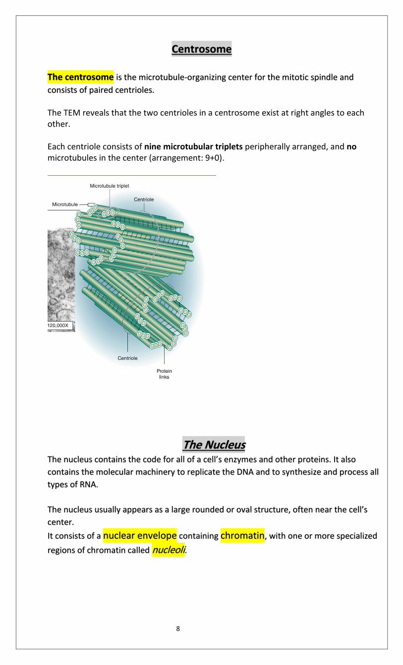
Centrosome
The centrosome is the microtubule-organizing center for the mitotic spindle and
consists of paired centrioles.
The TEM reveals that the two centrioles in a centrosome exist at right angles to each other. Each centriole consists of nine microtubular triplets peripherally arranged, and no microtubules in the center (arrangement: 9+0).
The Nucleus The nucleus contains the code for all of a cell’s enzymes and other proteins. It also
contains the molecular machinery to replicate the DNA and to synthesize and process all
types of RNA.
The nucleus usually appears as a large rounded or oval structure, often near the cell’s
center.
It consists of a nuclear envelope containing chromatin, with one or more specialized
regions of chromatin called nucleoli.
9
Functions of the Nucleus
1. Cellular regulation: Houses genetic material, which directs all cellular activities
and regulates cellular structure
2. Production: Produces ribosomal subunits in nucleolus and
Chromatin *The mass of DNA and its associated proteins
*Microscopically two categories of chromatin can be distinguished:
1-Euchromatin is visible as finely dispersed granular material in the electron microscope and as lightly
stained basophilic areas in the light microscope.
→ associated with active cells
2-Heterochromatin appears as coarse, electron-dense material in the electron microscope and as
intensely basophilic clumps in the light microscope.
→ associated with inactive cells
Nucleolus
The nucleolus is a spherical, highly basophilic subdomain of nuclei in cells actively engaged in protein
synthesis
It is the location of ribosomal subunit assembly and transcription of ribosomal RNA (rRNA).
*The intense basophilia of nucleoli is due not to heterochromatin but to the presence of densely
concentrated rRNA.
The Nuclear Envelope
The nuclear envelope is a double set of membranes with a narrow perinuclear space, which separates
the cytoplasm from nucleoplasm.
*The outer membrane binds ribosomes and is continuous with the RER.
*It is penetrated by nuclear pore complexes.
*It is supported internally by a meshwork, the nuclear lamina, composed of intermediate filament
subunits called lamins.
10
Nuclear pore complexes
Nuclear pore complexes (nuclear pores) contain more than 30 core proteins (nucleoporins), span both
membranes of the nuclear envelope, and regulate the bidirectional transfer of macromolecular complexes
between the nucleus and cytoplasm.
Epithelial Tissue: SUMMARY OF KEY POINTS
■ An epithelium is a tissue in which cells are bound tightly together structurally and functionally to
form a sheetlike or tubular structure with little extracellular material between the cells.
■ Cells in epithelia each have an apical side facing the sheet’s free surface and a basal side facing a
basement membrane and underlying connective tissue.
■ Epithelia are often specialized for absorption or transcytosis, pinocytosis of material at the apical
side and exocytosis at the basolateral side (or vice versa).
■ Cells of most epithelia exhibit continuous renewal, with the locations of stem cells and rates of cell
turnover variable in various specialized epithelia.
Basement Membrane.
■ The basement membrane of all epithelia is a thin extracellular layer of specialized
proteins, usually having two parts: a basal lamina and a more fibrous reticular lamina.
■ The basal lamina is a thin meshwork of type IV collagen and laminin produced by the
epithelial cells.
■ The reticular lamina contains type III collagen and anchoring fibrils of VII collagen, all
secreted by cells of the immediately adjacent connective tissue.
11
■ Together, these components attach epithelia to connective tissue, regulate (filter)
substances passing from connective tissue into epithelia, provide a guide or scaffold during tissue regeneration after injury, and compartmentalize epithelial cells from other tissues.
Intercellular Junctions
■ Intercellular junctions are well developed in epithelia and consist of three major types, with different functions.
■ Tight or occluding junctions are formed by interacting trans- membrane proteins
such as claudin and occludin; linear arrangements of these linked proteins surround
the apical ends of the cells and prevent paracellular passage of substances
(between the cells.)
■ Adherent or anchoring junctions, formed by interacting proteins of the
cadherin family, are points of strong attachment holding together cells of the epithelium.
■ Adherent junctions may form zonula adherens that encircle epithelial cells just
below their tight junctions or scattered, spot-like attachment sites called desmosomes
or maculae adherens, both of which are attached to cytoplasmic keratins.
■ Adherent junctions may form zonula adherens that encircle epithelial cells just below their tight junctions or scattered, spot-like attachment sites called desmosomes
or maculae adherens, both of which are attached to cytoplasmic keratins. ■ Hemidesmosomes composed of transmembrane integrins attach cells to proteins of the basal lamina.
■ Gap or communicating junctions are points of cell contact where both plasma membranes have numerous hexameric complexes of transmembrane connexons, each forming a channel allowing passage of small molecules from one cell to the other.
Apical Structures of Epithelial Cells
■ Microvilli are small membrane projections with cores of actin filaments that
generally function to increase epithelial cells’ apical surface area for absorption.
■ Stereocilia are long microvilli with specialized mechanosensory function in cells of the inner ear and for absorption in tissues of the male reproductive tract.
■ Cilia are larger projecting structures with a well-organized core of microtubules (in a 9 + 2 arrangement called the axoneme) in which restricted, dynein-based sliding of microtubules causes ciliary movement that propel material along an epithelial surface.
12
Morphological Types of Epithelia
■ An epithelium in which the basement membrane has one cell layer is simple; the cells of different simple epithelia range widely in height, from very thin or squamous, to roughly cuboidal, to very tall or columnar. ■ Epithelia with two or more layers of cells are stratified and almost all such epithelia are stratified squamous, in which the outer cell layers are thin and flattened. ■ Cells of stratified squamous epithelia move gradually from the basal to the surface layers, changing shape and becoming filled with keratin intermediate filaments. ■ Stratified squamous epithelia such as the epidermis cover the body surface, protecting underlying tissues from excess water loss (dehydration) and microbial invasion.
■ Pseudostratified epithelia are thick and appear to have several cell layers; all cells
attach to the basal lamina but not all extend to the free epithelial surface.
■ Transitional epithelium or urothelium, found only in the lining of the urinary system, is
stratified, with large rounded surface cells protective against urine.
Epithelial Secretion/Glands
■ The major function in many epithelial cells is synthesis and secretion of specialized
products; organs composed primarily of such epithelia are called glands.
■ Exocrine glands have epithelial ducts carrying secretions to specific sites; the ducts
of simple glands are unbranched and those of compound glands are branched.
■ The secretory portions of exocrine glands may form round, saclike acini (also called
alveoli) or elongated tubules; both types of secretory units may themselves branch.
■ Endocrine glands lack ducts; secreted substances are hormones carried throughout
the body by the interstitial fluid and blood, with specificity produced by the hormone
receptors of target cells.
■ Glands have three basic secretory mechanisms: merocrine, which uses
exocytosis; holocrine, in which terminally differentiated cells filled with lipid product are
released; and apocrine, in which apical, product-filled areas of cells are extruded.
■ Exocrine glands producing mucus, or similar individual cells called goblet cells, are called
mucous glands; oligosaccharide components of mucus stain poorly with routine dyes but stain
well with PAS stain.
■ Exocrine glands producing largely enzymes (proteins) are called serous glands and stain
darkly with H&E due to the cells’ content of RER and secretory granules.
■ Exocrine glands producing largely enzymes (proteins) are called serous glands and stain
darkly with H&E due to the cells’ content of RER and secretory granules.
13
Connective Tissue
■ Connective tissue is specialized to physically support and connect other tissues and
maintain the water required for metabolite diffusion to and from cells.
■ Connective tissues all consist primarily of extracellular material rather than cells.
■ Within most organs connective tissue proper form the supportive stroma, which supports
the organ’s unique functional components or parenchyma.
■ The extracellular matrix (ECM) of connective tissue proper usually consists of both large
protein fibers and nonfibrous areas of unstained ground substance rich in various GAGs and
water.
■ All adult connective tissues are derived from an embryonic form of connective tissue called
mesenchyme, which contains uniformly undifferentiated cells scattered in a gel-like matrix
.
Cells of Connective Tissue
■ Fibroblasts (fibrocytes), the major cells of connective tissue proper, are elongated, irregularly
shaped cells with oval nuclei that synthesize and secrete most components of the ECM.
■ Adipocytes (fat cells) are very large cells specialized for storage of triglycerides; they
predominate in a specialized form of connective tissue called adipose tissue.
■ Macrophages are short-lived cells that differentiate in connective tissue from precursor cells
called monocytes circulating in the blood; they function in ECM turnover, phagocytosis of dead cells
and debris, and antigen presentation to lymphocytes.
■ Mast cells also originate from blood cell precursors and are filled with granules for the release of
various vasoactive agents and other substances during inflammatory and allergic reactions.
■ Plasma cells are short-lived cells that differentiate from B lymphocytes and are specialized for the
abundant secretion of specific anti- bodies (immunoglobulins).
■ Besides macrophages and plasma cells, other leukocytes normally wander through all types of
connective tissue proper, providing surveillance against bacterial invaders and stimulating tissue
repair.
Fibers of Connective Tissue
■ The most important and abundant fibers of connective tissue are composed of the protein
collagen, of which there are some 20 related types.
■ Synthesis of collagen by fibroblasts and certain other cells involves posttranslational modifications
in the RER, notably hydroxylation of the numerous prolines and lysines, and formation of helical
trimeric subunits of procollagen.
14
■Upon exocytosis, the nonhelical ends of the procollagen subunits are removed , forming trimeric
collagen molecules that aggregate and become covalently bound together in large collagen fibrils.
■ The highly regular assembly of collagens in the fibrils produces a characteristic pattern of
crossbanding visible ultrastructurally along the fibrils of some collagen types.
■ Fibrils of type I collagen are bundled together by other forms of non- fibrillar, linking collagens to
produce large collagen bundles.
■ Collagen fibrils are degraded by collagenase enzymes classified as matrix metalloproteinases
(MMPs), produced primarily by macrophages.
■ Type III collagen produces a network of delicate reticular fibers, which stain very dark with silver
stains and are abundant in immune and lymphoid tissues.
■ Elastic fibers, or sheets called elastic lamellae, are composed of the proteins elastin and fibrillin,
which exist in a stretchable conformation that provides elastic properties to connective tissues rich in
this material.
Ground Substance
■ Ground substance is the watery, largely unstained extracellular material that is more abundant than fibers
in some types of connective tissue proper.
■ Ground substance is rich in hydrated glycosaminoglycans (GAGs), proteoglycans, and
multiadhesive glycoproteins.
■ The major types of GAGs are hyaluronan (hyaluronic acid), which is a very long polymer of the
disaccharide glucosamine-glucuronate, and various shorter chains of sulfated GAGs composed of other
disaccharide polymers.
■ Sulfated GAGs such as chondroitin sulfate and keratan sulfate have various sizes and
compositions, but they are all bound to the core proteins of proteoglycans and are produced in the
Golgi apparatus before secretion.
■ Proteoglycans attach to polymers of HA via linker proteins to form huge complexes in ground substance
that bind water and other sub- stances, including certain polypeptide growth factors that help regulate fibroblast
proliferation.
■ Multiadhesive glycoproteins such as fibronectin and laminin have binding sites for collagens and for
integrin proteins in cell membranes, thus allowing temporary attachments between cells and the ECM required
for cell migration and positioning.
15
Types of Connective Tissue
■ Connective tissue proper is usually classified as loose or dense according to the amount of collagen
and ground substance present.
■ Loose connective tissue (or areolar tissue) has relatively more ground substance than collagen, and it
typically surrounds small blood vessels and occupies areas adjacent to other types of epithelia.
■ Dense irregular connective tissue is filled primarily with randomly distributed bundles of type I collagen,
with some elastic fibers, providing resistance to tearing from all directions as well as some elasticity.
■ Dense regular connective tissue, prominent in tendons and ligaments, features bundles of essentially
parallel type I collagen, providing great strength (but little stretch) in binding together components of the
musculoskeletal system.
■ Reticular tissue consists of delicate networks of type III collagen and is most abundant in certain lymphoid
organs where the fibers form attachment sites for lymphocytes and other immune cells.
■ Mucoid tissue is a gel-like connective tissue with few cells found most abundantly around blood vessels in
the umbilical cord.
.
Adipose Tissue
■ The defining cells of adipose tissue (fat), adipocytes, are very large cells derived from mesenchyme and specialized for energy storage in lipid droplet(s) with triglycerides. ■ Adipocytes store lipids from three sources: from dietary fats packaged as chylomicrons in the intestine; from triglycerides produced in the liver and circulating as very-low-density lipoproteins (VLDLs); and from fatty acids synthesized locally. ■ Lipids are mobilized from adipocytes by hormone-sensitive lipase activated by norepinephrine released from the adrenal gland and various peptide hormones. ■ Cells of adipose tissue are supported by reticular fibers, with connective tissue septa dividing the tissue into lobules of various sizes. ■ There are two types of adipose tissue: white fat and brown fat
White Adipose Tissue
■ White adipose tissue is found in many organs throughout the body, typically forming about 20% of the body weight in adults. ■ Adipocytes of white fat are typically very large cells, ranging in diameter from 50 to 150 μm ■ These cells each contain primarily one large lipid droplet (they are unilocular), causing the nucleus and remaining cytoplasm to be pushed against the plasmalemma. ■ Fatty acids are released from white adipocytes by lipase activity when nutrients are needed and carried throughout the body on plasma proteins such as albumin. ■ Leptin is a polypeptide hormone with target cells in the hypothalamus that is released from white adipocytes and helps regulate eating behavior.
16
Brown Adipose Tissue ■ Brown fat comprises up to 5% of the newborn body weight but smaller amounts in adults . ■ Adipocytes of this tissue are typically smaller than those of white fat and contain primarily many small lipid droplets (they are multi-locular ) in cytoplasm containing many mitochondria and a central nucleus. ■ Fatty acids released in adipocytes of brown fat are metabolized in mitochondria of these cells for thermogenesis rather than ATP synthesis, using uncoupling protein-1.