155
Limiting factors for the enzymatic accessibility of soybean protein
Limiting factors for the enzymatic accessibility of soybean protein

Promotoren: Prof. dr. ir. H. Gruppen Persoonlijk hoogleraar bij de leerstoelgroep
Levensmiddelenchemie. Prof. dr. ir. A.G.J. Voragen
Hoogleraar in de Levensmiddelenchemie.
Promotiecommissie: Prof. dr. ir. Wouter H. Hendriks (Wageningen Universiteit)
Dr. Hanne Froekiaer (Danish Technical University, Lyngby, Denmark) Dr. Knud Erik Bach Knudsen (Research Centre Foulum, Tjele, Denmark) Dr. Marc J.E.C van der Maarel (TNO/Rijksuniversiteit Groningen)
Dit onderzoek is uitgevoerd binnen de onderzoekschool VLAG
Limiting factors for the
enzymatic accessibility
of soybean protein
Morten Fischer
Proefschrift
ter verkrijging van de graad van doctor op gezag van de rector magnificus
van Wageningen Universiteit Prof. dr. M.J. Kropff
in het openbaar te verdedigen op maandag 23 oktober 2006
des namiddags te half twee in de Aula

The research described in this thesis was performed at the Laboratory of Food Chemis-try, Department of Agrotechnology and Food Sciences, Wageningen University, The Netherlands and in the laboratories of Novozymes A/S, Denmark. Fischer, Morten Limiting factors for the enzymatic accessibility of soybean protein Ph.D. Thesis, Wageningen University ISBN 90-8504-496-0

Preface
I would like to express my sincere gratitude to those who contributed, directly and indi-
rectly, to accomplishing all the laborious work presented in this thesis. Many more peo-
ple should probably have been mentioned. To the ones forgotten, please forgive me.
I would like to thank Lene Venke Kofod, my supervisor from Novozymes, my scientific
inspirator in the early days, and former department manager of Feed Applications. Lene,
since 1996 when I met you for the very first time in the present Department of Food
Functionality you have been a source of inspiration with your enthusiasm for science,
your magic brains that seem to recall every little detail of scientific matters, and your
capability as a manager to make people sweat while smiling and being joyful at the
same time.
Very special thanks to all the present and former Feed Applications staff and to my
former and present directors Lars Dalgaard Andersen and Anders Østergaard for always
being so supportive, to Lone for the gentle push and your support to get this thesis com-
pleted, and to Pia for her constant support and encouragement. I am also grateful to Per
Munk Nielsen and Gerda Jensen for their support along the way.
Thanks to all of you for your incredibly enthusiastic working style - without all of you
going to work would have been much less inspiring and fun.
Sincere appreciation should go to my promoters Harry Gruppen and Fons Voragen from
the Laboratory of Food Chemistry at Wageningen University for their supervision, as-
sistance, suggestions and criticism to my daily research activities and to the manuscripts
that form the basis of this thesis. Thanks to Henk Schols who played an important and
committed role in the early phase of the project. Furthermore, I would, in particular like
to mention Sander Piersma for his supportive personality and for his ‘un-nerdy’ ap-
proach to science and Jolan de Groot for her extraordinary efforts to resolve technical
problems, strong advices and for always being there when help was urgently needed.
Thanks to Jan Cozijnsen for help with GC analysis. ‘Afstudeervak’ students Geerten de
Rooij and Bas Kuipers, now Masters of Soy, are thanked for performing experiments
that helped support this work. To my department room mates Stephanie, Laurice, Mir-

jam, Jolanda and all the other present and former colleagues at the Laboratory of Food
Chemistry and within the department of Agrotechnology and Food Sciences I would
like to express my thankfulness for providing a pleasant working atmosphere.
Thanks to all my ‘Dutch’ friends - in particular to Harold, Carmen, Gabriela, Vesna,
Francesca and Stefano, and Bjarne for always being there ‘for good, – and for bad’,–
when the grey, rainy sky was hanging low above the dike-embraced Dutch country.
I extend my gratitude to Novozymes A/S and Academy of Technical Sciences (ATV,
Denmark) for their financial support to this study and to Professor Mogens Jakobsen
from Royal Veterinary and Agricultural University, Copenhagen, Denmark.
Family and friends should not go unmentioned for their constant encouragement and for
patiently listening to my lengthy monologues about ‘exciting’ science when luck was on
my side – and long hours of complaints when motivation was out. Finally, kisses to my
beautiful wife and life companion, Ditte Louise for giving me the unique and special
chance of experiencing The Netherlands with you by my side. This added a completely
new dimension to my life – which I shall always carry in my heart.
In memory of my fantastic mother
– January 2006

Abstract Fischer, Morten Limiting factors for the enzymatic accessibility of soybean
protein Ph.D. Thesis Wageningen University, The Netherlands, 2006 Key words: Glycine max, cell wall, carbohydrate, protein, protease, car-
bohydrase, peptide, aggregation, solubility The research described in this thesis deals with the efficacy of enzymatic extraction of
protein and carbohydrates from soybean meals subjected to different heat treatments.
The meals were extracted by a repeated hydrolysis procedure using excessive concen-
trations of different combinations of commercial protease and carbohydrase prepara-
tions. For all soybean meals enzymatic treatment extracted most of the protein (89 -
94%). The use of carbohydrase preparations did not improve protein extraction. High
humidity heat treatment led to an effective enzymatic extraction, which seemed to cor-
relate to the extent of protein denaturation. A protein solubilisation model was devel-
oped and tested. Experiments indicated that the efficacy of the hydrolysis is to some ex-
tent negatively affected by the degradation products of the proteins. The ratio of the
endo/exo proteases in mixture was found to be an important parameter, which could af-
fect both protein solubilisation and degree of hydrolysis in both negative and positive
direction. Results indicated that a combined objective of high protein solubilisation and
high degree of hydrolysis in a single hydrolysis product is conflicting.
Analysis of the enzyme-unextractable proteinaceous material indicated that a large pro-
portion of the enzyme-unextractable material was composed of aggregated peptides of
low molecular weight. The largest aggregates were observed for meals heat-treated at
high humidity. Following solubilization, aggregates were fully degraded upon addi-
tional proteolytic treatment. Similar results were found for the insoluble components of
a digesta sample obtained from pigs fed a feed consisting of only soybean meal.
The results of this thesis show that the importance of peptide aggregation upon enzy-
matic degradation of proteins as a limiting factor for enzymatic accessibility is not lim-
ited to in vitro laboratory situations, but is also occurring in the digestive system of
animals.


Contents
Preface
Abstract
Chapter 1
Chapter 2
Chapter 3
Chapter 4
Chapter 5
Chapter 6
Summary
Curriculum Vitae
List of Publications
General introduction Enzymatic extractability of soybean meal proteins and car-bohydrates: Heat and humidity effects Optimising conditions for hydrolysis of soy proteins with exo- and endo-proteinase preparations Aggregation of peptides during hydrolysis as a cause of reduced enzymatic extractability of soybean meal proteins Presence of indigestible peptide aggregates of soybean meal in pig ileal digesta residue
General discussion
1
31
53
77
103
127

1
CHAPTER 1
GENERAL INTRODUCTION
Background and Perspective
This thesis deals with the effects of proteases and carbohydrases on the composition of
the enzyme-extractable and the enzyme-unextractable fractions of soybean meal (SBM)
following enzymatic treatment. The focus is primarily on the soy proteins, but the com-
position of the cell wall polysaccharides in different types of samples is also given some
attention.
Little is known about the composition of enzyme-resistant soy material in general.
Knowledge on this material is a prerequisite for selection of appropriate enzymes to op-
timise and increase the utilisation of soybean meal (derived) products. Mechanistic
understanding is also valuable for the discovery of novel enzymes and for development
of tailor-made proteases and carbohydrases. Therefore, this research project is focussed
on gaining understanding of enzymatic hydrolysis of SBM with emphasis on proteins
and identification of barriers to enzyme activities.
The Soybean and its Application in Foods and Feed
Soybean (Glycine max) is the most commonly grown of all oil crops throughout the
world. It belongs to the legume seeds (Leguminosae) family. The crop has a history,
which spans 4000-5000 years. The bean originated in Asia where it is widely grown due
to the favorable climate. The soybean was brought to Europe and was introduced in the
United States early on in the nineteenth century where large scale production started
during the 1850’s (1, 2).
Soy is a good source of protein for humans and animals even though soy protein is lim-
iting in methionine (3). Among cereals and other legume species the soybean has the
highest protein content (above 40%). Other legumes, e.g. canola, have protein contents
between 20% and 30%, whereas cereals have protein contents in the range of 8–15%.
Other valuable components found in soybeans include phospholipids, vitamins, and
isoflavones.

2
Soy is applied in a variety of products including bread, cakes and snacks. In spite of its
widespread use in foods, only a small percentage of global soy protein production goes
into such products. Today soybeans are grown primarily for the production of vegetable
oil for human consumption but, as a by-product, soybean meal (SBM) is becoming in-
creasingly important. On a global scale, soy is dominating the market for protein meals
due to its high protein content and good availability. This makes soy an excellent ingre-
dient in feed formulations and soybean accounts for 70-80% of all protein rich meals
fed to livestock in the US. The EU is one of the leading markets for import of SBM. A
large part of this SBM goes into feed for the production of monogastric animals. Soy is
particularly important for poultry production, constituting approximately 40% of a stan-
dard soy/maize diet since broilers and layers require a high proportion of protein in their
diets. For ruminants protein-rich diets are less important because these animals primar-
ily derive protein in an indirect manner through rumen fermentation of roughage.
A number of studies have shown that exogenous enzymes can be highly effective in en-
hancing animal performance (4, 5, 6, 7, 8). The digestive tract must break down the
feed so that animals can absorb and utilize nutrients. The main benefits of supplement-
ing feed with enzymes are better feed utilization (feed conversion ratio), faster growth
of the animal, more standardised production, better health status and improved envi-
ronmental impact of production (8). The nutritional value of several main feed ingredi-
ents can be improved using exogenous enzymes. Pigs and poultry benefit significantly
from enzymes in feed because their digestive systems cannot break down plant cell
walls (9, 10). The addition of commercial enzymes to a corn/SBM broiler diet signifi-
cantly improved weight gain and feed conversion ratio (4, 11). The increase in perform-
ance is related to an increase in ileal digestibility of crude protein (12), starch and fat
(4), as well as to an improvement in ileal digestibility of non-starch polysaccharides
(NSP) (7, 13, 14). Disruption of the cell wall matrix released assumingly entrapped pro-
tein and led to a more effective degradation of diet proteins by the digestive proteases
(5, 8). Enzyme effects depend on the activities present in the enzyme preparation and
also on the inclusion level of enzyme in the diet (6).

3
Processing of Soybeans
To obtain SBM and oil a commonly used method of soybean processing is shown in
Figure 1 (15). The hulls are separated from the cotyledons by seed cracking. After de-
hulling a preconditioning step (65 - 70°C; 10-30 min) is used to make the beans less
rigid during flaking (16). Flaking increases the speed and effectiveness of oil extraction.
A solvent, usually hexane, is commonly used to extract the soybean oil from the meal
(17). Toasting or desolventization is performed at a temperature of 70–80°C for about
20 min (16, 18, 19). Heat treatment is generally considered the most critical stage in the
processing of soybeans. Control of processing parameters such as temperature, moisture
content, pressure, and processing time is essential to maintain a high solubility of the
final soybean meal product (20). The last step includes grinding and classification of the
final products to meet specific standards for products such as soy grits and defatted
SBM.
Figure 1. Processing of soybeans.
Composition of Soybean Meal
The chemical and nutritional characteristics of SBM are subject to large variation de-
pending to a large degree on genetic variation and growing conditions which have large
effect on the composition of carbohydrates and protein (3, 21, 22). On dry weight basis
soybean meal is composed of 30-40% carbohydrates, 45-55% protein, less than 1 % fat,
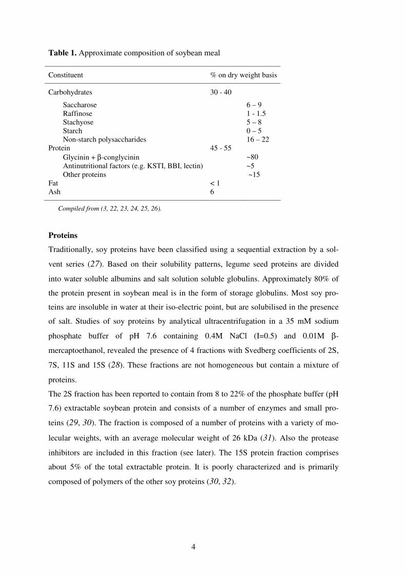
and 6% ash. An average composition of SBM is given in Table 1.
Soybean
↓↓↓↓ Cleaning, cracking
↓↓↓↓ Dehulling →→→→ Hulls
↓↓↓↓ Precondit ioning
↓↓↓↓ Flaking
↓↓↓↓ Oil extract ion →→→→ Crude oil
↓↓↓↓ ↓↓↓↓ Desolvent izat ion/toast ing Ref ining
↓↓↓↓ ↓↓↓↓ Grinding and classif icat ion Soy oil
↓↓↓↓
Soybean meal
4
Table 1. Approximate composition of soybean meal
Constituent % on dry weight basis
Carbohydrates 30 - 40
Saccharose 6 – 9 Raffinose 1 - 1.5 Stachyose 5 – 8 Starch 0 – 5 Non-starch polysaccharides 16 – 22
Protein 45 - 55
Glycinin + β-conglycinin ~80 Antinutritional factors (e.g. KSTI, BBI, lectin) ~5 Other proteins ~15
Fat < 1 Ash 6
Compiled from (3, 22, 23, 24, 25, 26).
Proteins
Traditionally, soy proteins have been classified using a sequential extraction by a sol-
vent series (27). Based on their solubility patterns, legume seed proteins are divided
into water soluble albumins and salt solution soluble globulins. Approximately 80% of
the protein present in soybean meal is in the form of storage globulins. Most soy pro-
teins are insoluble in water at their iso-electric point, but are solubilised in the presence
of salt. Studies of soy proteins by analytical ultracentrifugation in a 35 mM sodium
phosphate buffer of pH 7.6 containing 0.4M NaCl (I=0.5) and 0.01M β-
mercaptoethanol, revealed the presence of 4 fractions with Svedberg coefficients of 2S,
7S, 11S and 15S (28). These fractions are not homogeneous but contain a mixture of
proteins.
The 2S fraction has been reported to contain from 8 to 22% of the phosphate buffer (pH
7.6) extractable soybean protein and consists of a number of enzymes and small pro-
teins (29, 30). The fraction is composed of a number of proteins with a variety of mo-
lecular weights, with an average molecular weight of 26 kDa (31). Also the protease
inhibitors are included in this fraction (see later). The 15S protein fraction comprises
about 5% of the total extractable protein. It is poorly characterized and is primarily
composed of polymers of the other soy proteins (30, 32).

5
The 11S fraction comprises approx. 30 – 50% of the soluble soy proteins (33, 34). Gly-
cinin is the major globulin and composes about 60 – 80% of the soy proteins (33). Gly-
cinin consists of an acidic polypeptide, A (~40.000 Da) and a basic polypeptide, B
(~20.000 Da), linked by a single disulfide bridge and thereby forming an individual AB
subunit (35, 36). At least six acidic (A1a, A1b, A2 – A5) polypeptides and five basic (B1a,
B1b, B2 – B4) polypeptides have been identified (37). The acidic polypeptides have
isoelectric points varying from 4.75 to 5.4. The basic polypeptides can be separated into
three groups having isoelectric points of 8.0, 8.25 and 8.5, respectively (38). At neutral
pH and ionic strengths of above 0.35 glycinin has an hexameric structure (320.000 –
375.000 Da) consisting of a heterogeneous population of subunits (39). Glycinin has on
average 2 –SH and 18-20 S-S bonds per hexamer (27). An overview of the constituents
of glycinin and β-conglycinin is given in Table 2.
Table 2. Physicochemical properties of glycinin and β-conglycinin
Property Glycinin β-Conglycinin
Molecular weight (Da) of subunits/polypeptides A: 37.000-45.000
B: 22.500 α: 57.000-72.000
α’: 57.000-68.000
β: 42.000-52.000 Glycosylation (%) 0 ~5 SH-groups 0-2 / hexameric molecule 0 S-S bonds 18-20 / hexameric molecule 2 / trimeric molecule Isolelectric pH (average) 4.9 (hexameric molecule) 4.6 (trimeric molecule)
Compiled from (37, 38, 40, 41).
The 7S fraction of soy protein comprises about 35% of the soluble protein. This fraction
contains enzymes, a number of hemagglutinins (SBA) and predominantly a protein frac-
tion known as 7S globulins. About 85% of the 7S fraction is made up of ββββ-conglycinin
(33, 37, 42). This heterogeneous protein has trimeric quaternary structure and is glyco-
sylated for about 5%. β-Conglycinin is composed of seven different combinations of
three subunits. The subunits consist of three subunit proteins labelled α, α’ and β. The
α’ and α subunits have molecular weights of 57.000–72.000 Da. The β subunit has a
molecular weight of 42.000-52.000 Da. The seven combinations B0-B6 are, βββ, ββα’,
ββα, βαα’, βαα, ααα’, ααα, respectively (27). The subunits are non-covalently asso-
ciated by hydrophobic and hydrogen bonding (41). At low ionic strengths β-conglycinin
6
exists as a trimer having molecular weights ranging from 140.000 to about 170.000 Da.
β-conglycinin has no free –SH groups and on average two disulfide bonds per trimeric
molecule. At high ionic strengths, the β-conglycinin forms oligomers with a molecular
weight of about 280.000 to 350.000 Da and a sedimentation coefficient of 9S. The
isoelectric point of the trimeric β-conglycinin is 4.64 (38).
Protease Inhibitors
The nutritive value of unprocessed SBM is negatively affected by the presence of
antinutritional factors (ANF) (3, 43). The best characterised ANF are protease inhibitors
(44, 45), lectins (46, 47, 48), phytate (44, 49, 50), and phenolic compounds (51, 52).
In addition, oligosaccharides (22, 53) and allergenic epitopes of storage proteins (54)
are also considered among the antinutritional factors of soybean meal. Protease inhibi-
tors in soybean include trypsin and chymotrypsin inhibitors. Trypsin inhibitors (TI) are
proteins with the ability to inhibit most serine proteases (45, 55). Two families are
known: the Kunitz soy trypsin inhibitor (KSTI) and the Bowman Birk (BBI) trypsin in-
hibitor.
The BBI molecule consists of 71 amino acids and has a molecular weight of 7.8 kDa.
The protein can form dimers and trimers in solution which explains its association with
the other 2S proteins (31). The BBI molecule is highly symmetrical and composed of a
number of rings held together by the presence of 7 disulfide bonds. While many soy
proteins are low in sulfur-containing amino acids, this inhibitor has 14 of its 71 amino
acids composed of cysteine. The trypsin inhibiting site is the bond between lysine-16
and serine-17. While trypsin would normally cleave a Lys-Ser bond, it appears that the
rigid ring structure in which this bond exists often prevents cleavage. Although trypsin
binds to this protein, it cannot cleave the bond and is not readily released to cleave other
molecules. At the opposite end of the molecule, the bond between leucine-43 and ser-
ine-44 interacts with chymotrypsin. This band is also not cleaved. While named the
Bowman-Birk trypsin inhibitor, the protein can also inhibit the activity of chymotrypsin
depending on the experimental conditions (44).
The KSTI molecule is composed of 181 amino acids with a molecular weight of ap-
proximately 21.5 kDa (44). It has two disulfide bridges making it a less rigid molecule
than the Bowman-Birk inhibitor. Arginine-63 and isoleucine-64 form the bond at the

7
active site of the inhibitor. Trypsin cleaves this bond, but the enzyme is not released
from the inhibitor once contact has been made (45, 56, 57, 58).
Carbohydrates
The carbohydrates of soybean meal can be divided into water-extractable and water-
unextractable carbohydrates (59). The main water-extractable carbohydrates of defat-
ted soy flour consist of oligosaccharides, saccharose (~8% w/w), stachyose (~5%), raf-
finose (~1%), maltose (~0.5%), and verbascose (trace), together with the monosaccha-
rides, glucose (0.3%), arabinose (0.1%), and ribose (0.1%) (60, 61). Oligosaccharides
are responsible for the flatulence problems that are often associated with consumption
of soy products. However, most of the soluble carbohydrates are removed during the
manufacture of more refined soy products such as protein concentrates (>70% protein)
or isolates (>85% protein). The proportion of water-extractable NSP in soy is negligible
(<5%) (59). The water- and enzyme-unextractable plant cell wall polysaccharides are
the subject of interest of this thesis.
The water-unextractable carbohydrates comprise ~50% of total saccharides of soy
and are present as cell wall polysaccharides (25, 59, 62, 63). Compared to soy proteins,
characterisation of the cell wall polysaccharides is more difficult because the polymers
form a complex matrix of pectic substances, hemi-celluloses, celluloses and structural
proteins. Direct comparison between individual studies is difficult due to different
methods of extraction, separation, fractionation, and analysis. The analyses are based on
chemical or enzymatic hydrolysis of sugar linkages to release both monomeric and oli-
gomeric degradation products. The monomers are then identified by chromatographic
techniques, the oligomers are purified by chromatographic techniques and their fine
chemical structure established by e.g. NMR and MS (24).
The monosaccharide compositions of NSP of soybean meal and the water-
unextractable, de-proteinized solids (WUS) of soybean meal obtained in a study by Hu-
isman and co-workers (59) are presented in Table 3.
8
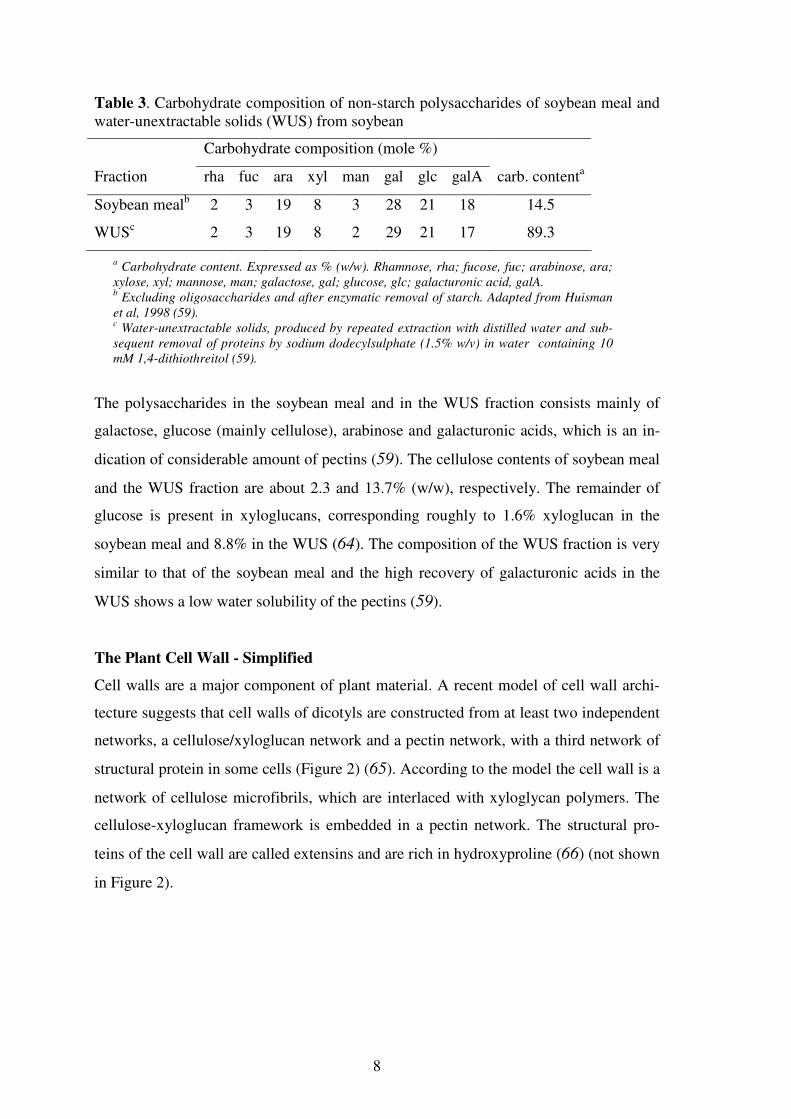
Table 3. Carbohydrate composition of non-starch polysaccharides of soybean meal and water-unextractable solids (WUS) from soybean
Carbohydrate composition (mole %)
Fraction rha fuc ara xyl man gal glc galA carb. contenta
Soybean mealb 2 3 19 8 3 28 21 18 14.5
WUSc 2 3 19 8 2 29 21 17 89.3
a Carbohydrate content. Expressed as % (w/w). Rhamnose, rha; fucose, fuc; arabinose, ara;
xylose, xyl; mannose, man; galactose, gal; glucose, glc; galacturonic acid, galA. b Excluding oligosaccharides and after enzymatic removal of starch. Adapted from Huisman
et al, 1998 (59). c Water-unextractable solids, produced by repeated extraction with distilled water and sub-
sequent removal of proteins by sodium dodecylsulphate (1.5% w/v) in water containing 10
mM 1,4-dithiothreitol (59).
The polysaccharides in the soybean meal and in the WUS fraction consists mainly of
galactose, glucose (mainly cellulose), arabinose and galacturonic acids, which is an in-
dication of considerable amount of pectins (59). The cellulose contents of soybean meal
and the WUS fraction are about 2.3 and 13.7% (w/w), respectively. The remainder of
glucose is present in xyloglucans, corresponding roughly to 1.6% xyloglucan in the
soybean meal and 8.8% in the WUS (64). The composition of the WUS fraction is very
similar to that of the soybean meal and the high recovery of galacturonic acids in the
WUS shows a low water solubility of the pectins (59).
The Plant Cell Wall - Simplified
Cell walls are a major component of plant material. A recent model of cell wall archi-
tecture suggests that cell walls of dicotyls are constructed from at least two independent
networks, a cellulose/xyloglucan network and a pectin network, with a third network of
structural protein in some cells (Figure 2) (65). According to the model the cell wall is a
network of cellulose microfibrils, which are interlaced with xyloglycan polymers. The
cellulose-xyloglucan framework is embedded in a pectin network. The structural pro-
teins of the cell wall are called extensins and are rich in hydroxyproline (66) (not shown
in Figure 2).

9
Figure 2. Simplified structural model of the primary cell wall (65).
The Cellulose/Xyloglucan Network: Cellulose ((1,4)-linked β-D-glucan) is the major
component of the primary cell wall. The cellulose chains are associated into microfibrils
by intermolecular hydrogen-bonding. The microfibrils are coated with hemicelluloses to
prevent them from aggregating. The principal hemicelluloses are xyloglucans, which
have also been identified in the cell walls of soy (59). Xyloglucan is thought to form a
tightly bound molecular monolayer on the surface of cellulose of which part can interact
with cellulose microfibrils via multiple hydrogen bonds thereby cross-linking the mi-
crofibrils (67).
The Pectin Network: The second polysaccharide network is composed of pectic poly-
saccharides, of which some of the known structures are very abundant in soy (59). Ho-
mogalacturonan is the most well known part of pectic substances consisting of (1,4)-
linked α-galacturonic acids residues but in soy this polymer is not a common structural
element (68). Xylogalacturonan is a relatively recently discovered sub-unit of soy pec-
tic substances (69, 70). The backbone consists of (1,4)-linked α-D-galacturonic resi-
dues. Xylose residues are β-(1,3)-linked to part of the galacturonic acid residues which
can be partly methyl esterified. Xylogalacturonan is probably associated with rhamno-
galacturonan regions (70, 71).
Rhamnogalacturonan (RG) is another major type of pectic polysaccharide. Polymers
containing this backbone are present in most if not all higher plant cell walls. Two types
of rhamnogalacturonan exist, namely rhamnogalacturonan I and II. In recent literature
10
RG II is termed highly branched galacturonan (70). Type I is by far the most abundant
(70). It consists of a long chain of alternating α-(1,4)-linked galacturonosyl, and α-
(1,2)-linked rhamnose units called rhamnogalacturonan. Next to rhamnose and galactu-
ronic acid residues in the backbone, RG I is composed of arabinofuranosyl-, galac-
topyranosyl-, and minor quantities of fucopyranosyl residues (70, 72).
Bound to the backbone at the rhamnose unit are sidechains of arabinan, galactan, and
highly branched arabinogalactans. They look like hairs and highly substituted rhamno-
galacturonans are called the hairy regions (66). Pectic L-arabinans consist of (1,5)-
linked α-L-arabinose residues, which are substituted with mainly monomeric arabinose
but also oligomeric arabinose chains mainly at O-3 but also at O-2. Pectic D-galactans
contain primarily β-(1,4)- linked galactose units, with little or no additional saccharide
material present in the molecule. Generally, the galactan sidechains are longer than ara-
binan and less branched (70, 73).
Arabinogalactans are divided into two types, type I and II. Pectin mostly contains type I
(74). Type I arabinogalactan has a backbone of β-(1,4)-linked galactans with branches
of α-(1,5)-linked arabinose or galactose chains bound α-(1,6) to the galactan backbone
(66). The ratio of arabinose and galactose in arabinogalactan, and the branching of ara-
binose show considerable variation within different plant species (70).
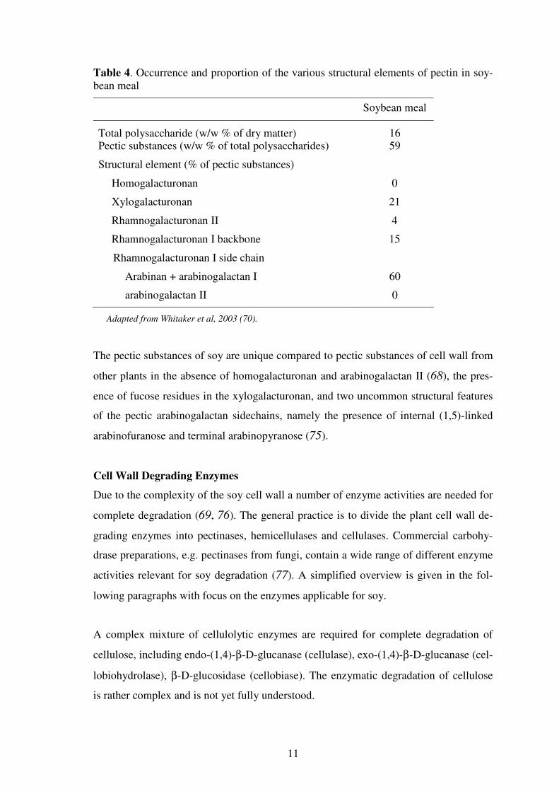
An approximate composition of pectin in soybean meal is presented in Table 4.
11
Table 4. Occurrence and proportion of the various structural elements of pectin in soy-bean meal
Soybean meal
Total polysaccharide (w/w % of dry matter) 16 Pectic substances (w/w % of total polysaccharides) 59
Structural element (% of pectic substances)
Homogalacturonan 0
Xylogalacturonan 21
Rhamnogalacturonan II 4
Rhamnogalacturonan I backbone 15
Rhamnogalacturonan I side chain
Arabinan + arabinogalactan I 60
arabinogalactan II 0
Adapted from Whitaker et al, 2003 (70).
The pectic substances of soy are unique compared to pectic substances of cell wall from
other plants in the absence of homogalacturonan and arabinogalactan II (68), the pres-
ence of fucose residues in the xylogalacturonan, and two uncommon structural features
of the pectic arabinogalactan sidechains, namely the presence of internal (1,5)-linked
arabinofuranose and terminal arabinopyranose (75).
Cell Wall Degrading Enzymes
Due to the complexity of the soy cell wall a number of enzyme activities are needed for
complete degradation (69, 76). The general practice is to divide the plant cell wall de-
grading enzymes into pectinases, hemicellulases and cellulases. Commercial carbohy-
drase preparations, e.g. pectinases from fungi, contain a wide range of different enzyme
activities relevant for soy degradation (77). A simplified overview is given in the fol-
lowing paragraphs with focus on the enzymes applicable for soy.
A complex mixture of cellulolytic enzymes are required for complete degradation of
cellulose, including endo-(1,4)-β-D-glucanase (cellulase), exo-(1,4)-β-D-glucanase (cel-
lobiohydrolase), β-D-glucosidase (cellobiase). The enzymatic degradation of cellulose
is rather complex and is not yet fully understood.

12
Hemicellulases are divided into groups which designate the type of hemicellulose they
are capable of degrading. Typical hemicellulases are endo-β-(1,4)-D-xylanases, endo-β-
(1,4)-D-mannanases, and endo-β-(1,3)-(1,4)-glucanases (70, 78). More relevant for soy
degradation are the galactan and arabinan degrading enzymes endo-α-(1,5)-L-
arabinanases, endo-β-(1,4)- and -β-(1,3)/(1,6)-D-galactanases are categorized both as
hemicellulases and as pectin degrading enzymes (70). With respect to chemical struc-
ture there is no sharp distinction between hemicelluloses and the side chains of the pec-
tic substances (70, 77). They differ in solubility and hemicelluloses are only soluble in
alkaline conditions (70). The activity of these endo-enzymes is generally enhanced by
the presence of glycosidases like β-xylosidase, β-galactosidase, and α-
arabinofuranosidases, particularly by removing side chains.
The homogalacturonan degrading enzymes include xylogalacturonan degrading endo
and exo-polygalacturonases (70), pectin lyases, pectate lyase (77), endo-
xylogalacturonase, and the group of esterases, which split of substituents ester-linked to
pectins, like pectin methyl esterases, pectin acetyl esterases and feruloyl esterases (70).
The rhamnogalacturonan degrading enzymes include endo-rhamnogalacturonase, endo-
rhamnogalacturonan lyase, rhamnogalacturonan rhamnohydrolyase, rhamnogalactu-
ronan galacturonohydrolyase (25, 79), and since arabinogalactans type I and arabinans
are side chains of the rhamnogalacturonan backbone, also arabinanases, α-
arabinofuranosidases, galactanases and galactosidases can be classified into this group
(70, 80). A rhamnogalacturonan acetyl esterases specific for rhamnogalacturonan struc-
tures has also been identified (80).
An extensive degradation of the soy cell wall polysaccharides is known to demand sev-
eral enzyme activities (70). Multicomponent enzyme preparations from e.g. Aspergillus
aculeatus (used in Chapter 2) are known to be effective for degradation of soy cell walls
(77).
Heat Treatment
Feeding of unheated soy proteins to animals has been demonstrated to have a detrimen-
tal affect on growth due to the inhibitory effects of trypsin inhibitors (81). Therefore, as

13
shown in Figure 1, the most common processing technology applied to the soybean is
heat treatment, which has proven effective for reducing levels of trypsin inhibitors and
soybean lectin. Commercial heat treatment is carefully controlled: underheating often
results in adequate inactivation of ANF while overheating can reduce availability of im-
portant meal components (e.g. lysine) through the occurrence of Maillard reactions (81,
82, 83, 84).
Heat treatment of soybean meals affects the solubility of the carbohydrates. High tem-
perature and low water content enhance Maillard reactions. The Maillard reaction is not
a clearly defined single reaction but encompasses a family of reaction pathways that
have a common first step: the condensation of an amino group with a reducing sugar.
Following the initial condensation, the product undergoes rearrangement, fragmenta-
tion, degradation, dehydration, and other reactions resulting in a large number of com-
pounds that eventually impart coloration to the soybean meal. Numerous factors deter-
mine the outcome of the Maillard reaction including the abundance of carbonyl and
amino groups, pH, temperature, water activity, and reaction time. The extent to which
Maillard reactions occur during heating of soybean meals can be estimated by measur-
ing decrease in total soluble sugar content (85). The above mentioned reactions nega-
tively affect the solubility and accessibility of the carbohydrates and proteins by carbo-
hydrases and proteases (14, 84, 86, 87, 88, 89).
KSTI and BBI in purified preparations respond differently to thermal treatment then
when they are embedded in the soybean meal matrix (45). Purified protease inhibitors
(PI) are less susceptible to heat treatment indicating that the higher sensitivity seen in
soybean meal is caused by the presence of other constituents of the soybean such as free
thiol agents (90) or cell wall polysaccharides that somehow promote inactivation of the
inhibitors (45). Literature generally agrees that antinutritional factors (ANF) such as
lectins and protease inhibitors are more rapidly inactivated at high water activities than
at lower water activities. For intact soybeans, residual PI activity was only 3% of origi-
nal after only 20 minutes of cooking in water (91). Comparable results were found for
raw defatted soybean meal subjected to autoclaving at 121°C for 10 min (45).
Heating of β-conglycinin causes dissociation of subunits which can then unfold, re-
associate and aggregate to form precipitates making them less accessible for enzymatic
degradation than in their native condition. At concentrations below 7% protein and low
14
ionic strength, β-conglycinin is reported as being relatively heat stable. The proteins are
stable toward heat treatments at temperatures in excess of 100°C (41, 92).
Heating of glycinin at 100°C at around neutral pH rapidly converted approx. 50% of the
protein into a buffer soluble aggregate (93). With continued heating, the soluble aggre-
gates increased in size and precipitated. The precipitated fraction consisted of the basic
polypeptides, whereas the acidic polypeptides remained soluble. However, when soy-
bean meal is heated at comparable conditions, the basic polypeptides of glycinin re-
mains soluble and no precipitate was observed (94).
Accessibility of Soybean Meal Constituents
When soybean meal is used industrially, a certain proportion of the protein remains in-
accessible or insoluble or indigestible to the animals. From both a financial and envi-
ronmental perspective this material represents a loss of an important and valuable nutri-
ent. For many years it has been anticipated that a major reason for the observed inacces-
sibility of the protein was the presence of plant cell wall polymers, which supposedly
trapped or shielded the proteins from enzymatic degradation (66). This has led to inves-
tigations with extensive use of plant cell wall degrading enzymes for improved protein
release and digestibility (8). In these studies it was, however, established that plant cell
wall degrading enzymes have only minor effects on the digestibility/extractability of
soybean meal proteins, even when the viscosity of soybean meal slurries can be drasti-
cally reduced by these enzymes. This viscosity reducing effect is beneficial to the capa-
bility of most animals to utilize the feed effectively (8). Some proteins and carbohy-
drates remain insoluble even when large amounts of enzymes are added to the diet (95).
The major reasons for this phenomenon are thought to be partly inherent, i.e. interac-
tions between the different cell wall components, and partly induced by the molecular
cross-linking (protein/protein, protein/carbohydrate) caused by the heat treatment. Ef-
fects of exogenous enzymes vary considerably, and accordingly there is a need for more
research to pursue the understanding of the complex structural changes that occur when
exogenous enzymes are used for SBM at in vivo as well as in vitro (96).
Enzymatic Digestibility of Soy Proteins
In vitro digestibility of soybean proteins has received little attention in the literature. In
particular, the number of studies describing the digestibility of isolated soybean proteins

15
is limited. Proteolytic enzymes reported include pepsin, papain, trypsin, chymotrypsin
plus various bacterial and fungal proteases (97). Without being complete, an overview
of different studies on this subject is given in Table 5, including SBM proteins, protein
isolates and purified proteins.

16
Table 5. Short literature overview on effects of proteolytic treatment on soybean meal protein, protein isolates and purified soy proteins.
Reference Substrate Enzymes used Conditions of hydrolysis and pos-
sible additional treatment
Main results/and or conclusions (hydrolysate com-
pared to intact protein
Marsman et al, 1997 (14) Untoasted, toasted and extruded SBM
Esperase, Neu-trase and Bio-Feed Pro
SBM suspensions (10%) in 0.05M Na-acetate buffer (pH 5), for Es-perase also carbonate buffer (pH 9). Analysis: DSC, SDS-PAGE.
Toasted SBM: β-conglycinin well degraded – glycinin more resistant (polypeptide B more than A). Possible explanation: primarily non-covalent bonds broken, disulfide bonds more or less intact. Unheated: high resistance against degradation ex-plained by native structure. Extruded: Both proteins well degraded – both non-covalent and disulfide bonds broken
Romagnolo et al, 1990 (98) Processed SBM
Rumen mi-crobes
Rumen bacterial fermentation, time course. Analysis: SDS-PAGE
β-conglycinin more susceptible to rumen degradation than glycinin that showed particular resistance within B-polypeptide.
Lallés et al, 1998 (99) Heated SBM Rumen mi-crobes
Rumen bacterial fermentation (nylon bag placed in the rumen). Analysis: Western blotting (antibod-ies)
Glycinin: Sharp decrease in immunoreactive glycinin 2 h post feeding. Early outflow of glycinin composed of nearly intact B-polypeptides and partially degraded A-polypeptides. However, intact A-polypeptide and B-polypeptide detected at both 2 h and 6 h post feeding.
β-Conglycinin: Rate considerably slower than glycinin. Lee et al 2001 (100) Defatted soy-
bean meal Alcalase and Flavourzyme
Acid pretreatment (0.05-0.2 N HCl), hydrolysis (0-24 h), pH 6.5. Analy-sis: DH and gel filtration
Protein hydrolysed primarily during first 5 h. DH and
α-amino nitrogen increased after acid pre-treatment. Average peptide chain length: Alcalase (3 h), 7-8 amino acids; Alcalase/Flavourzyme (21 h): 3-5 amino acids.
Tsumura et al, 2004 (101) Native soy pro-tein isolate
Pepsin and pa-pain
Dispersion in water (5% w/v). Pepsin incub: pH range 1.5-4.0, reaction
37°C for 30min.
Papain incub: pH 7.0, 37-80°C for 5 min. Analysis: SDS-PAGE, DSC.
Glycinin fraction in native isolate selectively hydro-lysed by pepsin in the range from pH 1.5-2.5.
β-Conglycinin fraction selectively hydrolysed by pa-
pain at 70°C. Proteolysis significantly correlated with onset of dena-
turation of glycinin and β-conglycinin in SPI.

17
Table 5 continued
Bernardi-Don et al, 1991 (97)
Soy protein concentrate, denatured
Aspergillus
oryzae and
Bacillus subtilis
Protein conc. 6%, pH 6.8, 1 h, differ-ent enzyme/substrate ratios between 0.1-1:100. Analysis: DH and NSI
Solubility (NSI) greatly enhanced by both proteases (no discrimination between different soy proteins). Greatest effect up to DH 10. Bacterial protease more effective protein solubiliser than fungal protease – effect most remarkable at the highest DH values.
Shutov et al, 1996 (102) Glycinin and
β-conglycinin, purified
Trypsin Substrate concentration (5 mg/ml) in 0.05 M Tris-HCl pH 8.0. Enzyme
substrate ratio 1:600, 30°C. Analysis: SDS-PAGE
Four cleavage points identified in the A polypeptide of
glycinin as well as the α’-chain of β-conglycinin.
Shutov et al, 1991 (103) Glycinin Trypsin, chy-
motrypsin
Protein conc. (5 mg/mL), heating to different degrees, incubation 60 min,
25°C, pH 8.2. Polypeptides separated by gel filtration.
Overall hexameric polypeptide structure retained. B polypeptides remained intact, A polypeptides reduced in size indicating that B is buried within the interior of the protein molecule (104, 105).
Kim et al, 2003 (106) Glycinin Trypsin Glycinin concentration 1% (w/w).
Incubation with enzyme at 50°C, pH 7 for 4 h. Analysis: gel permeation, reverse phase chromatography, and DH.
Glycinin hydrolysate DH 12. Molecular size distribu-tion from 200da to 1400 Da (~2-12 amino acids) show-ing varying hydrophobic character.
Hajos et al, 1996 (107) Soy albumins fraction
Pepsin Pepsin substrate ratio (100:1), pH 2, 2 h. Analysis: ELISA and SDS-PAGE
Immuno activity of KSTI significantly reduced. No significant reduction of BBI activity. One quarter of original KSTI content excluded from SDS-PAGE gel. BBI not reduced in molecular size.
Jensen et al, 1996 (108) BBI, unheated Bovine trypsin BBI + 5% (molar) trypsin in different buffers, pH ranges (3.5-10.3). Primary analysis: HPCE
Primary sequence almost fully conserved. Cleavage of trypsin reactive site and more slowly in chymotrypsin reactive subdomains
Vaintraub and Yattara, 1995 (109)
KSTI, com-mercial
Papain, subtil-
isin, pepsin
KSTI 0.5% solution + 0.05M acetate buffer, pH 4.5 / 0.05 M Tris-HCl, pH 8.5. Enzyme substrate ratio 1:200 for pepsin and 1: 100 for papain and sub-tilisin. Analysis: SDS-PAGE and gel per-meation
KSTI initially degraded into fragment retained in the molecule by covalent and disulfide bands. After ex-tended hydrolysis degraded into smaller peptides. Fragment size (many retained inhibitor activity): Pa-pain, (2.7 – 6.7 KDa); subtilisin, (3.9 – 8.8 kDa); pep-sin (6.9 – 18.4).

18
Although it is difficult to compare the various results found for hydrolysis of soy pro-
teins (different authors study different proteases on different substrates and at different
conditions; Table 5), some general conclusions can be drawn.
Firstly, resistance to proteolysis of the peptide bonds of the major soy proteins depends
largely on the extent of protein denaturation : native (high resistance, intact structure) >
toasted (medium resistance, non-covalent bonds broken) > extruded (low resistance,
non-covalent and disulfide bonds broken) (14). In toasted SBM β-conglycinin is effec-
tively degraded by different endo-proteases (14). Heat treated β-conglycinin was also
effectively degraded by proteases of rumen microbes (98), although Lallés and co-
workers reported a considerable slower degradation rate for β-conglycinin than for gly-
cinin by rumen microbes (99). Glycinin polypeptide B is more resistant to proteolysis
than polypeptide A (14, 98, 99) possibly because polypeptide B is buried within the
interior of the protein molecule (104, 105).
Also for soy protein isolates proteolysis significantly correlated with the onset of dena-
turation of glycinin and β-conglycinin (101). As shown in studies with Alcalase and
Flavourzyme, protein degradation progresses most rapidly within the first 5 h of incuba-
tion and a high degree of hydrolysis is only obtained for combinations of the two en-
zymes (100). Bacterial proteases are generally more powerful solubilisers than fungal
proteases (97).
Secondly, the degradation of purified glycinin and β-conglycinin by subtilisin types of
proteases (14) has many similarities to the degradation of these proteins in SBMs by
rumen microbes (98, 99). After heating at slightly alkaline pH the hexameric polypep-
tide structure of glycinin is conserved and B polypeptides remained largely intact (103).
The α- and α’ chains of β-conglycinin were also readily degraded by trypsin (102).
Finally, results of tryptic digestion of Bowman-Birk inhibitor showed that its primary
sequence was almost fully conserved (108) whereas purified KSTI after prolonged in-
cubation was degraded into smaller peptides by papain, subtilisin, and pepsin (109).
However, in a pepsin-treated soy albumin fraction the immuno reactivity of KSTI was
significantly reduced, whereas the molecule was only partially degraded. The BBI was
not reduced in size and retained its immuno activity (107). The primary sequence of
unheated BBI was also almost fully conserved after tryptic digestion (108).

19
In Vitro Testing of Enzymes
In vitro test systems are valuable complementary tools for the development of new en-
zyme applications for food and feed. In vitro models can be used to compare and rank
new enzymes according to parameters of particular interest, including the capacity to
degrade the target substrates at relevant conditions. The obvious strength of in vitro
models is that experiments can be repeated at exactly the same conditions in series. In
contrast, in vivo studies are subject to large physiological variations between individual
animals and also within the same animal during its physiological development stages
(8). Consequently a considerable number of animals are needed for in vivo testing in
order to obtain statistically valid results. The capacity of in vitro models decreases
greatly with increasing complexity of the model (i.e. how well it compares to an in vivo
situation). Dynamic models such as the IFR Model Gut (Institute of Food Research,
Norwich, UK) and the gastro intestinal model (GIT) from TNO (110) are used to study
several parameters simultaneously, but have limited capacity. Naturally, the technical
feasibility (and simplicity) inevitably plays a role when the need for high capacity is
critical. Therefore, relatively simple batch models with a high number of replicates and
a standardized set of conditions have been used for the experiments of this thesis.
Aim and Outline of Thesis
From the above, it is evident that much is known about what the different enzymes can
do – but not so much about what they cannot do. Accordingly, there is a lack of knowl-
edge about the molecular structure of the different soy polymers following enzymatic
digestion and a lack on knowledge about the composition of enzyme resistant structures.
A better knowledge on the latter is valuable for understanding if enzymes of interest in
principle are capable of degrading resistant structures or the causes for their ineffi-
ciency. This could also help understanding if certain enzyme activities are lacking
which might improve performance. Research in these areas should enable more efficient
use of the current enzymes and the development of more efficient enzymes.
The first aim of this thesis is to broaden the knowledge about factors affecting the effi-
cacy of enzymatic extraction of protein and carbohydrates from SBMs. The second aim
is to investigate the effects of heat treatments on protein composition of enzyme-
unextractable soybean meal fractions produced in vitro and in vivo.
20
In Chapter 2, the amounts and compositions of residues obtained after enzymatic
treatment of unheated SBM and SBMs heated at different humidities are presented.
High concentrations of commercial proteases and carbohydrases are used in a two-step
hydrolysis procedure to obtain enzyme-unextractable material. The extractability of pro-
tein and carbohydrates from the meals is subsequently examined, and the unextractable
residues are quantified and characterised with respect to amino acid and carbohydrate
composition.
In Chapter 3, the enzymatic hydrolysis conditions are optimised for two commercial
preparations, Alcalase and Flavourzyme, in order to increase protein solubilisation in a
single step hydrolysis of SBM.
The experiments of Chapter 4 further characterize the proteins in the enzyme-
unextractable residues which resist extraction from the soybean matrix by enzymatic
treatment. The residues are subjected to extraction by various solvents and the extracted
proteins are analyzed by gel electrophoresis, chromatographic techniques and mass
spectrometry. The solvent extractability of protein and carbohydrates from the residues
is determined and the resulting residues are quantified and characterized for amino acid
and carbohydrate composition.
Chapter 5 presents the molecular size and composition of proteinaceous material ex-
tracted from the insoluble components of a digesta sample obtained from pigs fed a feed
consisting entirely of soybean meal. The molecular size of the alkali-extractable protein
fraction is subsequently studied using gel permeation chromatography, gel electropho-
resis, RPLC-MS, and MALDI-ToF MS. In vitro proteolysis of extracted, aggregated
proteinaceous material is also studied.
In Chapter 6, the results obtained in the previous chapters are discussed.

21
References
1. Carpenter,K.J. Protein and Energy - A Study of Changing Ideas in Nutrition,
Cambridge University Press: Cambridge, UK, 1994.
2. Smith,A.K.; Circle,S.J. Soybean: Chemistry and Nutrition, Avi: Westport CT:
1972.
3. Liu,K.S. Soybeans. Chemistry, Technology, and Utilization, Chapman & Hall:
New York, US, 1997.
4. Zanella,I.; Sakomura,N.K.; Silversides,F.G.; Fiqueirdo,A.; Pack,M. Effect of en-
zyme supplementation of broiler diets based on corn and soybeans. Poult Sci
1999, 78, 561-568.
5. Thorpe,J.; Beal,J.D. Vegetable protein meals and the effects of enzymes. In En-
zymes in farm animal nutrition; Bedford,M.R.; Partridge,G.G., Eds.; CABI Pub-
lishing: Wallingford, UK, 2000; 125-143.
6. Kocher,A.; Choct,M.; Morrisroe,L.; Broz,J. Effects of enzyme supplementation on
the replacement value of canola meal for soybean meal in broiler diets. Aus J Ag-
ric Res 2001, 52, 447-452.
7. Pettersson,D.; Graham,H.; Aman,P. Enzyme supplementation of low or high crude
protein concentration diets for broiler chickens. Anim Prod 1990, 51, 399-404.
8. Bedford,M.R. Exogenous enzymes in monogastric nutrition - their current value
and future benefits. Anim Feed Sci Technol 2000, 86, 1-13.
9. Haberer,B.; Schulz,E.; Flachowsky,G. Effects of beta-glucanase and xylanase
supplementation in pigs fed a diet rich in nonstarch polysaccharides: disappear-
ance and disappearance rate of nutrients including the nonstarch polysaccharides
in stomach and small intestine. J Anim Physiol Anim Nutr 1998, 78, 95-103.
10. Kim,S.W.; Knabe,D.A.; Hong,K.J.; Easter,R.A. Use of carbohydrases in corn-
soybean meal-based nursery diets. J Anim Sci 2003, 81, 2496-2504.
11. Brufau,J.; Francesch,M.; Pérez-Vendrell,A.M. Exogenous enzymes in poultry
feeding. Recent developments. In 2002 Annual Animal Nutrition Conference; Fa-
yetteville: Arkansas, USA, 2002.
12. Simbaya,J.; Slominski,B.A.; Guenter,W.; Morgan,A.; Campbell,L.D. The effects
of protease and carbohydrase supplementation on the nutritive value of canola
meal for poultry: In vitro and in vivo studies. Anim Feed Sci Technol 1996, 61,
219-234.

22
13. Cowieson,A.J.; Adeola,O. Carbohydrases, protease, and phytase have an additive
beneficial effect in nutritionally marginal diets for broiler chicks. Poult Sci 2005,
84, 1860-1867.
14. Marsman,G.J.P.; Gruppen,H.; Mul,A.J.; Voragen,A.G.J. In-vitro accessibility of
untreated, toasted, and extruded soybean meals for proteases and carbohydrases. J
Agric Food Chem 1997, 45, 4088-4095.
15. Procter,A. Soybean oil extraction and processing. In Soybeans. Chemistry, tech-
nology and utilization; Liu,K., Ed.; Chapman & Hall: New York, US, 1997; 297-
346.
16. Ghazi,S.; Rooke,J.A.; Galbraith,H. Improvement of the nutritive value of soybean
meal by protease and alpha-galactosidase treatment in broiler cockerels and broiler
chicks. Br Poult Sci 2003, 44, 410-418.
17. Bennett,J.O.; Krishnan,A.H.; Wiebold,W.J.; Krishnan,H.B. Positional effect on
protein and oil content and composition of soybeans. J Agric Food Chem 2003,
51, 6882-6886.
18. Hettiarachchy,N.; Kasapathy,U. Soybean protein products. In Soybeans. Chemis-
try, technology and utilization; Liu,K., Ed.; Chapman & Hall: New York, US,
1997; 379-411.
19. De Meester,J.; Kempener,S.; Mollee,P. Production and isolation of soy proteins.
Industrial Proteins 2000, 8, 5-7.
20. Becker,K.W. Processing of oil seeds to meal and protein flakes. JAOCS 1971, 48,
299-304.
21. Grieshop,C.M.; Kadzere,C.T.; Clapper,G.M.; Flickinger,E.A.; Bauer,L.L.; Fra-
zier,R.L.; Fahey,G.C. Chemical and nutritional characteristics of United States
soybeans and soybean meals. J Agric Food Chem 2003, 51, 7684-7691.
22. Dandanell,D.Y.; Aman,P. Chemical composition of certain dehulled legume seeds
and their hulls with special reference to carbohydrates. Swed J Agr Res 1993, 23,
133-139.
23. Bernardini,E. Oilseeds, oils and fats: Volume 1. Planning a factory raw materials
and extraction techniques., 2nd ed.; Roma, B.E. Publishing House: 1985.
24. Voragen,A.G.J.; Schols,H.A.; Gruppen,H. Structural studies of plant cell-wall
polysaccharides using enzymes. In Plant polymeric carbohydrates; Meuser,F.,
Ed.; Royal Society of Chemistry: Cambridge, UK, 1993; 3-15.

23
25. Schols,H.A.; Lucas-Lokhorst,G.; Voragen ,A.G.J.; Niessen,W.M.A. Isolation and
characterization of cell wall polysaccharides from soybeans. Carbohydrates in the
Netherlands 1993, 9, 7-10.
26. Macrae,R.; Robinson,R.K.; Sadler,M.J. Encyclopedia of food science, food tech-
nology and nutrition, Academic Press: London, UK, 1993.
27. Fukushima,D. Structures of plant storage proteins and their functions. Food Rev
Int 1991, 7, 353-381.
28. Koshiyama,I. Distribution of the 7S proteins in soy bean globulins by gel filtration
with Sephadex G-200. Agric Biol Chem 1969, 33, 281-284.
29. Catsimpoolas,N.; Ekenstam,C. Isolation of alpha, beta, and gamma conglycinins.
Arch Biochem Biophys 1969, 129, 490-497.
30. Wolf,W.J. Soy bean proteins: Their functional, chemical and physical properties. J
Agric Food Chem 1970, 18, 969-976.
31. Gueguen,J.; van Oort,M.A.; Quillien,L.; Hessing,M. The composition, biochemi-
cal characteristics and analysis of proteinaceous antinutritional factors in legume
seeds. A review. In Recent advances of research in antinutritional factors in leg-
ume seeds; van der Poel,A.F.B.; Saini,H.S., Eds.; Wageningen Press: Wagenin-
gen, The Netherlands, 1993; 9-30.
32. Parkkonen,T.; Tervilawilo,A.; Hopeakoskinurminen,M.; Morgan,A.; Poutanen,K.;
Autio,K. Changes in wheat microstructure following in-vitro digestion. Acta Agric
Scand B - Soil Plant Sci 1997, 47, 43-47.
33. Thanh,V.H.; Shibasaki,K. Major proteins of soybean seeds. A straightforward
fractionation and their characterization. J Agric Food Chem 1976, 24, 1117-1121.
34. Wolf,W.J. Soybean proteins: their functional, chemical, and physical properties. J
Agric Food Chem 1970, 18, 969-976.
35. Nielsen,N.C. The chemistry of legume storage proteins. Philos Trans R Soc Lond
B Biol Sci 1984, 304, 287-296.
36. Staswick,P.E.; Hermodson,M.A.; Nielsen,N.C. Identification of the cystines which
link the acidic and basic components of the glycinin subunits. J Biol Chem 1984,
259, 13431-13435.
37. Brooks,J.R.; Morr,C.V. Current aspects of soy protein fractionation and nomen-
clature. J Am Oil Chem Soc 1985, 62, 1347-1354.

24
38. Koshiyama,I. Storage proteins of soybean. In Seed Proteins Biochemistry, Genet-
ics, Nutritive Value; Gottschalk,W.; Müller,H.P., Eds.; Martinus Nijhoff/Dr W.
Junk Publisher: The Hague, 1983; 427-450.
39. Wolf,W.J.; Briggs,D.R. Studies on the cold-insoluble fraction of the water-
extractable soybean proteins. II. Factors influencing conformation changes in the
11 S component. Arch Biochem Biophys 1958, 76, 377-393.
40. Thanh,V.H.; Shibasaki,K. Major proteins of soybean seeds. Subunit structure of
beta-conglycinin. J Agric Food Chem 1978, 26, 692-698.
41. Yamauchi,F.; Yamagishi,T.; Iwabuchi,S. Molecular Understanding of Heat-
Induced Phenomena of Soybean Protein. Food Rev Int 1991, 7, 283-322.
42. Bogracheva,T.Y.; Bespalove,N.Y.; Leont'ev,A.L. Isolation of 11S and 7S Globu-
lins from Seeds of Glycine max. Appl Biochem Microb 1996, 32, 429-433.
43. Garcia,M.C.; Torre,M.; Marina,M.L.; Laborda,F. Composition and characteriza-
tion of soyabean and related products. Crit Rev Food Sci Nutr 1997, 37, 361-391.
44. Liener,I.E. Implications of antinutritional components in soybean foods. Crit Rev
Food Sci Nutr 1994, 34, 31-67.
45. DiPietro,C.M.; Liener,I.E. Heat inactivation of the Kunitz and Bowman-Birk soy-
bean protease inhibitors. J Agric Food Chem 1989, 37, 39-44.
46. Grant,G.; van Driessche,E. Legume lectins: physicochemical and nutritional prop-
erties. In Recent advances of research in antinutritional factors in legume seeds.;
van Driessche,E.; van der Poel,A.F.B.; Huisman,J.; Saini,H.S., Eds.; Wageningen
Press: Wageningen, Netherlands, 1993; 219-233.
47. Kennedy,J.F.; Palva,P.M.G.; Corella,M.T.S.; Cavalcanti,M.S.M.; Coelho,L.C.B.B.
Lectins, versatile proteins of recognition: a review. Carbohyd Polym 1995, 26,
219-230.
48. Vasconcelos,L.M.; Trentim,A.; Guimaraes,J.A.; Carlini,C.R. Purification and
physicochemical characterization of soyatoxin, a novel toxic protein isolated from
soybeans (Glycine max). Arch Biochem Biophys 1994, 312, 357-366.
49. Honig,D.H.; Wolf,W.J.; Rackis,J.J. Phytic-Acid and Phosphorus Content of Vari-
ous Soybean Protein Fractions. Cereal Chem 1984, 61, 523-526.
50. Prattley,C.A.; Stanley,D.W. Protein-phytate interactions in soybeans. I. Localiza-
tion of phytate in protein bodies and globoids Antinutritional components of
foods. J Food Biochem 1982, 6, 243-253.
25
51. Eraso,F.; Hartley,R.D. Monomeric and Dimeric Phenolic Constituents of Plant
Cell Walls Possible Factors Influencing Wall Biodegradability. J Sci Food Agric
1990, 51, 163-170.
52. Ritter,M.A.; Morr,C.V.; Thomas,R.L. In-Vitro Digestibility of Phytate-Reduced
and Phenolics- Reduced Soy Protein Isolates. J Food Sci 1987, 52, 325-327, 341.
53. Asp,N.G. Dietary carbohydrates: Classification by chemistry and physiology.
Food Chem 1996, 57, 9-14.
54. Tukur,H.M.; Lalles,J.P.; Plumb,G.W.; Mills,E.N.C.; Morgan,M.R.A.; Toullec,R.
Investigation of the relationship between in vitro ELISA measures of immunore-
active soy globulins and in vivo effects of soy products. J Agric Food Chem 1996,
44, 2155-2161.
55. Liener,I.E. Effects of processing on antinutritional factors in legumes: The soy-
bean case. Arch Latinoam Nutr 1994, 44, 48-54.
56. Bau,H.M.; Villaume,C.; Nicolas,J.P.; Mejean,L.; Bau,H.M. Effect of germination
on chemical composition, biochemical constituents and antinutritional factors of
soya bean (Glycine max) seeds. J Sci Food Agric 1997, 73, 1-9.
57. Friedman,M.; Brandon,D.L.; Bates,A.H.; Hymowitz,T. Comparison of a commer-
cial soybean cultivar and an isoline lacking the Kunitz trypsin inhibitor: composi-
tion, nutritional value, and effects of heating. J Agric Food Chem 1991, 39, 327-
335.
58. Hessing,M.; Bleeker,H.; Van Biert,M.; Vlooswijk,H.A.A.; Hamer,R.J. Enzymatic
hydrolysis of soya products and analysis of antinutritional factors. Meded - Fac
Landbouwkd Toegepaste Biol Wet (Univ Gent) 1994, 59, 2257-2262.
59. Huisman,M.M.H.; Schols,H.A.; Voragen,A.G.J. Cell wall polysaccharides from
soybean (Glycine max.) meal. Isolation and characterisation. Carbohydr Polym
1998, 37, 87-95.
60. Eldridge,A.C.; Black,L.T.; Wolf,W.J. Carbohydrate composition of soybean flours
protein concentrates and isolates. J Agric Food Chem 1979, 27, 799-802.
61. MacLeod,G.; Ames,J. Soy flavor and its improvement. CRC Crit Rev Food Sci
Nutr 1988, 27, 219-400.
62. Huisman,M.M.H.; Schols,H.A.; Voragen,A.G.J. Isolation and sequential extrac-
tion of cell wall polysaccharides from soy meal. In Pectins and Pectinases. Pro-
ceedings of an International Symposium; J.Visser; A.G.J.Voragen, Eds.; Elsevier
Science B.V: The Netherlands, 1996; 511-515.

26
63. Kikuchi,T.; Sugimoto,H. Studies on polysaccharides from soy. Part V. Detailed
structure of an acidic polysaccharide in soy sauce, confirmed by use of two kinds
of purified pectinases. Agric Biol Chem 1976, 40, 87-92.
64. Huisman,M.M.H.; Weel,K.G.C.; Schols,H.A.; Voragen,A.G.J. Xyloglucan from
soybean (Glycine max) meal is composed of XXXG-type building units. Carbo-
hyd Polym 2000, 42, 185-191.
65. McCann,M.C.; Roberts,K. The Cytoskeletal Basis of Plant Growth and Form,
Academic Press: New York, USA, 1991.
66. Carpita,N.C.; Gibeaut,D.M. Structural models of primary cell walls in flowering
plants: consistency of molecular structure with the physical properties of the walls
during growth. Plant J 1993, 3, 1-30.
67. Levy,S.; York,W.S.; Stuike-Prill,R.; Meyer,B.; Staehelin,L.A. Simulations of the
static and dynamic molecular conformations of xyloglucan. The role of the fuco-
sylated sidechain in surface-specific sidechain folding. Plant J 1991, 1, 195-215.
68. Huisman,M.M.H.; Fransen,C.T.M.; Kamerling,J.P.; Vliegenthart,J.F.G.;
Schols,H.A.; Voragen,A.G.J. The CDTA-soluble pectic substances from soybean
meal are composed of rhamnogalacturonan and xylogalacturonan but not homoga-
lacturonan. Biopolymers 2001, 58, 279-294.
69. Huisman,M.M.H.; Schols,H.A.; Voragen,A.G.J. Enzymatic degradation of cell
wall polysaccharides from soybean meal. Carbohydr Polym 1999, 38, 299-307.
70. Whitaker,J.R.; Voragen ,A.G.J.; Wong,D.W.S. Handbook of Food Enzymology,
Marcel Dekker: New York, USA, 2003.
71. Schols,H.A.; Bakx,E.J.; Schipper,D.; Voragen,A.G.J. A xylogalacturonan subunit
present in the modified hairy regions of apple pectin. Carbohyd Res 1995, 279,
265-279.
72. Fry,S.C. The growing plant cell wall: Chemical and metabolic analysis, Long-
mann Scientific and Technical: New York, USA, 1988.
73. Stephen,A.M. Other Plant Polysaccharides. In The Polysaccharides; Aspi-
nall,G.O., Ed.; Academic Press Inc. London Ltd.: London, UK; 1983; 97-193.
74. De Vis,J.W. Characterization and mode of action of enzymes degrading galactan
structures of arabinogalactans, PhD thesis, Wageningen University: Wageningen,
The Netherlands, 1994.
75. Huisman,M.M.H.; Brull,L.P.; Thomas-Oates,J.E.; Haverkamp,J.; Schols,H.A.;
Voragen,A.G.J. The occurrence of internal (1 -> 5)-linked arabinofuranose and

27
arabinopyranose residues in arabinogalactan side chains from soybean pectic sub-
stances. Carbohyd Res 2001, 330, 103-114.
76. Ouhida,I.; Perez,J.F.; Gasa,J. Soybean (Glycine max) cell wall composition and
availability to feed enzymes. J Agric Food Chem 2002, 50, 1933-1938.
77. Kofod,L.V.; Mathiasen,T.E.; Heldt-Hansen,H.P.; Dalboege,H. Application of
monocomponent carbohydrases for modification of plant materials. In Carbohy-
drate Bioengineering; Petersen,S.B.; Svensson,B.; Pedersen,S., Eds.; Elsevier Sci-
ence B.V.: Amsterdam, Netherlands, 1995; 321-342.
78. Dekker,R.F.H. The Hemicellulase Group of Enzymes. In Polysaccharides in
Food; Blanshard,J.M.V.; Mitchell,J.R., Eds.; Butterworths: London, UK, 1979;
93-108.
79. Kofod,L.V.; Kauppinen,S.; Christgau,S.; Andersen,L.N.; Heldt-Hansen,H.P.; Dor-
reich,K.; Dalboge,H. Cloning and characterization of two structurally and func-
tionally divergent rhamnogalacturonases from Aspergillus aculeatus. J Biol Chem
1994, 269, 29182-29189.
80. Kauppinen,S.; Christgau,S.; Kofod,L.V.; Halkier,T.; Dorreich,K.; Dalboege,H.
Molecular cloning and characterization of a rhamnogalacturonan acetylesterase
from Aspergillus aculeatus. Synergism between rhamnogalacturonan degrading
enzymes. J Biol Chem 1995, 270, 27172-27178.
81. Araba,M.; Dale,N.M. Evaluation of protein solubility as an indicator of under-
processing of soybean meal. Poult Sci 1990, 69, 1749-1752.
82. Liener,I.E. Effects of processing on antinutritional factors (ANF) and nutritional
value of legume seeds for non-ruminant feeding. In Recent Advance of Research
in Antinutritional Factors (ANF) in Legume Seeds; Huisman,J.; van der
Poel,A.F.B.; Liener,I.E., Eds.; Pudoc: Wageningen, The Netherlands, 1989; 213-
227.
83. Huisman,J.; Tolman,G.H. Antinutritional factors in the plant proteins of diets for
non-ruminants. In Recent advances in Animal Nutrition; Garnsworthy,P.C.; Hare-
sign,W.; Cole,D.J.A., Eds.; Buttersworth-Heinemann: Oxford, UK, 1992; 3-32.
84. Araba,M.; Dale,N.M. Evaluation of protein solubility as an indicator of overproc-
essing soybean meal. Poult Sci 1990, 69, 76-83.
85. Nagodawithana,T.W. Savoury Flavours , Esteekay Associates, Inc.: Wisconsin,
USA, 1995.

28
86. Marsman,G.J.P.; Gruppen,H.; Van der Poel,A.F.B.; Huisman,J.; Saini,H.S. Effect
of extrusion on the in vitro digestibility of toasted and untoasted soybean meal. In
Recent Advances of Research in Antinutritional Factors in Legume Seeds. Pro-
ceedings of the Second International Workshop; Wageningen Press: Wageningen,
The Netherlands, 1993; 461-465.
87. Marsman,G.J.P.; Gruppen,H.; de Groot,J.; Voragen,A.G.J. Effect of toasting and
extrusion at different shear levels on soy protein interactions. J Agric Food Chem
1998, 46, 2770-2777.
88. Ljokjel,K.; Harstad,O.M.; Skrede,A. Effect of heat treatment of soybean meal and
fish meal on amino acid digestibility in mink and dairy cows. Anim Feed Sci Tech
2000, 84, 83-95.
89. Clatterbuck,K.L.; Kehrberg,N.L.; Marable,N.L. Solubility and in vitro digestibility
of soy flours, concentrates, and isolates. J Food Sci 1980, 45, 931-935.
90. Friedman,M.; Grosjean,O.K.; Zahnley,J.C. Inactivation of soya bean trypsin in-
hibitors by thiols. J Sci Food Agric 1982, 33, 165-172.
91. Leontowicz,H.; Kostyra,H.; Leontowicz,M.; Kulasek,G.W. The inactivation of
legume seed haemagglutinin and trypsin inhibitors by boiling. In Recent Advances
of Research in Antinutritional Factors in Legume Seeds and Rapeseed; Jans-
man,A.J.M.; Hill,G.D.; Huisman,J.; van der Poel,A.F.B., Eds.; Wageningen Press:
Wageningen, The Netherlands, 1998; 429-432.
92. Iwabuchi,S.; Watanabe,H.; Yamauchi,F. Observations on the dissociation of beta-
conglycinin into subunits by heat treatment. J Agric Food Chem 1991, 39, 34-40.
93. Mori,T.; Nakamura,T.; Utsumi,S. Gelation Mechanism of Soybean Glycine-Max
Cultivar Tsuro-No- Ko 11s Globulin Formation of Soluble Aggregates as Tran-
sient Intermediates. J Food Sci 1982, 47, 26-30.
94. German,B.; Damodaran,S.; Kinsella,J.E. Thermal dissociation and association be-
havior of soy proteins [Thermal gelation]. J Agric Food Chem 1982, 30, 807-811.
95. Fischer,M.; Kofod,L.V.; Schols,H.A.; Piersma,S.R.; Gruppen,H.; Voragen,A.G.J.
Enzymatic extractability of soybean meal proteins and carbohydrates: heat and
humidity effects. J Agric Food Chem 2001, 49, 4463-4469.
96. Kocher,A. Use of oilseed meals in broiler diets: Effects of feed enzymes. In 2002
Proc Aust Poult Sci Sym; Fayetteville: Arkansas, USA, 2002; 89-96.
29
97. Bernardi-Don,L.S.; Pilosof,A.M.R.; Batholomai,G.B. Enzymatic modification of
soy protein concentrates by fungal and bacterial proteases. J Am Oil Chem Soc
1991, 68, 102-105.
98. Romagnolo,D.; Polan,C.E.; Barbeau,W.E. Degradability of soybean meal protein
fractions as determined by SDS polyacrylamide gel electrophoresis. J Dairy Sci
1990, 73, 2379-2385.
99. Lallés,J.P.; Huet,A.; Quillien,L.; Plumb,G.W.; Mills,E.N.C.; Morgen,M.R.A.;
Toullec,R. Duodenal passage of immunoreactive glycinin and β−conglycinin from
soya bean in preruminant calves. In Recent advances of research in antinutritional
factors in legume seeds and rapeseed; Jansman,A.J.M.; Hill,G.D.; Huisman,J.;
van der Poel,A.F.B., Eds.; Wageningen Press: Wageningen, The Netherlands,
1998; 255-258.
100. Lee,J.Y.; Lee,H.D.; Lee,C.H. Characterization of hydrolysates produced by mild-
acid treatment and enzymatic hydrolysis of defatted soybean flour. Food Res In-
tern 2001, 34, 217-222.
101. Tsumura,K.; Saito,T.; Kugimiya,W.; Inouye,K. Selective proteolysis of the gly-
cinin and beta-conglycinin fractions in a soy protein isolate by pepsin and papain
with controlled pH and temperature. J Food Sci 2004, 69, C363-C367.
102. Shutov,A.D.; Kakhovskaya,I.A.; Bastrygina,A.S.; Bulmaga,V.P.; Horstmann,C.;
Muntz,K. Limited proteolysis of beta-conglycinin and glycinin, the 7S and 11S
storage globulins from soybean [Glycine max (L.) Merr.]: structural and evolu-
tionary implications. Eur J Biochem 1996, 241, 221-228.
103. Shutov,A.D.; Pineda,J.; Senyuk,V.I.; Reva,V.A.; Vaintraub,I.A. Action of trypsin
on glycinin: Mixed-type proteolysis and its kinetics; molecular mass of glycinin T.
Eur J Biochem 1991, 199, 539-544.
104. Lakemond,C.M.M.; Jongh,H.H.J.d.; Hessing,M.; Gruppen,H.; Voragen,A.G.J.
Soy glycinin: influence of pH and ionic strength on solubility and molecular struc-
ture at ambient temperatures. J Agric Food Chem 2000, 48, 1985-1990.
105. Lakemond,C.M.M.; Jongh,H.H.J.d.; Hessing,M.; Gruppen,H.; Voragen,A.G.J.
Heat denaturation of soy glycinin: influence of pH and ionic strength on molecular
structure. J Agric Food Chem 2000, 48, 1991-1995.
106. Kim,M.R.; Kawamura,Y.; Lee,C.H. Isolation and identification of bitter peptides
of tryptic hydrolysate of soybean 11S glycinin by reverse-phase high- perform-
ance liquid chromatography. J Food Sci 2003, 68, 2416-2422.

30
107. Hajos,G.; Gelencser,E.; Grant,G.; Bardocz,S.; Sakhri,M.; Duguid,T.J.; New-
man,A.M.; Pusztai,A. Effect of proteolytic modification and methionine enrich-
ment on the nutritional value of soya albumins for rats. J Nutr Biochem 1996, 7,
481-487.
108. Jensen,B.; Unger,K.K.; Uebe,J.; Gey,M.; Kim,Y.M.; Flecker,P. Proteolytic cleav-
age of soybean Bowman-Birk inhibitor monitored by means of high-performance
capillary electrophoresis. Implications for the mechanism of proteinase inhibitors.
J Biochem Biophys Methods 1996, 33, 171-185.
109. Vaintraub,I.A.; Yattara,H.B. Proteolysis of Kunitz soybean trypsin inhibitor, in-
fluence on its activity. J Agric Food Chem 1995, 43, 862-866.
110. Minekus,M.; Marteau,P.; Havenaar,R.; Huisintveld,J.H.J. A multicompartmental
dynamic computer-controlled model simulating the stomach and small-intestine.
Altern Lab Anim 1995, 23, 197-209.
31
CHAPTER 2
ENZYMATIC EXTRACTABILITY OF SOYBEAN MEAL PROTEINS AND
CARBOHYDRATES: HEAT AND HUMIDITY EFFECTS ♣
♣ Fischer,M.; Kofod,L.V.; Schols,H.A.; Piersma,S.R.; Gruppen,H.; Voragen,A.G.J. Enzymatic extracta-bility of soybean meal proteins and carbohydrates: heat and humidity effects.
Journal of Agricultural and Food Chemistry 2001, 49, 4463-4469.
32
ABSTRACT
To study the incomplete enzymatic extractability of proteins and carbohydrates of ther-
mally treated soybean meals, one unheated and three heat-treated soybean meals were
produced. To obtain truly enzyme resistant material the meals were extracted by a re-
peated hydrolysis procedure using excessive concentrations of different combinations of
commercial protease and carbohydrase preparations. The water extractability of protein
from the different meals varied considerably (13 - 67%). For all soybean meals enzy-
matic treatment extracted most of the original protein (89 - 94%). Carbohydrase prepa-
rations did not improve protein extraction. High humidity heat treatment led to a more
effective enzymatic extraction, which seemed to correlate to the extent of protein dena-
turation. Results with purified proteins indicated that the soybean meal matrix affects
the enzymatic extraction of protein from the meals. Interactions between protein and
other components (e.g. cellulose) may explain the incomplete enzymatic extractability
of protein from the meals.
Keywords: soybean meal; heat treatment; hydrolysis; extraction; enzymatic residue;
protease; carbohydrase; composition; protein; carbohydrate; amino acid

33
INTRODUCTION
The in vitro protein digestibility of soybean meals by enzymes has been shown to vary
with thermal processing conditions. As a result, enzymatic extraction by commercial
enzymes, i.e. the degradation and solubilization of SBM protein, is often incomplete (1).
Fractions containing enzyme unextractable protein represent a loss of valuable protein
for the manufacturers of enzymatic soy hydrolysates. An in-depth characterisation of the
unextractable residue is required to obtain a knowledge base to improve the protein
yield during hydrolysis of soybean meals.
Defatted soybean meal (SBM) contains approximately 50% protein (w/w) which is
mainly composed of glycinin and β-conglycinin. The nutritional value of unprocessed
soybean meal is limited by the presence of antinutritional factors (ANFs) such as trypsin
inhibitors, lectins and oligosaccharides (2). In addition to protein, SBM contains ap-
proximately 16% polysaccharides. A large part of the polysaccharides is cellulose and
more than half represents pectic substances. The latter can be divided into rhamnogalac-
turonans containing arabinan and arabinogalactan sidechains, xylogalacturonans, and
rhamnogalacturonans type II (3, 4). Together these structures form a complex matrix,
which form agglomerates with the cell wall proteins (5). The complex matrix composi-
tion of the native soybean meal is suspected to affect protein availability and extractabil-
ity by enzymes (1).
To improve the nutritional value of the unheated meal, soybeans are subjected to ther-
mal treatments such as toasting and extrusion (1, 6, 7, 8). Depending on temperature
and humidity conditions during heat treatment, the components of the soybean matrix
may interact resulting in a reduced enzymatic degradability and extractability of the pro-
teins (9, 10). Generally, the effects of heat treatment on solubility and the proteolytic
degradation of pure soy proteins, concentrates and isolates are well described in litera-
ture (7, 11, 12, 13, 14, 15). Purified native proteins may show some resistance towards
proteolytic degradation, but the enzymatic degradability of most proteins improves by
appropriate heat treatment at high humidity followed by proteolysis (16, 17). Hydrolys-
ates of soy isolates produced with single proteases (e.g. trypsin, pepsin, chymotrypsin,
and papain) often show a limited degree of hydrolysis (18, 19). A higher degree of hy-
drolysis can be obtained with combinations of endo- and exo protease containing
preparations such as Alcalase and Flavourzyme (20).

34
Less work has been published covering the effects of heat treatment and subsequent en-
zymatic proteolysis of SBM protein compared to purified proteins and protein isolates.
In most cases the composition of the extracted material is the subject of interest (11).
Marsman et al. (1) studied the in vitro accessibility of the water unextractable solids
(WUS) from untreated, toasted, and extruded soybean meals for different protease and
carbohydrase activities; they showed that the proteins in the extruded sample were more
rapidly and completely degraded compared to the toasted and untreated soybean meals.
Recently, Lee (21) characterized hydrolysates of protein dispersions, produced from de-
fatted SBM and treated with Alcalase and Flavourzyme. The amount of free amino acid,
dipeptide, and tripeptide accounted for almost half of the proteins in the hydrolysate,
and the oligopeptides (360 - 2,000 Da) constituted 40%. In both studies no attention was
given to the enzyme unextractable material.
Therefore, the aim of this study is to determine the amount and composition of residues
obtained after enzymatic treatment of unheated and SBMs heat-treated at different hu-
midities. High concentrations of commercial protease and carbohydrase enzyme prepa-
rations are used in a repeated hydrolysis procedure to obtain truly enzyme unextractable
material. The extractability of protein and carbohydrates from the meals is subsequently
examined, and the unextractable residues are quantified and characterised with respect
to amino acid and carbohydrate composition.
MATERIALS AND METHODS
Soybean Meal Preparation and Heat Treatment
Unheated soybean meal was produced according to Lakemond (22) from unheated Wil-
liams 82 soybeans (from the 1994 harvest, stored at -20 °C). The final product had a
protein content (N x 6.25) of 49% (w/w) and a particle size of 0.5 mm. It was denoted
USBM and stored at -20°C. Subsequently, two other meals were prepared from this
USBM. The first was heat-treated at high humidity: USBM (200 g) was mixed with
1800 mL water in 2-L screwcap bottles, stirred for 1 h and subsequently autoclaved with
closed lid for 15 min at 125 °C in a table autoclave. After the mixture was autoclaved
the suspension was cooled in an ice bath. The product was denoted SBM-H and stored
at -20 °C or used directly. The second heat-treated meal was produced at low humidity:
35
USBM (250 g) was freeze-dried overnight and subsequently transferred to a desiccator.
The water content was adjusted to 15% relative humidity by equilibrating the meal
above a saturated potassium nitrate solution (31.6 g in 100 mL water). Equilibration was
allowed for three days at 25 °C. The meal was placed in an airtight container and heat-
treated in a table oven at 125 °C for 15 min. The product was denoted SBM-L and
stored at -20 °C. A third heat-treated SBM, termed NN, was obtained from a local mill.
It had a protein content (N x 6.25) of 49% (w/w), was conditioned at 68 °C for 10 min
and pelletized (3 mm x 10 mm) at 85.2 - 86.9 °C. The meal was stored at -20 °C.
Isolation and Heat Treatment of Soybean Proteins
Glycinin and β-Conglycinin. Glycinin and β-conglycinin were isolated from USBM es-
sentially according to procedures described by Thanh and Shibasaki (23). The resulting
protein solution of 11S glycinin (12 mg/mL) showed a purity >90% by SDS-PAGE
analysis. The solution was stored at -20 °C.
The resulting protein solution of crude β-conglycinin (50% pure) was further purified by
affinity chromatography. A column (26 mm x 200 mm) was packed with 100 mL of
ConA Sepharose (Amersham Pharmacia Biosystems, Sweden) and washed with 35 mM
potassium phosphate buffer pH 7.6 containing 0.4 M NaCl and 2 mM β-
mercaptoethanol. Crude β-conglycinin (200 mL) was added at a flowrate of 2 mL/min.
The absorbance of the eluate was monitored at 280 nm. After removing unbound mate-
rial with the washing buffer the column was eluted isocratically with 0.5 M α-D-
glucopyranoside (Sigma, MO, USA) in the same buffer. The eluate was collected and
washed by diafiltration using a 400-mL ultrafiltration cell (Amicon, CA, USA) with a 3
kDa membrane at a nitrogen pressure of 2 Bar. The final protein concentration was 13
mg/mL. SDS-PAGE showed a purity of 85%. The solution was stored at -20 °C.
Kunitz Trypsin Inhibitor and Lectin. Kunitz trypsin inhibitor (KSTI) from soy was puri-
fied from a commercial product (Prod. no. 93618, Fluka Biochemica, Germany). An
ÄKTA Explorer system equipped with a Superdex 75 Hiload column (16 mm x 700
mm) (both from Amersham Pharmacia Biosystems, Sweden) was used. KSTI (1 g) was
dissolved in 30 mL of the 35 mM potassium phosphate elution buffer at pH 7.6. Several
injections of 5 mL were put onto the column, which was eluted at a flowrate of 10
mL/min. The absorbance of the eluate was monitored at 280 nm. Fractions eluting from

36
the column were analyzed by SDS-PAGE and the fractions containing essentially pure
protein were pooled. These pooled fractions were diafiltrated with Millipore purified
water using a 100 mL ultrafiltration cell (Amicon, CA, USA) with a 3 kDa membrane at
a nitrogen pressure of 2 Bar. The recovery of protein was 75% with a final concentration
of 5.5 mg/mL. The solution was stored at -20 °C.
Soybean lectin was purified from unheated soybean meal (Prod. no. S-9633, Sigma,
MO, USA) according to Gordon (24) with affinity chromatography using N-ε-
aminocaprooyl-β-D-galactopyranosylamine Sepharose (Sigma). The fractions eluting
from the column were analyzed by SDS-PAGE and fractions containing essentially pure
protein were pooled. The lectin protein solution (4.8 mg/mL) was stored at -20 °C.
Heat Treatment. The purified proteins were subjected to heat treatment at high or low
humidity. In each case, an amount equivalent to 5 mg protein was weighed into a 2 mL
eppendorf tube. For heat treatment at high humidity the volume was adjusted to 2 mL by
addition of 35 mM potassium phosphate buffer (pH 7.6). The tubes were heat-treated in
a table autoclave for 20 min at 125 °C. Before heat treatment at low humidity the puri-
fied proteins were freeze-dried in the tube and subsequently heat-treated in the eppen-
dorf tubes in an oven with closed lid for 20 min at 125 °C. Samples were cooled at room
temperature and stored at -20 °C.
Enzymes
Four enzyme preparations were selected for the experiments. Alcalase Food Grade (A)
is a preparation from Bacillus licheniformis. The main component, Subtilisin A is an
endoproteinase. The pH optimum is between pH 6.5 and 8.5. Flavourzyme (F) is a pro-
tease complex from Aspergillus oryzae. It contains endoproteinases and exopeptidases
and has a pH optimum of between pH 5.0 and 7.0. Energex (E) from Aspergillus acu-
leatus and Biofeed Plus (B) from Humicola insolens are carbohydrase preparations,
which hydrolyze a broad range of carbohydrate polymers. All enzymes were from No-
vozymes A/S (Bagsvaerd, Denmark).
Enzymatic Hydrolysis
Purified Proteins. The efficiency of the Alcalase and Flavourzyme protease combination
for hydrolysis of the unheated and the heat-treated purified proteins was examined (in

37
duplicate). Proteins subjected to high-humidity heat treatment were used directly after
heat processing (as described above). For these and for the unheated material an amount
equivalent to 5 mg protein was pipetted into an Eppendorf tube and made up to 2 mL by
35 mM potassium phosphate buffer (pH 7.6). The proteins subjected to heat treatment at
low humidity (see above) were taken from the freezer and solubilized in 50 µL 8 M
urea. Next, the volume was adjusted to 2 mL using 35 mM potassium phosphate buffer
(pH 7.6) to a final urea concentration of 0.2 M. Alcalase (2.5%) and Flavourzyme (5%)
were added (expressed as volume of enzyme product / weight of protein). Hydrolysis
took place in an Eppendorf Thermomixer comfort (Eppendorf, Germany) at 40 °C for
16 h at 700 rpm. The hydrolysates were analyzed by SDS-PAGE.
Efficiency of Different Enzyme Combinations. A repeated hydrolysis of the NN meal
was performed to find the enzyme combination, which could extract most protein from
the meals in subsequent experiments. Meal (200 g) was mixed with 1800 mL water in 2-
L screw cap bottles. Chloramphenicol (100 mg/L) was added to prevent microbial
growth. The suspensions were adjusted to pH 7 with 1 M NaOH and placed in a water
bath at 40 °C, and stirred for 1 h. At this point samples were taken to determine the ef-
fects of the different heat treatments on the extractability of proteins and carbohydrates
in water prior to enzymatic treatment of the meals. Subsequently, Alcalase (2.5%) and
Flavourzyme, Energex and Biofeed Plus (5%), respectively, were added (expressed as
volume of enzyme product / weight of protein). Different combinations of the four en-
zyme preparations were tested. The mixture containing Alcalase and Flavourzyme was
termed AF and the mixture containing Alcalase, Flavourzyme, Energex and Biofeed
Plus was termed AFEB. The used combinations were AF-AF, AF-AFEB, AFEB-AF,
and AFEB-AFEB. After enzyme addition hydrolysis took place at the pH of the suspen-
sion at 40 °C for 16 h with constant stirring. The pH was adjusted to 7, and the extracted
material was removed by centrifugation (20 min; 15.000 x g; 4 °C). Supernatants were
discarded. Residues were washed 3 times with 500 mL ice-cold deionised water and
freeze-dried before the second hydrolysis was started. The residues were dispersed in
water at same water/substrate ratio used for the first hydrolysis. The pH of the disper-
sion was adjusted to 7, and the residue was enzyme treated again at conditions similar to
the first hydrolysis. After hydrolysis the pH was adjusted to 7 and supernatants and resi-
dues were separated by centrifugation (20 min; 15.000 x g; 4 °C). Supernatants were
discarded. Residues were washed and freeze-dried as described above.
38
The enzyme combination resulting in the highest extraction of protein from NN was
subsequently used to hydrolyse the NN, USBM, SBM-H and SBM-L meals. A repeated
enzymatic hydrolysis was performed with the AFEB-AFEB enzyme combination ac-
cording to the procedures described above.
The enzymatic extraction of dry matter (EDM), protein (EP) and carbohydrates (EC)
from the different enzyme treatments was calculated by subtracting the values of the
residues from the values of the original material. All hydrolyses were performed in du-
plicate.
Statistical Variation: The reproducibility of the enzymatic hydrolysis has previously
been examined in triplicate. The enzymatic protein extraction (EP) has a standard devia-
tion of less than 1% and the standard deviation of EDM and EC is between 0.2% and
0.5%. This should be considered during interpretation of the results.
Differential Scanning Calorimetry (DSC)
The extent of protein heat denaturation in the meals was determined by differential
scanning calorimetry in a micro-DSC (Setaram, France). The four SBMs were sus-
pended in a 35 mM potassium phosphate buffer (pH 7.6), containing 0.1 M NaCl (I =
0.2). The protein concentration of the SBM suspensions was 50 mg/mL. The stainless
steel vessels contained 0.9 mL of suspension. The samples were scanned from 20 °C to
115 °C at a scanning rate of 1.2 K min-1 and subsequently cooled to 20 °C at the same
rate. The peak denaturation temperature (Tp), the temperature of the maximum heat ca-
pacity, was read from the curves. The peak areas of the unheated meal (USBM) were
used to calculate the extent of protein heat denaturation in the other meals.
Dry Matter (DM)
Dry matter was defined as the weight (Mettler AE 240, Switzerland) of meals and resi-
dues after freeze-drying.
Protein
The protein content (N x 6.25) of the final residues was determined by a semi-
automated Kjeldahl method. Approximately 25 mg of the dry samples was destructed in
concentrated sulfuric acid at 200-385 °C according to the Kjeldahl method (25). The

39
released NH3 was determined with an ammonia-hypochlorite-salicylate reaction on a
Skalar 5101 auto-analyzer (Skalar, The Netherlands).
Protein contents are the mean of three determinations.
Carbohydrate Composition
Neutral Carbohydrates. The polysaccharides were hydrolyzed by pretreatment with
72% (w/w) H2SO4 for 1 h at 30 °C, followed by hydrolysis with 1 M H2SO4 for 3 h at
100 °C. The neutral carbohydrates were converted to their alditol acetates and analyzed
by gas chromatography (26). Inositol was used as internal standard. Alditol acetates
were separated on a 3 m x 2 mm (i.d.) glass column (packed with Chrome WAW 80-
100 mesh coated with 3% OV275) in a Carlo Erba Fractovap 2300 GC operated at 200
°C and equipped with a flame ionization detector (FID) set at 270 °C.
Uronic Acids. The uronic acid content was determined using an automated m-hydroxy
biphenyl assay (27). For the procedure 96% (w/w) H2SO4 containing 0.0125 M sodium
tetraborate was used to quantify glucuronic as well as galacturonic acid residues.
Carbohydrate contents are the mean of three determinations.
Amino Acid Analysis
The amino acid composition was determined using an automated derivatizer analyzer
system (Model 420A) with a 130A separation unit and a 920 data module. All equip-
ment was from Applied Biosystems (Foster City, CA, USA). Each sample (10 mg) was
hydrolyzed in 100 µL 6 M HCl for 16 h and subsequently derivatized by PITC. Separa-
tion was performed using a C18 reversed-phase column (21 mm x 220 mm) at a
flowrate of 300 µL / min. The absorbance of the eluate was monitored at 254 nm.
Amino acid standard H (Pierce, IL, USA) was used for identification.
Amino acid contents are the mean of six determinations.
SDS-PAGE
SDS-PAGE was performed on a Protean-system (Bio-Rad, CA, USA) using 10-20%
pre-cast gels (Bio-Rad). Gels were processed at 200 V, 20 mA for 50 min, stained by
Coomassie Brilliant Blue over night, and then destained with 30% methanol and 10%
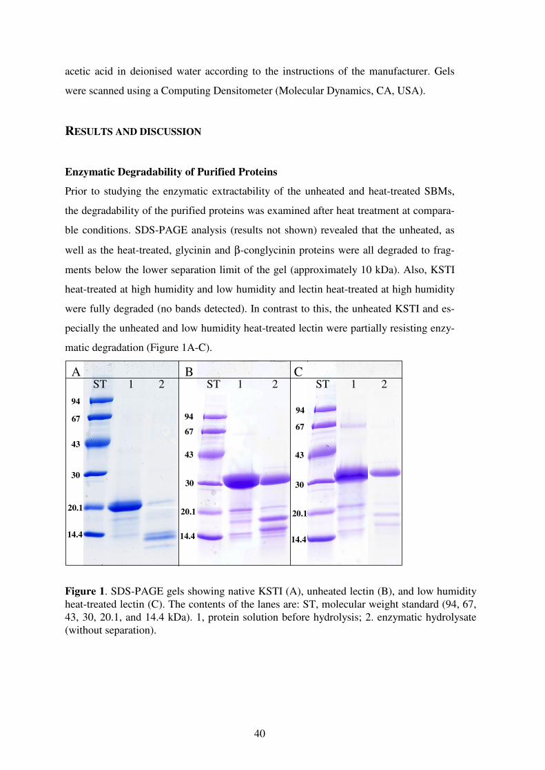
40
acetic acid in deionised water according to the instructions of the manufacturer. Gels
were scanned using a Computing Densitometer (Molecular Dynamics, CA, USA).
RESULTS AND DISCUSSION
Enzymatic Degradability of Purified Proteins
Prior to studying the enzymatic extractability of the unheated and heat-treated SBMs,
the degradability of the purified proteins was examined after heat treatment at compara-
ble conditions. SDS-PAGE analysis (results not shown) revealed that the unheated, as
well as the heat-treated, glycinin and β-conglycinin proteins were all degraded to frag-
ments below the lower separation limit of the gel (approximately 10 kDa). Also, KSTI
heat-treated at high humidity and low humidity and lectin heat-treated at high humidity
were fully degraded (no bands detected). In contrast to this, the unheated KSTI and es-
pecially the unheated and low humidity heat-treated lectin were partially resisting enzy-
matic degradation (Figure 1A-C).
94
67
43
30
20.1
14.4
A B CST 1 2 ST ST1 12 2
14.4
20.1
30 30
20.1
14.4
43 43
6767
9494
Figure 1. SDS-PAGE gels showing native KSTI (A), unheated lectin (B), and low humidity heat-treated lectin (C). The contents of the lanes are: ST, molecular weight standard (94, 67, 43, 30, 20.1, and 14.4 kDa). 1, protein solution before hydrolysis; 2. enzymatic hydrolysate (without separation).
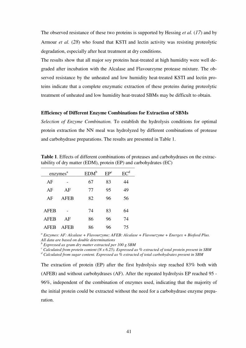
41
The observed resistance of these two proteins is supported by Hessing et al. (17) and by
Armour et al. (28) who found that KSTI and lectin activity was resisting proteolytic
degradation, especially after heat treatment at dry conditions.
The results show that all major soy proteins heat-treated at high humidity were well de-
graded after incubation with the Alcalase and Flavourzyme protease mixture. The ob-
served resistance by the unheated and low humidity heat-treated KSTI and lectin pro-
teins indicate that a complete enzymatic extraction of these proteins during proteolytic
treatment of unheated and low humidity heat-treated SBMs may be difficult to obtain.
Efficiency of Different Enzyme Combinations for Extraction of SBMs
Selection of Enzyme Combination. To establish the hydrolysis conditions for optimal
protein extraction the NN meal was hydrolyzed by different combinations of protease
and carbohydrase preparations. The results are presented in Table 1.
Table 1. Effects of different combinations of proteases and carbohydrases on the extrac-tability of dry matter (EDM), protein (EP) and carbohydrates (EC)
enzymesa EDMb EPc ECd
AF - 67 83 44
AF AF 77 95 49
AF AFEB 82 96 56
AFEB - 74 83 64
AFEB AF 86 96 74
AFEB AFEB 86 96 75 a Enzymes: AF: Alcalase + Flavourzyme; AFEB: Alcalase + Flavourzyme + Energex + Biofeed Plus.
All data are based on double determinations b Expressed as gram dry matter extracted per 100 g SBM
c Calculated from protein content (N x 6.25). Expressed as % extracted of total protein present in SBM
d Calculated from sugar content. Expressed as % extracted of total carbohydrates present in SBM
The extraction of protein (EP) after the first hydrolysis step reached 83% both with
(AFEB) and without carbohydrases (AF). After the repeated hydrolysis EP reached 95 -
96%, independent of the combination of enzymes used, indicating that the majority of
the initial protein could be extracted without the need for a carbohydrase enzyme prepa-
ration.
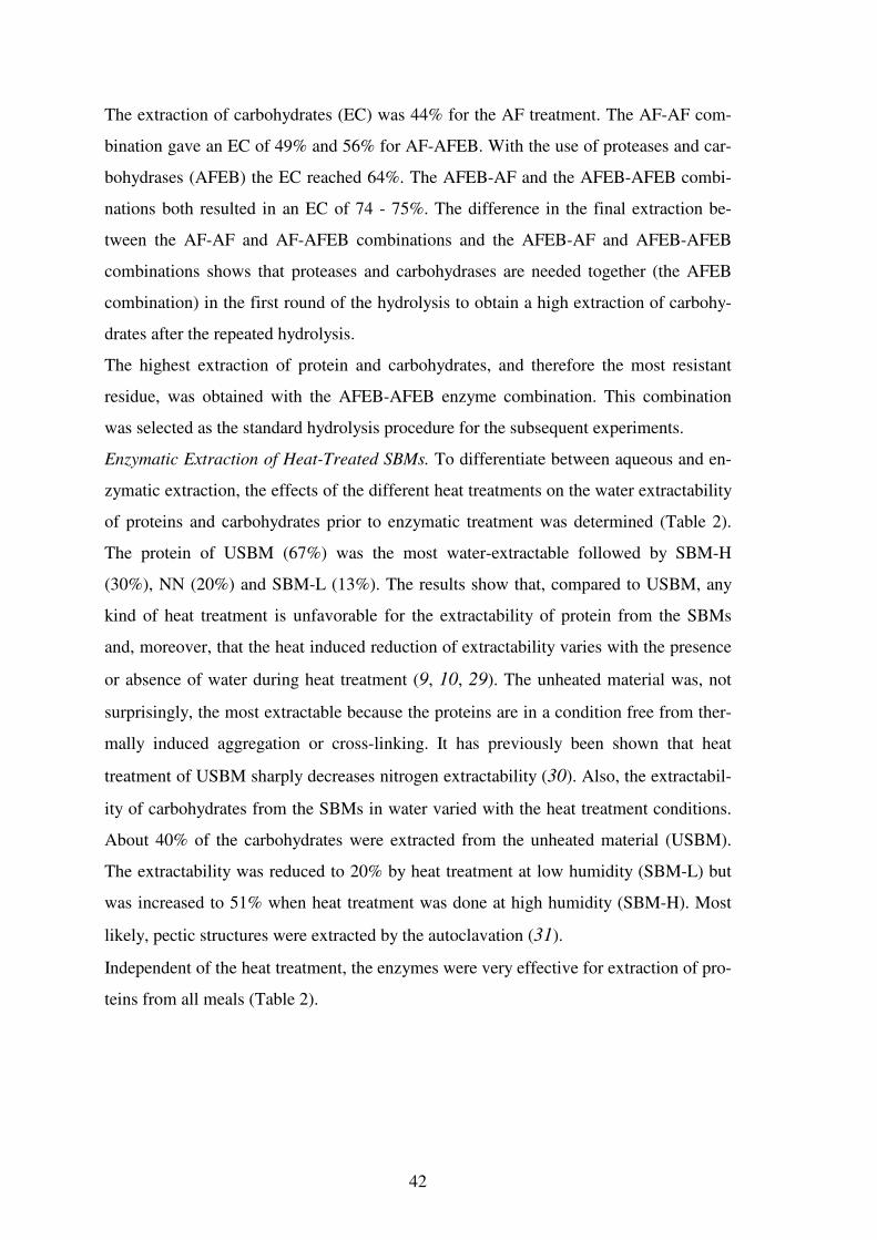
42
The extraction of carbohydrates (EC) was 44% for the AF treatment. The AF-AF com-
bination gave an EC of 49% and 56% for AF-AFEB. With the use of proteases and car-
bohydrases (AFEB) the EC reached 64%. The AFEB-AF and the AFEB-AFEB combi-
nations both resulted in an EC of 74 - 75%. The difference in the final extraction be-
tween the AF-AF and AF-AFEB combinations and the AFEB-AF and AFEB-AFEB
combinations shows that proteases and carbohydrases are needed together (the AFEB
combination) in the first round of the hydrolysis to obtain a high extraction of carbohy-
drates after the repeated hydrolysis.
The highest extraction of protein and carbohydrates, and therefore the most resistant
residue, was obtained with the AFEB-AFEB enzyme combination. This combination
was selected as the standard hydrolysis procedure for the subsequent experiments.
Enzymatic Extraction of Heat-Treated SBMs. To differentiate between aqueous and en-
zymatic extraction, the effects of the different heat treatments on the water extractability
of proteins and carbohydrates prior to enzymatic treatment was determined (Table 2).
The protein of USBM (67%) was the most water-extractable followed by SBM-H
(30%), NN (20%) and SBM-L (13%). The results show that, compared to USBM, any
kind of heat treatment is unfavorable for the extractability of protein from the SBMs
and, moreover, that the heat induced reduction of extractability varies with the presence
or absence of water during heat treatment (9, 10, 29). The unheated material was, not
surprisingly, the most extractable because the proteins are in a condition free from ther-
mally induced aggregation or cross-linking. It has previously been shown that heat
treatment of USBM sharply decreases nitrogen extractability (30). Also, the extractabil-
ity of carbohydrates from the SBMs in water varied with the heat treatment conditions.
About 40% of the carbohydrates were extracted from the unheated material (USBM).
The extractability was reduced to 20% by heat treatment at low humidity (SBM-L) but
was increased to 51% when heat treatment was done at high humidity (SBM-H). Most
likely, pectic structures were extracted by the autoclavation (31).
Independent of the heat treatment, the enzymes were very effective for extraction of pro-
teins from all meals (Table 2).
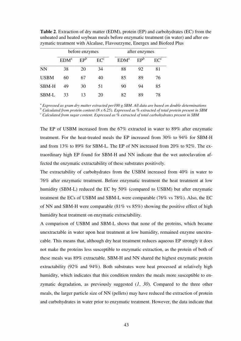
43
Table 2. Extraction of dry matter (EDM), protein (EP) and carbohydrates (EC) from the unheated and heated soybean meals before enzymatic treatment (in water) and after en-zymatic treatment with Alcalase, Flavourzyme, Energex and Biofeed Plus
before enzymes after enzymes
EDMa EPb ECc EDMa EPb ECc
NN 38 20 34 88 92 81
USBM 60 67 40 85 89 76
SBM-H 49 30 51 90 94 85
SBM-L 33 13 20 82 89 78
a Expressed as gram dry matter extracted per100 g SBM. All data are based on double determinations b Calculated from protein content (N x 6.25). Expressed as % extracted of total protein present in SBM
c Calculated from sugar content. Expressed as % extracted of total carbohydrates present in SBM
The EP of USBM increased from the 67% extracted in water to 89% after enzymatic
treatment. For the heat-treated meals the EP increased from 30% to 94% for SBM-H
and from 13% to 89% for SBM-L. The EP of NN increased from 20% to 92%. The ex-
traordinary high EP found for SBM-H and NN indicate that the wet autoclavation af-
fected the enzymatic extractability of these substrates positively.
The extractability of carbohydrates from the USBM increased from 40% in water to
76% after enzymatic treatment. Before enzymatic treatment the heat treatment at low
humidity (SBM-L) reduced the EC by 50% (compared to USBM) but after enzymatic
treatment the ECs of USBM and SBM-L were comparable (76% vs 78%). Also, the EC
of NN and SBM-H were comparable (81% vs 85%) showing the positive effect of high
humidity heat treatment on enzymatic extractability.
A comparison of USBM and SBM-L shows that none of the proteins, which became
unextractable in water upon heat treatment at low humidity, remained enzyme unextra-
cable. This means that, although dry heat treatment reduces aqueous EP strongly it does
not make the proteins less susceptible to enzymatic extraction, as the protein of both of
these meals was 89% extractable. SBM-H and NN shared the highest enzymatic protein
extractability (92% and 94%). Both substrates were heat processed at relatively high
humidity, which indicates that this condition renders the meals more susceptible to en-
zymatic degradation, as previously suggested (1, 30). Compared to the three other
meals, the larger particle size of NN (pellets) may have reduced the extraction of protein
and carbohydrates in water prior to enzymatic treatment. However, the data indicate that

44
the difference in particle size did not prevent enzymatic extraction of proteins from this
meal. This observation confirms the efficiency of the enzyme combination for protein
extraction.
Heat Denaturation and Enzymatic Extraction
The extent of protein denaturation of the meals after heat treatment was monitored by
DSC. The DSC thermograms of the four SBMs are shown in Figure 2.
Temperature (°C)
60 70 80 90 100
Hea
t F
low
(A
.U.)
A
B
C
D
Figure 2. DSC-thermograms of soybean meal suspensions (50 mg protein / mL) in 35 mM potassium phosphate buffer containing 0.1 M NaCl (pH 7.6), I = 0.2 M. A, USBM; B, NN; C, SBM-L; D, SBM-H. Scanning rate was 1.2 K min-1.
The curve of USBM shows two endothermic transitions with peak temperatures (Tp) of
74 °C and 95 °C corresponding to denaturation of the major proteins, 7S β-conglycinin
and 11S glycinin, respectively (32, 33). Whereas the SBM-H (high humidity) resulted in
complete denaturation of both 7S and 11S globulins, incomplete denaturation was found
after low humidity heat treatment (SBM-L). Compared to that of USBM, the β-
conglycinin peak area of NN and SBM-L were reduced by 79% and 86%, respectively.
Compared to USBM the glycinin peak area of NN was reduced by 18%; the glycinin
45
peak area of SBM-L was unchanged after the heat treatment. The absence of transition
peaks in SBM-H shows the efficient, irreversible protein unfolding occurring during
high humidity heat treatment.
Comparison of the enzymatic extractability of the four meals presented in Table 2 and
the denaturation state of the proteins suggests a correlation between the reduced enzy-
matic extractability of the proteins and the observed incomplete protein denaturation.
The protein of the fully heat denatured meal (SBM-H) was the most enzyme extractable
(94%) and the unheated USBM and the poorly denatured SBM-L meals were the least
extractable (89%).
The previous results on purified proteins showed that they were fully degraded after ap-
propriate heat treatment at high humidity. In contrast, even after heat processing which
resulted in complete protein denaturation (SBM-H), a complete enzymatic extraction of
the meal protein (94% → 100%) was never reached. Therefore, the composition of the
enzyme unextractable residues was analysed in more detail.
Composition of Enzyme Unextractable Material
Protein. The amino acid composition of the two original meals and the obtained enzyme
unextractable residues are presented in Table 3.
The residues contained 15 - 20% protein. The amino acid compositions of the meals
were comparable to those reported in literature (7). Enzymatic extraction of NN caused
an increase in the proportion of the hydrophobic amino acids (34) in the residue. Gly,
Ala, Val, and Leu increased from 30% of total in the NN to 41% in the enzymatic resi-
due. This change was accompanied by a decrease in the proportion of hydrophilic amino
acids Lys, Arg, His (34) from 14% in NN to 10% in the residue.
The proportion of hydrophobic amino acids in the USBM increased from 30% in the
SBM to 35% in the residue. This change was accompanied by a minor decrease in the
proportion of the hydrophilic amino acids in the residue. For SBM-H the content of hy-
drophobic amino acids in the residue was 38%. The proportion of hydrophilic amino
acids decreased insignificantly. The proportion of hydrophobic and hydrophilic amino
acids for SBM-L was more or less unchanged.

46
Table 3. Protein content and amino acid composition (molar %) of the NN and USBM soybean meals (SBMs) and the four residues obtained after enzymatic extraction
SBMsa residuesa SPIb
NN USBM NN USBM SBM-H SBM-L av. SD
Proteinc 49.4 49.2 15.3 19.9 17.3 18.3 n.d. n.d.
Ala 7.5 7.4 10.1 8.3 10.1 6.7 5.96 0.27
Arg 5.7 5.5 4.0 5.4 4.0 6.6 5.73 0.43
Asxd 10.1 10.8 7.5 9.4 8.1 7.8 10.96 0.44
Cys 0.9 0.8 1.5 0.4 0.5 1.0 1.00 0.14
Glxe 12.0 13.3 7.7 11.4 7.8 7.3 16.88 0.14
Gly 8.9 8.3 11.2 9.4 10.4 9.9 7.00 0.21
His 2.3 2.2 1.5 1.7 1.8 2.7 2.21 0.26
Ile 4.5 4.4 4.7 4.6 4.8 4.9 4.74 0.34
Leu 8.1 8.4 11.8 10.4 11.4 6.2 8.12 0.34
Lys 6.2 6.5 4.6 6.6 4.7 7.0 5.44 0.44
Met 0.6 0.5 0.6 0.6 1.0 1.6 1.38 0.22
Phe 4.4 4.3 4.7 4.4 4.5 6.1 4.40 0.26
Pro 6.9 6.4 7.4 6.5 9.6 6.6 6.42 0.51
Ser 8.3 7.8 7.8 7.2 7.1 9.2 6.47 0.26
Thr 5.1 4.8 5.3 4.6 4.1 6.8 4.17 0.17
Trpf - - - - - - 0.83 0.09
Tyr 2.7 2.7 2.3 2.4 2.7 3.3 2.95 0.29
Val 5.9 5.8 7.4 6.5 7.4 6.5 5.37 0.40
All data are based on six determinations a Standard deviations were generally between 1 and 5 %, except for Asx and Glx (3-10 %) and Met (10-
20 %) b Average of thirteen isolates (SPI), adapted from Henn and Netto, 1998 (7).
c Calculated from Kjeldahl N x 6.25. Expressed in % of dry matter
d The sum of asparagine + aspartic acid
e The sum of glutamine + glutamic acid
f Fully destroyed during hydrolysis
Generally, the increased hydrophobic character of the enzymatic residues suggests that
the residues were enriched in hydrophobic amino acids or hydrophobic peptides. Previ-
ous studies on the degradability of the purified proteins showed that KSTI and lectin
might resist complete enzymatic degradation if they were not heat-treated at high hu-
midity. A comparison of the amino acid composition of KSTI and lectin (Swissprot,
Switzerland) to the amino acid composition of the residues did not indicate that the resi-
dues were enriched in either of these two proteins.

47
Carbohydrates. The carbohydrate composition of the meals and enzyme unextractable
solids was analysed as seen in Table 4.
Table 4. Carbohydrate content and molar carbohydrate composition of soybean meals (SBMs) and the four enzyme unextractable residues (RES)
Carbohydratesd
sample DMa Cb (%) Cc (g) Rha Fuc Ara Xyl Man Gal Glc UA
SBMs
NN 100.0 29.4 29.4 3 (0.8) 0 (0.1) 7 (1.8) 8 (1.8) 4 (1.1) 22 (6.5) 43 (12.8) 13 (4.4)
USBM 100.0 25.6 25.6 2 (0.5) 1 (0.2) 8 (1.0) 6 (1.3) 3 (1.1) 31 (8.2) 31 (8.3) 18 (5.1)
RES
NN 12.4 44.0 5.5 3 (0.1) 0 (0.0) 6 (0.3) 16 (0.7) 2 (0.1) 3 (0.2) 61 (3.4) 9 (0.6)
USBM 15.5 40.9 6.3 3 (0.2) 3 (0.2) 7 (0.4) 11 (0.6) 2 (0.2) 7 (0.5) 52 (3.4) 15 (1.1)
SBM-H 10.0 38.0 3.8 3 (0.1) 0 (0.0) 4 (0.1) 9 (0.3) 3 (0.1) 4 (0.1) 64 (2.5) 12 (0.5)
SBM-L 18.0 33.0 5.9 3 (0.2) 2 (0.1) 7 (0.3) 11 (0.5) 2 (0.1) 8 (0.5) 51 (3.0) 16 (1.1)
All data are based on triple determinations a Dry matter (expressed in grams of 100 g soybean meal)
b Carbohydrate content (expressed in % w/w of DM). Determined as the sum of neutral sugars + uronic acids
c Carbohydrate content (expressed in grams)
d Composition of carbohydrates, shown as molar % (yield in grams /100 g meal is shown in parentheses)
The meals were generally rich in Glc, Gal, UA, Ara and contained low levels of Man,
Rha and Fuc. Compared to NN, USBM had 25% more uronic acid, 30% more Gal and
25% less Glc. The enzymatic treatment of the meals extracted a large proportion of the
carbohydrates. Compared to that of the meals the absolute carbohydrate content of the
residues was reduced by 75 - 85%. After enzymatic extraction, Glc was the most abun-
dant carbohydrate constituent in the enzymatic residues (51% to 64%), followed by UA
and Xyl. The level of Gal and Ara was low in all residues showing that arabinans and
arabinogalactans were well extracted. Little change was detected for the Rha, Fuc, and
Man content of the meals and residues.
The residues of NN and SBM-H had the highest proportion of Glc. The proportion of
cellulosic and non-cellulosic glucose was determined for the meal and enzymatic resi-
due of NN. Approximately 36% of the Glc in the meal originated from cellulose (4) and
more than 90% of the Glc in the residue was cellulose showing that an estimated, 20%
of the initial cellulose had been extracted during the enzymatic treatment. The results

48
show that the cellulose was only extracted to a minor extent by the cellulases of the two
carbohydrase preparations.
In conclusion, the enzymatic residues can be divided into two groups: 'high humidity'
and 'unheated + low humidity', based on the conditions for heat treatment. High-
humidity heat treatment leads to a more effective enzymatic extraction. However, in all
cases the protein extraction from the meals was incomplete and, taking into account the
results of the purified proteins, it is clear that the soybean meal matrix affects the enzy-
matic extraction of protein from the meals. We speculate that interactions between pro-
tein and other components e.g. the cellulose, which a major component of the residues)
may explain the incomplete enzymatic extractability of protein from the meals. This is a
topic of future investigation.
ACKNOWLEDGEMENTS
This research was financially supported by Danish Academy of Technical Sciences,
(Lyngby, Denmark). Gerrit van Koningsveld is thanked for helpful suggestions to the
manuscript. Jolan de Groot and Jan Cozijnzen are thanked for help with protein purifi-
cation and carbohydrate analysis. Ina Nørgaard from Novozymes A/S is thanked for
skillful assistance with the amino acid analysis.
ABBREVIATIONS USED
A, Alcalase; ANFs, antinutritional factors; B, Biofeed Plus; DM, dry matter; DSC, dif-
ferential scanning calorimetry; E, Energex; F, Flavourzyme; KSTI, Kunitz trypsin in-
hibitor; SBM, soybean meal; SBM-H, soybean meal heat-treated at high humidity;
SBM-L, soybean meal heat-treated at low humidity; EC, extraction of carbohydrate;
EDM, extraction of dry matter; EP, extraction of protein; Tp, transition temperature;
USBM, unheated soybean meal

49
REFERENCES
1. Marsman,G.J.P.; Gruppen,H.; Mul,A.J.; Voragen,A.G.J. In-vitro accessibility of un-
treated, toasted, and extruded soybean meals for proteases and carbohydrases. J Ag-
ric Food Chem 1997, 45, 4088-4095.
2. Liener,I.E. Implications of antinutritional components in soybean foods. Crit Rev
Food Sci Nutr 1994, 34, 31-67.
3. Voragen ,A.G.J.; Beldman,G.; Schols,H.A. Chemistry and enzymology of pectins. In
Advanced Dietary Fibre Technology; McCleary B.V.; Prosky,L., Eds.; Blackwell
Science: Oxford, UK, 2000; 379-396.
4. Huisman,M.M.H.; Schols,H.A.; Voragen,A.G.J. Cell wall polysaccharides from
soybean (Glycine max.) meal. Isolation and characterisation. Carbohydr Polym
1998, 37, 87-95.
5. Carpita,N.C.; Gibeaut,D.M. Structural models of primary cell walls in flowering
plants: consistency of molecular structure with the physical properties of the walls
during growth. Plant J 1993, 3, 1-30.
6. Camire,M.E. Protein functionality modification by extrusion cooking. J Am Oil
Chem Soc 1991, 68, 200-205.
7. Henn,R.L.; Netto,F.M. Biochemical-characterization and enzymatic-hydrolysis of
different commercial soybean protein isolates. J Agric Food Chem 1998, 46, 3009-
3015.
8. Marsman,G.J.P.; Gruppen,H.; de Groot,J.; Voragen,A.G.J. Effect of toasting and
extrusion at different shear levels on soy protein interactions. J Agric Food Chem
1998, 46, 2770-2777.
9. Araba,M.; Dale,N.M. Evaluation of protein solubility as an indicator of overprocess-
ing soybean meal. Poult Sci 1990, 69, 76-83.
10. Araba,M.; Dale,N.M. Evaluation of protein solubility as an indicator of underproc-
essing of soybean meal. Poult Sci 1990, 69, 1749-1752.
11. Clatterbuck,K.L.; Kehrberg,N.L.; Marable,N.L. Solubility and in vitro digestibility
of soy flours, concentrates, and isolates. J Food Sci 1980, 45, 931-935.
12. Bernardi-Don,L.S.; Pilosof,A.M.R.; Batholomai,G.B. Enzymatic modification of
soy protein concentrates by fungal and bacterial proteases. J Am Oil Chem Soc 1991,
68, 102-105.

50
13. Kim,S.Y.; Park,P.S.W.; Rhee,K.C. Functional properties of proteolytic enzyme
modified soy protein isolate. J Agric Food Chem 1990, 38, 651-656.
14. Lopez,L.M.I.; Brullo,A.; Natalucci,C.L.; Caffini,N.O.; Sorgentini,D.A.; Wagner,J.R.
Thermal behavior, solubility and structural properties of soy concentrate hydrolyzed
by new plant proteases. J Food Biochem 1998, 22, 125-141.
15. Lynch,C.J.; Rha,C.K.; Catsimpoolas,N. Tryptic hydrolysis of glycinin and its sub-
units. J Sci Food Agric 1977, 28, 971-979.
16. Shutov,A.D.; Pineda,J.; Senyuk,V.I.; Reva,V.A.; Vaintraub,I.A. Action of trypsin on
glycinin: Mixed-type proteolysis and its kinetics; molecular mass of glycinin T. Eur
J Biochem 1991, 199, 539-544.
17. Hessing,M.; Bleeker,H.; Van Biert,M.; Vlooswijk,H.A.A.; Hamer,R.J. Enzymatic
hydrolysis of soya products and analysis of antinutritional factors. Meded - Fac
Landbouwkd Toegepaste Biol Wet (Univ Gent) 1994, 59, 2257-2262.
18. Plumb,G.W.; Mills,E.N.C.; Tatton,M.J.; D'Ursel,C.C.M.; Lambert,N.; Mor-
gan,M.R.A. Effect of thermal and proteolytic processing on glycinin, the 11S globu-
lin of soy (Glycine max): a study utilizing monoclonal and polyclonal antibodies. J
Agric Food Chem 1994, 42, 834-840.
19. Vaintraub,I.A.; Yattara,H.B. Proteolysis of Kunitz soybean trypsin inhibitor, influ-
ence on its activity. J Agric Food Chem 1995, 43, 862-866.
20. Chiang,W.D.; Shih,C.J.; Chu,Y.H. Functional properties of soy protein hydrolysate
produced from a continuous membrane reactor system. Food Chem 1999, 65, 189-
194.
21. Lee,J.Y.; Lee,H.D.; Lee,C.H. Characterization of hydrolysates produced by mild-
acid treatment and enzymatic hydrolysis of defatted soybean flour. Food Res Intern
2001, 34, 217-222.
22. Lakemond,C.M.M.; Jongh,H.H.J.d.; Hessing,M.; Gruppen,H.; Voragen,A.G.J. Soy
glycinin: influence of pH and ionic strength on solubility and molecular structure at
ambient temperatures. J Agric Food Chem 2000, 48, 1985-1990.
23. Thanh,V.H.; Shibasaki,K. Major proteins of soybean seeds. A straightforward frac-
tionation and their characterization. J Agric Food Chem 1976, 24, 1117-1121.
24. Gordon,J.A.; Blumberg,S.; Lis,H.; Sharon,N. Purification of soybean agglutinin by
affinity chromatography on sepharose N-ε-aminocaproyl-β-D galacto pyrano-
sylamine. FEBS Lett 1972, 24, 193-196.

51
25. AOAC. Official Methods of Analysis, 13th ed., Association of Official Analytical
Chemists: Washington DC, USA, 1988.
26. Englyst,H.N.; Cummings,J.H. Simplified method for the measurement of total non-
starch polysaccharides by gas-liquid chromatography of constituent sugars as alditol
acetates. Analyst 1984, 109, 937-942.
27. Thibault,J.F. An automated method for the determination of pectic substances. Le-
bensm Wiss Tech 1979, 12, 247-251.
28. Armour,J.C.; Perera,R.L.C.; Buchan,W.C.; Grant,G. Protease inhibitors and lectins
in soya beans and effects of aqueous heat-treatment. J Sci Food Agric 1998, 78, 225-
231.
29. Mohammed,Z.H.; Hill,S.E.; Mitchell,J.R. Covalent cross-linking in heated protein
systems. J Food Sci 2000, 65, 221-226.
30. Marsman,G.J.P.; Gruppen,H.; Van der Poel,A.F.B.; Huisman,J.; Saini,H.S. Effect of
extrusion on the in vitro digestibility of toasted and untoasted soybean meal. In Re-
cent Advances of Research in Antinutritional Factors in Legume Seeds. Proceedings
of the Second International Workshop; Wageningen Press: Wageningen, The Neth-
erlands, 1993; 461-465.
31. Fry,S.C. The growing plant cell wall: Chemical and metabolic analysis, Longmann
Scientific and Technical: New York, USA, 1988.
32. Hermansson,A.M. Physico-chemical aspects of soy proteins structure formation. J
Texture Stud 1978, 9, 33-58.
33. Renkema,J.M.S.; Lakemond,C.M.M.; de Jongh,H.H.J.; Gruppen,H.; van Vliet,T.
The effect of pH on heat denaturation and gel forming properties of soy proteins. J
Biotechnol 2000, 79, 223-230.
34. Creighton,T.E. Proteins. Structures and Molecular Properties, 2nd ed.; W. H.
Freeman and Company: New York, USA, 1996.

52
53
CHAPTER 3
OPTIMISING CONDITIONS FOR HYDROLYSIS OF SOYBEAN MEAL PROTEINS
WITH EXO- AND ENDO-PROTEINASE PREPARATIONS ♣
♣
Fischer,M.; Voragen,A.G.J.; Kofod,L.V.; de Rooij,G.; Gruppen,H. Optimising conditions for hydrolysis of soybean meal proteins with exo- and endo-proteinase preparations. Submitted 2006.

54
ABSTRACT
With the purpose of obtaining a high solubilisation of protein and/or high degree of hy-
drolysis from soybean meal by two commercial protease preparations the effects of time,
pH and dose of the two enzyme products were studied. Protein solubilisation was favored
by a relatively high pH (7.5) reflecting the pH optimum of the endo-proteinase and em-
phasizing the importance of this type of enzyme to reach a final high level of protein
solubilisation. Experiments indicated that the efficacy of the hydrolysis is to some extent
negatively affected by the degradation products of the proteins. The ratio of the endo/exo
proteases in mixture was found to be an important parameter, which could affect both
protein solubilisation and degree of hydrolysis in both negative and positive direction. A
combined objective of high protein solubilisation and high degree of hydrolysis in a sin-
gle hydrolysis product may be conflicting.
Finally, two models were developed in order to predict protein solubilisation and degree
of hydrolysis as function of dose of the proteases. The protein solubilisation model was
tested and results indicated a slight improvement of solubilisation using the prediction of
the model although further optimization is needed. Models of this kind could have a po-
tential in commercial production of enzyme hydrolysed vegetable proteins to control
development of protein solubilisation or degree of hydrolysis depending on the objective
of the hydrolysis.
Keywords: degree of hydrolysis; endo-proteinase; exo-proteinase, protein; solubilisation,
soybean meal,

55
INTRODUCTION
Soybean is a rich source of protein and is, therefore, widely used as an ingredient in
foods and as a protein source for animal feed. Appropriate processing of the soybean to
produce soybean meal (SBM) is crucial for the final quality and nutritional value of soy
proteins. Different heat treatments may explain differences in protein solubility of
SBMs, but also differences in processing procedures for dehulling and flaking condi-
tions are known to affect the solubility of the proteins and their enzymatic degradability.
Toasting of soybean meal results in completely different protein properties compared to
pelleting and extrusion (1, 2, 3, 4, 5). The Protein Dispersibility Index (PDI) is often
used to determine the solubility of vegetable protein preparations (6, 7, 8). The PDI for
raw materials is generally between 70 and 90% and decreases generally to about 50%
after 20 min at 85°C (2).
The in vitro degradability and in vivo digestibility of processed soy proteins is increased
by enzymes. Enzymes have multiple advantages compared to other agents including a
high specificity, effectiveness at low concentrations and under mild conditions. In addi-
tion, being in general considered as safe, it is not necessary to remove the enzymes from
the finished products (9, 10).
The solubilisation of protein from defatted SBM by enzymes is generally between 84%
and 90% (11). Of the different soy proteins β-conglycinin is in generally readily de-
graded enzymatically, whereas glycinin shows more resistance to proteolysis (3, 12).
The basic polypeptides of glycinin are less readily degraded than the acidic polypeptides
(3). The relatively high resistance of the basic polypeptides against proteolytic attack is
possibly due to the tendency of these polypeptides to from large insoluble complexes
(13) that render them less prone to enzymatic hydrolysis (14).
Among the protease preparations that are commonly used for enzymatic hydrolysis of
vegetable proteins are Alcalase and Flavourzyme (15, 16). Although Alcalase, derived
from Bacillus sp., is most active at slight alkaline pH, the enzyme remains fairly active
down to ~pH 5. Most food/feed systems, including soybean meals, have a neutral or
slightly acidic pH. In most of these systems Alcalase still has an activity of 80% or
higher (17). Flavourzyme is derived from Aspergillus oryzae (15) and has a pH opti-
mum of about 5-7 (9), hence a high activity in most food/feed systems.
The effects of Alcalase and Flavourzyme on protein solubilisation from SBMs have
been previously subject of study (18). The initial pH of hydrolysis was pH 7 but during

56
enzymatic processing the pH was allowed to float freely resulting in a gradual pH de-
crease to about pH 6.5. Upon incubation, the protein solubilisation reached 83% of the
total protein present in the meal. By reincubating the residue obtained from the first hy-
drolysis with fresh enzymes the total protein solubilisation reached 95%. The results
indicated an inhibition or inactivation of protease activity and it was hypothesized that
components inherently present in the meal or proteolytic degradation products of the
SBM protein were affecting the activity of the proteases (18).
Since enzymatic solubilisation of protein from SBM is influenced by several parame-
ters, including quality of the substrate, choice of enzymes, doses and ratios of the en-
zyme products and various other conditions such as the pH applied in the process it ap-
pears that a one-fits- all solution is improbable. In addition, the economical perspective
(cost-benefit) of the process is often limiting to the amount of enzymes which can be
used in industrial protein hydrolysis.
Our specific objectives were to enhance the solubilisation of protein from SBM using
two commercial protease preparations and to determine the effects of time, pH and en-
zyme doses on the efficacy of the solubilisation and degree of hydrolysis in a single step
hydrolysis of SBM protein using Alcalase and Flavourzyme.
MATERIALS
Substrates. The soybean meal used was a pelletised SBM processed in a local pilot
plant. The meal was conditioned at 68°C and pelletised at about 85 °C for 10 min. The
meal contained 42.7% crude protein (N x 6.25).
A casein isolate (Merck, Germany) containing 89.2% crude protein according to the
manufacturer was used for protease inhibition studies.
Enzymes. Two commercial protease preparations were selected for the experiments.
Alcalase Food grade (Alcalase) is a protease preparation from Bacillus licheniformis
composed of mainly endoproteinases. Its main component is Subtilisin A. The product
has an activity optimum from pH 6.5−8.5. Flavourzyme (Flavourzyme) is a protease
complex derived from Aspergillus oryzae. Flavourzyme contains several proteolytic ac-

57
tivities, mainly endoproteinases and exopeptidases and has an activity optimum from
pH 5−7 (18). Both enzymes were from Novozymes A/S (Bagsvaerd, Denmark).
METHODS
Protein Hydrolysis
The efficacy of protein solubilisation from the SBM by Alcalase and Flavourzyme was
optimized in a series of experiments. The first experiments were conducted on a rela-
tively large scale. Later experiments were downscaled to ease sample handling.
Chloramphenicol (0.3 mM) was added to all SBM preparations to reduce the influence
of proteolytic activities originating from microbial contaminants in the meals.
General Hydrolysis Procedure. Unless stated otherwise, SBM was suspended in water
in a ratio of 1:9 (w/w). After stirring for 1h the pH was adjusted to 7.0, which is about
the natural pH of the soybean meal suspended in water. A representative sample was
taken while stirring. The suspension was transferred to a water bath (40°C) to equili-
brate for 30 min before enzymes were added (pH was still ~7). Different doses of the
two enzyme preparations were applied in separate experiments. Enzymes dosages were
calculated as percentage of enzyme solution relative to approximate amount of protein
in the samples (v/w). Different enzyme/substrate combinations were used (details under
results). After enzyme addition the hydrolysis proceeded for 16h with constant stirring
(300 rpm). When the incubation was completed the pH was recorded and all samples
were adjusted back to pH 7 prior to separation by centrifugation (8000g; 4°C; 25 min-
utes). Supernatants were stored at -20°C and residues were washed with Millipore water
in a ratio of 1:9 (w/w). Residues were recovered by centrifugation (8000g; 4°C; 25 min-
utes) and were freeze-dried. The protein content of the residues and the degree of hy-
drolysis of the solubilised proteins were subsequently determined (see analyses).
Time Optimization. To optimize the incubation time, 50 g of SBM in 450 g water was
treated according to General Hydrolysis Procedure using 2.5% Alcalase and 5% Fla-
vourzyme. After enzyme addition aliquots (10 mL) were taken from the suspension at
10, 20, 30, 40, 60, 90, 120, 150, 180, 210, 240, 300, 360, 420, and 1200 min. Immedi-

58
ately after sampling, the enzymes in the aliquots were inactivated by heat treatment
(90°C; 15 min) This is sufficient to inactivate proteolytic activities in the protease
preparations (19, 20). Further separation of the samples by centrifugation and washing
of the residues were as described under General Hydrolysis Procedure. Experiments
were conducted in duplicate.
pH Optimization. The effect of fluctuating pH around neutral (pH 7) was studied for the
narrow range between 6.5 and 7.5. SBM (10g) was suspended in 90 mL MilliQ water.
The pH was adjusted using 1M NaHCO3. After adjustment the pH remained constant
within 0.1 pH unit over the entire incubation period. The pH was adjusted to 7 prior to
separation of the soluble material fraction from the solid particles by centrifugation.
Further treatment of the samples was as described under General Hydrolysis Procedure
and protein solubilisation and degree of hydrolysis was determined. All pH experiments
were conducted in triplicate.
Sequential Hydrolysis: To study the stability of protease activities the effect of a stan-
dard hydrolysis of the SBM by Alcalase and Flavourzyme was compared to a sequential
hydrolysis procedure. For the “standard” treatment SBM (50 g) was suspended in 450 g
water and enzymes (2.5% Alcalase and 5% Flavourzyme) were added. The conditions
for the “sequential” treatment were as “standard” during the first 10h but with another
(equal) dose of the two enzymes added at 10h. The start pH for hydrolysis was 7 and
hydrolysis and separation was performed essentially as described under General Hy-
drolysis Procedure. Aliquots were collected at 10 and 20 h. Experiment was conducted
in triplicate.
Effect of Soluble Peptides on Efficacy of Protein Solubilisation: The effect of SBM deg-
radation products released into the supernatant by the two protease preparations on pro-
tease efficacy was studied. Casein isolate, generally known as a readily hydrolysable
substrate (21), and SBM were used as substrates. A supernatant produced according to
General Hydrolysis Procedure (2.5% Alcalase and 5% Flavourzyme) was isolated by
centrifugation and inactivated by heat treatment (90°C; 15 min) as previously described.
Cooled supernatant or water (45 mL) was mixed with either casein (5 g) or SBM (5 g)
before Alcalase (2.5%) and Flavourzyme (5%) were added to the samples. The start pH
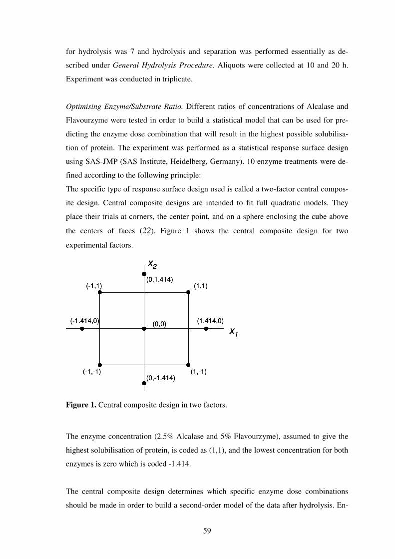
59
for hydrolysis was 7 and hydrolysis and separation was performed essentially as de-
scribed under General Hydrolysis Procedure. Aliquots were collected at 10 and 20 h.
Experiment was conducted in triplicate.
Optimising Enzyme/Substrate Ratio. Different ratios of concentrations of Alcalase and
Flavourzyme were tested in order to build a statistical model that can be used for pre-
dicting the enzyme dose combination that will result in the highest possible solubilisa-
tion of protein. The experiment was performed as a statistical response surface design
using SAS-JMP (SAS Institute, Heidelberg, Germany). 10 enzyme treatments were de-
fined according to the following principle:
The specific type of response surface design used is called a two-factor central compos-
ite design. Central composite designs are intended to fit full quadratic models. They
place their trials at corners, the center point, and on a sphere enclosing the cube above
the centers of faces (22). Figure 1 shows the central composite design for two
experimental factors.
x1
x2
(-1.414,0) (1.414,0)
(0,1.414)
(0,-1.414)
(1,1)(-1,1)
(-1,-1) (1,-1)
(0,0)x
1
x2
(-1.414,0) (1.414,0)
(0,1.414)
(0,-1.414)
(1,1)(-1,1)
(-1,-1) (1,-1)
(0,0)
Figure 1. Central composite design in two factors.
The enzyme concentration (2.5% Alcalase and 5% Flavourzyme), assumed to give the
highest solubilisation of protein, is coded as (1,1), and the lowest concentration for both
enzymes is zero which is coded -1.414.
The central composite design determines which specific enzyme dose combinations
should be made in order to build a second-order model of the data after hydrolysis. En-
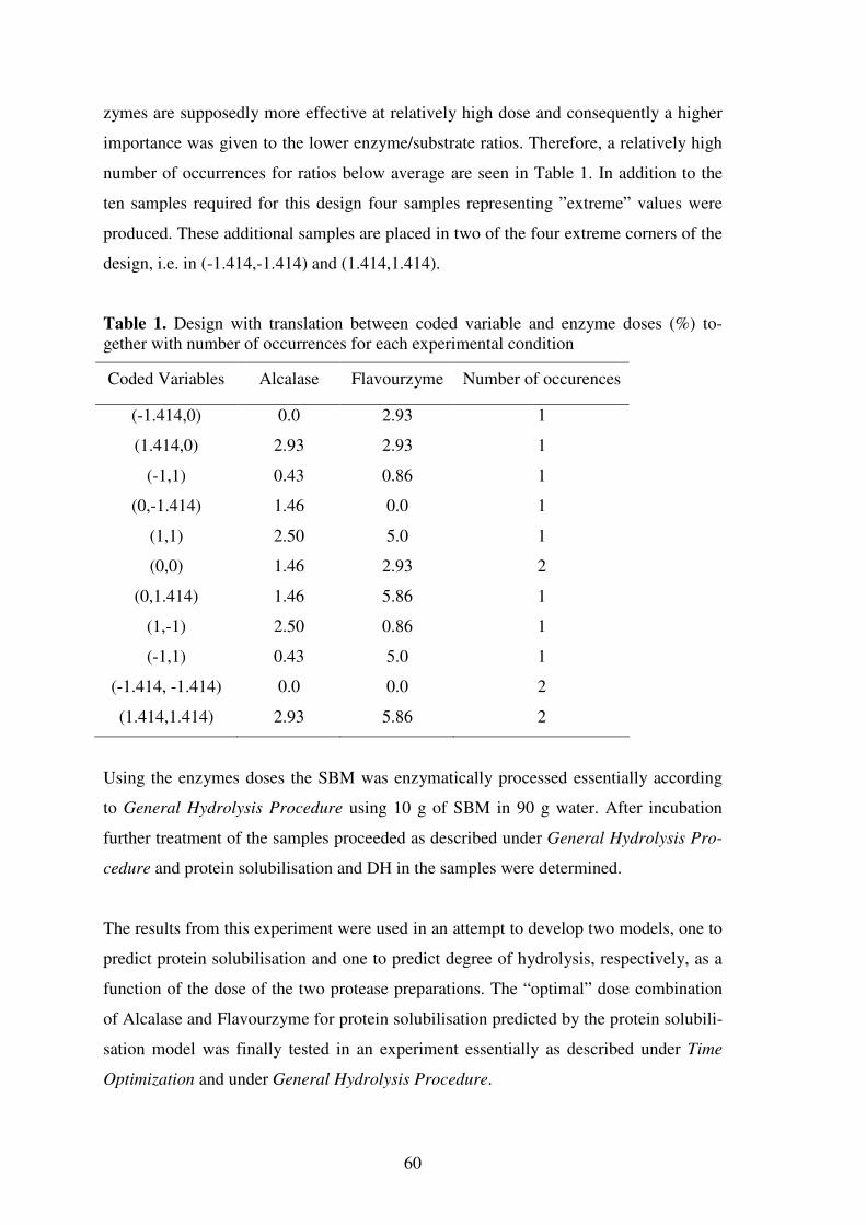
60
zymes are supposedly more effective at relatively high dose and consequently a higher
importance was given to the lower enzyme/substrate ratios. Therefore, a relatively high
number of occurrences for ratios below average are seen in Table 1. In addition to the
ten samples required for this design four samples representing ”extreme” values were
produced. These additional samples are placed in two of the four extreme corners of the
design, i.e. in (-1.414,-1.414) and (1.414,1.414).
Table 1. Design with translation between coded variable and enzyme doses (%) to-gether with number of occurrences for each experimental condition
Coded Variables Alcalase Flavourzyme Number of occurences
(-1.414,0) 0.0 2.93 1
(1.414,0) 2.93 2.93 1
(-1,1) 0.43 0.86 1
(0,-1.414) 1.46 0.0 1
(1,1) 2.50 5.0 1
(0,0) 1.46 2.93 2
(0,1.414) 1.46 5.86 1
(1,-1) 2.50 0.86 1
(-1,1) 0.43 5.0 1
(-1.414, -1.414) 0.0 0.0 2
(1.414,1.414) 2.93 5.86 2
Using the enzymes doses the SBM was enzymatically processed essentially according
to General Hydrolysis Procedure using 10 g of SBM in 90 g water. After incubation
further treatment of the samples proceeded as described under General Hydrolysis Pro-
cedure and protein solubilisation and DH in the samples were determined.
The results from this experiment were used in an attempt to develop two models, one to
predict protein solubilisation and one to predict degree of hydrolysis, respectively, as a
function of the dose of the two protease preparations. The “optimal” dose combination
of Alcalase and Flavourzyme for protein solubilisation predicted by the protein solubili-
sation model was finally tested in an experiment essentially as described under Time
Optimization and under General Hydrolysis Procedure.

61
ANALYSES
Dry Matter. Dry matter was defined as the weight of meals and residues immediately
after freeze-drying. The solubilisation of dry matter by the different treatment was cal-
culated as: (DMtotal – DMresidue) / DMtotal x 100%.
Protein Determination. The protein content of SBM (N x 6.25) and casein (N x 6.38)
and of the different enzymatic residues was determined by a semi-automated Kjeldahl
method (23). Dry samples (100 mg) were destructed in concentrated sulfuric acid at
200-385 °C. The released NH3 was determined with an ammonia-hypochloritesalicylate
reaction on a Skalar 5101 autoanalyzer (Skalar, Breda, The Netherlands). The solubili-
sation of protein by the different treatments was calculated as: (Ptotal – Presidue) / Ptotal x
100%.
The protein contents determined have a standard deviation of about 0.2%, which should
be considered during interpretation of the results. Protein contents are the means of
three determinations.
Degree of Hydrolysis. The degree of hydrolysis (DH) of the protein hydrolysates in dif-
ferent samples was determined using an semi-automated micro titer plate based colori-
metric OPA method (24). The OPA reagent was prepared as follows: 7.620 g di-sodium
tetraborate decahydrate and 200 mg sodium dodecyl sulphate (SDS) were dissolved in
150 mL deionised water. 160 mg o-phthaldialdehyde 97% (OPA) was dissolved in 4
mL ethanol. The OPA solution was transferred quantitatively to the above-mentioned
solution. 176 mg dithiothreitol 99% (DTT) was added to the solution that was made up
to 200 mL with deionised water. A serine standard (0.9516 meqv/L) was prepared by
solubilizing 50 mg serine (Merck, Germany) in 500 mL deionized water.
The sample solution was prepared by diluting each sample to an absorbance (280 nm)
of about 0.5. Generally, supernatants were diluted (100 ×) using an automated Tecan
dilution station (Männedorf, Schwitzerland). All other spectrophotometer readings were
performed at 340 nm using deionized water as the control. Aliquots (25 µL), were dis-
pensed into a micro titer plate. The micro titer plate was inserted into an iEMS MF
reader (Labsystems, Finland) and 200µL of OPA reagent was automatically dispensed.
Plates were shaken (2 min; 700 rpm) before measuring absorbance at 340 nm. Finally,

62
the DH (total number of peptide bonds htot=7.8 (17) ) was calculated using L-serine for
the standard curve (25). Eightfold determination of all samples was carried out.
RESULTS AND DISCUSSION
Effects of Time
The time course of solubilisation of protein, dry matter and degree of hydrolysis is pre-
sented in Figure 2.
Time (min)
0 200 400 600 800 1000 1200
Pro
tein
and d
ry m
att
er
solu
bili
sation (
%)
0
20
40
60
80
100
Degre
e o
f hyd
roly
sis
(%)
Figure 2. Solubilisation of protein ( ), dry matter ( ) and development of DH ( ) plotted against time (n=2). Enzyme dose, 2.5% Alcalase and 5% Flavourzyme.
The figure shows that enzymatic solubilisation of the protein progresses rapidly during
the first hours of the hydrolysis. At the start of the hydrolysis ~20% of the protein in the
SBM was water soluble, whereas 10 min after addition of the proteases 59% of protein
was soluble and after 60 min solubilisation exceeded 70%. At 240 min the solubilisation
had reached 80% increasing to 86% at 420 min. Maximum solubilisation of protein
(90%) was reached at 1200 min, indicating that proteases were still active at the end of
the experiment. However, between 420 and 1200 min only a limited amount (4%) of
protein was solubilised. A decrease of pH (results not shown) from pH 6.4 to 6.1 was
63
observed between 800 and 1200 min. This pH decrease could partly be explained by the
increased DH.
In contrast to the rapid solubilisation of protein from the beginning the degree of hy-
drolysis increased at a more moderate rate. In the time span from 10 min to 1200 min
DH increased steadily from DH 13 to 66 showing that development of very high DH in
protein hydrolysates is a relatively time demanding process.
From the data it is obvious that the rate of proteolytic solubilisation of protein proceeds
very fast in the early phase of the incubation but becomes steadily slower over time.
This could indicate either a gradual inhibition or inactivation of proteolytic activities by
degradation products released over time or/and that the remaining fraction of insoluble
SBM protein bebecomes increasingly more difficult to access for the proteases (3, 18,
26). Due to the increasing risk of microbial contamination over time it was not consid-
ered relevant to increase the incubation time further. Therefore, subsequent experiments
were focused on the effects on protein solubilisation and degree of hydrolysis by higher
enzyme dose and optimized pH conditions to better match the pH optima of the differ-
ent activities of the protease preparations.
Optimization of pH
A study was conducted to compare the influence of pH on the protein solubilising ef-
fect of Alcalase and Flavourzyme in the narrow pH range between 6.5 and 7.5. The re-
sults are presented in Table 2.
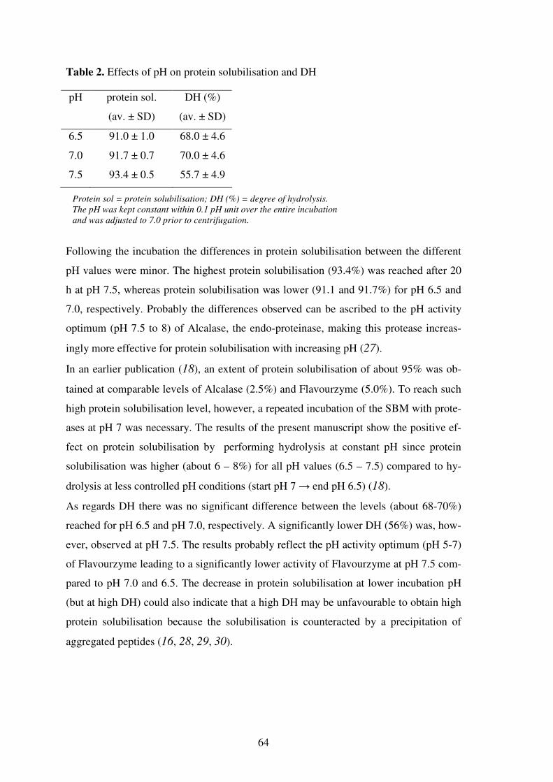
64
Table 2. Effects of pH on protein solubilisation and DH
pH protein sol.
(av. ± SD)
DH (%)
(av. ± SD)
6.5 91.0 ± 1.0 68.0 ± 4.6
7.0 91.7 ± 0.7 70.0 ± 4.6
7.5 93.4 ± 0.5 55.7 ± 4.9
Protein sol = protein solubilisation; DH (%) = degree of hydrolysis.
The pH was kept constant within 0.1 pH unit over the entire incubation
and was adjusted to 7.0 prior to centrifugation.
Following the incubation the differences in protein solubilisation between the different
pH values were minor. The highest protein solubilisation (93.4%) was reached after 20
h at pH 7.5, whereas protein solubilisation was lower (91.1 and 91.7%) for pH 6.5 and
7.0, respectively. Probably the differences observed can be ascribed to the pH activity
optimum (pH 7.5 to 8) of Alcalase, the endo-proteinase, making this protease increas-
ingly more effective for protein solubilisation with increasing pH (27).
In an earlier publication (18), an extent of protein solubilisation of about 95% was ob-
tained at comparable levels of Alcalase (2.5%) and Flavourzyme (5.0%). To reach such
high protein solubilisation level, however, a repeated incubation of the SBM with prote-
ases at pH 7 was necessary. The results of the present manuscript show the positive ef-
fect on protein solubilisation by performing hydrolysis at constant pH since protein
solubilisation was higher (about 6 – 8%) for all pH values (6.5 – 7.5) compared to hy-
drolysis at less controlled pH conditions (start pH 7 → end pH 6.5) (18).
As regards DH there was no significant difference between the levels (about 68-70%)
reached for pH 6.5 and pH 7.0, respectively. A significantly lower DH (56%) was, how-
ever, observed at pH 7.5. The results probably reflect the pH activity optimum (pH 5-7)
of Flavourzyme leading to a significantly lower activity of Flavourzyme at pH 7.5 com-
pared to pH 7.0 and 6.5. The decrease in protein solubilisation at lower incubation pH
(but at high DH) could also indicate that a high DH may be unfavourable to obtain high
protein solubilisation because the solubilisation is counteracted by a precipitation of
aggregated peptides (16, 28, 29, 30).

65
Sequential Hydrolysis
The stability of Alcalase and Flavourzyme activity was studied by comparing the effect
of a standard hydrolysis to a sequential hydrolysis for which fresh enzymes were added
at 10h. The results are presented in Table 3.
Table 3. Protein solubilisation and DH for two conditions of hydrolysis of SBM by Al-calase and Flavourzyme
10 h 20 h
treatment protein sol.
(av. ± SD)
DH (%) protein sol.
(av. ± SD)
DH (%)
”standard” 83.0 ± 0.2 51 89.4 ± 0.1 68
”sequential” 83.0 ± 0.1 52 89.0 ± 0.5 75
Protein sol = protein solubilisation; DH (%) = degree of hydrolysis.
Sample pH: start, pH 7.0; final, pH ~6.5.
No significant differences (89.4 vs. 89.0%) in protein solubilisation was observed be-
tween the two treatments at the endpoint (20 h) of the incubation. Accordingly there
was no additional protein solubilising effect of adding fresh enzymes at 10h (sequen-
tial). The conditions for the first 10 h of incubation are identical for the treatments,
which are also reflected in the protein solubilisation level (83%) at this time.
While having no effect on protein solubilisation the sequential treatment resulted in a
DH increase of about 7% (from 68 – 75%). This is higher than the DH (66%) obtained
in Figure 2 and, since protein solubilisation was not affected by the additional dose of
the enzymes, it is speculated that the increase is primarily due to the addition of fresh
exo-acting proteolytic activities from Flavourzyme.
Even with a double dose of proteases, of which half the amount was added after 10h to
boost the reaction, the protein solubilisation in the present experiment was not increased
to a higher level. This might indicate that protease activities are to some extent affected
by degradation products of the intact soy proteins which are brought into solution by
means of the action of Alcalase and Flavourzyme (18). This hypothesis was studied fur-
ther

66
Effect of Soluble Peptides on Efficiency of Protein Solubilisation
The effects of the addition of soluble peptidesderived from one of the previous trials
(2.5% Alcalase and 5% Flavourzyme) on the enzymatic solubilisation of protein from
SBM and a casein isolate was studied. The results are presented in Table 4.
Table 4. Development of protein solubilisation and DH during hydrolysis of SBM and casein isolate by Alcalase and Flavourzyme in water and in supernatant (containing soluble SBM peptides)
10h 20 h
conditions protein sol.
(av. ± SD)
protein sol.
(av. ± SD)
DH (%)
Casein + supernatant 95.2 ± 1.1 91.8 ± 0.8 54
Casein + water 97.7 ± 0.4 96.5 ± 0.4 47
SBM + supernatant 84.0 ± 0.2 92.9 ± 0.2 68
SBM + water 86.4 ± 0.1 93.5 ± 0.1 66
Protein sol = protein solubilisation; DH (%) = degree of hydrolysis.
Protein numbers are corrected for the contribution of protein from the supernatant.
After 20 h incubation the highest solubilisation of casein (96.5%) was seen for the ca-
sein + water treatment. With a protein solubilisation of 91.8% a negative effect (4.7%)
was seen when casein was hydrolysed in the presence of soluble SBM peptides (super-
natant). Also, at 10 h this effect is seen (decrease from 97.7 to 95.2%). A higher protein
solubilisation level from the casein isolate at 10 h compared to 20 h show that this sub-
strate is readily hydrolysed and that the reaction can be optimized by reducing the reac-
tion time. This decrease could be explained by increasing aggregation of peptides be-
tween 10 and 20 h (31, 32).
Also for the SBM differences in protein solubilisation were seen between the SBM +
water and SBM + soluble SBM peptides treatments. At 10 h, protein solubilisation was
86.4 and 84% for the two treatments, respectively. After 20 h, however, protein solubi-
lisation had increased to 93.5% for the water treatment and 92.9% for the soluble SBM
peptides treatment. The final levels of protein solubilisation in this experiment were un-
usually high for the SBM hydrolysis. We have no obvious explanation for this. The re-
sults show that enzymatic solubilisation of SBM protein in water progresses at lower
rate (86.4%) compared to casein (97.7%) after 10 h. The same is true for the treatment
in the presence of soluble SBM peptides (84 vs. 95.2%). After 20 h in water, however,

67
the final difference in solubilisation between the two substrates was only 3% (casein
96.5% vs. SBM 93.5%).
From this experiment it can be concluded that the solubilisation of protein (20 h) from
casein isolate as well as SBM is to some extent negatively affected by the soluble SBM
peptides present in the supernatant. However, the earlier results on the effects of pH in-
dicated that this effect may be less pronounced or can be compensated for when pH is
set and held at 7.5.
Prediction of Protein Solubilisation
The protein solubilisation and the DH values achieved by the various combinations of
different concentrations of Alcalase and Flavourzyme (Table 1) are presented in Table
5.
Table 5. Protein solubilisation and DH for different doses of Alcalase and Flavourzyme
enzyme dose (%)
sample Alcalase Flavourzyme protein sol. DH (%)
1 0.0 2.93 84.5 41
2 2.93 2.93 90.1 57
3 0.43 0.86 86.1 31
4 1.46 0.0 82.3 15
5 2.50 5.0 85.3 62
6 1.46 2.93 83.6 48
7 1.46 5.86 85.3 66
8 2.50 0.86 82.7 41
9 1.46 2.93 89.2 52
10 0.43 5.0 88.6 59
11 0.0 0.0 20.2 0
12 0.0 0.0 19.3 0
13 2.93 5.86 84.7 67
14 2.93 5.86 86.3 68
Protein sol = protein solubilisation; DH (%) = degree of hydrolysis.
The different combinations of Alcalase and Flavourzyme resulted in a solubilisation of
protein ranging from 82 to 90%. The solubilisation of the soy protein in water prior to

68
enzyme addition was about 20% (samples 11 and 12). The highest solubilisation
(90.1%) was seen for 2.93% of both Alcalase and Flavourzyme. For Flavourzyme alone
(sample 1, 2.93%) protein solubilisation was 84.5%. Protein solubilisation by a rela-
tively small amount of only Alcalase (sample 4, 1.46%) was 82.3%. With 1.46% Alca-
lase and higher concentration of Flavourzyme (sample 9, 2.93%) protein solubilisation
increased from 82 to 89%. However, when the dose of Flavourzyme was further in-
creased to 5.86% protein solubilisation decreased to 85.3% indicating that Flavourzyme
doses above 2.93% have a negative effect on protein solubilisation (in combination with
Alcalase).
With respect to DH, the highest DH values (66-68%) were reached in the samples (7,
13, and 14) with the highest Flavourzyme dose (5.86%). When only one of the proteases
was used Flavourzyme (sample 1, 2.93%) reached a DH of 41, whereas Alcalase (sam-
ple 4, 1.46%), not surprisingly, had a limited effect (15%) on DH. In sample 3 with an
approximately 3 times lower Alcalase dose (0.43%) compared to sample 1 and a low
Flavourzyme dose (0.86%) a DH of 31 was reached making the DH increasing effect of
the exopeptidase in the combination obvious. The results clearly reflect the capability
of Flavourzyme to increase DH in protein hydrolysates (21). They also emphasize that
high doses of Flavourzyme are not necessarily beneficial to the protein solubilisation.
From the data obtained (Table 5) a protein solubilisation model was developed and the
capability of the model to predict protein solubilisation as a function of Alcalase and
Flavourzyme doses was subsequently tested. According to the surface response analysis
of SAS-JMP protein solubilisation can be predicted using the following equation (also
presented as a chart in Figure 3):
P sol = 33.6881 + 30.3628*Alc + 18.0238*Flav – 4.5961*Alc2 – 1.5565*Flav
2 –
3.2979*Alc*Flav
Where P sol = protein solubilisation (%), Alc = Alcalase dose; and Flav = Flavourzyme
dose (%). The terms Alc, Flav, Flav2, and Alc*Flav in the model are all significant. Alc
2
is not significant but is used to get a better fit of the model. The fact that the interaction
effect between Alcalase dose and Flavourzyme dose is significant indicates that effect
of one enzyme depends on the level of the other. Furthermore, the fact that the second-
order effect of Flavourzyme is significant whereas the second-order effect of Alcalase is
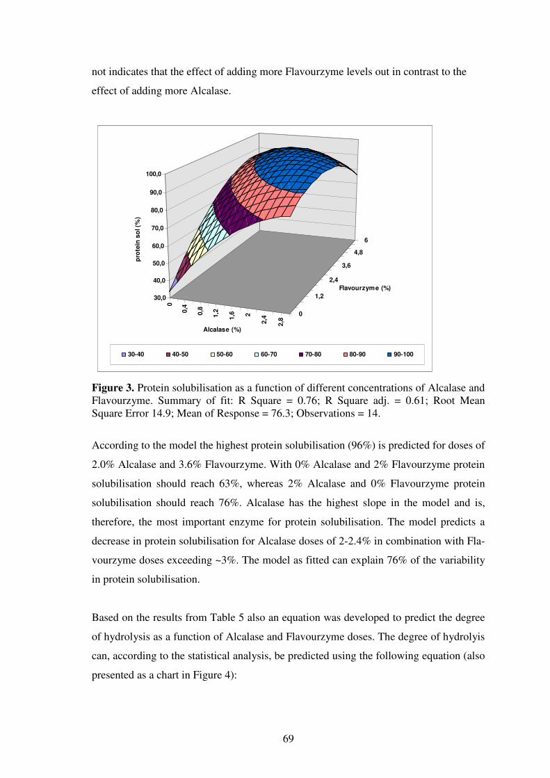
69
not indicates that the effect of adding more Flavourzyme levels out in contrast to the
effect of adding more Alcalase.
0
0,4
0,8
1,2
1,6 2
2,4
2,8
0
1,2
2,4
3,6
4,8
6
30,0
40,0
50,0
60,0
70,0
80,0
90,0
100,0
pro
tein
so
l (%
)
Alcalase (%)
Flavourzyme (%)
30-40 40-50 50-60 60-70 70-80 80-90 90-100
Figure 3. Protein solubilisation as a function of different concentrations of Alcalase and Flavourzyme. Summary of fit: R Square = 0.76; R Square adj. = 0.61; Root Mean Square Error 14.9; Mean of Response = 76.3; Observations = 14.
According to the model the highest protein solubilisation (96%) is predicted for doses of
2.0% Alcalase and 3.6% Flavourzyme. With 0% Alcalase and 2% Flavourzyme protein
solubilisation should reach 63%, whereas 2% Alcalase and 0% Flavourzyme protein
solubilisation should reach 76%. Alcalase has the highest slope in the model and is,
therefore, the most important enzyme for protein solubilisation. The model predicts a
decrease in protein solubilisation for Alcalase doses of 2-2.4% in combination with Fla-
vourzyme doses exceeding ~3%. The model as fitted can explain 76% of the variability
in protein solubilisation.
Based on the results from Table 5 also an equation was developed to predict the degree
of hydrolysis as a function of Alcalase and Flavourzyme doses. The degree of hydrolyis
can, according to the statistical analysis, be predicted using the following equation (also
presented as a chart in Figure 4):

70
DH=0.82848 + 12.8980*Alc + 17.2649*Flav –1.1603*Flav2–0.8721*Alc
2–1.5724*Alc*Flav
Where DH = Degree of Hydrolysis Alc = Alcalase dose; and Flav = Flavourzyme dose
(%).
As was the case for protein solubilisation the terms Alc, Flav, Flav2, and Alc*Flav in the
model are all significant. Alc2 is not significant but is used to get a better fit of the
model. The fact that the interaction effect between Alcalase dose and Flavourzyme dose
is significant indicates that effect of one enzyme depends on the level of the other.
0 0,4 0,8 1,2 1,6 2 2,4 2,8
0
0,8
1,6
2,4
3,2
4
4,8
5,6
0
10
20
30
40
50
60
70
Deg
ree o
f H
yd
roly
sis
(%
)
Alcalase (%)
Flavourzyme (%)
0-10 10-20 20-30 30-40 40-50 50-60 60-70
Figure 4. DH as a function of different concentrations of Alcalase and Flavourzyme. Summary of fit: R Square = 0.97; R Square adj. = 0.95; Root Mean Square Error 5.09; Mean of Response = 43.04; Observations = 14.
The figure clearly shows the larger effect of Flavourzyme on DH compared to Alcalase.
In combination with Alcalase the DH increases steadily with increasing doses of Fla-
vourzyme but the effect levers out for the higher dose region of Flavourzyme. This
could possibly be explained by an increased level of aggregation and precipitation of the
peptides released, which may in turn also affect protein solubilisation negatively. Lack
of potential hydrolysable peptide bonds may also be part of the cause. The model as fit-
ted explains 97% of the variability in the DH.

71
The chart of Figure 3 predicts that maximum protein solubilisation (96%) is reached
with doses of 2.0% Alcalase and 3.6% Flavourzyme. The time courses of solubilisation
of protein and degree of hydrolysis for these doses together with those of 2.5% Alca-
lase and 5% Flavourzyme are presented in Figure 5.
Time (min)
0 200 400 600 800 1000 1200
Pro
tein
solu
bili
sation (
%)
0
10
20
30
40
50
60
70
80
90
100
Degre
e o
f hyd
roly
sis
(%
)
Figure 5. Protein solubilisation and degree of hydrolysis plotted against time (n=2) and compared to data from Figure 2. = protein sol. for 2.0% Alcalase + 3.6% Fla-vourzyme; = protein sol. for 2.5% Alcalase + 5% Flavourzyme); = DH for 2.0% Alcalase + 3.6% Flavourzyme; = DH for 2.5% Alcalase + 5% Fla-vourzyme.
From the two protein solubilisation curves it appears that protein solubilisation pro-
ceeded somewhat faster in the beginning for 2.5% Alcalase and 5% Flavourzyme com-
pared to 2.0% Alcalase and 3.6% Flavourzyme. This is in line with the assumptions of
the model. At the endpoint of the experiment (1200 min) the solubilisation of protein by
2% Alcalase and 3.6% Flavourzyme was 92%. This is only insignificantly higher than
the 90% reached using 2.5% Alcalase and 5% Flavourzyme, but evidently not as high as
the 96% predicted by Figure 3 although it still shows that the model has potential.
The results show that protein solubilisation can potentially reach >90% in a single step
if hydrolysis conditions are set correctly and when enzyme dosing is optimized. Evi-

72
dently optimisation of SBM protein hydrolysis is a complicated topic and the final level
of either protein solubilisation or degree of hydrolysis in the end product can be largely
influenced by careful adjustment of the different process parameters. Economical con-
siderations were not part of this study but the cost of enzymes will in many cases put a
limitation to the degrees of freedom when designing the process. In this connection
models offer approximation of solubilisation of soybean proteins by different doses of
Alcalase and Flavourzyme in real conditions and could be useful tools in commercial
production of enzyme hydrolysed vegetable protein. However, due to the complexity of
the process, further evaluation of the here presented models is needed.
CONCLUSIONS
The presented data show that effective protein solubilisation is not only a matter of se-
lecting efficient endo- and exo-proteinases. The enzymes can be overdosed and the ratio
between the enzymes is equally important depending of the need for high protein solu-
bilisation or high DH in the final hydrolysed protein product. The control of pH was
found to be an important parameter for protein solubilisation as well as DH. To better
understand the limitations of the enzymes, further attention could be given to the frac-
tion of the soy proteins resisting enzymatic solubilisation.
73
REFERENCES
1. Marsman,G.J.P.; Gruppen,H.; Van der Poel,A.F.B.; Huisman,J.; Saini,H.S. Effect of
extrusion on the in vitro digestibility of toasted and untoasted soybean meal. In
Recent Advances of Research in Antinutritional Factors in Legume Seeds.
Proceedings of the Second International Workshop; Wageningen Press:
Wageningen, The Netherlands, 1993; 461-465.
2. Marsman,G.J.P.; Gruppen,H.; de Groot,J.; Voragen,A.G.J. Effect of toasting and
extrusion at different shear levels on soy protein interactions. J Agric Food Chem
1998, 46, 2770-2777.
3. Marsman,G.J.P.; Gruppen,H.; Mul,A.J.; Voragen,A.G.J. In-vitro accessibility of
untreated, toasted, and extruded soybean meals for proteases and carbohydrases. J
Agric Food Chem 1997, 45, 4088-4095.
4. Marsman,G.J.P.; Gruppen,H.; Vanzuilichem,D.J.; Resink,J.W.; Voragen,A.G.J. The
influence of screw configuration on the in-vitro digestibility and protein solubility of
soybean and rapeseed meals. J Food Eng 1995, 26, 13-28.
5. Liu,K.S. Soybeans. Chemistry, Technology, and Utilization, Chapman & Hall: New
York, US, 1997.
6. Batal,A.B.; Douglas,M.W.; Engram,A.E.; Parsons,C.M. Protein dispersibility index
as an indicator of adequately processed soybean meal. Poult Sci 2000, 79, 1592-
1596.
7. Araba,M.; Dale,N.M. Evaluation of protein solubility as an indicator of
overprocessing soybean meal. Poult Sci 1990, 69, 76-83.
8. Araba,M.; Dale,N.M. Evaluation of protein solubility as an indicator of
underprocessing of soybean meal. Poult Sci 1990, 69, 1749-1752.
9. Bernardi-Don,L.S.; Pilosof,A.M.R.; Batholomai,G.B. Enzymatic modification of
soy protein concentrates by fungal and bacterial proteases. J Am Oil Chem Soc
1991, 68, 102-105.
10. Bernardini,E. Oilseeds, oils and fats: Volume 1. Planning a factory raw materials
and extraction techniques., 2nd ed.; Roma, B.E. Publishing House: 1985.
11. Bressani,R. The role of soybeans in food systems. J Am Oil Chem Soc 1981, 58,
392-400.
12. Kim,S.Y.; Park,P.S.W.; Rhee,K.C. Functional properties of proteolytic enzyme
modified soy protein isolate. J Agric Food Chem 1990, 38, 651-656.

74
13. Wolf,W.J.; Tamura,T. Heat Denaturation of Soy Bean-D 11s Protein. Cereal Chem
1969, 46, 331-344.
14. Wolf,W.J. Soybean proteins: their functional, chemical, and physical properties. J
Agric Food Chem 1970, 18, 969-976.
15. Aaslyng,M.D.; Martens,M.; Poll,L.; Nielsen,P.M.; Flyge,H.; Larsen,L.M. Chemical
and sensory characterization of hydrolyzed vegetable protein, a savory flavoring. J
Agric Food Chem 1998, 46, 481-489.
16. Conde,J.M.; Escobar,M.D.Y.; Jimenez,J.J.P.; Rodriguez,F.M.; Patino,J.M.R. Effect
of enzymatic treatment of extracted sunflower proteins on solubility, amino acid
composition, and surface activity. J Agric Food Chem 2005, 53 , 8038-8045.
17. Adler-Nissen,J. Enzymatic hydrolysis of food proteins, Elsevier Publishing Co.:
New York, 1986.
18. Fischer,M.; Kofod,L.V.; Schols,H.A.; Piersma,S.R.; Gruppen,H.; Voragen,A.G.J.
Enzymatic extractability of soybean meal proteins and carbohydrates: heat and
humidity effects. J Agric Food Chem 2001, 49, 4463-4469.
19. Severin,S.; Xia,W.S. Enzymatic hydrolysis of whey proteins by two different
proteases and their effect on the functional properties of resulting protein
hydrolysates. J Food Biochem 2006, 30, 77-97.
20. Claver,I.P.; Zhou,H.M. Enzymatic hydrolysis of defatted wheat germ by proteases
and the effect on the functional properties of resulting protein hydrolysates. J Food
Biochem 2005, 29, 13-26.
21. Byun,T.; Kofod,L.; Blinkovsky,A. Synergistic action of an X-prolyl dipeptidyl
aminopeptidase and a non-specific aminopeptidase in protein hydrolysis. J Agric
Food Chem 2001, 49, 2061-2063.
22. Montgomery,D.C. Design and Analysis of Experiments, Fourth edition ed.; John
Wiley & Sons: NJ, USA, 1997.
23. AOAC. Official Methods of Analysis, 13th ed., Association of Official Analytical
Chemists: Washington DC, USA, 1988.
24. Kocher,A.; Choct,M.; Ross,G.; Broz,J.; Chung,T.K. Effects of enzyme
combinations on apparent metabolizable energy of corn-soybean meal-based diets in
broilers. J Appl Poult Res 2003, 12, 275-283.
25. Nielsen,P.M.; Petersen,D.; Dambmann,C. Improved method for determining food
protein degree of hydrolysis. J Food Sci 2001, 66, 642-646.
75
26. Fischer,M.; Gruppen,H.; Piersma,S.R.; Kofod,L.V.; Schols,H.A.; Voragen,A.G.J.
Aggregation of peptides during hydrolysis as a cause of reduced enzymatic
extractability of soybean meal proteins. J Agric Food Chem 2002, 50, 4512-4519.
27. Cone,J.W. The influence of pH on in vitro protein solubility and enzymatic
hydrolysis of protein in feedstuffs. J Anim Feed Sci 1993, 2, 67-72.
28. Spellman,D.; Kenny,P.; O'Cuinn,G.; FitzGerald,R.J. Aggregation properties of
whey protein hydrolysates generated with Bacillus licheniformis proteinase
activities. J Agric Food Chem 2005, 53, 1258-1265.
29. Kuipers,B.J.H.; van Koningsveld,G.A.; Alting,A.C.; Driehuis,F.; Gruppen,H.;
Voragen,A.G.J. Enzymatic hydrolysis as a means of expanding the cold gelation
conditions of soy proteins. J Agric Food Chem 2005, 53, 1031-1038.
30. Doucet,D.; Foegeding,E.A. Gel formation of peptides produced by extensive
enzymatic hydrolysis of β-lactoglobulin. Biomacromolecules 2005, 6, 1140-
1148.
31. Otte,J.; Ju,Z.Y.; Faergemand,M.; Lomholt,S.B.; Qvist,K.B. Protease-induced
aggregation and gelation of whey proteins. J Food Sci 1996, 61, 911-915.
32. Otte,J.; Lomholt,S.B.; Ipsen,R.; Stapelfeldt,H.; Bukrinsky,J.T.; Qvist,K.B.
Aggregate formation during hydrolysis of beta-lactoglobulin with a Glu and Asp
specific protease from Bacillus licheniformis. J Agric Food Chem 1997, 45, 4889-
4896.

76

77
CHAPTER 4
AGGREGATION OF PEPTIDES DURING HYDROLYSIS AS A CAUSE OF REDUCED
ENZYMATIC EXTRACTABILITY OF SOYBEAN MEAL PROTEINS ♣
♣ Fischer,M.; Gruppen,H.; Piersma,S.R.; Kofod,L.V.; Schols,H.A.; Voragen,A.G.J. Aggregation of pep-tides during hydrolysis as a cause of reduced enzymatic extractability of soybean meal proteins. Journal of Agricultural and Food Chemistry 2002, 50, 4512-4519.

78
ABSTRACT
With the purpose of analyzing the size and composition of enzyme-unextractable pro-
teins in differently heat-treated soybean meals, a selection of extractants was screened
for their ability to extract these proteins from enzyme-unextractable residues. The larg-
est effects were obtained with urea, urea plus β-mercaptoethanol, and dilute alkali; the
latter extracted up to 87% of the enzyme-unextractable protein. Gel permeation chroma-
tography indicated that a large proportion of the extracted material was of high molecu-
lar weight. However, the combined results from gel electrophoresis, LC-MS, and
MALDI-ToF MS showed that the extracted protein material was composed of aggre-
gated peptides. The largest aggregates were observed in the enzymatic residues originat-
ing from meals heat-treated at high humidity. Extracted aggregates were fully degraded
upon subsequent proteolytic treatment.
Keywords: soybean meal; heat treatment; enzymatic hydrolysis; extraction; protein;
amino acids; peptides; mass spectrometry.
79
INTRODUCTION
The proteins of soybean meals can be extracted enzymatically to a high extent, but even
with excessive enzyme concentrations the enzymatic extraction of soybean meal protein
remains incomplete (1, 2). The unextractable material represents a loss of valuable pro-
tein, for example for manufacturers of enzymatic soy protein hydrolysates.
We have previously analyzed the composition of residues of unheated meal and soy-
bean meals heat-treated at controlled humidity conditions after extensive enzymatic ex-
traction with commercial protease and carbohydrase preparations (1). Before the enzy-
matic treatment, the extractability in water of the proteins of the different meals varied
between 13% and 67%. However, after enzymatic treatment, an almost equally high ex-
tractability of protein from the meals (89-94%) was observed. Protein comprised 15-
20% (w/w) of the residues and had a hydrophobic amino acid profile. Depending on the
heat treatment conditions the cellulose content of the residues accounted for 17-27%
(w/w) (1). Although the basic polypeptide of glycinin has been reported to be more re-
sistant toward proteolytic breakdown than the acidic polypeptide (3, 4, 5) the observed
resistance to enzymatic extraction could not be ascribed to resistance of a single poly-
peptide to proteolytic breakdown. The cause of the observed resistance of the protein to
further enzymatic extraction has not yet been elucidated (1).
The aim of the present work is to further characterize these, previously obtained, en-
zyme-unextractable residues and possibly identify proteins that resist extraction from
the soybean matrix by enzymatic treatment. The residues are subjected to extraction by
various solvents, and the molecular weight of the extracted proteinaceous material is
determined by gel electrophoresis, different chromatographic techniques, and mass
spectrometry. The extractability of protein and carbohydrates from the residues by dif-
ferent solvents is determined, and the resulting residues are quantified and characterized
with respect to amino acid and carbohydrate composition.
80
MATERIALS AND METHODS
Materials
Enzyme-unextractable solids (EUS) were prepared from four differently heat-treated
soybean meals by a repeated hydrolysis of the meals using excessive concentrations of
Alcalase (A), Flavourzyme (F), Energex (E), and Biofeed Plus (B) as previously de-
scribed (1). After separation the obtained EUS fractions were washed and freeze-dried.
The enzymes used were commercial protease and carbohydrase preparations from No-
vozymes A/S (Bagsvaerd, Denmark). The meals were USBM, a defatted unheated soy-
bean meal, SBM-H, a meal heat-treated at high humidity, and SBM-L, a meal heat-
treated at low humidity (the two latter were prepared by heating of USBM). NN, a de-
fatted pelletized meal, was obtained from a local supplier (1).
Extractions
Comparison of Extractants. Five extractants, including a chaotrophic reagent, a reduc-
ing agent, a detergent, acid, and alkali, were tested for their ability to extract protein
from the EUS. The extractants were 6 M urea in 35 mM potassium phosphate buffer,
pH 7.6; 6 M urea in 35 mM potassium phosphate buffer, pH 7.6 plus 5 mM β-
mercaptoethanol; detergent Triton X100 (1%); pH 12 (dilute NaOH) and pH 2 (dilute
HCl). For extraction by urea, urea plus β-mercaptoethanol, and detergent, the four resi-
dues (150 mg) were suspended in 1500 µL extractant in an Eppendorf tube and shaken
in an Eppendorf Thermomixer comfort (Hamburg, Germany) for 1 h at 700 rpm and 25
°C. Supernatants were separated from the residues by centrifugation (20 min; 12000g;
25 °C).
For extraction at pH 2 the residues (150 mg) were suspended in 1200µL Millipore wa-
ter. Subsequently, droplets of 0.1 M HCl were added with simultaneous shaking. After
reaching pH 2 the volumes were made up to 1500 µL with water. Samples were shaken
for 10 min in an Eppendorf Thermomixer comfort (700 rpm) and were separated by
centrifugation (20 min; 12000g; 25 °C). The supernatants were quickly adjusted to pH
7.6 using 0.1 M NaOH and stored at −20 ˚C for further analyses. Samples were denoted
pH 2 (10 min).
For extraction at pH 12 the effect of incubation time was examined. To reach pH 12 ±
0.1 each of the four EUS samples (500 mg) was mixed with 20 mL 0.1 M NaOH. The
81
alkaline solutions contained 50 mM NaBH4 to prevent peeling of the polysaccharides
(6). Extraction took place for 10 min and 1 h, respectively, at 25 °C. The suspensions
were centrifuged (10 min; 12000g; 25 °C), and the residues were washed and freeze-
dried. The supernatants from the alkaline extractions were adjusted to pH 7.6 using 0.1
M HCl and stored at -20 °C for further analysis. Samples were denoted pH 12 (10 min)
and pH 12 (1 h).
All residues obtained after extraction were freeze-dried, denoted extractant unex-
tractable solids (EXUS), and stored in a dessicator at 4 °C for further analysis. The ex-
tractability of protein (EP) and carbohydrates was calculated by subtracting the respec-
tive amount present in the EXUS from the amount present in the EUS. All extractions
were performed in duplicate.
Statistical Variation. The extraction procedures have a standard deviation of ~0.5%. De-
terminations of proteins and carbohydrates have a standard deviation of 0.2% and 0.5%,
respectively. This should be considered during interpretation of the results.
Enzymatic Degradability of Extracted Proteins
The enzymatic degradability of the extracted proteins was examined by incubating an
extract with commercial protease preparations. A pH 12 extract (10 min) of NN was
adjusted to pH 7.6 and subjected to enzymatic hydrolysis. Alcalase (2.5 µL) and Fla-
vourzyme (5 µL) were added to an extract containing 100 mg of protein. Reaction was
allowed for 5 h. After centrifugation (10 min; 12000g; 25 °C), the extract and the hydro-
lysate were analyzed by gel permeation chromatography.
Dry Matter
Dry matter was defined as the weight of meals and residues immediately after freeze-
drying.
Protein
The protein content (N x 6.25) of the final residues was determined by a semi-
automated Kjeldahl method (7). The dry samples (25 mg) were destructed in concen-
trated sulfuric acid at 200-385 °C. The released NH3 was determined with an ammonia-
hypochlorite-salicylate reaction on a Skalar 5101 auto-analyzer (Skalar, Breda, The
Netherlands). Protein contents are means of three determinations.

82
Carbohydrate Composition
Neutral Carbohydrates. Polysaccharides were hydrolyzed by pre-treatment with 72%
(w/w) H2SO4 for 1 h at 30 °C, followed by hydrolysis with 1 M H2SO4 for 3 h at 100
°C. The neutral sugars were converted to their alditol acetates and analyzed by gas
chromatography (8). Inositol was used as internal standard. Alditol acetates were sepa-
rated on a DB-225 [5 m × 0.53 mm internal diameter; film thickness 1.0 µm] (J&W
Scientific Folsom, CA, USA) on a CE Instruments GC 8000 TOP (ThermoQuest Italia,
Milan, Italy). It was operated at 200 ºC and equipped with a flame ionization detector
(ThermoQuest Italia) set at 270 ºC.
Uronic Acids. The uronic acid content was determined using an automated m-hydroxy
biphenyl assay (9). For the procedure, 96% (w/w) H2SO4 was used containing 0.0125 M
sodium tetraborate in order to quantify glucuronic as well as galacturonic acid residues.
Carbohydrate contents are means of three determinations.
Amino Acid Analysis
The amino acid composition was determined using an automated Model 420A derivat-
izer analyzer system with a 130A separation unit and a 920 data module. All equipment
was from Applied Biosystems (Foster City, CA, USA). Each sample (10 mg) was hy-
drolyzed in 100 µL of 6 M HCl for 16 h at 100 °C and subsequently derivatized by
phenyl isothiocyanate. Separation was performed using a C18 reversed phase Brownlee
Speri-5 PTC column [2.1 mm x 220 mm] (Perkin Elmer, Wellesley, MA) at a flow rate
of 300 µL/min. The absorbance of the eluate was monitored at 254 nm. Amino acid
standard H from Pierce (Rockford, IL, USA) was used for identification. Amino acid
contents are means of six determinations.
Statistical Analysis. Effects of experimental conditions and intrinsic amino acid proper-
ties on the molar proportions of amino acids in the residues were tested using a “re-
peated measures analyses of variance” (10). Within-subject factors were ”before (EUS)
and after (EXUS) alkali extraction”, denoted “extraction” and ”enzymatic SBM resi-
dues” (NN, USBM, SBM-H, and SBM-L), denoted ”residues”. Three groups of amino
acids (hydrophobic, hydrophilic, and ”neutral”) (11) constituted a between subjects fac-
tor “level of amino acid hydrophobicity”, denoted ”hydrophobicity”. Prior to this analy-
sis, proportions were arcsine transformed to correct for non-linearity of the data. Analy-

83
ses were performed using SPSS software (SPSS Version 8.0, SPSS Inc, Chicago IL,
USA). The significance level was 5%.
SDS-PAGE
SDS-PAGE was performed on a Protean-system (BioRad, Hercules, CA, USA) using
10-20% Tris-HCl and 10-20% Tris-Tricine pre-cast gels (BioRad). According to the
manufacturer the gels have an optimized separation range of 10-100 kDa and 1-40 kDa,
respectively. The two gel types were processed at 200 V, 20 mA for 50 min. The Tris-
HCl gels were stained by Coomassie Brilliant Blue o/n and destained with 30% (v/v)
methanol and 10% (v/v) acetic acid in deionised water according to the instructions of
the manufacturer. Tris-Tricine gels were developed using a PlusOne protein silver stain-
ing kit (Amersham Pharmacia Biotech, Uppsala, Sweden) according to the instructions
of the manufacturer. Gels were scanned using a Computing Densitometer from Molecu-
lar Dynamics (Sunnyvale, CA, USA).
N-terminal Sequencing
Sequencing was used for identification of proteins, which could be extracted from the
EUS. The extracts were boiled in reducing sample buffer and loaded onto a 10-20%
gradient gel (BioRad, Hercules, CA, USA). The gel was processed according to the in-
structions of the manufacturer. Blotting was performed using a Mini Trans-Blot Cell
(BioRad) and Hybond-P, a PVDF transfer membrane (Amersham Pharmacia Biotech,
Uppsala, Sweden). The protein transfer buffer was a 40 mM Tris buffer containing 40
mM boric acid and 1 mM EDTA, pH 8.3. The gel and the membrane were prepared for
transfer according to the instructions of the manufacturer and subsequently processed
for 1 h at 100 V / 0.3 A with cooling. The membrane was stained for 5 min using a
standard Coomassie Brilliant Blue staining solution, with 5% acetic acid, followed by
destaining for 1 min with 50% methanol containing 10% acetic acid. The membrane
was air-dried, and bands at 20 and 14 kDa were selected for sequencing, which was per-
formed at the E.C. Slater Institute (Amsterdam, The Netherlands) using an automated
N-terminal Edman degradation method. Analysis was performed using a Procise 494A
from Applied Biosystems (San José, CA, USA)

84
Molecular Weight Determination
Extractions. The molecular weight distribution of the urea, urea plus β-
mercaptoethanol, and pH 12 (1 h) extracts was determined using a Superdex 75 column
[3.2 mm x 300 mm] connected to an ÄKTA purifier system (all equipment from Amer-
sham Pharmacia Biotech, Uppsala, Sweden). Samples were diluted (10 times) and
eluted using three different buffers: buffer a), potassium phosphate buffer (35 mM) con-
taining 0.1 M NaCl, pH 7.6; buffer b), buffer a + 6 M urea; and buffer c), buffer a con-
taining 6 M urea and 5 mM β-mercaptoethanol.
For analysis, the pH 12 extracts (1 h) were diluted in buffers a-c. The extracts obtained
with urea and urea plus β mercaptoethanol were diluted in urea (buffer b). The diluted
samples were allowed to equilibrate for 3 h before injection (25 µL) onto the column.
Elution was performed with the respective buffers at a flow rate of 80 µL/min. The ab-
sorbance of the eluate was monitored at 214 and 280 nm.
Proteolysis of Extracts. The molecular weight distribution before and after hydrolysis of
a pH 12 (10 min) extract and a urea extract of SBM-H was determined using a Superdex
75 column [3.2 mm x 300 mm] connected to a SMART-system (Amersham Pharmacia
Biotech). Extracts and hydrolysates were diluted 20 times with the elution buffer before
50 µL was applied onto the column. Separation took place at a flow rate of 80 µL/min
using a 35 mM potassium phosphate buffer containing 0.1 M NaCl, pH 7.6. The ab-
sorbance of the eluate was monitored at 214 and 280 nm. All presented chromatograms
were verified by duplicate injections.
Mass Spectrometry
Liquid Chromatography−Mass Spectrometry (LC-MS). The size of the proteins ex-
tracted by urea, urea plus β-mercaptoethanol and pH 12 (1 h) was determined by LC-
MS using a C18 column [2.1 mm x 250 mm] from Vydac (Hesperia, CA, USA) con-
nected to HPLC system from Spectra Physics (San José, CA, USA). Solutions of 0.03%
(v/v) trifluoroacetic acid (TFA) in water and 0.03% (v/v) TFA in acetonitrile were used
for elution. The extracts were diluted two times with the first eluent. A flow rate of 0.2
mL/min was used in a linear gradient with the second eluent rising from 0.8 to 80%
over a period of 60 min. The absorbance of the eluate was monitored at 214 and 280
nm. Mass spectrometric analysis was performed using a MAT 95 LCQ ion trap mass
spectrometer from Thermo Finnigan (San José, CA, USA). Analysis was run in the
85
positive electrospray mode using a spray voltage of 2.5 kV and a capillary temperature
of 200 °C. The apparatus was controlled and data were processed by Xcalibur software.
Matrix Assisted Laser Desorption Ionization Time of Flight Mass Spectrometry
(MALDI-ToF MS). Protein and peptides were cleaned up from the urea and pH 12 ex-
tracts using ZipTip C18 reversed phase tips from Millipore (Bedford, MA, USA) ac-
cording to the instructions of the manufacturer. MALDI-ToF MS spectra were recorded
on a PerSeptive Voyager DE-RP (PerSeptive Biosystems, Framingham, MA, USA)
mass spectrometer equipped with delayed extraction technology. The spectra were ac-
quired in linear positive-ion mode. Per spectrum 256 laser shots were summed. The fol-
lowing instrumental settings were used: accelerating voltage 25000 V; grid voltage,
91.5%; guide wire, 0.3%; and extraction time, 200 ns. Sinapinic acid and 2,5-
dihydroxybenzoic acid were used as matrices. The sinapinic acid matrix solution was
prepared by dissolving 10 mg matrix in 1 mL 50% (v/v) aqueous acetonitrile containing
0.3% (v/v) TFA. The 2,5-dihydroxybenzoic acid was dissolved in 1 mL distilled water
containing 0.3% (v/v) TFA. Samples were prepared using the dried droplet method by
mixing 9 µL matrix solution with 1 µL sample solution. Aliquots of 1µL were trans-
ferred to a gold-coated welled sample plate and were allowed to crystallize under at-
mospheric pressure at room temperature. All samples were spotted in duplicate. Spectra
were calibrated externally using the [M+H+]+ and [M+2H+]2+ peaks in the spectrum of a
mixture of bovine insulin (5734.6 Da), thioredoxin (11674.5 Da) and apo-myoglobin
(16952.6 Da) (calibration mixture 3, PerSeptive Biosystems). For identification of
smaller peptides a mixture of angiotensin (1297.5 Da), ACTH (2094.5 Da, 2466.7 Da,
and 3660.2 Da, respectively), and bovine insulin (5734.6) was used for external calibra-
tion (calibration mixture 2, PerSeptive Biosystems). The spectra were recorded under
identical conditions.
RESULTS AND DISCUSSION
Effects of Different Extractants
Extractability of Protein from Residues. The extractability of protein (EP) was deter-
mined after incubation with the different extractants as shown in Table 1.
86
Table 1. Extractability of protein from the enzyme-unextractable solids of differently heated soybean meals by the various extractants
extractability of protein (%) 1
urea urea +
β-ME
Triton pH 12
(10 min)
pH 2
(10min)
pH 12
(1h)
NN 18 14 4 50 4 66
USBM 20 32 28 50 20 74
SBM-H 17 21 2 49 3 87
SBM-L 25 39 15 49 15 72
1 Expressed as proportion (%) of total protein originally present in the residue.
The standard deviation of the extraction procedures was ~0.5%.
On the basis of the extractability of protein from the four enzymatic residues by either
urea, urea plus β-mercaptoethanol, detergent Triton X100, or pH 2 (10 min), two groups
could be distinguished. The residues of unheated plus low humidity heat-treated meals
(USBM and SBM-L) constituted one group, and the residues of meals heat-treated at
high humidity (SBM-H and NN) constituted the other group. Urea extracted 20-25% of
the protein of the USBM and SBM-L residues, increasing to 32-39% in the presence of
β-mercaptoethanol. Conversely, urea had less effect on NN and SBM-H residues (17-
18%), and for these residues the extraction did not improve significantly with addition
of β-mercaptoethanol. The results show that, by elimination of hydrogen bonds (cha-
otropic reagent), protein material could be extracted from both groups of residues. Fur-
thermore, this suggests that disulfide bonds were retaining protein in the enzymatic
residues of the unheated and low-humidity heat-treated meals. The detergent solution
extracted 15-27% of the protein from the SBM-L and USBM residues. For the SBM-H
and NN residues it was only 2-4%. The acid treatment extracted 15-20% of the protein
from SBM-L and USBM residues, whereas little protein was extracted from the SBM-H
and NN residues (3-4%).
The highest extraction of protein from the residues was obtained with the alkali. By pH
12 (10 min) between 49 and 50% of the enzyme-unextractable protein was extracted,
increasing to 66-87% with pH 12 (1 h). As much as 89-93% of the protein could be ex-
tracted by increasing the concentration to 1 M and 6 M NaOH, respectively (results not
shown). However, these harsh alkaline conditions resulted in splitting of the peptide
backbone as observed by SDS-PAGE using purified soy glycinin and Kunitz protease
87
inhibitor as reference proteins. With pH 12 (1 h) no splitting of the backbone was ob-
served by SDS-PAGE (no further results shown). It should, however, be realized that
even at relatively low concentrations of alkali, deamidation of proteins occurs (12).
Extraction with urea and urea plus β-mercaptoethanol does not affect the primary
structure of proteins (13). Therefore, these two extracts were included with the pH 12 (1
h) extracts for further characterization of the molecular weight of the extracted material.
Composition of Residues
Protein Composition. The molar amino acid composition of the enzyme-unextractable
solids (EUS) and the extractant unextractable solids (EXUS) resulting from pH 12 (1 h)
extraction was determined as shown in Table 2.

88
Table 2. Protein content and amino acid composition (molar %) of the enzyme-unextractable solids (EUS) and the extractant-unextractable solids (EXUS) obtained af-ter extraction of EUS with pH 12 (1 h)
EUSa EXUSa
NN USBM SBM-H SBM-L NN USBM SBM-H SBM-L
proteinb 15.3 19.9 17.3 18.3 8.1 7.3 4.5 7.1
Ala 10.1 8.3 10.1 6.7 10.6 11.4 7.3 11.2
Arg 4.0 5.4 4.0 6.6 2.4 3.2 2.4 3.0
Asxc 7.5 9.4 8.1 7.8 5.2 5.6 5.2 5.3
Cys 1.5 0.4 0.5 1.0 0.2 0.2 0.2 0.2
Glxd 7.7 11.4 7.8 7.3 8.2 5.8 16.8 6.6
Gly 11.2 9.4 10.4 9.9 14.0 11.8 10.4 11.8
His 1.5 1.7 1.8 2.7 1.4 1.1 2.7 1.3
Ile 4.7 4.6 4.8 4.9 4.1 5.2 2.6 4.7
Leu 11.8 10.4 11.4 6.2 13.0 15.1 9.0 15.2
Lys 4.6 6.6 4.7 7.0 3.9 3.6 7.9 4.4
Met 0.6 0.6 1.0 1.6 0.5 0.7 0.2 1.0
Phe 4.7 4.4 4.5 6.1 4.4 5.4 3.9 5.1
Pro 7.4 6.5 9.6 6.6 8.3 7.2 9.7 7.0
Ser 7.8 7.2 7.1 9.2 8.4 7.4 8.5 7.6
Thr 5.3 4.6 4.1 6.8 5.6 6.0 3.5 5.5
Trpe - - - - - - - -
Tyr 2.3 2.4 2.7 3.3 2.6 2.2 4.4 2.6
Val 7.4 6.5 7.4 6.5 7.3 8.0 6.0 7.7
All data are based on six determinations a Standard deviations were generally between 1 and 5%, except for Asx and Glx (3-10%) and
Met (10-20 %) b Calculated from Kjeldahl N x 6.25. Expressed as % of dry matter
c The sum of asparagine + aspartic acid
d The sum of glutamine + glutamic acid
e Fully destroyed during hydrolysis
To understand whether or not akaline treatment was selectively extracting proteinaceous
material containing specific types of amino acids, the proportions of hydrophilic (Arg,
His, and Lys), hydrophobic (Gly, Ala, Val, Leu, and Ile) and neutral amino acids (11) in
the residues before extraction were compared with the respective proportions after ex-
traction. A significant effect was found for the Extraction × Hydrophobicity interaction:
F(2,14) = 4.09, p = 0.040. This implies systematically changing proportions of the three
hydrophobicity categories upon alkaline extraction, regardless of the identity of the ini-

89
tial residue (NN, USBM, SBM-H, or SBM-L). As shown in Figure 1, this interaction is
caused by a general increase upon alkaline extraction of the proportions of hydrophobic
amino acids at the cost of a decrease of proportions of hydrophilic amino acids.
EUS EXUS
Am
ino
aci
d c
onte
nt
(%)
0
2
4
6
8
10
12
14 NN
EUS EXUS
USBM
EUS EXUS
SBM-LSBM-H
EUS EXUS
Am
ino
aci
d c
onte
nt
(%)
0
2
4
6
8
10
12
14SBM-H
Figure 1. Average molar percentage (± standard error) for each category of amino ac-ids for the NN, USBM, SBM-H, and SBM-L residues: (�) hydrophobic amino acids;
(O) neutral amino acids; (�), hydrophilic amino acids.
However, this general tendency does not apply for the SBM-H residue, which, upon ex-
traction, shows a decrease of the relative proportion of hydrophobic amino acids to-
gether with an increase of the relative proportion of hydrophilic amino acids. This con-
trasting effect is reflected by the significant three-way extraction × hydrophobicity ×
residue interaction: F(6, 42) = 4.71, p = 0.001.
The results did not show a significantly different composition of amino acids in the four
EUS residues: F(3, 42) = 0.071, p = 0.975. From this it should be concluded that com-
pared to the NN, USBM, and SBM-L residues, the high-humidity condition of heat
treatment used for SBM-H results in a different amino acid composition of the extrac-
tant-unextractable solids.

90
Carbohydrate Composition. The molar monosaccharide composition of carbohydrates
in the residues after hydrolysis (EUS) and the residues (EXUS) after extraction with pH
12 (1 h) is shown in Table 3.
Table 3. Yield of dry matter and carbohydrates after enzymic hydrolysis and extraction with pH 12 (1 h) and molar carbohydrate compositions of enzyme-unextractable solids (EUS) and extractant-unextractable solids (EXUS)
carbohydrates (molar %)
yielda Cb (%) Cc (g)
Rha Fuc Ara Xyl Man Gal Glc UA
EUS
NN 12.4 44.0 5.5 3 0 6 16 2 3 61 9 USBM 15.5 40.9 6.3 3 3 7 11 2 7 52 15 SBM-H 10.0 38.0 3.8 3 0 4 9 3 4 64 12 SBM-L 18.0 33.0 5.9 3 2 7 11 2 8 51 16
EXUS
NN 8.0 68.0 5.4 1 0 6 14 1 1 71 7
USBM 10.9 58.0 6.3 2 0 6 10 2 5 62 15
SBM-H 5.1 71.0 3.6 1 0 4 8 2 2 75 9
SBM-L 13.1 44.0 5.8 3 0 6 9 2 6 60 15
All data are based on double determinations a Yield of dry matter. Expressed in grams of 100g SBM
b Carbohydrate content (%). Calculated as the sum of neutral sugars + uronic acids. Expressed in per-
cent of DM c Carbohydrate content in absolute amounts (expressed in g)
The total carbohydrate content of the different EUS samples ranged from 33% (SBM-L)
to 44% (NN). In the EXUS samples the carbohydrate content ranged from 58%
(USBM) to 71% (SBM-H), primarily due to the effective extraction of protein by the
alkali. However, in absolute amounts, the treatment with pH 12 (1 h) did not extract
much carbohydrate. Chemical extraction of soy carbohydrates would demand higher
concentration of alkali (14). The molar composition of the EUS showed that glucose
was, by far, the most abundant of the constituent sugars (51-64%) followed by uronic
acid (9-16%) and xylose (9-16%). Upon extraction (EXUS) the molar proportion of
glucose increased to 60-75%, while the xylose content remained steady at 8-14%. It has
previously been shown that >90% of the glucose of the residues is cellulose (1).

91
Molecular Weight of Extracted Material
Gel Permeation Chromatography. The molecular weight distribution (280 nm) of the
protein material extracted by pH 12 (1 h) was determined after dilution and elution in
phosphate buffer, urea, and urea plus β-mercaptoethanol containing buffers as shown in
Figure 2A,B, curves a-c.
Figure 2. Gel permeation chromatograms (Superdex 75) of pH 12 (1 h) extracts of SBM-H (A) and USBM (B). Extracts were diluted and eluted in three different buffers: (a) 35mM potassium phosphate buffer containing 0.1M NaCl, pH 7.6; (b) buffer a + 6M
urea, pH 7.6; (c) buffer a + 6M urea + 5mM β-mercaptoethanol, pH 7.6. Eluting peaks are denoted 1 and 2, respectively [V0 = 0.9 mL and Vt = 1.9 mL].
Volume (mL)
0,0 0,5 1,0 1,5 2,0 2,5 3,0
mA
U 2
80
nm
0
200
400
600 b-ME
a
b
c
1
2
1
2
1
2
A
Volume (mL)
0,0 0,5 1,0 1,5 2,0 2,5 3,0
mA
U 2
80
nm
0
50
100
a
b
c
b-ME
1
2
1
1
2
2
B

92
After dilution and elution in phosphate buffer, two major peaks were eluting from the
pH 12 (1 h) extract of high-humidity heat-treated samples (SBM-H and NN). The
chromatogram obtained for SBM-H is shown in Figure 2A. Peak 1 had an apparent mo-
lecular weight >70 kDa (eluting at V0). The peak contained >75% of the extracted mate-
rial, based on 280 nm absorption. Peak 2 contained material with a molecular weight of
<14 kDa.
Compared to the chromatogram obtained with phosphate buffer, dilution and elution in
urea buffer or urea plus β-mercaptoethanol (Figure 2A, curves b and c) did, to some ex-
tent, dissolve part of the high molecular weight material (peak 1 was smaller for curves
b and c than for curve a). Furthermore, more material was recovered in Peak 2 and a
minor peak appeared at 1.2 mL (corresponding to a molecular weight of ~30 kDa). The
results indicate that, to some extent, hydrogen bonds (and possibly other non-covalent
bonds) were probably affecting the size of the extracted material (aggregation). The
comparable curves for urea and urea plus β-mercaptoethanol show that S-S bridges
were not affecting the size of the extracted material. The large peak observed in the urea
plus β-mercaptoethanol extract at retention volume 2.3 mL is from β-mercaptoethanol
itself.
Different proportions of the two major peaks were observed for the pH 12 (1 h) extracts
of the residues of unheated (USBM) plus low humidity heat-treated (SBM-L) meals af-
ter dilution and elution in phosphate buffer as shown by USBM in Figure 2B, curve a.
For USBM, peak 1 contained <25% of the extracted protein material. Consequently, the
proportion (~75%) of material smaller than 14 kDa (peak 2) was much larger than ob-
served for SBM-H (Figure 2A). In accordance with the results for SBM-H, dilution of
the USBM extract in urea and urea plus β-mercaptoethanol (Fig. 2B, curves b and c) did
to some extent affect the proportions of peaks 1 and 2 as seen by the appearance of two
minor peaks at 1.2 mL and ~2.1 mL. This indicates that hydrogen bonds (and possibly
other non-covalent bonds) were probably affecting the size of the extracted material. As
for SBM-H, the results did not indicate that S-S bridges were present in the material.
The chromatograms (280 nm) obtained for the urea and the urea plus β-mercaptoethanol
extracts of the SBM-H and USBM residues after dilution and elution in urea buffer are
shown in Figure 3A,B, curves I and II.
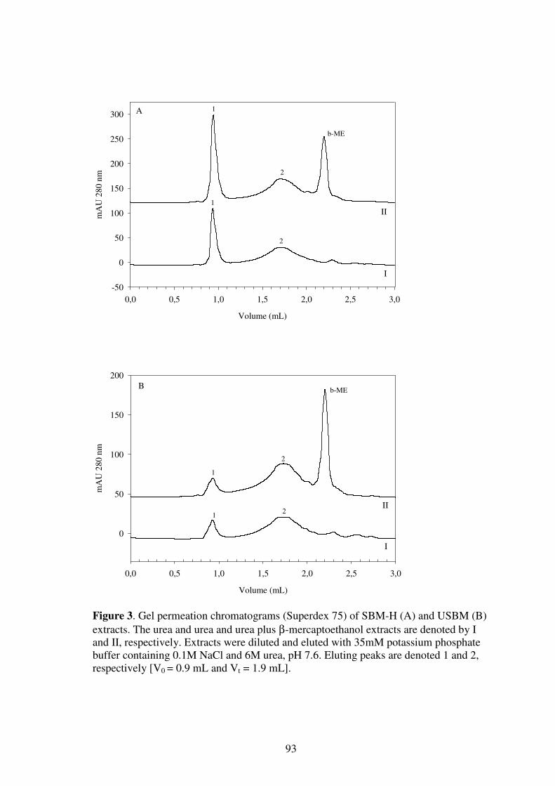
93
Volume (mL)
0,0 0,5 1,0 1,5 2,0 2,5 3,0
mA
U 2
80
nm
-50
0
50
100
150
200
250
300
I
II
b-ME
1
2
1
2
A
Volume (mL)
0,0 0,5 1,0 1,5 2,0 2,5 3,0
mA
U 2
80 n
m
0
50
100
150
200
I
II
b-ME
1
2
12
B
Figure 3. Gel permeation chromatograms (Superdex 75) of SBM-H (A) and USBM (B)
extracts. The urea and urea and urea plus β-mercaptoethanol extracts are denoted by I and II, respectively. Extracts were diluted and eluted with 35mM potassium phosphate buffer containing 0.1M NaCl and 6M urea, pH 7.6. Eluting peaks are denoted 1 and 2, respectively [V0 = 0.9 mL and Vt = 1.9 mL].

94
For both residues the peak profile was comparable to the pH 12 (1 h) extracts presented
in Figure 2. A large proportion of protein material was contained in peak 1 (>70 kDa) of
SBM-H. For the USBM the primary proportion of protein material eluted in peak 2 con-
taining material smaller than 14 kDa. For both types of residues extraction by urea plus
β-mercaptoethanol did not change the distribution of the peaks to any major extent.
The results indicate that the presence of high molecular weight proteinaceous material
in the residues correlates with the use of high-humidity heat treatment (SBM-H and
NN) of the soybean meals.
Gel Electrophoresis. The different extracts were subjected to SDS-PAGE as shown in
Figure 4.
Figure 4. SDS-PAGE gels showing pH 2 and pH 12 extracts of the NN, USBM, SBM-H, and SBM-L residues: (lanes 1-4) pH 2; (lanes 5-8) pH 12. ST (starting from above): molecular markers 97, 67, 43, 30, 21, and 14 kDa.
The material extracted by pH 12 was smaller than the lower separation limit (10 kDa) of
the gel as seen in lanes 5-8. For the NN and SBM-H samples a weak coloring was no-
ticed below the 14 kDa protein marker at the bottom of lanes 6 and 7. The peptide gel
contained no electrophoretically recognizable products (results not shown). Accord-
ingly, at least 50% (Table 1) of the protein contained in the four different enzymatic
residues had a molecular weight below 1 kDa (peptides < 8-10 amino acids). This find-
ing clearly contradicts the molecular weights determined by the gel permeation chroma-
tograms presented in Figures 2 and 3. No electrophoretically recognizable protein bands
were observed by SDS-PAGE in the material extracted with urea, urea plus β-
mercaptoethanol, and detergent Triton X 100 (results not shown). The results indicate
that the two major peaks in the chromatograms contain aggregated peptides, which can-
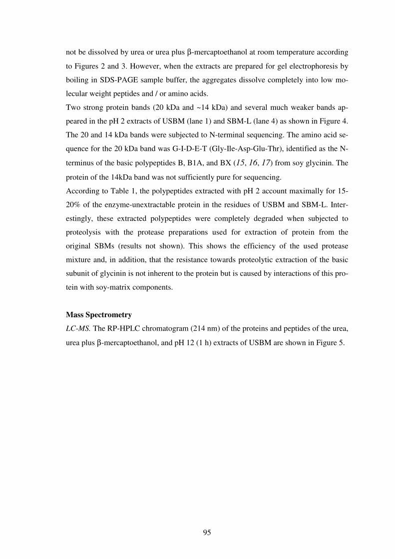
95
not be dissolved by urea or urea plus β-mercaptoethanol at room temperature according
to Figures 2 and 3. However, when the extracts are prepared for gel electrophoresis by
boiling in SDS-PAGE sample buffer, the aggregates dissolve completely into low mo-
lecular weight peptides and / or amino acids.
Two strong protein bands (20 kDa and ~14 kDa) and several much weaker bands ap-
peared in the pH 2 extracts of USBM (lane 1) and SBM-L (lane 4) as shown in Figure 4.
The 20 and 14 kDa bands were subjected to N-terminal sequencing. The amino acid se-
quence for the 20 kDa band was G-I-D-E-T (Gly-Ile-Asp-Glu-Thr), identified as the N-
terminus of the basic polypeptides B, B1A, and BX (15, 16, 17) from soy glycinin. The
protein of the 14kDa band was not sufficiently pure for sequencing.
According to Table 1, the polypeptides extracted with pH 2 account maximally for 15-
20% of the enzyme-unextractable protein in the residues of USBM and SBM-L. Inter-
estingly, these extracted polypeptides were completely degraded when subjected to
proteolysis with the protease preparations used for extraction of protein from the
original SBMs (results not shown). This shows the efficiency of the used protease
mixture and, in addition, that the resistance towards proteolytic extraction of the basic
subunit of glycinin is not inherent to the protein but is caused by interactions of this pro-
tein with soy-matrix components.
Mass Spectrometry
LC-MS. The RP-HPLC chromatogram (214 nm) of the proteins and peptides of the urea,
urea plus β-mercaptoethanol, and pH 12 (1 h) extracts of USBM are shown in Figure 5.

96
Time (min)
0 17 33 50 67
AU
21
4 n
m
0
1
2
Ace
ton
itri
le (
%)
0
20
40
60
80
100
b-ME
1
1
1
I
II
III
A 2
2
2
Figure 5. LC-MS reversed phase chromatograms for USBM after extraction by urea (I),
urea + b-ME (II), and pH 12 (1 h) (III): peaks are denoted b-ME (for the β-mercaptoethanol peak) and 1 and 2 (for the peaks eluting at 36 and 42% acetonitrile concentrations, respectively).
Comparable results were obtained for the NN, SBM-H, and SBM-L residues. Essen-
tially all proteinaceous material was eluting between 15 and 50% acetonitrile in line
with the general range of peptide hydrophobicity (18). Qualitatively, the three chroma-
tograms are comparable, though not identical. A complex mixture of peptides/proteins
was eluting between 15 and 35% acetonitrile, depending on the extractant used. At 36
and 42% acetonitrile concentrations two more distinct peaks (marked 1 and 2, respec-
tively) can be distinguished for all three extractants.
Mass spectra were acquired from 400-1800 Da over the whole chromatogram (Figure
6). In Figure 6, curve I, an example of a mass spectum of the USBM residue after ex-
traction with urea is shown for peak 1 of Figure 5.
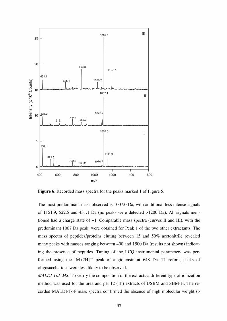
97
Figure 6. Recorded mass spectra for the peaks marked 1 of Figure 5.
The most predominant mass observed is 1007.0 Da, with additional less intense signals
of 1151.9, 522.5 and 431.1 Da (no peaks were detected >1200 Da). All signals men-
tioned had a charge state of +1. Comparable mass spectra (curves II and III), with the
predominant 1007 Da peak, were obtained for Peak 1 of the two other extractants. The
mass spectra of peptides/proteins eluting between 15 and 50% acetonitrile revealed
many peaks with masses ranging between 400 and 1500 Da (results not shown) indicat-
ing the presence of peptides. Tuning of the LCQ instrumental parameters was per-
formed using the [M+2H]2+ peak of angiotensin at 648 Da. Therefore, peaks of
oligosaccharides were less likely to be observed.
MALDI-ToF MS. To verify the composition of the extracts a different type of ionization
method was used for the urea and pH 12 (1h) extracts of USBM and SBM-H. The re-
corded MALDI-ToF mass spectra confirmed the absence of high molecular weight (>
m/z
400 600 800 1000 1200 1400 1600
Inte
nsity
(x
10
5 C
oun
ts)
0
5
10
15
20
25
1007.0
1151.9
1076.7762.3
431.1
522.5
431.2
431.1
863.2
I
II
III
1187.7
1007.1
863.3
1038.2685.1
1007.1
1076.7
863.3762.3
618.1
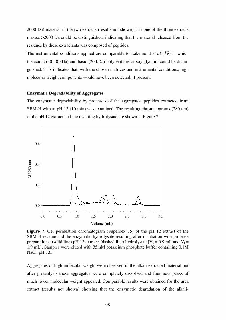
98
2000 Da) material in the two extracts (results not shown). In none of the three extracts
masses >2000 Da could be distinguished, indicating that the material released from the
residues by these extractants was composed of peptides.
The instrumental conditions applied are comparable to Lakemond et al (19) in which
the acidic (30-40 kDa) and basic (20 kDa) polypeptides of soy glycinin could be distin-
guished. This indicates that, with the chosen matrices and instrumental conditions, high
molecular weight components would have been detected, if present.
Enzymatic Degradability of Aggregates
The enzymatic degradability by proteases of the aggregated peptides extracted from
SBM-H with at pH 12 (10 min) was examined. The resulting chromatograms (280 nm)
of the pH 12 extract and the resulting hydrolysate are shown in Figure 7.
Volume (mL)
0,0 0,5 1,0 1,5 2,0 2,5 3,0 3,5
AU
280
nm
0,0
0,2
0,4
0,6
Figure 7. Gel permeation chromatogram (Superdex 75) of the pH 12 extract of the SBM-H residue and the enzymatic hydrolysate resulting after incubation with protease preparations: (solid line) pH 12 extract; (dashed line) hydrolysate [V0 = 0.9 mL and Vt = 1.9 mL]. Samples were eluted with 35mM potassium phosphate buffer containing 0.1M NaCl, pH 7.6.
Aggregates of high molecular weight were observed in the alkali-extracted material but
after proteolysis these aggregates were completely dissolved and four new peaks of
much lower molecular weight appeared. Comparable results were obtained for the urea
extract (results not shown) showing that the enzymatic degradation of the alkali-

99
extracted proteinaceous material should not be explained by alkali-induced deamidation
of proteins during extraction.
It thus seems that the aggregates end up in the enzyme-unextractable residues during
hydrolysis of the original meals simply because they are difficult to access for the en-
zymes and not because they resist enzymatic degradation. It could be speculated that
interactions between peptide aggregates, formed during hydrolysis and other compo-
nents of the enzyme-unextractable matrix somehow reduce the extractability of the pro-
tein material.
In conclusion, we believe that part of the extracted and hydrolyzed proteins become in-
soluble during hydrolysis because the peptides tend to aggregate after being released by
the proteases (20, 21). Aggregation of peptides is favored by the neutral pH conditions
that exist during hydrolysis of the original SBMs. At higher (>9) pH conditions the pep-
tides are more prone to solubilisation due to electrostatic repulsion (22), in line with the
strong effect of alkaline extractants for protein extraction. Once extracted, the proteina-
ceous material can be hydrolysed by the enzymes. A high proportion of cellulose in the
residues indicated a correlation between the insoluble cellulose and the formation of en-
zyme-unextractable peptides during hydrolysis of SBM proteins.
The mechanisms, which render the peptides insoluble during enzymatic hydrolysis
thereby preventing a complete extraction of protein from soybean meals, are the topic of
future experiments.
ACKNOWLEDGEMENTS
Harold Bult is greatly acknowledged for performing the statistical analysis of the amino
acid data. Bas Kuipers is acknowledged for performing part of the experiments. Jan Co-
zijnzen from Wageningen University is thanked for help with carbohydrate analysis.
This research was financially supported by Danish Academy of Technical Sciences,
(Lyngby, Denmark).
ABBREVIATIONS USED
A, Alcalase; B, Biofeed Plus; DM, dry matter; E, Energex; EP, extractability of protein;
EUS, enzyme-unextractable solids; EXUS, extractant unextractable solids; F, Fla-

100
vourzyme; SBM, soybean meal; SBM-H, soybean meal heat-treated at high humidity;
SBM-L, soybean meal heat-treated at low humidity; USBM, unheated soybean meal.
REFERENCES
1. Fischer,M.; Kofod,L.V.; Schols,H.A.; Piersma,S.R.; Gruppen,H.; Voragen,A.G.J.
Enzymatic extractability of soybean meal proteins and carbohydrates: heat and hu-
midity effects. J Agric Food Chem 2001, 49, 4463-4469.
2. Marsman,G.J.P.; Gruppen,H.; Mul,A.J.; Voragen,A.G.J. In-vitro accessibility of un-
treated, toasted, and extruded soybean meals for proteases and carbohydrases. J Ag-
ric Food Chem 1997, 45, 4088-4095.
3. Marsman,G.J.P.; Gruppen,H.; de Groot,J.; Voragen,A.G.J. Effect of toasting and
extrusion at different shear levels on soy protein interactions. J Agric Food Chem
1998, 46, 2770-2777.
4. Kim,S.Y.; Park,P.S.W.; Rhee,K.C. Functional properties of proteolytic enzyme
modified soy protein isolate. J Agric Food Chem 1990, 38, 651-656.
5. Lynch,C.J.; Rha,C.K.; Catsimpoolas,N. Tryptic hydrolysis of glycinin and its sub-
units. J Sci Food Agric 1977, 28, 971-979.
6. Fry,S.C. The growing plant cell wall: Chemical and metabolic analysis, Longmann
Scientific and Technical: New York, USA, 1988.
7. AOAC. Official Methods of Analysis, 13th ed., Association of Official Analytical
Chemists: Washington DC, USA, 1988.
8. Englyst,H.N.; Cummings,J.H. Simplified method for the measurement of total non-
starch polysaccharides by gas-liquid chromatography of constituent sugars as alditol
acetates. Analyst 1984, 109, 937-942.
9. Thibault,J.F. An automated method for the determination of pectic substances. Le-
bensm Wiss Tech 1979, 12, 247-251.
10. Stevens,J. Applied Multivariate Statistics for the Social Sciences, Lawrence Erl-
baum Associates: Mahwah, NJ, USA, 1996.
11. Creighton,T.E. Proteins. Structures and Molecular Properties, 2nd ed.; W. H.
Freeman and Company: New York, US, 1996.
12. Riha,W.E.; Izzo,H.V.; Zhang,J.; Ho,C.T. Nonenzymatic deamidation of food pro-
teins. Crit Rev Food Sci Nutr 1996, 36, 225-255.

101
13. Clatterbuck,K.L.; Kehrberg,N.L.; Marable,N.L. Solubility and in vitro digestibility
of soy flours, concentrates, and isolates. J Food Sci 1980, 45, 931-935.
14. Huisman,M.M.H.; Schols,H.A.; Voragen,A.G.J. Cell wall polysaccharides from
soybean (Glycine max.) meal. Isolation and characterisation. Carbohydr Polym
1998, 37, 87-95.
15. Mori,T.; Utsumi,S.; Inaba,H.; Kitamura,K.; Harada,K. Differences in subunit com-
position of glycinin among soybean cultivars. J Agric Food Chem 1981, 29, 20-23.
16. Utsumi,S.; Inaba,H.; Mori,T. Heterogeneity of soybean glycinin. Phytochem 1981,
20, 585-589.
17. Utsumi,S.; Kohno,M.; Mori,T.; Kito,M. An alternate cDNA encoding glycinin
A1aBx subunit. J Agric Food Chem 1987, 35, 210-214.
18. van der Ven,C.; Gruppen,H.; de Bont,D.B.A.; Voragen,A.G.J. Reversed phase and
size exclusion chromatography of milk protein hydrolysates: relation between elu-
tion from reversed phase column and apparent molecular weight distribution. Int
Dairy J 2001, 11, 83-92.
19. Lakemond,C.M.M.; de Jongh,H.H.J.; Gruppen,H.; Voragen,A.G.J. Differences in
denaturation of genetic variants of glycinin. J Agric Food Chem 2002, 50 (15),
4275-4281.
20. Otte,J.; Ju,Z.Y.; Faergemand,M.; Lomholt,S.B.; Qvist,K.B. Protease-induced aggre-
gation and gelation of whey proteins. J Food Sci 1996, 61, 911-915.
21. Otte,J.; Lomholt,S.B.; Ipsen,R.; Stapelfeldt,H.; Bukrinsky,J.T.; Qvist,K.B. Aggre-
gate formation during hydrolysis of beta-lactoglobulin with a Glu and Asp specific
protease from Bacillus licheniformis. J Agric Food Chem 1997, 45, 4889-4896.
22. Caessens,P.W.J.R.; Gruppen,H.; Visser,S.; van Aken,G.A.; Voragen,A.G.J. Plasmin
hydrolysis of β-casein: Foaming and emulsifying properties of the fractionated hy-
drolysate. J Agric Food Chem 1997, 45, 2935-2941.

102

103
CHAPTER 5
PRESENCE OF INDIGESTIBLE PEPTIDE AGGREGATES OF SOYBEAN MEAL IN
PIG ILEAL DIGESTA RESIDUE ♣ 1
♣ Fischer,M.; Voragen,A.G.J.; Piersma,S.R.; Kofod,L.V.; Joergensen,C.I.; Guggenbuhl,P.; Nunes,C.S.; Gruppen,H. Presence of indigestible peptide aggregates of soybean meal in pig ileal digesta residue Submitted to Journal of the Science of Food and Agriculture, 2006.

104
ABSTRACT
With the purpose of analyzing the molecular size and composition, proteinaceous mate-
rial was extracted from the insoluble components of a digesta sample obtained from
pigs fed a feed consisting of only soybean meal. Gel permeation chromatography indi-
cated that the alkali-extractable fraction of the proteinaceous material from the residue
was of relatively high apparent molecular weight. However, the combined results from
gel electrophoresis, RPLC-MS, and MALDI-ToF MS showed that the extracted protein
material was in fact, to a high extent, composed of aggregated peptides. To our knowl-
edge this has not previously been described. Aggregates extracted by dilute alkali were
fully degraded upon subsequent proteolytic treatment. N-terminal sequencing of se-
lected protein bands from SDS-PAGE gels indicated the presence of partly degraded β-
conglycinin α-subunits in the residue.
Keywords: amino acids; enzymatic hydrolysis; ileal digesta; mass spectrometry; soy-
bean meal; peptide aggregates; protein;

105
INTRODUCTION
Soybean meal (SBM) is a commonly used ingredient in animal nutrition. In feed appli-
cations the digestibility of the proteins is of primary importance. For this, the enzymatic
accessibility of proteins in the feed matrix is a key issue. The use of exogenous enzymes
in food and feed manufacturing has increased tremendously over the last decades. More
recently, the enzymatic extractability of proteins from unheated and heat-treated soy-
bean meals has been studied at in vitro conditions (1, 2). Even with high dosages of
carefully selected combinations of commercial endo- and exo-proteases, containing sig-
nificant amounts of carbohydrase activities as well, the enzymatic extraction of protein
remains incomplete (1). For heat-treated soybean meals (SBMs) it was shown that, in-
dependent of the heating conditions, a large proportion of the enzyme unextractable pro-
tein from SBM was composed of aggregated and precipitated peptides (2). Conse-
quently, the proportion of undegraded or partly degraded polypeptides in the non-
extracted fraction was limited. The majority of the aggregated soy peptides could be ex-
tracted by an alkaline treatment (0.1 mol litre-1 NaOH) and the aggregates solubilised
could subsequently be degraded by the same proteases (2). It was hypothesized that in-
teraction of the aggregates with other components of the soybean matrix could partly
explain the precipitation observed and the incomplete proteolytic extraction of proteina-
ceous material from the soybean meals (2).
In vivo studies have shown that soybean meal proteins cannot be fully utilized by pigs
(3, 4, 5, 6). Most of the work published in this research area presents protein mass bal-
ances or focuses on the composition of the soluble part or enzyme extractable protein
fractions of soybean meal in the gastro intestinal content. In view of that there is a scar-
city of information on the composition of the insoluble proteinaceous material in digesta
collected at the distal ileum from pigs fed a SBM feed. Obviously, this is proteinaceous
material, which is not extracted by the hydrolytic activities in the digestive tract of the
animal. In addition to unextracted dietary proteins, digesta of soybean meal might con-
tain (partly degraded) digestive enzymes and other proteins e.g. mucin secreted by the
animals. Mucin is a glycoprotein, secreted as part of the gastrointestinal mucus, that
contributes to endogenous protein and carbohydrate levels in digesta (7, 8, 9). Mucin
constitutes approximately 30, 7 to 22, 15 and 11 % of the endogenous threonine,
proline, serine and protein, respectively, in ileal digest of pigs fed a protein free diet
(10).

106
Mucin subunits, resulting from in vivo proteolysis of exposed regions of the protein,
have been identified (9, 11, 12). Exhaustive proteolysis in vitro resulted in degradation
of up to 30% of mucus protein (8, 13). Still little is known about the recovery of mucin,
or the extent of proteolytic digestion of mucin, at the distal ileum of pigs (10, 13).
In order to investigate the reason why proteins present in soybean meals can not be fully
utilized by pigs the present study focuses on the analysis of water-unextractable pro-
teinaceous material from a digesta sample representing an average of 7 pigs fed a feed
consisting only of SBM. The pooled digesta sample was analyzed with various analyti-
cal methods.
MATERIALS AND METHODS
Preparation of Pigs Digesta Residue
Digesta was collected from seven Large White growing pigs weighing on average 50.4
± 3.1 kg at the beginning of the experiment. The animals were obtained from GAEC
(Leclerc, Ostheim, France). Each animal was fitted with permanent cannulae in the gas-
trointestinal tract in the ileum 10 cm before the caecum as described by Nunes and co-
workers (14). This operation allowed non-painful withdrawal of kinetic ileum contents.
After surgery the animals were individually housed in cages allowing easy access to the
intestinal cannulae for sampling. Between experiments animals were kept on a normal
maintenance diet.
Only on the day of the trial each animal was fed 600 g of a SBM (Soy 44), a toasted
meal obtained from a local supplier. The SBM was given at 7.45 a.m., after a 24 hours
fasting period. Seven ileal contents samples were collected during the time intervals 30
minutes to 2 hours, 2 to 3 hours, 3 to 4 hours, 4 to 5 hours, 5 to 6 hours, 6 to 7 hours,
and 7 to 8 hours post feeding. All samples collected from the seven pigs were kept on
ice, pooled, mixed thoroughly and frozen at –80 °C.
To remove soluble digesta components the digesta was thawed and transferred to centri-
fuge tubes (500 ml) and centrifuged (30 min; 4000 g; 25 °C). The resulting residue was
washed (three times) with 350 ml tap water, with intermediate centrifugation steps (30
min; 4000 g; 25 °C), until a clear supernatant was obtained. The residue was freeze-
107
dried and homogenized using a mortar and pestle. It was denoted 'Pigs Digesta Residue'
(PDR) and stored at –20 °C for further analysis.
Extraction of Pigs Digesta Residue
PDR (500 mg) was suspended in 20 ml of 0.1 mol litre-1 NaOH. The suspension was
stirred at 25 °C for 1 h and centrifuged (20 min; 10000 g; 25 °C). The supernatant ob-
tained was adjusted to pH 7.6 with 1 mol litre-1 HCl and immediately subjected to fur-
ther analysis. The remaining residue was washed twice with 50 ml of deionised water
with intermediate centrifugation steps (20 min; 10000 g; 25 °C). The final residue was
freeze-dried denoted 'Alkali-Unextractable Residue' (AUR) and stored at –20 °C for fur-
ther analyses.
For LC-MS and MALDI-ToF MS analyses an additional extract was produced from
PDR. PDR (150 mg) was suspended in 1.5 ml of 6 mol litre-1 urea in 35 mM potassium
phosphate buffer, pH 7.6, in an Eppendorf tube and shaken in a Thermomixer comfort
(Eppendorf, Germany) for 1 h at 700 rpm and 25 °C. Supernatants were separated from
the residues by centrifugation (20 min; 12000 g; 25 °C) and were subjected to immedi-
ate analysis.
All extractions were performed in duplicate.
Enzymatic Degradability of Extracted Proteins
Two protease preparations were selected for the experiments. Alcalase Food Grade is a
preparation from Bacillus licheniformis. It has mainly endoproteinase activities and an
activity optimum between pH 6.5 and pH 8.5. Flavourzyme is a protease complex of
endoproteinases and exopeptidases from Aspergillus oryzae with an activity optimum
between pH 5.0 and pH 7.0. All enzymes were provided by Novozymes A/S (Bags-
vaerd, Denmark) (1).
To study the enzymatic degradability of the proteinaceous alkali-extracted PDR mate-
rial, an alkali extract was adjusted to pH 7.6 with 1 mol litre-1 HCl. Alcalase 2.4L FG
(2.5 µlitre) and Flavourzyme 1000L (5 µlitre) (Novozymes A/S, Bagsvaerd, Denmark),
were added to an alkali extract containing 100 mg of protein in a final volume of 1500
µlitre. Reaction was allowed for 5 h at 40 °C. The molecular size distribution of pro-
teinaceous material in the parental extract and the hydrolysate obtained were subse-
quently determined by gel permeation chromatography.

108
Experiments were performed in duplicate.
Chemical Composition of Samples
The protein content (N x 6.25) of the different samples was determined by the Dumas
combustion method using a NA 2100 Nitrogen and Protein Analyzer (ThermoQuest
Italia, Milan, Italy). Approximately 20 mg of each sample was used for analysis.
Protein contents are means of three determinations.
Carbohydrate and amino acid composition of the different samples were determined ac-
cording to previously described procedures (2). Carbohydrate contents are means of
three determinations and amino acid contents are means of six determinations.
Differential Scanning Calorimetry (DSC)
Differential scanning calorimetry of Soy 44 and PDR was performed using a micro-
DSC (Setaram, France). Samples were suspended in 35 mmol litre-1 mM potassium
phosphate buffer (pH 7.6) containing 0.1 mol litre-1 NaCl (I = 0.2). The protein concen-
tration of the suspensions was 50 g litre-1. Samples (0.9 mL) were scanned from 20 °C
to 115 °C at a scanning rate of 1.2 K min-1 and subsequently cooled to 20 °C at a similar
rate. The peak denaturation temperature (Tp), the temperature of the maximum heat
flow, was read from the curve. The area of the major peaks of the Soy 44 sample was
subsequently used to determine whether non-unfolded proteins, resisting proteolytic
degradation, were present in the PDR.
Molecular Size Determinations
Gel Permeation Chromatography. The molecular size distribution of the proteinaceous
material in the supernatants of the alkali-extracted PDR was determined using a Super-
dex 75 column (3.2 × 300 mm) connected to an ÄKTA-system. One set of samples was
diluted (10 times) using 35 mmol litre-1 potassium phosphate buffer (pH 7.6) containing
0.1 mol litre-1 NaCl. A volume of 50 µlitre was applied onto the column. Another set of
samples was diluted using 6 mol litre-1 urea in 35 mmol litre-1 potassium phosphate
buffer (pH 7.6) containing 0.1 mol litre-1 NaCl.
The molecular size distribution before and after enzymatic hydrolysis of an alkaline
PDR extract (25 °C; 1h) was determined using a Superdex 75 column (3.2 mm × 300
mm) connected to a SMART-system. Extract and hydrolysate were diluted (20 times)

109
using a 35 mmol litre-1 potassium phosphate buffer containing 0.1 mol litre-1 NaCl, pH
7.6.
For all samples separation took place at a flow rate of 80 µlitre min-1 using the respec-
tive dilution buffers. The absorbance of eluates was monitored at 214 and 280 nm. The
void volume (V0) and the total volume (Vt) of the column was determined using mo-
lecular weight standards. Presented chromatograms were verified by duplicate injections
and processed using Unicorn software.
Chromatographic equipment, GPC columns and molecular standards and software were
from GE Healthcare (Uppsala, Sweden).
SDS-PAGE. Analysis was performed using a Protean-system. Tris-HCl pre-cast gels
(10-20%) were used for separation of proteins (10−100 kDa) and Tris-Tricine pre-cast
gels (10-20%) were used for separation of peptides (1−40 kDa). Separation unit and gels
were from BioRad (Hercules, CA, USA).
The PDR and the supernatant from the alkaline extraction were both subjected to analy-
sis. The supernatant was mixed with sample buffer (1:1) and boiled for 5 min at 100 °C.
An aliquot 25 µlitre was loaded onto the gel. To extract proteinaceous material from the
PDR a sample (10 mg) was mixed in an Eppendorf tube with 500 µlitre of a reducing
sample buffer containing SDS (25 g kg-1) and β-mercaptoethanol (0.25 g kg-1). The effi-
ciency of protein extraction with this method was ~90%. The suspension was subse-
quently incubated (100 °C; 30 min) in a Thermomixer comfort (Eppendorf, Germany).
Finally, the suspension was separated by centrifugation (10 min; 15000 g; 25 °C). An
aliquot (25 µlitre) of the supernatant obtained was loaded onto the gel after appropriate
dilution.
The two gel types were processed at 200 V, 20 mA for 50 min. Tris-HCl gels were
stained by Coomassie Brilliant Blue and destained with 300 ml litre-1 methanol, 100 ml
litre-1 acetic acid in deionised water according to the instructions of the manufacturer.
The Tris-Tricine gels were developed by using a PlusOne protein silver staining kit (GE
Healthcare, Uppsala, Sweden) according to the instructions of the manufacturer. Gels
were scanned using a Computing Densitometer from Molecular Dynamics (Sunnyvale,
CA, USA).
110
N-terminal Sequencing
Sequencing was used to identify selected polypeptides/proteins which could be ex-
tracted from the PDR by SDS-PAGE sample buffer according to above described pro-
cedures.
After cooling, the extract was re-heated for 5 min at 100 °C in reducing sample buffer
and loaded onto a 10-20% Tris-HCl gradient gel (BioRad). The gel was processed ac-
cording to the instructions of the manufacturer. Blotting was performed using a Mini
Trans-Blot Cell (BioRad) and Hybond-P, a PVDF transfer membrane (GE Healthcare,
Uppsala, Sweden). The protein transfer buffer was a 40 mmol litre-1 Tris buffer (pH 8.3)
containing 40 mmol litre-1 boric acid, and 1 mmol litre-1 EDTA. The gel and the mem-
brane were prepared for transfer according to the instructions of the manufacturer and
subsequently processed for 1 h at 100 V/0.3 A with cooling. The membrane was stained
for 5 min using a standard Coomassie Brilliant Blue staining solution, with 5 ml litre-1
acetic acid, followed by destaining for 1 min with 500 ml litre-1 methanol containing
100 ml litre-1 acetic acid. The membrane was air-dried and bands were selected for se-
quencing. Due to N-terminal blockage of most of the extracted polypeptides selected
polypeptide bands were cut-out from the PVDF membrane and incubated (24 h) in 50
µlitre of a saturated solution of CNBr in 700 ml litre-1 formic acid. The hereby obtained
mixtures of peptides derived from the original polypeptide were still attached to the
membrane. Samples were applied to the sequencing apparatus and subjected to an
automated N-terminal Edman degradation method using a Procise 494A (Applied Bio-
systems, San José, CA, USA). Peptide sequences were identified using the FASTF pro-
gram (the ‘mixed peptides versus protein database’ (http://fasta.bioch.virginia.edu) that
compares an ordered peptide mixture, as obtained by Edman degradation of a CNBr
cleavage of a protein, against a protein (fastf) or DNA (tfastf) database - and by general
search in public sequence databases.
Liquid Chromatography Mass Spectrometry. The molecular mass of the alkali and urea
extracted proteinaceous material from PDR was determined by liquid chromatography
mass spectrometry (LC-MS). A 218MS C18 reversed phase column (2.1 mm × 250
mm) from Vydac (Hesperia, CA, USA) was connected to an HPLC system from Spectra
Physics (San José, CA, USA). Mass spectrometric analysis was performed using a MAT
95 LCQ ion trap mass spectrometer from Thermo Finnigan (San José, CA, USA).
111
MALDI-ToF Mass Spectrometry. Protein and peptides were cleaned up from the alkali
and urea extracts using ZipTip C18 reversed phase tips from Millipore (Bedford, MA,
USA) according to the instructions of the manufacturer. Matrix Assisted Laser Desorp-
tion Ionization Mass Spectra (MALDI-ToF MS) were recorded on a PerSeptive Voy-
ager DE-RP (PerSeptive Biosystems, Framingham, MA, USA) mass spectrometer
equipped with delayed extraction technology. Sinapinic acid and α-cyano-4-hydroxy-
cinnamic acid (Sigma, St. Louis, MO, USA) were used as matrices. All other instru-
mental settings, methods and procedures were performed as previously described (2).
Statistical Variation
The extraction procedures have a standard deviation of 0.5%. Determinations of pro-
teins and carbohydrates have standard deviations of 0.2% and 0.5%, respectively (2).
RESULTS & DISCUSSION
Composition of Residue
Effect of Alkaline Extraction. The PDR contained 117 g kg-1 protein of which 76 g kg-1
(~65% (w/w) was alkali-extractable. A comparable solubilising effect of proteinaceous
material by 0.1 mol litre-1 NaOH has previously been found when studying the compo-
sition of protein in residues from an in vitro hydrolysis of SBM using high concentra-
tions of proteases and carbohydrases (2). The carbohydrate content of PDR was 620 g
kg-1, of which 110 g kg-1 (~18% (w/w) was alkali-extractable. Accordingly, the carbo-
hydrate content of AUR was 710 g kg-1. In total 22% of the dry matter in PDR could be
extracted with alkali.
Protein. The protein contents and amino acid compositions of the original Soy 44 soy-
bean meal, PDR and AUR are presented in Table 1.

112
Table 1. Protein content (g kg-1) and amino acid composition (molar %) of Soybean meal 44, PDR, AUR and crude mucin (CM)
SBM SD PDR SD AUR SD CMe SD
Proteina 480 - 117 - 52 - n.d. -
Ala
7.5 ± 0.08 8.8 ± 0.24 8.5 ± 0.15 8.6 ± 0.35
Arg 5.8 ± 0.11 4.2 ± 0.42 4.8 ± 0.09 3.4 ± 0.14
Asxb 12.8 ± 0.09 7.6 ± 0.40 10.2 ± 0.50 10.1 ± 0.09
Cys 1.0 ± 0.07 0.6 ± 0.12 0.6 ± 0.06 n.d.
Glxc 16.2 ± 0.08 14.3 ± 0.33 15.7 ± 0.19 9.8 ± 0.06
Gly 8.4 ± 0.07 13.0 ± 1.30 10.4 ± 0.54 11.7 ± 0.35
His 2.4 ± 0.06 1.9 ± 0.45 2.1 ± 0.16 2.0 ± 0.06
Ile t - t - t - 4.5 ± 0.11
Leu 6.6 ± 2.95 6.5 ± 2.96 7.0 ± 3.14 6.8 ± 0.05
Lys 5.8 ± 0.16 5.4 ± 0.32 6.1 ± 0.17 5.6 ± 0.27
Met t - t - t - 1.1 ± 0.10
Phe 4.5 ± 0.06 3.9 ± 0.34 4.5 ± 0.14 3.2 ± 0.04
Pro 6.6 ± 0.11 9.1 ± 0.18 7.7 ± 0.18 9.5 ± 0.99
Ser 7.6 ± 0.07 8.1 ± 0.49 7.5 ± 0.14 10.2 ± 0.31
Thr 5.1 ± 0.07 4.7 ± 0.52 4.9 ± 0.08 12.9 ± 0.56
Trpd - - - - - - - -
Tyr 2.7 ± 0.06 2.6 ± 0.14 2.5 ± 0.07 2.8 ± 0.14
Val 5.6 ± 0.05 6.6 ± 0.19 6.1 ± 0.16 7.3 ± 0.13
All data are based on six determinations. t=trace amount a Calculated from Kjeldahl N x 6.25. Expressed as g kg
-1 of dry matter
b The sum of asparagine + aspartic acid
c The sum of glutamine + glutamic acid
d Fully destroyed during hydrolysis
e CM: crude mucin. Adapted from Lien et al,1997 (15)
Soy 44 had a protein content of 480 g kg-1. The protein content (w/w) of the PDR and
AUR was 117 g kg-1 and 52 g kg–1, respectively. The amino acid composition of the
soybean meal proteins was comparable to data reported in literature (16). The compa-
rable amino acid composition of Soy 44 and PDR (Table 1) indicated a non-specific ex-
traction/degradation of soybean meal proteins by the digestive enzymes. In literature
crude mucin (9, 10) is reported to have a high total content (30-40 mol/100 mol) of
threonine, serine and proline (Table 1). In the SBM and PDR residue the total content
of threonine, serine and proline was 19.3 mol/100 mol) and 21.9 mol/100 mol), respec-
tively. Based on these figures, there is no indication that the PDR is rich in proteina-
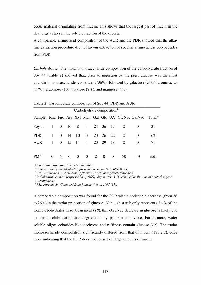
113
ceous material originating from mucin, This shows that the largest part of mucin in the
ileal digsta stays in the soluble fraction of the digesta.
A comparable amino acid composition of the AUR and the PDR showed that the alka-
line extraction procedure did not favour extraction of specific amino acids/ polypeptides
from PDR.
Carbohydrates. The molar monosaccharide composition of the carbohydrate fraction of
Soy 44 (Table 2) showed that, prior to ingestion by the pigs, glucose was the most
abundant monosaccharide constituent (36%), followed by galactose (24%), uronic acids
(17%), arabinose (10%), xylose (8%), and mannose (4%).
Table 2. Carbohydrate composition of Soy 44, PDR and AUR
Carbohydrate compositiona
Sample Rha Fuc Ara Xyl Man Gal Glc UAb GlcNac GalNac Total c
Soy 44 1 0 10 8 4 24 36 17 0 0 31
PDR 1 0 14 10 3 23 26 22 0 0 62
AUR 1 0 15 11 4 23 29 18 0 0 71
PM d 0 5 0 0 0 2 0 0 50 43 n.d.
All data are based on triple determinations a Composition of carbohydrates, presented as molar % (mol/100mol)
b UA (uronic acids) is the sum of glucuronic acid and galacturonic acid cCarbohydrate content (expressed as g /100g dry matter
-1). Determined as the sum of neutral sugars
+ uronic acids d PM: pure mucin. Compiled from Ronchetti et al, 1997 (17).
A comparable composition was found for the PDR with a noticeable decrease (from 36
to 26%) in the molar proportion of glucose. Although starch only represents 3-4% of the
total carbohydrates in soybean meal (18), this observed decrease in glucose is likely due
to starch solubilisation and degradation by pancreatic amylase. Furthermore, water
soluble oligosaccharides like stachyose and raffinose contain glucose (18). The molar
monosaccharide composition significantly differed from that of mucin (Table 2), once
more indicating that the PDR does not consist of large amounts of mucin.

114
As a result of alkaline extraction of protein material from PDR the proportion of carbo-
hydrates in AUR was increased by about 15% (from 620 g kg-1 to 710 g kg-1). Neverthe-
less a comparable molar composition of the PDR and AUR residues was found.
Protein Conformation
The DSC thermograms of Soy 44 and PDR are shown in Figure 1.
Temperature (°C)
0 20 40 60 80 100 120
Hea
t F
low
[m
W]
0
1
2
3
4
5
B
A
Figure 1. DSC-thermograms of Soy 44 (A) and PDR (B) protein suspensions.
Soy 44 (curve A) shows two endothermic transitions with peak temperatures (Tp) of
approximately 80 °C and 100 °C. These peaks correspond to the denaturation of the ma-
jor proteins, 7S β-conglycinin and 11S glycinin, respectively (19, 20) and show the
presence of intact proteins in the meal (1). The absence of transition peaks in PDR
(curve B), however, show that intact soy proteins were no longer present in the PDR
after passing the low pH conditions in the stomach and the action of the digestive en-
zymes through the stomach and small intestine. This finding is in line with previously
published results (1) showing that, with an appropriate selection of proteases, incom-
plete unfolding of soy proteins is not limiting for the efficiency of proteolytic extraction
of protein from SBMs by commercial protease preparations (1). This is in agreement
with the observation that the efficiency of degradation of the major soy proteins de-
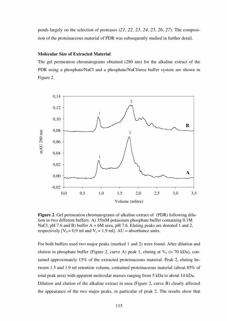
115
pends largely on the selection of proteases (21, 22, 23, 24, 25, 26, 27). The composi-
tion of the proteinaceous material of PDR was subsequently studied in further detail.
Molecular Size of Extracted Material
The gel permeation chromatograms obtained (280 nm) for the alkaline extract of the
PDR using a phosphate/NaCl and a phosphate/NaCl/urea buffer system are shown in
Figure 2.
Volume (mlitre)
0,0 0,5 1,0 1,5 2,0 2,5 3,0 3,5
mA
U 2
80
nm
-0,02
0,00
0,02
0,04
0,06
0,08
0,10
0,12
0,14
A
B
1
1
2
2
Figure 2. Gel permeation chromatograms of alkaline extract of (PDR) following dilu-tion in two different buffers: A) 35mM potassium phosphate buffer containing 0.1M NaCl, pH 7.6 and B) buffer A + 6M urea, pH 7.6. Eluting peaks are denoted 1 and 2, respectively [V0 = 0.9 ml and Vt = 1.9 ml]. AU = absorbance units.
For both buffers used two major peaks (marked 1 and 2) were found. After dilution and
elution in phosphate buffer (Figure 2, curve A) peak 1, eluting at V0 (> 70 kDa), con-
tained approximately 15% of the extracted proteinaceous material. Peak 2, eluting be-
tween 1.5 and 1.9 ml retention volume, contained proteinaceous material (about 85% of
total peak area) with apparent molecular masses ranging from 5 kDa to about 14 kDa.
Dilution and elution of the alkaline extract in urea (Figure 2, curve B) clearly affected
the appearance of the two major peaks, in particular of peak 2. The results show that
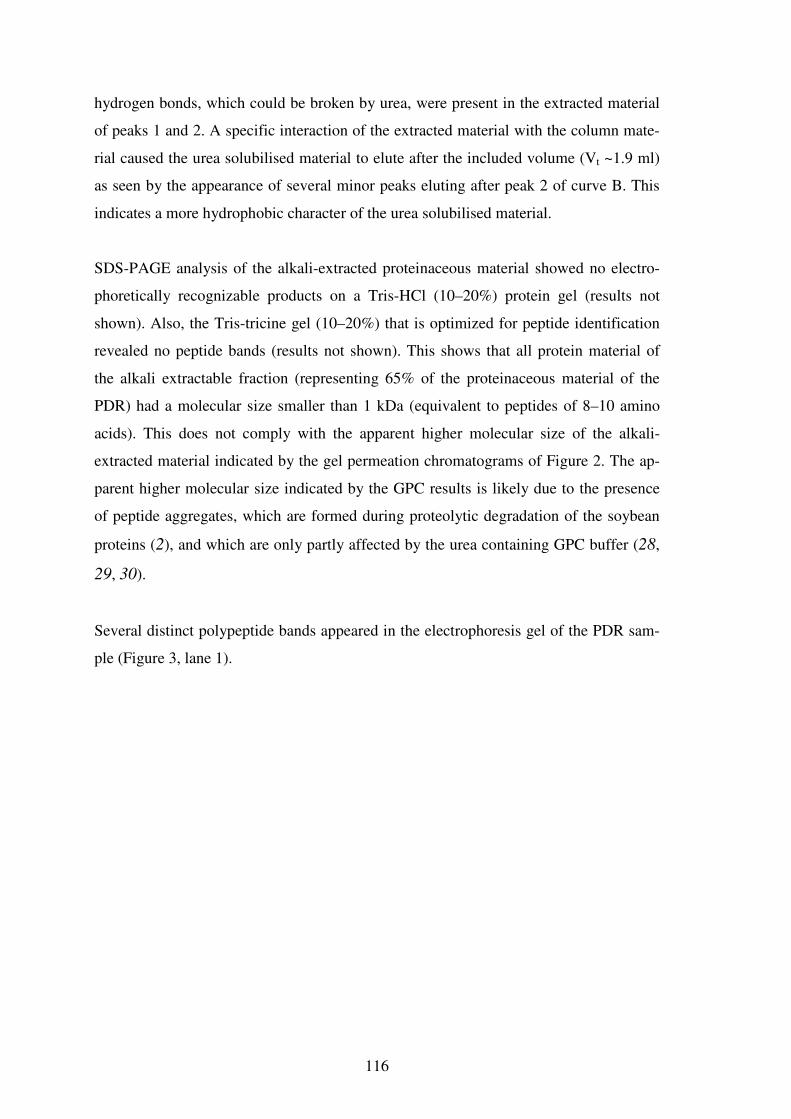
116
hydrogen bonds, which could be broken by urea, were present in the extracted material
of peaks 1 and 2. A specific interaction of the extracted material with the column mate-
rial caused the urea solubilised material to elute after the included volume (Vt ~1.9 ml)
as seen by the appearance of several minor peaks eluting after peak 2 of curve B. This
indicates a more hydrophobic character of the urea solubilised material.
SDS-PAGE analysis of the alkali-extracted proteinaceous material showed no electro-
phoretically recognizable products on a Tris-HCl (10–20%) protein gel (results not
shown). Also, the Tris-tricine gel (10–20%) that is optimized for peptide identification
revealed no peptide bands (results not shown). This shows that all protein material of
the alkali extractable fraction (representing 65% of the proteinaceous material of the
PDR) had a molecular size smaller than 1 kDa (equivalent to peptides of 8–10 amino
acids). This does not comply with the apparent higher molecular size of the alkali-
extracted material indicated by the gel permeation chromatograms of Figure 2. The ap-
parent higher molecular size indicated by the GPC results is likely due to the presence
of peptide aggregates, which are formed during proteolytic degradation of the soybean
proteins (2), and which are only partly affected by the urea containing GPC buffer (28,
29, 30).
Several distinct polypeptide bands appeared in the electrophoresis gel of the PDR sam-
ple (Figure 3, lane 1).
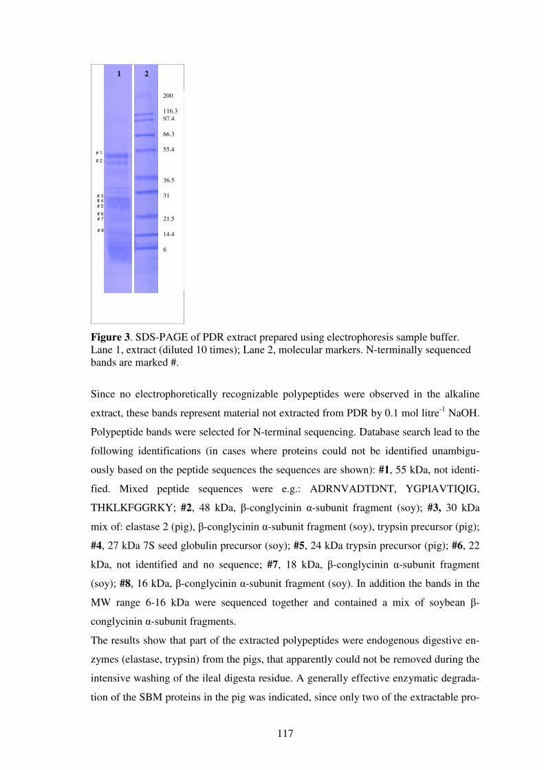
117
Figure 3. SDS-PAGE of PDR extract prepared using electrophoresis sample buffer. Lane 1, extract (diluted 10 times); Lane 2, molecular markers. N-terminally sequenced bands are marked #.
Since no electrophoretically recognizable polypeptides were observed in the alkaline
extract, these bands represent material not extracted from PDR by 0.1 mol litre-1 NaOH.
Polypeptide bands were selected for N-terminal sequencing. Database search lead to the
following identifications (in cases where proteins could not be identified unambigu-
ously based on the peptide sequences the sequences are shown): #1, 55 kDa, not identi-
fied. Mixed peptide sequences were e.g.: ADRNVADTDNT, YGPIAVTIQIG,
THKLKFGGRKY; #2, 48 kDa, β-conglycinin α-subunit fragment (soy); #3, 30 kDa
mix of: elastase 2 (pig), β-conglycinin α-subunit fragment (soy), trypsin precursor (pig);
#4, 27 kDa 7S seed globulin precursor (soy); #5, 24 kDa trypsin precursor (pig); #6, 22
kDa, not identified and no sequence; #7, 18 kDa, β-conglycinin α-subunit fragment
(soy); #8, 16 kDa, β-conglycinin α-subunit fragment (soy). In addition the bands in the
MW range 6-16 kDa were sequenced together and contained a mix of soybean β-
conglycinin α-subunit fragments.
The results show that part of the extracted polypeptides were endogenous digestive en-
zymes (elastase, trypsin) from the pigs, that apparently could not be removed during the
intensive washing of the ileal digesta residue. A generally effective enzymatic degrada-
tion of the SBM proteins in the pig was indicated, since only two of the extractable pro-
# 2 # 1
# 4 # 5 # 3
# 6 # 7 # 8
200
116.3 97.4
66.3
55.4
36.5
31
21.5
14.4
6
2 1
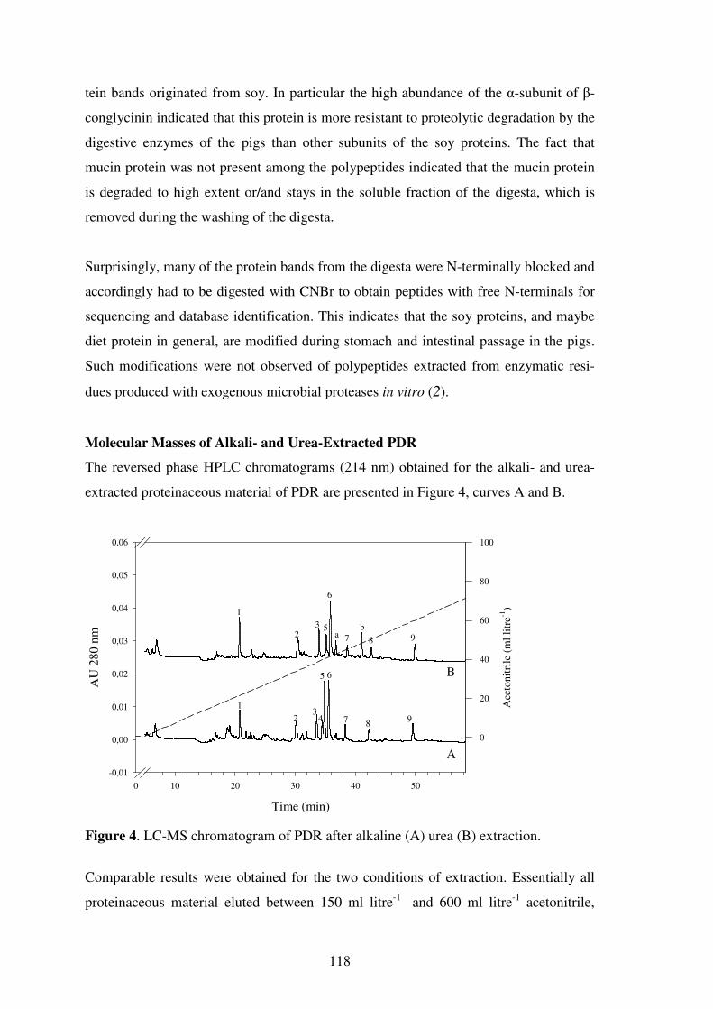
118
tein bands originated from soy. In particular the high abundance of the α-subunit of β-
conglycinin indicated that this protein is more resistant to proteolytic degradation by the
digestive enzymes of the pigs than other subunits of the soy proteins. The fact that
mucin protein was not present among the polypeptides indicated that the mucin protein
is degraded to high extent or/and stays in the soluble fraction of the digesta, which is
removed during the washing of the digesta.
Surprisingly, many of the protein bands from the digesta were N-terminally blocked and
accordingly had to be digested with CNBr to obtain peptides with free N-terminals for
sequencing and database identification. This indicates that the soy proteins, and maybe
diet protein in general, are modified during stomach and intestinal passage in the pigs.
Such modifications were not observed of polypeptides extracted from enzymatic resi-
dues produced with exogenous microbial proteases in vitro (2).
Molecular Masses of Alkali- and Urea-Extracted PDR
The reversed phase HPLC chromatograms (214 nm) obtained for the alkali- and urea-
extracted proteinaceous material of PDR are presented in Figure 4, curves A and B.
Time (min)
0 10 20 30 40 50
AU
280
nm
-0,01
0,00
0,01
0,02
0,03
0,04
0,05
0,06
Ace
tonit
rile
(m
l li
tre-1
)
0
20
40
60
80
100
A
B
1
23
4
5 6
7 89
1
23 5
6
7 8 9ab
Figure 4. LC-MS chromatogram of PDR after alkaline (A) urea (B) extraction.
Comparable results were obtained for the two conditions of extraction. Essentially all
proteinaceous material eluted between 150 ml litre-1 and 600 ml litre-1 acetonitrile,

119
which is in line with the general range of peptide hydrophobicity (31). Qualitatively, the
two chromatograms are comparable, although not identical. Except for one distinct
peak (peak 1) a complex mixture of low abundant peptide/protein material was eluting
between 150 ml litre-1 and 280 ml litre-1 acetonitrile. For the alkaline extract (curve A),
a range of distinct peaks (marked 2-9, respectively) is distinguished eluting between 300
ml litre-1 and 600 ml litre-1 acetonitrile. A comparable chromatogram was seen for the
urea extracted proteinaceous material (curve B) except from the appearance of two addi-
tional peaks (marked a and b, respectively).
Mass spectra from 400 - 1800 Da were acquired for all peaks in the chromatogram con-
firming the low molecular size of proteinaceous material indicated by SDS-PAGE
analysis. Typical mass spectra for selected peaks of PDR after extraction by 0.1 mol li-
tre-1 NaOH are shown in Figure 5 for peak 2 eluting at 33 ml litre-1 acetonitrile (Figure
5A), peak 6 eluting at 40 ml litre-1 acetonitrile (Figure 5B), and peak 9 eluting at ap-
proximately 60 ml litre-1 acetonitrile (Figure 5C).
The most predominant masses observed are 453.27, 566.40, 605.20, 718.33, 854.07,
984.00, and 1268.80 Da. A series of additional, less abundant, signals were observed
with masses ranging between the above mentioned masses. The 275.60 mass present in
all mass spectra is a contamination from the plastic container in which the PDR was
stored.

120
Figure 5. Recorded mass spectra of an alkaline extract of ileal pigs digesta residue (PDR). The three mass spectra shown (panels A-C) correspond to peaks 2, 6 and 9 of Figure 4.
Comparable mass spectra were acquired for the urea extract of PDR. Peak a of Figure 4,
curve B, contained two predominant masses of 943.1 Da and 912.9 Da. In peak b the
predominant masses were 230.2 Da and 459.3 Da (MS results not shown). The mass
spectra of peptides / proteins eluting between 15 ml litre-1 and 60 ml litre-1 acetonitrile
revealed many smaller peaks with masses ranging between 230 Da and approximately
1500 Da (results not shown) indicating the presence of low abundant peptides. The in-
A
B
C
1400 1600 1800 2000
m/z
0
5
10
15
20
25
30
35
40
45
50
55
60
65
70
75
80
85
90
95
100
Rela
tive
Abu
nda
nce
275.60
1268.80
334.73
259.67
541.13491.07 945.20635.87 759.73 1010.33340.13 1421.13814.33
1093.80 1509.20 1799.531635.93 1903.60
1400 1600 1800 2000m/z
0
5
10
15
20
25
30
35
40
45
50
55
60
65
70
75
80
85
90
95
100
Re
lati
ve A
bun
dan
ce
984.00
566.40
275.60
718.33283.67 854.07
334.73
732.13 968.07234.73 541.13453.33
756.13604.53352.40 1170.80947.13 1249.60
1013.93 1766.671525.401474.13 1604.20 1933.73
1400 1600 1800 2000
m/z
0
5
10
15
20
25
30
35
40
45
50
55
60
65
70
75
80
85
90
95
100
Re
lative
Ab
un
dance
453.27
605.20275.60
435.27 475.40
871.00334.73227.13
491.47
951.40634.80 855.20 1023.33 1322.801233.00 1387.40 1590.87 1815.67 1953.071676.13

121
strumental parameters of the LCQ were tuned using the [M+2H]2+ peak of angiotensin
at 648 Da. Therefore, peaks of oligosaccharides are not likely to be observed.
The low molecular size of the alkali- and urea-extracted proteinaceous material indi-
cated by mass spectrometric measurements was subsequently confirmed by MALDI-
ToF Mass Spectrometry, a different ionization method. The recorded MALDI-ToF mass
spectra confirmed that all proteinaceous material extracted at both conditions had a mo-
lecular size <1800 Da (results not shown). It has previously been shown that the differ-
ent subunits of soy glycinin are distinguishable at comparable instrumental settings
(32).
Enzymatic Degradability of Aggregates
The enzymatic degradability by protease preparations of the alkali-extracted aggregated
peptides in PDR was examined. The chromatograms (280 nm) of the extract and the hy-
drolysate produced are shown in Figure 6.
Volume (mlitre)
0 1 2 3
mA
U 2
80 n
m
0,00
0,02
0,04
0,06
0,08
0,10
Figure 6. Gel permation chromatograms of alkaline extract of PDR and the enzymatic hydrolysate obtained after incubation of the extract with two commercial protease preparations. Solid line, alkaline extract; dashed line, enzymatic hydrolysate.
Aggregates of high molecular weight were observed in the alkali-extracted material, but
after proteolysis these aggregates were almost completely degraded. As a result three

122
smaller peaks appeared with a retention volume corresponding to a lower molecular
size. A hydrophobic character of the enzymatically degraded material was indicated,
since all the new peaks were eluting after Vt (1.9 ml).
Peptide aggregates, of comparable molecular size to the ones from PDR, were also
highly abundant in residues from in vitro hydrolysis of SBM proteins using two com-
mercial protease preparations. Like for the protein material extracted from ileal digesta
these other aggregates could also be degraded by commercial protease preparations (2).
The comparable composition of the aggregated protein material from these two soy
residues indicate that the aggregates of the alkaline extract of PDR are primarily derived
from the soy proteins and do not contain much endogenous protein.
In conclusion, the results presented indicate a high efficiency of the digestive enzymes
of the pig for extraction/degradation of SBM proteins. Naturally, a large proportion of
the soluble protein material is readily absorbed by the animal or stays in the soluble
fraction of the ileal sample, which was not the subject for investigation in the current
study. From the present experiment, however, it is evident that insoluble peptide aggre-
gates are formed during hydrolysis of the soy proteins in the stomach or the ileal part of
the pigs gastro-intestinal system that become inaccessible to the digestive enzymes of
the animals. Poor accessibility (steric hindrance) of the digestive enzymes to these pep-
tide aggregates or interaction of the aggregated material with other components could
partly explain the incomplete enzymatic protein extraction observed. We are currently
investigating this subject.
ACKNOWLEDGEMENTS
This research was financially supported by Danish Academy of Technical Sciences,
(Lyngby, Denmark).
ABBREVIATIONS USED
AUR, alkali-unextractable residue; DSC, Differential Scanning Calorimetry; GPC, gel
permeation chromatography; HPLC, high performance liquid chromatography; RPLC-
MS, reversed phase liquid chromatography - mass spectrometry; MALDI-ToF MS, ma-
trix assisted laser desorption / ionization time-of-flight mass spectrometry; PDR, pigs
digesta residue; SBM, soybean meal.

123
REFERENCES
1. Fischer,M.; Kofod,L.V.; Schols,H.A.; Piersma,S.R.; Gruppen,H.; Voragen,A.G.J.
Enzymatic extractability of soybean meal proteins and carbohydrates: heat and hu-
midity effects. J Agric Food Chem 2001, 49, 4463-4469.
2. Fischer,M.; Gruppen,H.; Piersma,S.R.; Kofod,L.V.; Schols,H.A.; Voragen,A.G.J.
Aggregation of peptides during hydrolysis as a cause of reduced enzymatic extrac-
tability of soybean meal proteins. J Agric Food Chem 2002, 50, 4512-4519.
3. Li,S.Y.; Sauer,W.C.; Caine,W.R. Response of nutrient digestibilities to feeding diets
with low and high-levels of soybean trypsin-inhibitors in growing pigs. J Sci Food
Agric 1998, 76, 357-363.
4. Makkink,C.A.; Heinz,T.; Souffrant,W.B.; Verstegen,M.W.A. Endogenous N losses
at the terminal ileum of young piglets fed diets based on four different protein
sources. J Anim Feed Sci 1997, 6, 219-234.
5. de Lange,C.F.; Souffrant,W.B.; Sauer,W.C. Real ileal protein and amino acid di-
gestibilities in feedstuffs for growing pigs as determined with the 15N-isotope dilu-
tion technique. J Anim Sci 1990, 68, 409-418.
6. Grala,W.; Verstegen,M.W.; Jansman,A.J.; Huisman,J.; Wasilewko,J. Nitrogen utili-
zation in pigs fed diets with soybean and rapeseed products leading to different ileal
endogenous nitrogen losses. J Anim Sci 1998, 76, 569-577.
7. Mantle,M.; Allen,A. Isolation and characterization of the native glycoprotein from
pig small-intestinal mucus. Biochem.J. 1981, 195, 267-275.
8. Mantle,M.; Mantle,D.; Allen,A. Polymeric structure of pig small-intestinal mucus
glycoprotein. Biochem J 1981, 195, 277-285.
9. Allen,A. Structure and function of gastrointestinal mucus. In Physiology of the gas-
trointestinal tract; Johnson,L.R., Ed.; Raven Press: New York, US, 2001; 617-639.
10. Lien,K.A.; Sauer,W.C.; Fenton,M. Mucin output in ileal digesta of pigs fed a pro-
tein-free diet. Z.Ernahrungswiss 1997, 36, 182-190.
11. Ofosu,F.; Forstner,J.; Forstner,G. Mucin degradation in the intestine. Biochim Bio-
phys Acta 1978, 543, 476-483.
12. Mantle,M.; Allen,A. Gastrointestinal mucus. In Gastrointestinal secretion; Davi-
son,J.S., Ed.; Butterworth and Co Ltd.: London, UK, 1989; 202-229.
13. Scawen,M.; Allen,A. The action of proteolytic enzymes on the glycoprotein from
pig gastric mucus. Biochem J 1977, 163, 363-368.
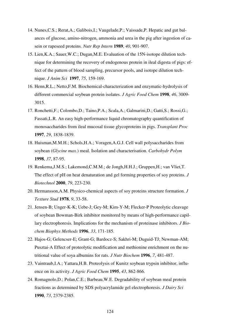
124
14. Nunes,C.S.; Rerat,A.; Galibois,I.; Vaugelade,P.; Vaissade,P. Hepatic and gut bal-
ances of glucose, amino-nitrogen, ammonia and urea in the pig after ingestion of ca-
sein or rapeseed proteins. Nutr Rep Intern 1989, 40, 901-907.
15. Lien,K.A.; Sauer,W.C.; Dugan,M.E. Evaluation of the 15N-isotope dilution tech-
nique for determining the recovery of endogenous protein in ileal digesta of pigs: ef-
fect of the pattern of blood sampling, precursor pools, and isotope dilution tech-
nique. J Anim Sci 1997, 75, 159-169.
16. Henn,R.L.; Netto,F.M. Biochemical-characterization and enzymatic-hydrolysis of
different commercial soybean protein isolates. J Agric Food Chem 1998, 46, 3009-
3015.
17. Ronchetti,F.; Colombo,D.; Taino,P.A.; Scala,A.; Galmarini,D.; Gatti,S.; Rossi,G.;
Fassati,L.R. An easy high-performance liquid chromatography quantification of
monosaccharides from ileal mucosal tissue glycoproteins in pigs. Transplant Proc
1997, 29, 1838-1839.
18. Huisman,M.M.H.; Schols,H.A.; Voragen,A.G.J. Cell wall polysaccharides from
soybean (Glycine max.) meal. Isolation and characterisation. Carbohydr Polym
1998, 37, 87-95.
19. Renkema,J.M.S.; Lakemond,C.M.M.; de Jongh,H.H.J.; Gruppen,H.; van Vliet,T.
The effect of pH on heat denaturation and gel forming properties of soy proteins. J
Biotechnol 2000, 79, 223-230.
20. Hermansson,A.M. Physico-chemical aspects of soy proteins structure formation. J
Texture Stud 1978, 9, 33-58.
21. Jensen-B; Unger-K-K; Uebe-J; Gey-M; Kim-Y-M; Flecker-P Proteolytic cleavage
of soybean Bowman-Birk inhibitor monitored by means of high-performance capil-
lary electrophoresis. Implications for the mechanism of proteinase inhibitors. J Bio-
chem Biophys Methods 1996, 33, 171-185.
22. Hajos-G; Gelencser-E; Grant-G; Bardocz-S; Sakhri-M; Duguid-TJ; Newman-AM;
Pusztai-A Effect of proteolytic modification and methionine enrichment on the nu-
tritional value of soya albumins for rats. J Nutr Biochem 1996, 7, 481-487.
23. Vaintraub,I.A.; Yattara,H.B. Proteolysis of Kunitz soybean trypsin inhibitor, influ-
ence on its activity. J Agric Food Chem 1995, 43, 862-866.
24. Romagnolo,D.; Polan,C.E.; Barbeau,W.E. Degradability of soybean meal protein
fractions as determined by SDS polyacrylamide gel electrophoresis. J Dairy Sci
1990, 73, 2379-2385.
125
25. Shutov,A.D.; Kakhovskaya,I.A.; Bastrygina,A.S.; Bulmaga,V.P.; Horstmann,C.;
Muntz,K. Limited proteolysis of beta-conglycinin and glycinin, the 7S and 11S stor-
age globulins from soybean [Glycine max (L.) Merr.]: structural and evolutionary
implications. Eur J Biochem 1996, 241, 221-228.
26. Plumb,G.W.; Mills,E.N.C.; Tatton,M.J.; D'Ursel,C.C.M.; Lambert,N.; Mor-
gan,M.R.A. Effect of thermal and proteolytic processing on glycinin, the 11S globu-
lin of soy (Glycine max): a study utilizing monoclonal and polyclonal antibodies. J
Agric Food Chem 1994, 42, 834-840.
27. Shutov,A.D.; Pineda,J.; Senyuk,V.I.; Reva,V.A.; Vaintraub,I.A. Action of trypsin
on glycinin: Mixed-type proteolysis and its kinetics; molecular mass of glycinin T.
Eur J Biochem 1991, 199, 539-544.
28. Caessens,P.W.J.R.; Daamen,W.F.; Gruppen,H.; Visser,S.; Voragen,A.G.J. β-
Lactoglobulin hydrolysis. 2. Peptide identification, SH/SS exchange, and functional
properties of hydrolysate fractions formed by the action of plasmin. J Agric Food
Chem 1999, 47, 2980-2990.
29. Otte,J.; Lomholt,S.B.; Ipsen,R.; Stapelfeldt,H.; Bukrinsky,J.T.; Qvist,K.B. Aggre-
gate formation during hydrolysis of beta-lactoglobulin with a Glu and Asp specific
protease from Bacillus licheniformis. J Agric Food Chem 1997, 45, 4889-4896.
30. Otte,J.; Lomholt,S.B.; Halkier,T.; Qvist,K.B. Identification of peptides in aggregates
formed during hydrolysis of beta-lactoglobulin B with a Glu and Asp specific mi-
crobial protease. J Agric Food Chem 2000, 48, 2443-2447.
31. van der Ven,C.; Gruppen,H.; de Bont,D.B.A.; Voragen,A.G.J. Reversed phase and
size exclusion chromatography of milk protein hydrolysates: relation between elu-
tion from reversed phase column and apparent molecular weight distribution. Int
Dairy J 2001, 11, 83-92.
32. Lakemond,C.M.M.; de Jongh,H.H.J.; Gruppen,H.; Voragen,A.G.J. Differences in
denaturation of genetic variants of glycinin. J Agric Food Chem 2002, 50 (15),
4275-4281.

126
127
CHAPTER 6
GENERAL DISCUSSION
Introduction
When this thesis work was initiated in 1998, it was based on the assumption that protein
solubilisation from soybean meal is to a certain extent limited by the complexity of the
matrix components of the soy cell wall (1, 2). This complexity, in combination with
varying forms of heat treatment, is leading to reduced accessibility of the substrate to
the enzymes (3, 4). Release of, assumingly, encapsulated protein has been explained by
enzymatic disruption of the cell wall matrix, which resulted in a more effective degrada-
tion of proteins by the digestive proteases or/and exogenous proteases (5, 6). Later on,
Ouhida and coworkers (7) reported that incubation of water insoluble polysaccharides
in soybean cotyledons with pectinase, cellulase, and xylanase resulted in the release of
only low amounts (not more than 5% bound basis) of monosaccharides, mostly uronic
acids, xylose, and arabinose. Protein extraction hardly increased after enzymatic incuba-
tion. Although recent literature has identified pectinases which are capable of degrading
residual pectin structures from okara (soybean milk residue) only a limited effect on
protein release was reported (8). In the recent work of Rosenthal and co-workers (9) and
Kasai and co-workers (10) proteases were the only enzymes that resulted in a definite
and effective increase in protein extraction.
In the mean time the results of this thesis showed that a large proportion of the soy pro-
teins are actually accessible to the proteolytic enzymes but the efficacy of proteolytic
extraction is to varying degree affected by peptide aggregation. In this chapter the most
important findings of the experiments presented in the preceding chapters are discussed.
Enzymatic Hydrolysis of SBM Proteins
Optimisation of the hydrolysis of soybean meal proteins is not straight-forward. The
optimization efforts are complicated by e.g. the variation of the composition of the soy-
bean meals due to e.g. different varieties, growth conditions and different conditions for
128
heat-treatment (11). In addition, several parameters (12), including pH, choice of en-
zyme(s) and their substrate specificity, enzyme dose, and incubation time affect the effi-
cacy of enzymatic processing of soybean proteins. In this thesis only the above men-
tioned parameters were studied.
For the first experiments (Chapter 2) the hydrolysis conditions (e.g. enzymes, dose and
pH) were selected based on previous experience with the enzymes within Novozymes.
The experiments were performed with selected proteases and carbohydrases to under-
stand the extent to which the given combinations and dose of the enzymes could de-
grade differently heated proteins in the SBM matrix. In addition, degradation of purified
proteins was studied to understand the effects of the SBM matrix (13). For this purpose
solubilisation of protein from unheated SBM (USBM) and differently heat-treated
SBMs: SBM-H (autoclaved at high humidity), SBM-L (heated at relatively low (15%)
humidity), and NN (pelleted at ~85°C) was studied following incubation with combina-
tions of two proteolytic enzymes (Alcalase, Flavourzyme) and two carbohydrase prepa-
rations (Energex and Biofeed Plus). Although the water extractability of protein from
the different meals varied between 13 and 67%, proteolytic treatment effectively ex-
tracted the proteins to a final protein solubilisation of 89 – 94%. An important observa-
tion is that the final protein solubilisation level in the different samples differed only to
a limited extent. This shows that the proteolytic enzymes could largely compensate for
the negative effects of, primarily, dry heat treatment on protein solubility. This means
that, although the nutritional implications of the different forms of heat treatment were
not considered in this connection, the choice of SBM (high versus low extent of protein
denaturation) for different foods and feeds could in some cases become less critical,
given that appropriate exogenous protease(s) are selected for the application.
Another important finding is that protein solubilisation was as effective (up to 94%)
with proteases alone as it was for the combination of proteases and carbohydrases in
spite of a relatively effective solubilisation of soybean pectins by the pectinase prepara-
tion used (13). This indicates that these 94% of the total soy proteins are not encapsu-
lated, at least not to extents that render them inaccessible to the proteases. When this
finding is combined with results from Chapter 4, which show the presence of aggregates
in the enzyme-unextractable residues, it becomes evident that only a limited proportion
of the proteinaceous material from the enzyme-unextractable material is present in the

129
form of polypeptides. This implies that the polypeptides are effectively extracted and
degraded by the enzymes. However, part of the SBM proteins that are hydrolysed in the
process later on become insoluble because the released peptides aggregate and precipi-
tate. At in vitro conditions the aggregation process is favored by the neutral pH condi-
tions (14) chosen for the hydrolysis. Therefore, larger aggregates are formed that coun-
teract the complete solubilisation of soy proteins. When alkali is used for extraction
electrostatic repulsion makes the peptides in the aggregate soluble. Once released from
the residues, the aggregated material can be degraded to lower molecular size by prote-
olytic enzymes.
Similarities Between In Vitro and In Vivo Samples
The comparable composition of proteinaceous material from the enzyme-unextracable
ileal digesta sample (Chapter 5) (15) and that of in vitro derived enzyme-unextractable
material (Chapter 4) (16) is another important discovery of this thesis. In contrast to the
in vitro residues, which are produced in a batch system, the ileal digesta sample is pro-
duced at dynamic conditions in the pig with a complementary range of digestive en-
zymes optimized through evolution. The nutrients released are continuously absorbed.
Consequently one would expect a lower probability of intermolecular interactions (e.g.
hydrophobic) between peptides released, interactions that eventually lead to aggregation
behavior. However, the results clearly show that peptide aggregation is also occurring in
the pigs.
As stated above, a surprisingly comparable composition of the enzyme-unextractable
material of the enzyme-unextractable in vitro and in vivo residues is observed. Proteins
constitute about 15-20% and carbohydrates about 50-60% of both residues. For both
conditions an effective proteolytic extraction/degradation of protein from the meals was
shown by the low abundance of “intact” polypeptides in the residues. However,
complete extraction of protein from SBMs seems extremely hard to reach. The primary
proportion of the enzyme unextractable proteinaceous material was composed of
peptides of relatively low molecular weight (≤ 1200 Da), which had aggregated and
precipitated during protein hydrolysis. These findings were rather surprising
considering the different conditions for protein hydrolysis in vitro and in vivo and the
different proteases concerned. Consequently, it was an inspiration to study the effects of
130
a selection of commercial proteases on the proteins of the intact ileal digesta sample,
which contains soluble and insoluble components.
Effects of Exogenous Proteases on Composition of Ileal Digesta Proteins
The proportion of residual material from the original soybean meal that ends up in the
ileal digesta sample after gastro-intestinal passage has only been in contact with prote-
olytic enzymes from the digestive tract of the pig (primarily pepsin and pancreatin).
Therefore, it was of relevance to study the potential further solubilisation/degradation of
the digesta proteins by commercial proteases, including exo- and endo enzymes. For
this purpose an ileal digesta sample collected from pigs fed a single meal of Soy 44 was
obtained (15). The complete digesta sample contained 2.19% (w/v) protein) and dry
matter comprised 16% (w/w) of the digesta. The carbohydrate content was 62% (w/w)
of DM and cellulose comprised about 50% of this (15). For hydrolysis different com-
mercial proteases were selected and incubated (pH 7; 40°C; 5h) with the digesta sample
according to Table 1. All enzymes were dosed at high concentrations. After incubation
the effects of the proteases on protein solubilisation and and digestible protein were
determined (Table 1).
Further solubilisation of protein from the digesta sample by the different proteases was
generally difficult to obtain for the different treatments. The solubilisation was nega-
tively affected by papain and PTN P (reduced to 93.6%) and also for Novo Pro D a mi-
nor negative effect (98.2%) is seen. Only Pronase and Flavourzyme had a limited (non-
significant) positive effect (101.2 and 101.0%, respectively, relative to blank). The
negative effects of PTN P and papain were also reflected on the level of digestible pro-
tein, as determined by gel permeation chromatography (peptide material below 1500
Da). The amount of digestible protein was significantly reduced to about 90% and 92%,
respectively. Digestible protein was significantly positively affected by Pronase
(104.7%), and numerically positively affected by Alcalase (104.2%) and Flavourzyme
(103.2%).

131
Table 1. Effects of different protease preparations on the level of solubilised and “di-gestible” protein in ileal digesta sample from pigs
Enzymes1 Organism Dosage 2 Sol P
(%)
CV Dig P
(%)
CV
Blank - 100.0 a 2.8 100.0 a 2.6 Alcalase (2,4 L)a Bacillus licheniformis 2.5% 99.6 a 2.1 104.2 a 2.6 Flavourzymea Aspergillus oryzae 5.0% 101.0 a 1.7 103.2 a 2.1 PTN P (pancreatin)a Porcine pancreas 4.8% 93.6 b 3.6 90.9 c 4.1 Pronaseb Streptomyces griseus 2 mg/mL 101.2 a 3.4 104.7 b 3.3 Novo Cor ABLa Rhizomucor orgamism 10% 100.2 a 3.6 101.1 a 3.6 Novo Pro Da Bacillus clausii 0.5 mg/g 98.2 a 2.9 99.9 a 4.8 Papaina Carica papaya 0.5 mg/mL 93.6 a 3.3 92.4 c 2.6
1Enzyme manufacturers:
a Novozymes A/S;
b Roche Molecular Biochemicals
2 Enzyme dosage. Percentages (v/w) should be read as: volume of enzyme preparation
relative to protein content in digesta sample
Sol P (solubilised protein): Calculated from the protein content (Dumas combustion method; N x
6.25) of the different fractions. Effects of protease treatments were calculated relative to the level of
solubilised protein in the blank digesta sample (set to 100). Positive protein solubilising effects are
seen as numbers higher than 100, for negative effects numbers are lower than 100. Dig P (digestible protein): Determined using the molecular size distribution (Superdex 30 peptide
column, 214 nm) of soluble protein material in the different protease treated digesta samples. Di-
gestible protein is defined as the relative change to the proportion of soluble protein with a molecular
size smaller than 1500 Da compared to blank.
Samples with different letters are significantly different.
In conclusion, the results show that primarily for the PTN P and papain treatments the
proportion of digestible protein (peptide material below 1500 Da) is reduced compared
to the blank (i.e. intestinal conditions). Based on the results of the previous chapters it is
highly likely that peptide aggregation is occurring during incubation as seen by the si-
multaneous reduction of protein solubilisation. Considering that pancreatic enzymes had
already been in contact with the digesta sample in the animals it was, in this respect, in-
teresting to observe that protein solubilisation and digestibility were still so negatively
affected by PTN P, which is a preparation of pancreatin complex from porcine pancreas.
The results imply that aggregation inducing behavior of protease preparations should be
considered as part of the performance evaluation of novel enzyme candidates for animal
feed. In future studies it would be interesting to study differences in aggregation behav-
ior of different proteases in vivo by including them with the soybean meal prior to in-
gestion and final collection of the ileal digesta.
Effects of Cell Wall Polysaccharides on Protein/Peptide Aggregation
Throughout the experiments of this thesis (Chapters 2, 3, and 5) it has been speculated
that the cell wall polysaccharides (e.g. cellulose or other cell wall components of the
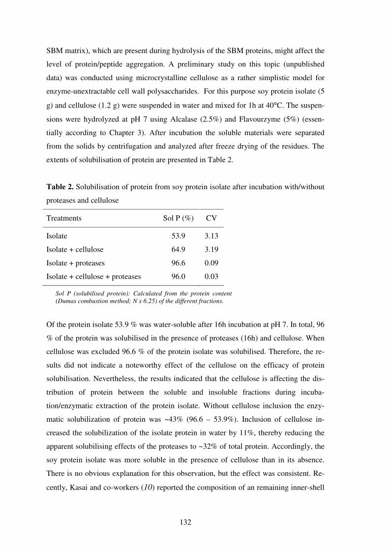
132
SBM matrix), which are present during hydrolysis of the SBM proteins, might affect the
level of protein/peptide aggregation. A preliminary study on this topic (unpublished
data) was conducted using microcrystalline cellulose as a rather simplistic model for
enzyme-unextractable cell wall polysaccharides. For this purpose soy protein isolate (5
g) and cellulose (1.2 g) were suspended in water and mixed for 1h at 40°C. The suspen-
sions were hydrolyzed at pH 7 using Alcalase (2.5%) and Flavourzyme (5%) (essen-
tially according to Chapter 3). After incubation the soluble materials were separated
from the solids by centrifugation and analyzed after freeze drying of the residues. The
extents of solubilisation of protein are presented in Table 2.
Table 2. Solubilisation of protein from soy protein isolate after incubation with/without
proteases and cellulose
Treatments Sol P (%) CV
Isolate 53.9 3.13
Isolate + cellulose 64.9 3.19
Isolate + proteases 96.6 0.09
Isolate + cellulose + proteases 96.0 0.03
Sol P (solubilised protein): Calculated from the protein content
(Dumas combustion method; N x 6.25) of the different fractions.
Of the protein isolate 53.9 % was water-soluble after 16h incubation at pH 7. In total, 96
% of the protein was solubilised in the presence of proteases (16h) and cellulose. When
cellulose was excluded 96.6 % of the protein isolate was solubilised. Therefore, the re-
sults did not indicate a noteworthy effect of the cellulose on the efficacy of protein
solubilisation. Nevertheless, the results indicated that the cellulose is affecting the dis-
tribution of protein between the soluble and insoluble fractions during incuba-
tion/enzymatic extraction of the protein isolate. Without cellulose inclusion the enzy-
matic solubilization of protein was ~43% (96.6 – 53.9%). Inclusion of cellulose in-
creased the solubilization of the isolate protein in water by 11%, thereby reducing the
apparent solubilising effects of the proteases to ~32% of total protein. Accordingly, the
soy protein isolate was more soluble in the presence of cellulose than in its absence.
There is no obvious explanation for this observation, but the effect was consistent. Re-
cently, Kasai and co-workers (10) reported the composition of an remaining inner-shell
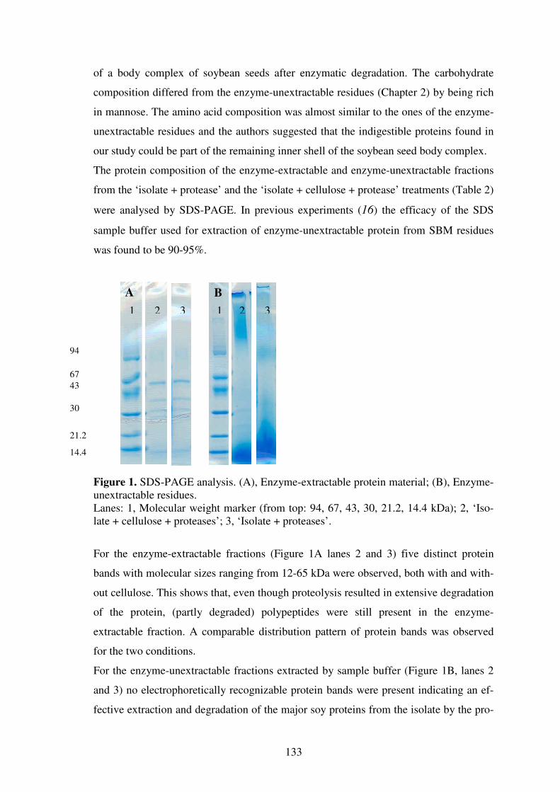
133
of a body complex of soybean seeds after enzymatic degradation. The carbohydrate
composition differed from the enzyme-unextractable residues (Chapter 2) by being rich
in mannose. The amino acid composition was almost similar to the ones of the enzyme-
unextractable residues and the authors suggested that the indigestible proteins found in
our study could be part of the remaining inner shell of the soybean seed body complex.
The protein composition of the enzyme-extractable and enzyme-unextractable fractions
from the ‘isolate + protease’ and the ‘isolate + cellulose + protease’ treatments (Table 2)
were analysed by SDS-PAGE. In previous experiments (16) the efficacy of the SDS
sample buffer used for extraction of enzyme-unextractable protein from SBM residues
was found to be 90-95%.
Figure 1. SDS-PAGE analysis. (A), Enzyme-extractable protein material; (B), Enzyme-unextractable residues. Lanes: 1, Molecular weight marker (from top: 94, 67, 43, 30, 21.2, 14.4 kDa); 2, ‘Iso-late + cellulose + proteases’; 3, ‘Isolate + proteases’.
For the enzyme-extractable fractions (Figure 1A lanes 2 and 3) five distinct protein
bands with molecular sizes ranging from 12-65 kDa were observed, both with and with-
out cellulose. This shows that, even though proteolysis resulted in extensive degradation
of the protein, (partly degraded) polypeptides were still present in the enzyme-
extractable fraction. A comparable distribution pattern of protein bands was observed
for the two conditions.
For the enzyme-unextractable fractions extracted by sample buffer (Figure 1B, lanes 2
and 3) no electrophoretically recognizable protein bands were present indicating an ef-
fective extraction and degradation of the major soy proteins from the isolate by the pro-
B A
1 2 3 1 2 3
94
67
43
30
21.2
14.4
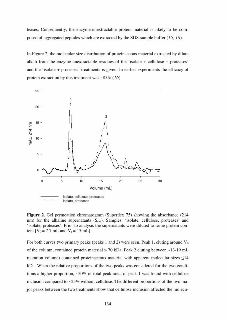
134
teases. Consequently, the enzyme-unextractable protein material is likely to be com-
posed of aggregated peptides which are extracted by the SDS-sample buffer (15, 16).
In Figure 2, the molecular size distribution of proteinaceous material extracted by dilute
alkali from the enzyme-unextractable residues of the ‘isolate + cellulose + proteases’
and the ‘isolate + proteases’ treatments is given. In earlier experiments the efficacy of
protein extraction by this treatment was ~85% (16).
Volume (mL)
0 5 10 15 20 25 30
mA
U 2
14
nm
0
5
10
15
20
25
Isolate, cellulose, proteasesIsolate, proteases
1
2
Figure 2. Gel permeation chromatogram (Superdex 75) showing the absorbance (214 nm) for the alkaline supernatants (Sext). Samples: ‘isolate, cellulose, proteases’ and ‘isolate, proteases’. Prior to analysis the supernatants were diluted to same protein con-tent [V0 = 7.7 mL and Vt = 15 mL].
For both curves two primary peaks (peaks 1 and 2) were seen. Peak 1, eluting around V0
of the column, contained protein material > 70 kDa. Peak 2 eluting between ~13-19 mL
retention volume) contained proteinaceous material with apparent molecular sizes ≤14
kDa. When the relative proportions of the two peaks was considered for the two condi-
tions a higher proportion, ~50% of total peak area, of peak 1 was found with cellulose
inclusion compared to ~25% without cellulose. The different proportions of the two ma-
jor peaks between the two treatments show that cellulose inclusion affected the molecu-

135
lar size distribution of the alkali extractable proportion of proteinaceous material in the
two enzyme-unextractable residues. Comparable curves were recorded for the two
treatments at 280 nm indicating that the increased size of the peak (214 nm) at the void
volume was not (partly) due to solubilisation of cellulose.
Since the protein solubilisation level (Table 3) was about the same (~96%) in these two
treatments the different size of the aggregates did not seem to translate into a reduced
solubilisation for the sample containing cellulose – only the molecular size distribution
was affected. The effective solubilisation and degradation of protein from the isolate is
in line with the generally effective degradation of, unheated as well as heat-treated puri-
fied glycinin and β-conglycinin by Alcalase and Flavourzyme presented in Chapter 2
(13).
In conclusion, the experiment indicated that the size of the aggregates formed during
protein hydrolysis increases in the presence of cellulose. Although this affected the ap-
parent molecular size distribution (or aggregate size) in the enzyme-unextractable pro-
tein material for the conditions tested, it did not lead to a reduced efficacy of protein
solubilisation from the protein isolate. This topic clearly warrants further investigations.
In future it would be interesting to investigate the effects of different types of cellulose
and/or cell wall material and different hydrolysis conditions followed by a detailed
analysis of peptides from the enzyme-unextractable residues.
Concluding Remarks
The results of this thesis have highlighted that the importance of peptide aggregation
upon enzymatic hydrolysis of proteins as a limiting factor for enzymatic accessibility is
not limited to in vitro situations, but is also occuring in the digestive system of animals.
Enzymatic hydrolysis of plant proteins is generally perceived as a way of increasing
protein solubilisation. The soluble fraction of an extensive hydrolysate produced from a
rapeseed protein isolate by Alcalase and Flavourzyme was characterized by Vioque and
co-workers (17). The final hydrolysate had a degree of hydrolysis of 60%. The hydro-
lysate was almost 100% soluble between pH values 2.5 and 7. The peptide characteris-
tics of sunflower protein hydrolysates prepared using Alcalase and Flavourzyme was
studied by Villanueva and co-workers (18). The combined use of the two proteases
136
generated the highest degree of hydrolysis (54%) and the highest solubility (90%) be-
tween pH 2.5 and 7. In addition, Conde and co-workers (19) reported that solubility of
sunflower protein hydrolysates increased with increasing degree of hydrolysis (DH)
over the range from DH 0 to 46%.
In none of the above-mentioned papers on hydrolysis of rapeseed and sunflower aggre-
gation behavior of the intensively hydrolysed degradation products were reported.
However, the results of this thesis provide clear evidence that peptide aggregation plays
a larger role as a limiting factor for effective proteolytic solubilisation of soy proteins
than anticipated when the project was initiated. During hydrolysis of proteins in vitro
and in vivo the structure of the solubilised soy proteins is altered, and hydrophobic
groups become exposed, which might result in aggregate formation. In vitro, during ex-
tensive hydrolysis of bovine whey proteins with Alcalase and limited hydrolysis and
limited hydrolysis with Bacillus licheniformis protease BLP (20, 21) these aggregates
can form gel networks.
For soy proteins it has been shown that different, mainly alkaline and neutral pro-
teinases of microbial origin can induce coagulation of proteins in soy milk (22). Inouye
and co-workers (23, 24) reported that subtilisin Carlsberg induces aggregation during
the hydrolysis of soy protein isolates. In vivo the experimental settings are more or less
given, but in vitro it may be possible to identify the conditions that are less favorable for
aggregate formation by controlling the parameters known to influence this behavior e.g.
degree of hydrolysis, pH and ionic strength. This may, however, may not be uncompli-
cated. Kuipers and co-workers (14) reported that aggregation (cold gelation) behavior
of soy proteins is not simply a balance of repulsive electrostatic and attractive hydro-
phobic interaction, but is more complex. Possibly, specific electrostatic attractions play
a role, making the aggregation mechanism less predictable and difficult to control e.g.
on an industrial scale. A PhD project on this topic is currently ongoing at the Laboratory
of Food Chemistry at Wageningen University.

137
References
1. Bedford,M.R. The Role of Carbohydrases in Feedstuff Digestion. In Poultry
Feedstuffs: Supply, Composition and Nutritive Value; McNab,J.M.; Boorman,K.N.,
Eds.; CABI Publishing: New York, US, 2002; 317-336.
2. Bedford,M.R.; Autio,K. Microscopic examination on feed and digesta from wheat-
fed broiler chickens and its relation to bird performance. Poult Sci 1996, 75, 14-
1996.
3. Marsman,G.J.P.; Gruppen,H.; Mul,A.J.; Voragen,A.G.J. In-vitro accessibility of
untreated, toasted, and extruded soybean meals for proteases and carbohydrases. J
Agric Food Chem 1997, 45, 4088-4095.
4. Marsman,G.J.P.; Gruppen,H.; de Groot,J.; Voragen,A.G.J. Effect of toasting and
extrusion at different shear levels on soy protein interactions. J Agric Food Chem
1998, 46, 2770-2777.
5. Thorpe,J.; Beal,J.D. Vegetable protein meals and the effects of enzymes. In
Enzymes in farm animal nutrition; Bedford,M.R.; Partridge,G.G., Eds.; CABI
Publishing: Wallingford, UK, 2000; 125-143.
6. Bedford,M.R. Exogenous enzymes in monogastric nutrition - their current value and
future benefits. Anim Feed Sci Technol 2000, 86, 1-13.
7. Ouhida,I.; Perez,J.F.; Gasa,J. Soybean (Glycine max) cell wall composition and
availability to feed enzymes. J Agric Food Chem 2002, 50, 1933-1938.
8. Kasai,N.; Murata,A.; Inui,H.; Sakamoto,T.; Kahn,R.I. Enzymatic high digestion of
soybean milk residue (okara). J Agric Food Chem 2004, 52, 5709-5716.
9. Rosenthal,A.; Pyle,D.L.; Niranjan,K.; Gilmour,S.; Trinca,L. Combined effect of
operational variables and enzyme activity on aqueous enzymatic extraction of oil
and protein from soybean. Enzyme Microb Technol 2001, 28, 499-509.
10. Kasai,N.; Satake,R.; Ikehara,H. Isolation and enzymatic digestion of body complex
of soybean seed. J Agric Food Chem 2005, 53, 10026-10033.
11. Grieshop,C.M.; Kadzere,C.T.; Clapper,G.M.; Flickinger,E.A.; Bauer,L.L.;
Frazier,R.L.; Fahey,G.C. Chemical and nutritional characteristics of United States
soybeans and soybean meals. J Agric Food Chem 2003, 51, 7684-7691.
12. Liu,K.S. Soybeans. Chemistry, Technology, and Utilization, Chapman & Hall: New
York, US, 1997.

138
13. Fischer,M.; Kofod,L.V.; Schols,H.A.; Piersma,S.R.; Gruppen,H.; Voragen,A.G.J.
Enzymatic extractability of soybean meal proteins and carbohydrates: heat and
humidity effects. J Agric Food Chem 2001, 49, 4463-4469.
14. Kuipers,B.J.H.; van Koningsveld,G.A.; Alting,A.C.; Driehuis,F.; Gruppen,H.;
Voragen,A.G.J. Enzymatic hydrolysis as a means of expanding the cold gelation
conditions of soy proteins. J Agric Food Chem 2005, 53, 1031-1038.
15. Fischer,M.; Voragen ,A.G.J.; Piersma,S.R.; Kofod,L.; Joergensen,C.I.;
Guggenbuhl,P.; Simões Nunes,C.; Gruppen,H. Presence of indigestible peptide
aggregates of soybean meal in pig ileal digesta residue. Submitted for publication in
J Sci Food Agric 2006.
16. Fischer,M.; Gruppen,H.; Piersma,S.R.; Kofod,L.V.; Schols,H.A.; Voragen,A.G.J.
Aggregation of peptides during hydrolysis as a cause of reduced enzymatic
extractability of soybean meal proteins. J Agric Food Chem 2002, 50, 4512-4519.
17. Vioque,L.; Sanchez-Vioque,P.; Clemente,A.; Pedroche,U.; Bautista,U.; Millan,F.
Production and characterization of an extensive rapeseed protein hydrolysate. J Am
Oil Chem Soc 1999, 76, 819-823.
18. Villanueva,A.; Vioque,J.; Sanchez-Vioque,R.; Clemente,A.; Pedroche,J.;
Bautista,J.; Millan,F. Peptide characteristics of sunflower protein hydrolysates. J Am
Oil Chem Soc 1999, 76, 1455-1460.
19. Conde,J.M.; Escobar,M.D.Y.; Jimenez,J.J.P.; Rodriguez,F.M.; Patino,J.M.R. Effect
of enzymatic treatment of extracted sunflower proteins on solubility, amino acid
composition, and surface activity. J Agric Food Chem 2005, 53 , 8038-8045.
20. Doucet,D.; Otter,D.E.; Gauthier,S.F.; Foegeding,E.A. Enzyme-induced gelation of
extensively hydrolyzed whey proteins by Alcalase: peptide identification and
determination of enzyme specificity. J Agric Food Chem 2003, 51, 6300-6308.
21. Otte,J.; Ju,Z.Y.; Faergemand,M.; Lomholt,S.B.; Qvist,K.B. Protease-induced
aggregation and gelation of whey proteins. J Food Sci 1996, 61, 911-915.
22. Murata,K.; Kusakabe,I.; Kobayashi,H.; Kiuchi,H.; Murakami,K. Selection of
commercial enzymes suitable for making soy milk-curd. Agric Biol Chem 1987, 51,
2929-2934.
23. Inouye,K.; Nagai,K.; Takita,T. Coagulation of soy protein isolates induced by
subtilisin Carlsberg. J Agric Food Chem 2002, 50, 1237-1242.

139
24. Nagai,K.; Inouye,K. Insights into the reaction mechanism of the coagulation of soy
protein isolates induced by subtilisin Carlsberg. J Agric Food Chem. 2004, 52,
4921-4927.

Summary
Despite all the research performed soybean proteins during the last decades, and despite
a widespread use of exogenous enzymes in food and feeds to enhance protein diges-
tion/utilization, the composition of proteinaceous enzyme-unextractable residues has
never gained much attention. Consequently, there is a lack of knowledge about the
structure of the different soy polymers resisting enzymatic digestion. Therefore, the aim
of this thesis was to broaden the knowledge about factors affecting the efficacy of en-
zymatic extraction of protein and carbohydrates from soybean meals (SBM). Also, the
composition of protein in enzyme-unextractable soybean meal fractions produced in vi-
tro and in vivo were investigated because this knowledge contributes to a better under-
standing of the characteristics of this protein fraction.
In Chapter 1, background information is given about SBM and its application in foods
and animal feed. The processing applied to soybeans for the production of defatted
SBM is presented. The protein composition of SBM is discussed with focus on the pro-
teolytic degradation of the major soy proteins following different heat treatments. Car-
bohydrates, in particular the cell wall polysaccharides of soy, and carbohydrate degrad-
ing enzymes of relevance are also discussed
In Chapter 2, one unheated and three heat-treated soybean meals were produced with
the purpose of studying the incomplete enzymatic extractability of proteins and carbo-
hydrates of thermally treated soybean meals. To obtain enzyme resistant material, the
meals were extracted by a repeated hydrolysis procedure using excessive concentrations
of different combinations of commercial protease and carbohydrase preparations. For all
soybean meals enzymatic treatment extracted most of the protein (89 - 94%). Addition
of carbohydrase preparations did not improve protein extraction. Based on the condi-
tions for heat treatment the enzymatic residues can be divided into two groups: 'high
humidity' and 'unheated + low humidity'. High-humidity heat treatment led to a more
effective enzymatic extraction. Nevertheless, in all cases the protein extraction from the
meals was incomplete. In addition, the major proteins present in soybean meal, glycinin,
β-conglycinin, and protease inhibitors were purified and subjected to enzymatic degra-
dation using the same conditions. Taking into account the more efficient degradation of
purified soy proteins, it is clear that the soybean matrix affects the enzymatic extraction
of protein from the meals. It is speculated that interactions between protein and other

components e.g. the cellulose, a major constituent of the enzyme-unextractable residues,
may reduce the efficacy of protein extraction from the meals.
In Chapter 3, the effects of time, pH and dose of two commercial protease preparations
s were studied with the purpose of obtaining a high solubilisation of protein and/or high
degree of hydrolysis from soybean meal by these two preparations. Protein solubilisa-
tion was favored by a relatively high pH (7.5) reflecting the pH optimum of the endo-
proteinase and emphasizing the importance of this type of enzyme to reach a final high
level of protein solubilisation. Experiments indicated that the efficacy of the hydrolysis
is to some extent negatively affected by degradation products formed in the protein di-
gests. The ratio of the endo/exo proteases in the enzyme mixture was found to be an im-
portant parameter, which could affect both protein solubilisation and degree of hydroly-
sis in both a negative or positive direction. The presented data illustrate that effective
protein solubilisation is not solely a matter of selecting efficient endo- and exo-
proteases. The enzymes can be overdosed and the ratio between endo- and exo-enzymes
is equally important depending on the need for high protein solubilisation or high DH in
the final hydrolysed protein product. Therefore, a combined objective of high protein
solubilisation and high degree of hydrolysis in a single hydrolysis product may be con-
flicting. Two models were developed with the purpose of predicting protein solubilisa-
tion and degree of hydrolysis as function of the dosage of the proteases. The protein
solubilisation model was tested and results indicated a slight improvement of protein
solubilisation using the prediction of the model. Although further optimization is
needed, it is clear that such models have a potential in commercial production of hydro-
lysed vegetable proteins.
The reasons for the limitations of the enzymes to enable complete solubilisation of soy
proteins were further studied in chapters 4 and 5 by analyzing the fraction of the soy
proteins, which resisted enzymatic extraction. In Chapter 4, different extractants were
screened for their ability to extract proteins from enzyme-unextractable residues of dif-
ferent heat-treated soybean meals with the purpose of analyzing the size and composi-
tion of enzyme-unextractable proteins. The largest solubilising effects were obtained
with urea, urea plus β-mercaptoethanol, and dilute alkali; the latter extracted up to 87%
of the enzyme-unextractable protein. Gel permeation chromatography indicated that a
large proportion of the urea-extracted enzyme-unextractable material was of high appar-
ent molecular weight. However, the combined results from gel electrophoresis, LC-MS,
and MALDI-ToF MS showed that the extracted protein material was composed of ag-
gregated peptides. The largest aggregates were observed in the enzyme-unextractable
residues originating from meals heat-treated at high humidity. Extracted aggregates
were fully degraded upon subsequent proteolytic treatment. It was concluded that part
of the extracted and hydrolyzed proteins assumingly become insoluble during hydroly-
sis because part of the peptides released by the proteases are obviously prone to aggre-
gation. Aggregation of peptides is favored by the neutral pH conditions that exist during
hydrolysis of the original SBMs.
In Chapter 5, the molecular size and composition of proteinaceous material extracted
from the insoluble components of a digesta sample obtained from pigs fed a single soy-
bean meal was analyzed. Gel permeation chromatography indicated that the alkali-
extractable fraction of the proteinaceous material from the residue was of relatively high
apparent molecular weight. However, the combined results from gel electrophoresis,
RPLC-MS, and MALDI-ToF MS showed that the extracted protein material was in fact,
to a high extent, composed of aggregated peptides. To our knowledge this has not pre-
viously been described. Aggregates extracted by dilute alkali were fully degraded upon
subsequent proteolytic treatment. N-terminal sequencing of selected protein bands from
SDS-PAGE gels indicated the presence of partly degraded β-conglycinin α-subunits in
the residue. The results indicated a high efficiency of the digestive enzymes of the pig
for extraction/degradation of SBM proteins. Naturally, a large proportion of the soluble
protein material is readily absorbed by the animal or stays in the soluble fraction of the
ileal sample. However, during digestion insoluble peptide aggregates are formed when
the soy proteins are hydrolysed in the stomach or the ileal part of the pig’s gastro-
intestinal tract and these aggregates become inaccessible to the digestive enzymes of the
animals.
In Chapter 6, the most important results from the previous chapters are discussed. Spe-
cial attention is given to the possible role of cell wall polysaccharides in aggregate for-
mation.
In conclusion, the results of this thesis show that the importance of peptide aggregation
upon enzymatic hydrolysis of proteins as a limiting factor for enzymatic accessibility is
not limited to in vitro laboratory situations, but aggregation of peptides is also occurring
in the digestive system of animals.

Curriculum Vitae
Morten Fischer was born on November 14th 1968 in Copenhagen. In 1991 he began
studies of agricultural science at The Royal Veterinary and Agricultural University
(RVAU), Copenhagen. Due to a large interest in applied enzyme science the
experimental work for, and writing of, the master thesis was performed at Novozymes
A/S (former Novo Nordisk A/S) in Department of Food Functionality. He graduated in
1997 with a specialisation in plant biology and plant biochemistry. From 1997 he
worked, primarily with molecular biology, at Department of Plant Biology, in the
laboratory of Professor Birger Lindberg Møller. In Spring 1998 he joined the Feed
Applications Department at Novozymes working on a so-called International Industrial
Ph.D. Fellowship, administered by the Danish Academy of Technical Sciences and
supported The Danish Ministry of Economic and Business Affairs. In connection with
the project research collaboration was established with the Laboratory of Food
Chemistry at Wageningen University, The Netherlands, headed by Professor Fons
Voragen. The project was dealing with aspects of protein and carbohydrate science in
relation to the application of enzymes. It also involved the Centre for Protein
Technology, led by Professor Harry Gruppen and the carbohydrate group, led by Dr.
Henk Schols. A fifty-fifty time-split between the company research facilities in
Bagsvaerd, Denmark and Wageningen University was agreed upon for the four years
time frame of the project. The experiments performed during this period are presented
in this thesis. Since January 2002 Morten has been working as research scientist and
project manager in the Feed Applications department of Novozymes dealing with in
vitro application development enzymes for animal feed.

List of Publications
Papers
Fischer,M.; Kofod,L.V.; Schols,H.A.; Piersma,S.R.; Gruppen,H.; Voragen,A.G.J. En-
zymatic extractability of soybean meal proteins and carbohydrates: heat and humidity
effects. J Agric Food Chem 2001, 49, 4463-4469.
Fischer,M.; Gruppen,H.; Piersma,S.R.; Kofod,L.V.; Schols,H.A.; Voragen,A.G.J. Ag-
gregation of peptides during hydrolysis as a cause of reduced enzymatic extractability of
soybean meal proteins. J Agric Food Chem 2002, 50, 4512-4519.
Fischer,M.; Voragen ,A.G.J.; Piersma,S.R.; Kofod,L.; Joergensen,C.I.; Guggenbuhl,P.;
Simões Nunes,C.; Gruppen,H. Presence of indigestible peptide aggregates of soybean
meal in pig ileal digesta residue. Submitted for publication in J Sci Food Agric 2006.
Wittstock,U.; Fischer,M.; Svendsen,I.; Halkier,B.A. Cloning and characterization of
two cDNAs encoding sulfatases in the Roman snail, Helix pomatia. IUBMB life 2000,
49, 71-76.
Patents
Lassen,S.F.; Sjoholm,C.; Ostergaard,P.R.; Fischer,M. WO2005123911-A2. New poly-
peptide having protease activity, useful in detergents or in animal feed or animal feed
additives, in increasing digestible or soluble protein in animal feed, or in increasing the
degree of hydrolysis of proteins in animal diets. Novozymes A/S.
Svendsen,A.; Kaasgaard,S.; Borch,K.; Fischer,M.; Pettersson,D.; Gregory,P.C.
WO2005115445-A1. New protease derived from Nocardiopsis sp., NRRL 18262, useful
for treating digestive disorders, pancreatic insufficiency, pancreatitis, cystic fibrosis,
diabetes type I and/or diabetes type II. Novozymes A/S.
Lassen,S.F.; Sjoholm,C.; Ostergaard,P.R.; Andersen,C.; Fischer,M.; Sjoholm,C.; Ap-
pelman,B.; Fish,E.J. WO2004111220-A1. New polypeptide having protease activity
and 78 degrees C melting temperature, useful for improving nutritional value of animal
feed, for increasing digestible or soluble protein in animal diet or as detergent compo-
nent. Novozymes A/S.
Kofod,L.V.; Fischer,M.; Nielsen,P.M.; Pommer,K. WO9818343-A. Production of a
food flavouring agent from plant protein and insoluble plant material - useful for im-
parting a variety of flavour notes, e.g. a roasted character, a smoked and pork like note,
or a vegetable flavour. Novozymes A/S.
Kofod,L.V.; Fischer,M.; Nielsen,P.M.; Pommer,K. WO9818342-A. Production of a
food flavouring agent by hydrolysis of plant proteins and carbohydrates - useful for im-
parting a variety of flavour notes, in the same manner as conventional hydrolysed vege-
table protein (HVP). Novozymes A/S.


