Gordon V. WolfeCollege of Oceanic and Atmospheric Sciences, 104 Oceanography Administration Bldg., Oregon State University,Corvallis 97331-5503
Michael SteinkeMarine Botany FB2, University of Bremen NW2, POB 330440, 28334 Bremen, Germany
Abstract
Emiliania h uxleyi clones CCMP 370 and CCMP 373 produced similar amounts of dimethylsulfoniopro-pionate (DMSP) during axenic exponential growth, averaging 109 mM internal DMSP. Both clones haddetectable DMSP lyase activity, as measured by production ofdimethyl sulfide (DMS) during in vitro assaysof crude cell preparations, but activities and conditions differed considerably between clones. Clone 373 hadhigh activity; clone 370 had low activity and required chloride. For both strains, enzyme activity per cellwas constant during exponential growth, but little DMS was produced by healthy cells. Rather, DMS pro-duction was activated when cells were subjected to physical or chemical stresses that caused cell lysis. Wepropose that DMSP lyase and DMSP are segregated within these cells and reaction only under conditionsthat result in cell stress or damage. Such activation occurs during microzooplankton grazing. When theseclones were grazed by the dinoflagellate Oxyrrhis marina, DMS was produced; ungrazed cells, as well asthose exposed to grazer exudates and associated bacteria, generated no DMS. Grazing of clone 373 producedmuch more DMS than grazing of clone 370, consistent with their relative in vitro DMSP lyase activities.DMS was only generated when cells were actually being grazed, indicating that ingested cells were responsiblefor the DMS formation. We suggest that even low levels of grazing can greatly accelerate DMS production.
Many marine phytoplankton synthesize dimethylsul-foniopropionate (DMSP) (Keller et al. 1989), a sulfonium
compound that seems to be the main biological precursorfor dimethyl sulfide (DMS). DMSP is widespread among
taxa but seems to be particularly abundant in specificgroups, such as the dinophyceae and prymnesiophyceae
(Keller et al. 1989). It may accumulate to high concen-
trations (mM-M) within cells and may be the dominantsulfur compound by mass in some species (Matrai and
Keller 1994).
The biological function of DMSP, and especially of itscleavage to DMS, acrylate, and a proton, is still not clear.
AcknowledgmentsWe thank Brian Palenik, Barry and Evelyn Sherr, Gunter O.
Kirst, and Ronald Kiene for helpful discussions and criticalcomments on the manuscript. Mark de Souza provided adviceon enzyme assays, a gift of DMSA, and ran the DMSP lyaseantibody tests. Claudia Daniel synthesized DMSP-CI.
This work was supported by NASA grant NAGW-3737 andEuropean Community Project 930326. M. Steinke was provideda travel grant by the Bremer Studien-Fonds.
High concentrations of DMSP contr_ibute to the osmoticbalance of cells, and DMSP belongs to a class of com-pounds known as "compatible solutes," which seem to
be less damaging to cellular activities than are inorganic
ions (K_irst 1990). However, studies have found little ev-idence for short-term modulation of DMSP in response
to osmotic stress (Dickson and K_irst 1986; Edwards et
al. 1988), and DMSP is only one of a number of suchsolutes, all of which contribute toward overall osmoreg-
ulation. Specifically, it is not yet established that thecleavage of DMSP to DMS and acrylate has a primary
role in osmotic adjustment or maintenance. Anotherfunction of very high solute concentrations, cryoprotec-tion, has also been suggested for ice-algae that contain
DMSP (Kirst et al. 1991), but this function does not seemto be general for the many nonpolar species that contain
large amounts of this compound. It is likely that marinephytoplankton may utilize DMSP for other biochemical
reactions, such as methyl transfer, as was suggested forthe heterotrophic flagellate Crypthecodinium (Gyrodi-
niurn) cohnii (Ishida and Kadota 1968).
Studies ofphytoplankton DMS production have largely
1152 Wolfe and Steinke
been motivated by its potential climatic impact (Charlsonet al. 1987). Culture studies have focused mainly on en-vironmental cues that may result in increased DMS emis-
sions (Baumann et al. 1994; Vairavamurthy et al. 1985;Vetter and Sharp 1993), and field studies have focused
on large blooms of high-DMSP titer species (Holligan etal. 1993; Matrai and Keller 1993; Stefels et al. 1995). Inculture, production of DMS by healthy, axenic phyto-plankton during exponential growth, such as by Hymen-omonas carterae (Vairavamurthy et al. 1985) and Phaeo-cystis pouchetii (Stefels and van Boekel 1993), seems tobe relatively rare. It is not clear that all algae that syn-thesize DMSP are able to cleave it to DMS (Steinke et
1 t_al. 9_6). The observations of DMS production with non-
axenic clones are complicated by evidence that manybacteria utilize DMSP and produce DMS (Kiene 1992;Kiene and Service 1991). Other microbial processes, suchas mesozooplankton grazing ofhigh-DMSP species, havebeen shown to generate DMS through zooplankter or bac-
terial enzymatic action (Dacey and Wakeham 1986).Studies of microzooplankton grazing have shown contra-dictory results: Wolfe et al. (1994) found that little DMSwas produced during grazing by the dinoflagellate Ox-yrrhis marina on Emiliania huxleyi (strain CCMP 370),but a similar study with E. huxleyi strain CCMP 379showed increased production of DMS during grazing(Malin et al. 1994). Without an understanding of the func-tion and mechanism of DMS production from DMSP, ithas been difficult to predict when and where DMS isproduced.
In this study we compare two axenic clones ofE. hux-
leyi, CCMP 370 and CCMP 373, which both synthesizeDMSP but differ in their abilities to convert it to DMS.We measured DMS and DMSP as well as in vivo and in
vitro DMSP lyase activity during batch growth, followingcell stress and injury, and also when cells were grazed bythe dinoflagellate O. marina in order to gain insights intothe mechanism and function of DMS formation by phy-toplankton in the marine environment.
Methods
Culture growth conditions--Axenic E. huxleyi Cultureswere obtained from the Provasoli-Guillard National Cen-
ter for the Cultivation of Marine Phytoplankton (CCMP,West Boothbay Harbor, Maine). Cultures were inoculatedinto l-liter volumes of filtered, autoclaved seawater en-
riched with nutrients (f/2, Guillard and Ryther 1962) inpolycarbonate bottles and incubated at 80-100 #molm -2s- _ under a 16 : 8 L/D cycle at 15°C. Cells were checked
for bacterial contamination throughout experiments byepifluorescence microscopy following staining with acri-dine orange and by plating on 1% peptone agar plates.No bacteria were detected by either method, except intreatments where bacteria were introduced intentionallyor with grazers. Bottles were capped and maintained with
minimal headspace to avoid degassing of DMS duringsampling. Bottles were rotated gently before sampling todistribute cells but were otherwise unshaken, and DMSsamples were not taken until at least 5 min after rotation
to allow gas equilibration between water and headspace.Typical cell densities during grazing experiments were 5-30 x 103 ml -_. During growth studies, cell densities reached5-8 x 10⁵ ml-_ in stationary phase.
Sulfur determinations--Sulfur analyses were made bygas chromatography using a Shimadzu GC- 14 chromato-graph equipped with a flame photometric detector. Thecolumn packing was Chromosil 330 (Supelco), operatedisothermally at 60°C. Helium was the carrier gas and wasalso used for sample sparging. DMSP was analyzed asDMS by alkaline hydrolysis. DMS was introduced viaheadspace samples (0.1-I00 #M samples) or followingcryotrapping (0.1-1,000 nM samples). Detection limit was
1 pmol sulfur. Other analytical details were the sameas those reported by Wolfe et al. (1994), except that sam-ples for DMSP (2 ml) were filtered under low vacuum(<5 mm of Hg) rather than by syringe to minimize cellbreakage.
DMSP lyase assays--Phytoplankton cells were con-
centrated by cer_trifugation (in vivo tests: 4,000 × g for20 min; in vitro tests: 20,000 × g for 10 min) at 15°C.The supernatent was removed by pipette, and the pelletwas resuspended by gentle pipetting into 0.3-1 ml f/2 orbuffer based on 50 mM 2-[N-morpholino] ethanesulfonicacid (MES) with 13-20 mM CaC12-2H20. For cloneCCMP 370, this buffer was amended with 600 mM NaCI
and 2 mM DL-dithiothreitol (DTT or Cleland's Reagent)and adjusted to pH 6.5. For clone CCMP 373, the bufferwas amended with 0.1-0.5% (v/v) of the nonionic deter-gent polyoxyethylenesorbitan monooleate (Tween 80) andadjusted to pH 6.2. For storing frozen extracts, 10% (v/v) glycerol was also added; tests showed extracts werestable under such storage. For in vitro assays, cells re-suspended in buffer were sonicated by brief (2 x 10 s)bursts while on ice.
DMSP lyase was assayed by adding DMSP-C1 [syn-thesized by the method of Lather et al. (1977) or obtainedfrom Research Plus] to a sample of live-cells or cell extract
in buffer and incubating 295 #1 in 1.8-ml glass screwcapvials with Tefon-coated septa. Whole-cell (in vivo) assayswere incubated in the light at in situ temperatures (15°C).In vitro assays were incubated in a water bath at 30°C.DMS production was measured by headspace analysis(50 Izl). Before adding DMSP, samples were monitoredfor endogenous DMS production for 10-20 min, then thevials were uncapped, 5 #1 of a 60 mM stock DMSP-C1solution were added (1 mM final concn), and the sampleswere immediately recapped with fresh, unpunctured septaand monitored again for 30-60 min. Typically, only 1-5% of the DMSP was convened during this time, so rateswere nearly first-order even though substrate concentra-tiong were not saturating. When necessary, the pH of thefinal solution was checked to verify that the reaction prod-ucts did not acidify the solution. DMSP standards wereprepared in NaOH for headspace calibration.
Cellular chlorophyll and fluorescence-- Chlorophyll wasextracted from GF/F-filtered cells (5-10 rnl) with 90%
Grazing-activated DMS production 1 l 53
Table 1. Comparison of initial prey and predator densitiesfor four grazing experiments. Numbers are means (or ranges, inthe case of Oxyrrhis marina) of duplicate bottles.
D. tertiolecta 22,160O. marina 1,380 930 240--450 380-390
* O. marina feeding on Emiliania huxleyi CCMP 373.t O. marina feeding on E. huxleyi CCMP 370._i O. marina feeding on either E. huxle>,i 373 or 370.§ O. marina feeding on E. huxleyi 37.3 with or without Duna-
liella tertiolecta.
acetone for 24 h, then measured by a Turner Designs 10-
AU fluorometer (Strickland and Parsons 1972). In vivofluorescence was measured by fluorometer.
Cell enumerations-- Phytoplankton cells were enumer-ated by epifluorescence microscopy after staining with
acridine orange, as by Wolfe et al. (1994). Whole-cell (invivo) fluorescence was also used to monitor growth in
some experiments. O. marina cells were enumerated livewith a dissecting microscope (Wild M3Z) in 1-10/A drops.
Grazing experiments--A culture of O. marina wasmaintained on Dunaliella tertiolecta. This prey produces
minimal DMSP, can sustain high O. marina numbers (upto 40,000 ml-_), and can be removed from culture by
placing the prey and grazers in the dark at 15°C for severaldays, allowing O. marina to completely clear the preyfrom the bottles and reach a starved state.
E. huxleyi cultures were inoculated into f/2 and allowed
to grow for several days, until densities were _ 1-2 x 10⁴
ml-' as determined by epifluorescence microscopy or cal-culated from in vivo fluorescence. Concentrated grazer
cultures were added to prey bottles; typical grazer den-sities were 200-1,000 ml -_. Table 1 summarizes initial
prey and predator densities for four feeding experiments
utilizing clones 370, 373, and D. tertiolecta as prey. Be-cause O. marina and D. tertiolecta cultures contained
bacteria that might affect DMSP and DMS pools, filtratesof the concentrated O. marina and D. tertiolecta cultures
were prepared by gravity filtration through 3-um (D. ter-tiolecta) or 5-/xm (O. marina) Nuclepore filters and added
to controls in order to keep bacterial populations similar
in all treatments. A few D. tertiolecta passed through the3-urn filter, but no O. marina cells were observed to pass
through 5-urn filters.Grazing experiments were conducted at 80-100 #mol
m -2 s -_ under a 16 : 8 L/D cycle at 15°C. Prey and pred-ator cell numbers, DMS, and DMSP concentrations were
measured every 6-12 h for 24-48 h. Exponential growthrates were calculated from log-transformed cell densities
E
c
29
10 _
113 $
I0 a
0
I0 _
5 10
I O(X)
I(X) z_
.t::
I(J _
--t" ___o- / °_i°Ms_
I0 i i
5 l0
Day
Fig. 1. Emiliania huxleyi clone 370 during batch growth.
[a.] Cell density and chlorophyll a vs. time. [b.] DMS and par-ticulate DMSP vs. time. Numbers are means of duplicates, withranges shown by error bars.
- grazing) were calculated similarly from predator + preybottles, and grazing rates were deduced by difference.
Results
Production of DJ4SP and DMS during batch growth-
During exponential growth, clones 370 and 373 grew atrates of 0.70 and 0.47 d -j to final concentrations of
8.5x 10⁵ and 5.8x 10 s ml ', respectively (Figs. la. 2a,Table 2). Clone 370 reached stationary phase at day 6.
but clone 373 continued exponential growth until day 10
(Figs. 1, 2). Under our growth conditions, neither cultureproduced coccoliths. Clone 373 was larger than clone 370(5.1 um diameter vs. 3.9 um based on observations of
live cells) and had a correspondingly larger DMSP titer
per cell (7.6 vs. 3.6 fmol). These titers were constantduring exponential growth (Fig. 3a), similar to results
shown by Matrai and Keller (1994), who found _6 fmolDMSP cell -_ for clone 8613C. Because of the different
cell volumes, both clones produced similar concentra-
tions of internal DMSP during growth, averaging 109umol cm -3 cell volume. Dissolved DMSP, defined op-
erationally by passage through a GF/F filter during gentlefiltration, was consistently _ 6-7% of internal DMSP dur-
ing all stages of growth for both clones (data not shown).
Dissolved DMSP seemed to rise during stationary phasefor both clones, but this may have been an artifact of
t 154 Wolfeand Steinke
10 6
E
E 10 5
_u
10 4
10 4
(a)
Cell density
Chl a_
5 10
-tl
I
15
I fX30
_a-,
¢o
100 :=
ml
=
_0 oo
Z-
o=
or,O
10 3
10 2
10 _
I0 o
10 -i
0
(b)DMSPp
DMS
, , • , , i
Day
Fig. 2. As Fig. I, but for clone 373.
particulate DMSP per cell and chlorophyll a per cell de-creased during stationary phase.
In contrast to the high concentrations of internal DMSP,
very little DMS was produced by exponentially growingcells (Figs. l b, 2b). Clone 370 consistently produced more
10
:_ o.ir_
_z
e_ o.01
o
. 1.5
_-YK"
-_._ , o
e-, _ 0.5
= 0.0
(a) 373
DMSPp
5 10
i
15
370
... _--..-.-_ _ • _ , _- , -" " -" • • , r
5 10 I "_
Day
Fig. 3. Comparison ofEmiliania hu.v/eyl clones 370 (ll) and373 (IS])during batch growth. [a.] Particulate DMSP and DMSper cell vs. time. [b.] In vitro DMSP I_ase activity per cell vs.time. Numbers are means of duplicates, with ranges sho_vn b_error bars.
Table 2. Comparison of growth and DMS(P) characteristicsfor Emiliania hu.xleyi clones CCMP 370 and 373 during ex-ponential growth.
Parameter Clone 370 Clone 373
Growth rate _, d -i) 0.70 0.47Final cell density (ml- _) 8.5 x 10s 5.8 x 10 _Cell diam, #m (n = 20) 3.93+0.29 5.13_+0.53Cell vol., x 10 -_2 cm 3 31.8 70.7Chl a cell -_ (ng) 0.15 0.22DMSP cell -_ (fmol) 3.58 7.59Internal DMSP concn (raM) 113 107Dissolved DMSP cell -_ (fmol) 0.29 0.51DMS cell-_ (fmol) 0.07 0.03In vitro DMSP lyase activity
(fmol cell - _ rain- _) 0.05 1.03
DMS than clone 373 did on a per-cell basis during ex-
ponential growth (0.07 vs. 0.03 fmol cell _; Table 2, Fig.3a). During growth, DMS levels were a small fraction of
dissolved DMSP (_7%) for clone 373, but a significantfraction (51%) for clone 370. When cells reached station-
ary phase and stopped dividing, DMS production con-
tinued, so that DMS per cell increased (clone 370, Fig.3a). This increase was also seen for clone 373 in other
experiments (data not shown). However, DMS produc-
tion rates per cell during stationary phase were no higherthan during exponential phase.
In vitro production of D,_¢S in cell extracts--Intri-guingly, we found in vitro DMSP lyase activity in both
clones despite their limited DMS production duringgrowth. Clone 370, which produced more DMS during
growth, had low but detectable DMSP lyase activity (0.02-
0.05 fmol DMS min -_ cell7 _at 1 mM DMSP). Clone 373showed 20-fold higher in vitro DMSP lyase activities,averaging 1.03 fmol DMS min-t cell-_ at 1 mM DMSP;
however, this strain produced almost no DMS duringexponential growth. Furthermore, biochemical charac-terization of the crude cell extracts showed distinct dif-
ferences between the two clones (Table 3; Steinke et al.in prep.). In particular, clone 370 showed an absolute salt
requirement, and enzyme preparations were stabilized byaddition of a reducing agent (DTT or fl-mercaptoethanol).
In contrast, clone 373 lyase activity was unaffected by
NaCI concentration or reductant, but improved slightlyby addition of detergent. Solubilities of the two enzymes
were also quite different.
Despite these contrasts, in vitro enzyme activity percell did not change for either clone during exponential
growth or when cells reached stationary phase (Fig. 3b),suggesting that total enzyme titer was constitutive. Clone
373 cells grown in high-nitrate (883 uM) and low-nitrate(50 uM) f/2 showed similar in vitro rates (data not shown).
Neither enzyme exhibited any lytic ability with the closelyrelated sulfonium compound, dimethylsulfonioacetate
(DMSA), similar to a DMSP lyase isolated from a marinebacterium (de Souza and Yoch 1995). However, cell-free
extracts of either enzyme failed to cross-react by westernblot or ELISA with a polyclonal antibod? prepared against
Grazing-activated DMS production 1155
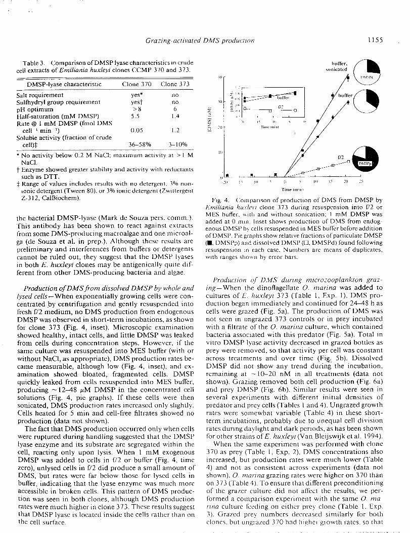
Table 3. Comparison ofDMSP lyase characteristics in crudecell extracts of Emiliania huxleyi clones CCMP 370 and 373.
DMSP-lyase characteristic Clone 370 Clone 373
Salt requirement yes*Sulthydryl group requirement yestpH optimum > 8Half-saturation (mM DMSP) 5.5Rate @ 1 mM DMSP (fmol DMS
cell -_ min -t) 0.05Soluble activity (fraction of crude
cell)_
no
no
61.4
1.2
36-58% 3-10%
* No activity below 0.2 M NaCI; maximum activity at > I MNaCI.
t Enzyme showed greater stability and activity with reductantssuch as DTT.
$ Range of values includes results with no detergent, 3% non-ionic detergent (Tween 80), or 3% ionic detergent (ZwittergentZ-312, CalBiochem).
the bacterial DMSP-lyase (Mark de Souza pets. comm.).This antibody has been shown to react against extracts
from some DMS-producing macroalgae and one microal-
ga (de Souza et al. in prep.). Although these results arepreliminary and interferences from buffers or detergents
cannot be ruled out, they suggest that the DMSP lyasesin both E. huxleyi clones may be antigenically quite dif-
ferent from other DMS-producing bacteria and algae.
Production of DMS frorn dissolved DMSP by whole and
l vsed cells--When exponentially growing cells were con-
centrated .by centrifugation and gently resuspended intofresh f/2 medium, no DMS production from endogenousDMSP was observed in short-term incubations, as shown
for clone 373 (Fig. 4, inset). Microscopic examination
showed healthy, intact cells, and little DMSP was leakedfrom cells during concentration steps. However, if the
same culture was resuspended into MES buffer (with or
without NaCI, as appropriate), DMS production rates be-came measurable, although low (Fig. 4, inset), and ex-
amination showed bloated, fragmented cells. DMSPquickly leaked from cells resuspended into MES buffer,
producing _ 12-48 uM DMSP in the concentrated cellsolutions (Fig. 4, pie graphs). If these cells were then
sonicated, DMS production rates increased only slightly.Cells heated for 5 min and cell-free filtrates showed no
production (data not shown).The fact that DMS production occurred only when cells
were ruptured during handling suggested that the DMSPtyase enzyme and its substrate are segregated within the
cell, reacting only upon lysis. When 1 mM exogenousDMSP was added to cells in t"/2 or buffer (Fig. 4, time
zero), unlysed cells in t"/2 did produce a small amount ofDMS, but rates were far below those for lysed cells in
buffer, indicating that the lyase enzyme was much moreaccessible in broken cells. This pattern of DMS produc-
tion was seen in both clones, although DMS production
rates were much higher in clone 373. These results suggestthat DMSP lyase is located inside the cells rather than onthe cell surface.
bull"er,
sonicated
I()
._ 20 l'imt" I mini
I0
20 1" !u 5 (} _ I(I I> 20 25
['ime (rain)
Fig. 4. Comparison of production of DMS from DMSP byErniliania hu.vlevi clone 373 during resuspension into 1"/2 orMES buffer, with and without sonication: 1 mM DMSP wasadded at 0 min. Inset shows production of DMS from endog-enous DMSP by cells resuspended in MES buffer before additionof DMSP. Pie graphs show relative fractions of particulate DMSP(i, DMSPp) and dissolved DMSP (E3, DMSPd) found followingresuspension in each case. Numbers are means of duplicates,with ranges shown by error bars.
Production qfl D:_IS during rnicrozooplankton graz-
ing-When the dinoflagellate O. marina was added tocultures of E huxleyi 373 (Table 1, Exp. 1), DMS pro-
duction began immediately and continued for 24-48 h ascells were grazed (Fig. 5a). The production of DMS was
not seen in ungrazed 373 controls or in prey incubatedwith a filtrate of the O. marina culture, which contained
bacteria associated with this predator (Fig. 5a). Total in
vitro DMSP lyase activity decreased in grazed bottles asprey were removed, so that activity per cell was constant
across treatments and over time (Fig:_ 5b). DissolvedDMSP did not show any trend during the incubation,remaining at -10-20 nM in all treatments (data not
shown). Grazing removed both cell production (Fig. 6a)and prey DMSP (Fig. 6b). Similar results were seen in
several experiments with different initial densities of
predator and prey cells (Tables I and 4). Ungrazed growthrates were somewhat variable (Table 4) in these short-
term incubations, probably due to unequal cell division
rates during daylight and dark periods, as has been shownfor other strains ofE. huxleyi (Van Bleijswijk et al. 1994).
When the same experiment was performed with clone
370 as prey (Table 1, Exp. 2), DMS concentrations alsoincreased, but production rates were much lower (Table
4) and not as consistent across experiments (data not
shown). O. marina grazing rates were higher on 370 thanon 373 (Table 4). To ensure that different preconditioning
of the grazer culture did not affect the results, we per-formed a comparison experiment with the same O. ma-
rina culture feeding on either prey clone (Table 1, Exp.
3). Grazed prey numbers decreased similarly for bothclones, but ungrazed 370 had higher growth rates, so that
1156 Wolfe and Steinke
50
4O
3O
2O
l0
2.0
e_
i .5
E
@
1.0
T_a
(}.5
E--__ 00.
0
[-0- h ,.iccM 3,o°,y /I
• . m , ;0 _.
6 12 18 24 30
(b)
i
36
a i a i i r
6 12 18 24 30 _6
Time (hi
Fig. 5. Production of DMS (a) and in vitro DMSP lyaseactivity per cell (b) during grazing of Oxyrrhis marina on Em-iliania huxleyi clone 373. Numbers are means of duplicates,with ranges shown by error bars.
40OOO
3OOOO
_a
_. 20OOO=
_a
i0000
(a)
------Q_ . huxleyi CCMP 373 only
+ + O. marina filtrate
0 I I I I i I
0 6 12 18 24 30 36
3O0
2(YO
100
(b)
i i i n I
f_ 12 18 24 t)
I'ime ( h }
i
_6
Table 4. Comparison of growth, grazing, and DMS produc-tion rates from Oxyrrhis marina grazing on Emiliania huxleyiand Dualiella tertiolecta. Numbers are averages with ranges inparentheses.
(prey predator -' d- _) 8.6(3.9)DMS production rate (nM d-t) 23.0(7.0)DMS produced per grazed prey
(fmol cell- ') 4.6(2.4)
0.61(0.13) 0.781.40(0.48) 2.10
2.31(0.41) 4.34
16.2(2.2) 53.23.5(1.5) 1.2
0.3(0.2) 0.0
* Range of values from three experiments; rates are averagedover experimental period.
t Range of values from two experiments; rates are averaged overexperimental period.
$ Values from one experiment.
grazing removed more "new production" for clone 370than for clone 373. Because predator numbers increased
similarly in both treatments, this implied that O. marina
cleared clone 370 at higher rates (Table 4).Because it was not clear whether the DMS observed in
the grazed cultures was produced by the grazed or un-
grazed prey, we performed an experiment in which clone
373 cells were present during grazing and exposed tochemical or physical cues due to grazing (grazing exu-
dates, shear stresses) but were not actually grazed (Table1, Exp. 4). To do this, we took advantage of the preference
of O. marina for the prey D. tertiolecta, a non-DMSP-
producing chlorophyte. We incubated clone 373 (10,000cells ml -_) with a 2-fold higher concentration of D. ter-
tiolecta (22,000 cells ml-_). When O. marina cells wereadded (500 cells ml-l), D. tertiolecta cells were rapidly
removed by grazing (Fig. 7a, 0-30 h). Clone 373 numbersincreased until D. tertiolecta cells had been grazed to
_5,000 ml -_ at 25 h, at which time O. marina began
grazing on clone 373 and its numbers decreased (Fig. 7a).DMS was not produced during the grazing olD. tertiolecta
(Fig. 7b, 0-25 h), but production began as soon as clone
373 began to be eaten (Fig. 7b, 25-55 h). In treatmentsthat contained only clone 373 as prey, DMS was produced
throughout the experiment (data not shown), as clone 373cells were grazed. In the clone 373-only treatment, both
grazing rates and DMS production rates were highest ini-
tially and decreased over the experiment as prey becamescarce. In contrast, when D. tertiolecta prey were also
{____
Fig. 6. Removal of prey cells (a) and prey DMSP (b) duringgrazing O.vyrrtus marina on Emiliania huxleyi clone 373. Num-bers are means of duplicates, with ranges shown by error bars.
Grazing-activated DMS production •
present, grazing rates and DMS production rates both 250o0_increased sharply at 25 h, when predators switched their
grazing to clone 373. Once again, control bottles of clone 2oooo373 incubated with D. tertiolecta and with O. marina
culture filtrate showed no DMS production. These ob- _ 1500oservations confirmed that production of DMS originates
from grazed clone 373 cells. _ 10o00(
¢/¢,.)
5O00
Discussion
The two E. huxleyi strains synthesized similar concen-trations of internal DMSP and also produced constitutive
DMSP-lyase enzymes. However, production of DMS
during growth was a trivial fraction of potential produc-tion given the measured rates of in vitro DMSP lyase.For example, clone 370, which produced -0.05 fmol
DMS cell- ' min- ' in vitro ( 1 mM DMSP), generated only6.5 x 10 .6 fmol DMS cell -t min -_ during growth--about
0.04% of potential production. For clone 373, the dis-
crepancy was even greater. Instead, DMS production was
clearly associated with damaged cells; as demonstrated
by increased DMS production when cells were lysed bychemical or physical means (Fig. 4) or when cells were
grazed (Figs. 5, 7). The very low cleavage of DMSP bygrowing E. huxleyi cells contrasts strongly with anotherimportant DMS-producing phytoplankter, P. pouchetii,
which produces large quantities of DMS during growth.That species averaged 3.05 fmol DMS cell- t rain- ' during
exponential growth in axenic culture (Stefels and van Boe-kel 1993) and cleaved exogenous DMSP at rates several
thousand-fold greater than we found for healthy, undam-
aged E. huxleyi. It seems likely that the DMSP lyaseenzyme, and possibly its physiological rote, is quite dif-
ferent for these species.
One explanation for the behavior we observed is thatDMSP and the DMSP lyase enzyme are physically seg-
regated within the cell and only react under conditionsthat rupture the compartments and allow mixing. Cell
manipulations clearly showed increased rates of DMSproduction from endogenous or exogenous DMSP when
cells were ruptured. One potential model for such a seg-
regated enzyme-substrate system is a cell-surface enzymewith the active site outside the cell or imbedded in the
plasma or cell membrane. Although we found that whole,
uninjured cells exposed to mM exogenous DMSP couldform DMS (Fig. 4), rates were much lower than for lysed
cell extracts. Application of proteinase K, shown to de-
grade other cell-surface proteins in clones of E. huxleyi
under similar growth conditions (Palenik and Morel 1990),did not decrease DMS production in our whole-cell tests
(data not shown). Therefore, we believe that the enzymeis internal to the cell. We were not able to detect significant
uptake of the exogenous DMSP into cells (data not shown),
so cleavage of exogenous DMSP by whole cells is stillsomewhat mysterious. It is possible that external DMSPinitiates conversion of internal DMSP pools through somesignal mechanism, and it is conceivable that DMSP-lyase
may play some role in detecting external stress or envi-ronmental cues.
0 I
0 60
(a)
_ D. tertiolecta
, l , I i i I
I 0 20 30 40 50
4O(b)
3O
20
10
0 i i l J i i
0 I0 20 _0 40 50 60
Time (h)
Fig. 7. Experiment with Oxyrrhis marina grazing both Dun-aliella tertiolecta and Erniliania huxleyi clone 373. [a.] Prey celldensities. [b.] DMS concentrations. Numbers are means of du-plicates, with ranges shown by error bars.
The dramatic contrast in lyase activity and functionbetween two clones of the same species is surprising, but
there is precedent for other biochemical and genetic di-
versity among E. huxleyi. Van Bleijswijk et al. (1991)found two distinct morphotypes of E. huxleyi based on
an antibody test to a coccolith polysaccharide, and Conte
et al. (1995) found different biomarker compounds anddifferent amounts of fucoxanthin in oceanic and neritic
strains. These studies suggested that E. huxleyi may infact be multispecific. Although genetic testing using DNA
sequence variation information (Medtin et al. 1994)showed little difference among widely distributed isolates,
preliminary evidence from amplified polymorphic DNA
(RAPD) analysis (Barker et al. 1994) in mesocosm and
bloom studies suggests that there may be genetic varia-tions at the subspecies level that are not detected by DNAmethods. There is also biochemical .evidence for phe-
notypic diversity among E. huxleyi clones. Palenik andKoke (1995) found that a cell-surface enzyme expressed
under nitrogen limitation was present in some but not allof five axenic E. huxleyi clones, suggesting that closely
related clones may have significantly different enzyme
systems.Wood and Leatham (1992) pointed out that many stud-
ies on diverse marine phytoplankton have shown intra-
species phenotypic variation and suggested that straindesignation should be considered essential information
when experimental results are reported. We therefore pre-
1158 Wolfe and Steinke
dict that other phytoplankton species will also show di-versity among strains with respect to DMSP lyase be-
havior. There is already evidence for DMSP lyase diver-
sity among related macroalgae. Steinke et al. (1996) foundthat the DMSP lyase enzyme seems to be widespread,
but activity can vary greatly between species. Three spe-cies of Enterornorpha (E. clathrata, E. intestinalis, and
E. cornpressa) had high specific lyase activities, but an-other species (E. bulbosa) had very low activity. Although
the assay of Steinke et al. was developed and optimizedfor E. clathrata and may not have detected other enzymes
that operate under different conditions, it is likely that
DMSP lyase activity is often species- or strain-specific.It is even possible that the genetic ability to cleave DMSP
to form DMS may not always be related to the ability tosynthesize DMSP. Thus, these results reinforce the notion
that DMSP may serve other biochemical functions insidecells aside from DMS-acrylate production.
Production of DMS during rnicrozooplankton graz-ing- DMS can be formed during grazing when lyase en-
zymes ai-e present in either the prey or predator. Previous
work with predators such as copepods (Dacey and Wake-ham 1986) and fish (Dacey et al. 1994) has suggested that
either the grazer or bacteria associated with grazer diges-tive tracts or fecal material could be responsible for DMSP
cleavage during grazing. Our work shows that algal DMSP
lyases may also be activated during grazing.We were not able to perform grazing treatments without
bacteria, but we believe their contribution to DMS pro-
duction was minimal. Although bacteria were likely pres-ent in the O. marina culture that cleaved or demethylatedDMSP, activities were probably low because treatments
without grazers but with grazer exudates and bacteriaproduced little or no DMS (Fig. 5a) even when substantial
pools of dissolved DMSP (10-20 nM) were present. Fur-
thermore, in grazed treatments with the same grazer-bac-teria populations and different prey, DMS production
varied greatly but was always correlated with prey DMSPlyase in vitro activity (Table 4). We also believe bacterial
DMS consumption was minimal. Once grazing had re-
moved E. huxleyi cells and DMS production stopped,DMS levels usually remained steady over many hours(not shown).
It is clear that our lyase assay measured DMSP lyaseactivity in live, ungrazed cells. In vitro DMS production
rates were proportional to live cell numbers, decreasingas cells were grazed and as grazer populations increased,
so that rates per live cell were constant, as they were forungrazed cultures (Fig. 5b). However, we believe that the
DMS produced during grazing came not from live E.
huxleyi cells but only from those which had been ingestedby O. marina, as was seen clearly in the experiment in
which clone 373 was exposed to grazers but was not grazeddue to the presence of an alternate prey, D. tertiolecta.
Until D. tertiolecta cells were grazed to low numbers, noDMS was formed, but as soon as consumption of clone
373 began, DMS levels rose sharply (Fig. 7). During graz-ing, degradation of the prey cells inside O. marina di-
gestive vacuoles must briefly allow the enzyme-substrate
reaction to proceed before prey enzymes are destroyedby predator digestion. For example, with clone 373 we
found that DMS production rates in three grazing exper-
iments averaged 4.6 fmol DMS per grazed cell (Table 4).If we assume that production rates by grazed cells were
similar to in vitro rates (- 1.0 fmol DMS cell _ rain-J),then the lyase need only have been active for 3-5 rain
following ingestion. Clone 373 had a titer of -7.6 fmolDMSP cell -_ (Table 2), so roughly 60% of prey DMSP
was converted to DMS following grazing. Because O. ma-rina grazed clone 373 at low rates (-0.4 prey predator -_
h -_, Table 4), it seems reasonable that digestion would
have taken longer than a few minutes, allowing slightlydigested or broken prey cells to produce DMS for a short
period following ingestion. Similar calculations for clone
370 yield similar time estimates for DMS productionfollowing ingestion, but because enzyme activities werelower, a much smaller fraction of cellular DMSP was
converted to DMS during grazing.Such a lysis-activated reaction has analogs among mar-
cophytic defense reactions, such as the hydrolysis ofglu-
cosinolates (Chew 1988) and the rapid conversion uponinjury of halimedatetraacctate to the feeding deterrent
halimedatrial in the marine macroalga Halirneda (Pauland van Alstyne 1992). We hypothesize that this reaction
may also serve as a chemical deterrent against protozoan
herbivory. DMS is merely a byproduct, and the acrylateproduced acts as a toxin, as has long been suggested (Sie-
burth t 960). Obviously, since E. huxleyi cells were readilygrazed by O. marina, the reaction is not grossly toxic.
However, for clone 373, cleavage of 60% of the prey
DMSP following ingestion would leave the grazer foodvacuole with 65 mM acrylic acid (neglecting dilution).
and we often observed multiple prey inside protozoanfood vacuoles. O. marina repeatedly cleared clone 373 at
lower rates than clone 370. which produced the sameamount of DMSP but much less DMS and, presumably,
acrylate. Furthermore, both E. huxleyi clones were grazed
at lower rates than was the non-DMSP-producing D. ter-tiolecta prey (Table 4). Whether this reaction might func-tion for defense in natural situations is unknown. There
is no indication that E. huxleyi is particularly resistant
to grazing pressure, and one study found evidence for
preferential grazing of this species compared to all phy-toplankton (Holligan et al. 1993). We are currently testing
this hypothesis with other E. huxleyi strains and withgrazers more representative of surface marine waters
(Wolfe et al. in prep.).
The differing production of DMS during microzoo-plankton grazing on these two clones helps explain some
of the diversity seen in previous experiments. When O.marina grazed E. huxleyi clone 370 (Wolfe et al. 1994)
some DMS was formed, but only a small fraction of theprey DMSP that was metabolized during grazing. That
study suggested that the DMS production was bacterial,but it now seems that at least some of the DMS produced
was due to a low-activity prey DMSP tyasc, activated
during grazing. However, another stud?: using the samegrazer species with a different E. hu.v/evi clone (strain PLY
379) found significant DMS production (Malin ctal. 1994).
Grazing-activated DMS production 1159
quite similar to our results with clone 373. These results
suggest that production of DMS by grazed E. huxleyi willbe strain-specific, and we believe it is critical to specify
the clones used in experiments.
Implications for DAIS production in natural waters--
Our results yield some insight into the patterns of DMSPand DMS seen in the field. First, if the results we observed
in our two E. huxleyi clones are representative of other
strains, there is significant intraspecies phenotypic vari-
ability, and we will need to know not just which speciesare present but which strains. Two E. huxleyi blooms
might show very different temporal patterns of DMS pro-duction. Second, our work reinforces the diverse nature
of DMS formation, because grazing-activated prey pro-duction of DMS must now be added to other known DMS
production mechanisms, including production by growth-active phytoplankton DMSP lyases as in P. pouchetii (Ste-
fels and van Boekel 1993), inducible bacterial DMSP ly-ases (de Souzaand Yoch 1995), and heterotroph (Ishida
1968) or predator-associated (Dacey et al. 1994) DMSPlyases.
Our experiments reinforce the importance of grazing
processes to the production of DMS. During growth, DMSproduction rates for both clones were very low. Over the
life cycle of an individual cell (1.0-1.5 d), only-0.01fmol of DMS was produced by either clone. However, in
the few minutes following ingestion, 0.3-4.6 fmol DMS
was produced from clones 370 or 373. Thus, productionper celt increased 30-fold to 400-fotd during grazing. Al-
though herbivory rates in our experiments were high,these results suggest that even low rates of herbivory will
result in greatly increased DMS production. Furthermore,
cells that have the segregated enzyme-substrate lyase sys-tem do not have to be grazed to become ruptured, and
senescent cells might produce DMS in the absence ofgrazing. DMS has been observed to be highest in the older
parts ofE. huxleyi blooms (Matrai and Keller 1993). Thispattern is consistent with the mechanism we have ob-
served, but could also be explained by bacterial or me-
sozooplankton actions. Our results clearly need to be ex-tended to other grazers and prey, including other E. hux-leyi strains and other DMSP-producing phytoplankton.
References
BARKER, G. L. A., J. C. GREEN, P. K. HAYES, AND L. K. MEDLIN.1994. Preliminary results using the RAPD analysis to screenbloom populations ofEmiliania huxleyi (Haptophyta). Sar-sia 79: 301-306.
BAUMANN, M. E. M., F. P. BRANDINI, AND R. STAUBES. 1994.The influence of light and temperature on carbon-specificDMS release by cultures of Phaeocystis antarctica and threeantarctic diatoms. Mar. Chem. 45: 129-136.
CHARLSON, R. J., J. E. LOVELOCK, M. O. ANDREAE, AND S. G.WARREN. 1987. Oceanic phytoplankton, atmospheric sul-phur, cloud albedo and climate. Nature 326: 655-661.
CHEW, F.S. 1988. Biological effects ofglucosinolates, p. 155-181. In H. G. Cutler [ed.], Biologically active natural prod-ucts. ACS.
CONTE, M. H., A. THOMPSON, G. EGLINTON, AND J. C. GREEN.
1995. Lipid biomarker diversity in the coccolithophoridErniliania huxleyi (Prymnesiophyceae) and the related Ge-phyrocapsa oceanica. J. Phycol. 31: 272-282.
DACEY, J. W. H., G. M. KING, AND P. S. LOBEL. 1994. Her-bivory by reef fishes and the production ofdimethylsulfideand acrylic acid. Mar. Ecol. Prog. Ser. 112: 67-74.
--, AND S. G. WAKEHAM. 1986. Oceanic dimethylsulfide:
Production during zooplankton grazing on phytoplankton.
Science 233:1314-1316.
DE SOUZA, M. P.,.AND D. C. YOCH. 1995. Purification andcharacterization of dimethylsulfoniopropionate lyase froman Alcaligenes-like dimethyl sulfide producing marine iso-late. Appl. Environ. Microbiol. 61:21-26.
DICK.SON, D. M., AND G. O. KIRST. 1986. The role of 3-di-methylsulphoniopropionate, glycine betamc and homarinein the osmoacclimation of Plat)_mona__ subcord_)rmis.Planta 167: 536-543.
EDWARDS, D. M., R. H. REED, AND W. D. P. STEWART. 1988.Osmoacclimalion in Enteromorpha intestinali.s': Long-termeffects of osmotic stress on organic solute accumulation.Mar. Biol. 98: 467_176.
GUILLARD, R. R. L., AND J. H. RYTHER. 1962. Studies ofmarine planktonic diatoms. 1. Cvclotella plapm Hustedt andDetonula confervacea Cleve. Can. J. Microbiol. 8: 229-239.
HOLLIGAN, P. M., AND OTHERS. 1993. A biogeochemical study
of the coccolithophore, Emlhania hu.vlcw, in the NorthAtlantic. Global Biogeochem. Cycles 7: 879-900.
ISHIDA, Y. 1968. Physiological studies on evolution of di-methyl sulfide from unicellular marine algae. Mere. Coll.Agric. Kyoto Univ. 94: 47-82.
--, AND H. KADOTA. 1968. Participation of dimethyl-._-
propiothetin in transmethylation reaction in Gyrodiniumcohnii. Bull. Jpn. Soc. Sci. Fish. 34: 699-705.
KELLER, M. D., W. K. BELLOWS, AND R. R: [.. GUILLARD. 1989.
Dimethyl sulfide production in marine phytoplankton, p.167-183. In E. S. Saltzman and W. J. Cooper [eds.], Bio-genic sulfur in the environment. ACS.
Y-dENE, R. P. 1992. Dynamics ofdimethyl sulfide and dime-thylsulfoniopropionate in oceanic water samples. Mar.Chem. 37: 29-52.
--, ANDS. K. SERVICE. 1991. Decomposition of dissolved
DMSP and DMS in estuarine waters: Depe.ndence on tem-perature and substrate concentration. Mar. Ecol. Prog. Ser.76:1-11.
KIRST, G. O. 1990. Salinity tolerance of eukaryotic marinealgae. Annu. Rev. Plant Physiol. Plant Mol. Biol. 41: 21-53.
--, 'AND OTHERS. 1991. Dimethylsulfoniopropionate
(DMSP) in ice-algae and its possible biological role. Mar.Chem. 35:381-388.
LARHER, F., J. HAMELIN, AND G. R. STEWART. 1977. L'aclde
dim6thylsulfonium-3 propanioYque de 5"partina anglica.Phytochemistry 16:2019-2020.
MALIN, G., P. S. LISS, AND S. M. TURNER. 1994. Dimethyl
sulphide: Production by prymnesiophytes and atmosphericconsequences, p. 303-320. ltz J. C. Green and B. S. C.Leadbeater [eds.], The haptophyte algae. Clarendon.
MATRAI, P. A., AND M. D. KELLER. 1993. Dimethylsulfide ina large-scale coccolithophore bloom in the Gulf of Maine.Cont. Shelf Res. 13:831-843.
--, AND -- 1994. Total organic sulfur and dime-thylsulfoniopropionate in marine phytoplankton: Intracet-lular variations. Mar. Biol. 119:61-68.
MEDLIN, L. K., O. L. A. BARKER, N|. E. ['vI. BAUMANN, P. K.
HAYES, AND _'_. LANGE. 1904. Molecular biology and svs-
1160 Wolfe and Steinke
tematics, p. 393-411. In J. C. Green and B. S. C. Leadbeater[eds.], The haptophyte algae. Clarendon.
P_zt,rIK, B., AND J. A. Kog3_. 1995. Characterization of anitrogen-regulated protein identified by cell surface bioti-nylation of a marine phytoplankton. Appl. Environ. Mi-crobiol. 61:3311-3315.
, AND F. M. M. MOREL. 1990. Amino acid utilizationby marine phytoplankton: A novel mechanism. Limnol.Oceanogr. 35: 260-269.
PAUL, V. J., AND K. L. w_q ALSTVI_. 1992. Activation ofchemical defenses in the tropical green algae Halimeda spp.J. Exp. Mar. Biol. Ecol. 160:191-203.
SmBLrRTH, J. M. 1960. Acrylic acid, an "antibiotic" principlein Phaeocystis blooms in Antarctic waters. Science 132:676-677.
STEFZLS, J., L. DrJ_, AND W. W. C. GXrSKV_S. 1995.DMSP-lyase activity in a spring phytoplankton bloom offthe Dutch coast, related to Phaeocystis Sp. abundance. Mar.Ecol. Prog. Ser. 123: 235-243.
--,ANDW. H. M. vANBOEKEL. 1993. ProductionofDMSfrom dissolved DMSP in axenic cultures of the marine
phytoplankton species Phaeocystis sp. Mar. Ecol. Prog. Set.97:11-18.
STEINKE, M., C. DANIEL, ANDG. O. KIRST. 1996. DMSPlyasein marine macro- and microalgae: Intraspecific differencesin cleavage activity, p. 317-324. In R. P. Kiene et al. [eds.],Biological and environmental chemistry of DMSP and re-lated sulfonium compounds. Plenum.
STRICKLAND, J. D. H., arm T. R. PARSONS. 1972. A practical
handbook of seawater analysis, 2nd ed. Bull. Fish Res. Bd.Can. 167.
V_dRAVAMtmTHy, A., M. O. ANDRF._E, AND R. L. IVERSON. 1985.Biosynthesis of dimethylsulfide and dimethylpropiothetinby Hymenomonas carterae in relation to sulfur source andsalinity variations. Limnol. Oceanogr. 30: 59-70.
VAN BLEIJSWlJK, J., R. KEMPERS, AND M. VELDHUIS. 1994. Cell
and growth characteristics of types A and B Emiliania hux-leyi (Prymnesiophyceae) as determined by flow cytometryand chemical analyses. J. Phycol. 30:230-241.
--, P. VAN DER WAL, R. KEMPERS, AND M. VELDHUIS.1991. Distribution of two types of Emiliania huxleyi(Prymnesiophyceae) in the northeast Atlantic region as de-termined by immunofluoresence and coccolith morpholo-gy. J. Phycol. 27: 566-570.
VH'rER, Y.-A., AND J. H. SHARP. 1993. The influence of lightintensity on dimethylsulfide production by a marine dia-tom. Limnol. Oceanogr. 38: 419-425.
WOLVE, G. V., E. B. SHERa, AND B. S. SHEAR. 1994. Releaseand consumption of DMSP from Emiliania huxleyi duringgrazing by Oxyrrhis marina. Mar. Ecol. Prog. Ser. 111:1 l 1-119.
WOOD, A. M., AND T. LEA-rnAM. 1992. The species conceptin phytoplankton ecology. J. Phycol. 28: 723-729.