Page 1
University of Rhode Island University of Rhode Island
DigitalCommons@URI DigitalCommons@URI
Open Access Dissertations
2016
Lipid Nanoparticle Interactions and Assembles Lipid Nanoparticle Interactions and Assembles
Matthew Ryan Preiss University of Rhode Island, [email protected]
Follow this and additional works at: https://digitalcommons.uri.edu/oa_diss
Recommended Citation Recommended Citation Preiss, Matthew Ryan, "Lipid Nanoparticle Interactions and Assembles" (2016). Open Access Dissertations. Paper 459. https://digitalcommons.uri.edu/oa_diss/459
This Dissertation is brought to you for free and open access by DigitalCommons@URI. It has been accepted for inclusion in Open Access Dissertations by an authorized administrator of DigitalCommons@URI. For more information, please contact [email protected] .
Page 2
LIPID NANOPARTICLE INTERACTIONS AND ASSEMBLIES
BY
MATTHEW RYAN PREISS
A DISSERTATION SUBMITTED IN PARTIAL FULFILLMENT OF THE
REQUIREMENTS FOR THE DEGREE OF
DOCTOR OF PHILOSOPHY
IN
CHEMICAL ENGINEERING
UNIVERSITY OF RHODE ISLAND
2016
Page 3
DOCTOR OF PHILOSHOPHY DISSERTATION
OF
MATTHEW RYAN PREISS
APPROVED:
Dissertation Committee:
Geoffrey Bothun
Arijit Bose
Stephen Kennedy
Nasser H. Zawia
DEAN OF THE GRADUATE SCHOOL
UNIVERSITY OF RHODE ISLAND
2016
Page 4
ABSTRACT
Novel liposome-nanoparticle assemblies (LNAs) provide a biologically
inspired route for designing multifunctional bionanotheranostics. LNAs combine the
benefits of lipids and liposomes to encapsulate, transport, and protect hydrophilic and
hydrophobic therapeutics with functional nanoparticles. Functional nanoparticles
endow LNAs with additional capabilities, including the ability to target diseases,
triggered drug release, controlled therapeutic output, and diagnostic capabilities to
produce a drug delivery system that can effectively and efficiently deliver therapeutics
while reducing side effects. Not only could LNAs make existing drugs better, they
could also provide an avenue to allow once promising non-approved drugs (rejected
due to harmful side effects, inadequate pharmacokinetics, and poor efficacy) to be
safely used through targeted and controlled delivery directly to the diseased site.
LNAs have the potential to be stimuli responsive, delivering drugs on command by
external (ultrasound, RF heating, etc.) or internal (pH, blood sugar, heart rate, etc.)
stimuli. Individually, lipids and nanoparticles have been clinically approved for
therapy, such as Doxil (a liposomal doxorubicin for cancer treatment), and diagnosis,
such as Feridex (an iron oxide nanoparticle an MRI contrast enhancement agent for
liver tumors).
In order to engineer these multifunctional LNAs for theranostic applications,
the interactions between nanoparticles and lipids must be better understood. This
research sought to explore the formation, design, structures, characteristics, and
functions of LNAs. To achieve this goal, different types of LNAs were formed,
Page 5
specifically magnetoliposomes, bilayer decorated LNAs (DLNAs), and lipid-coated
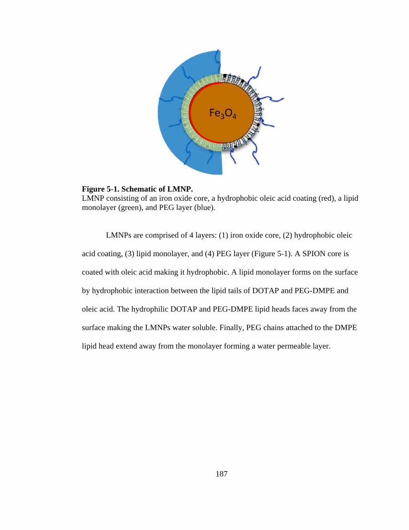
magnetic nanoparticles (LMNPs).
A fluorescent probe was embedded in the lipid bilayer of magnetoliposomes
allowing the local temperature and membrane fluidity to be observed. When subjected
to an electromagnetic field that heated the encapsulated iron oxide nanoparticles
encapsulated in the lipid bilayer, the local temperature and membrane fluidity could be
observed.
DLNAs were encapsulated with different sized nanoparticles and
concentrations in order to observe the effect of the bilayer nanoparticles on the lipid
bilayer’s phase behavior and leakage. Two different sized nanoparticles were used, a 2
nm gold nanoparticle (GNP) much smaller than the thickness of the bilayer and a 4 nm
GNP near the thickness of the lipid bilayer. The 2 nm GNPs were shown to affect the
lipid bilayer differently than the 4 nm GNP. Specifically, the two nanoparticles altered
the phase behavior and leakage differently in a temperature dependent fashion,
demonstrating that embedded nanoparticle size can be used induce or inhibit bilayer
leakage.
A dual solvent exchange method was used to control the lipid surface
composition of an iron oxide nanoparticle with a cationic lipid and a polyethylene
glycol (PEG) lipid to produce lipid coated magnetic nanoparticles (LMNPs). PEG is
well known for its ability to enhance the pharmacokinetics of nanostructures by
preventing uptake by the immune system. By controlling the lipid surface
composition, the surface charge and PEG conformation can be controlled which
Page 6
allowed the LMNPs to be used as an MRI contrast agent and a delivery system for
siRNA that could be triggered with temperature.
Page 7
v
ACKNOWLEDGMENTS
I would like to extend my deepest gratitude to my whole family: Mom, Dad,
Jenn, Jess, Amy, Tom, Scott, Ben, Marcus, Jake, Jessica Rose, Jackson, Jeff, Liz, and
Chris without your loving support, I would not have been able to accomplish this. I
love you with all my heart.
I would like to especially recognize my Father, who is my best friend and role
model. Without your support, guidance, time, and love, I would not be the person I am
today. You have been the most influential person in my life. I hope someday I can
become the man you are. Everything I accomplish I owe to you. Thank you for
everything. I cannot express how much I love you.
I would like to also thank my Mother for her support in all aspects of my life.
No matter what time of day or night, you have always been there for me and I cannot
thank you enough for that. I truly appreciate everything you have done for me. Thank
you for always being there willing to listen and help when I needed it most. I love you
with all my heart and am grateful to have a mother like you.
I would also like to extend a heartfelt thank you to Dr. Geoffrey Bothun. You
are not just a teacher or mentor to me, you are also a friend. Thank you for allowing
me to be your graduate student and having the opportunity to learn from you. You
have had a profound influence on my life, not just as a student and researcher, but also
as a person. I am a better person because of you. I truly appreciate your willingness to
always been there for me, regardless if it is school related or not. You are and always
will be one of the most influential people in my life. You have touched my life more
Page 8
vi
than you will ever know or realize. I will always have a special place in my heart for
you. Thank you for everything.
I would like to acknowledge Providence College, Columbia University, and
University of New Hampshire School of Law and their faculty, the education I have
received from each of these institutions have had an enormous influence on me as a
person. I especially would like the University of Rhode Island and the Department of
Chemical Engineering for giving me the opportunity to learn from such great faculty
members and allowing me to study for my Ph.D. I would like to especially thank the
following professors for their influence on me: Dr. Harry Knickle (my M.S. advisor),
Dr. Stanley Kowalski (ITTI and UNH Law), and Jon Cavicchi (UNH Law).
Page 9
vii
PREFACE
This dissertation was prepared in manuscript format. Chapter 1 is an overview
into the various different lipid-nanoparticle assemblies (LNAs) structures that can be
formed combining lipids and nanoparticles for therapeutic and diagnostic (theranostic)
applications. Chapter 2 investigates LNAs responsive to stimuli, such as magnetic,
temperature, pH, light, ultrasound, etc., which can allow them to be used for triggered
and controlled release of therapeutics. Chapters 1 and 2 provide a summary of the
current status of LNA research and development. Chapter 3 is a manuscript related to
measuring the fluidity of the lipid bilayer of a magnetoliposomes (liposomes with iron
oxide encapsulated in its aqueous core) when the nanoparticles are heated with an
electromagnetic field at radio frequency. Chapter 4 is a manuscript that investigated
the effect nanoparticle size and concentration on the phase behavior and permeability
of bilayer decorated LNAs (DLNAs). Chapter 5 is a manuscript that examines how
lipid surface concentration of an iron oxide nanoparticle effects the MRI relaxivity and
siRNA binding and release.
The first chapter, entitled “Liposome-Nanoparticle Assemblies”, is Chapter 11
(pg. 273-307) in the book Bionanotechnology: Biological Self-Assembly and its
Applications published by Caister Academic Press (Norfolk, UK) in 2013. This
chapter was invited submission by the editor, Bernd H.A. Rehm. Also, flatteringly, the
front cover of the book (displayed on the Chapter 1 title page) was selected from
Figure 1-2 that we submitted.
Page 10
viii
The second chapter, entitled “Stimuli-Responsive Liposome-Nanoparticle
Assemblies”, was an invited article published in Expert Opinion on Drug Delivery in
2011 (Expert Opinion on Drug Delivery 8(8) 1025-1040 (2011)).
The third chapter, entitled “Local Heating in Magnetite Nanoparticle-
Liposome Dispersions via Fluorescence Anisotropy”, was published in the Journal of
Colloid and Interface Science in 2011 (Journal of Colloid and Interface Science
357(1) 70-74 (2011)).
The fourth chapter, entitled “Hydrophobic Nanoparticles Embedded in
Liposomes Modify the Thermal Release Behavior of Encapsulated
Carboxyfluorescein”, is a manuscript currently in preparation for submission to ACS
Nano.
The fifth chapter, entitled “MRI Relaxivity and siRNA Binding Capacity of
Lipid-Coated Magnetic Nanoparticles Controlled by PEG Confirmation”, is a
manuscript currently in preparation for submission to Nano Letters.
Page 11
ix
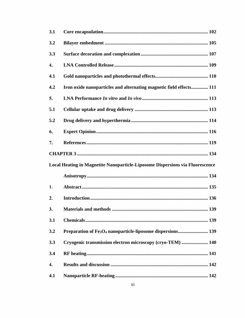
TABLE OF CONTENTS
ABSTRACT .................................................................................................................. ii
ACKNOWLEDGMENTS ........................................................................................... v
PREFACE ................................................................................................................... vii
TABLE OF CONTENTS ............................................................................................ ix
LIST OF TABLES .................................................................................................... xiv
LIST OF FIGURES ................................................................................................... xv
CHAPTER 1 ................................................................................................................. 1
Liposome-Nanoparticle Assemblies ............................................................................ 1
Abstract ............................................................................................................. 2
Introduction ...................................................................................................... 3
Liposome ........................................................................................................... 9
Nanoparticles .................................................................................................. 13
4.1 Quantum Dots ................................................................................................ 15
4.2 Gold Nanoparticles ........................................................................................ 16
4.3 Superparamagnetic Iron Oxide Nanoparticles ........................................... 17
Formation, Structure, and Design Strategies .............................................. 18
5.1 Encapsulated Liposome-Nanoparticle Assembly ........................................ 24
5.2 Bilayer-Decorated Liposome-Nanoparticle Assembly ................................ 31
5.3 Surface-Coupled Liposome-Nanoparticle Assembly and Complexation .. 34
Controlled Release ......................................................................................... 37
6.1 Gold Nanoparticles and Photothermal Effects ............................................ 38
6.2 Iron Oxide Nanoparticles and Alternating Magnetic Fields ...................... 41
Page 12
x
Targeted Therapy .......................................................................................... 43
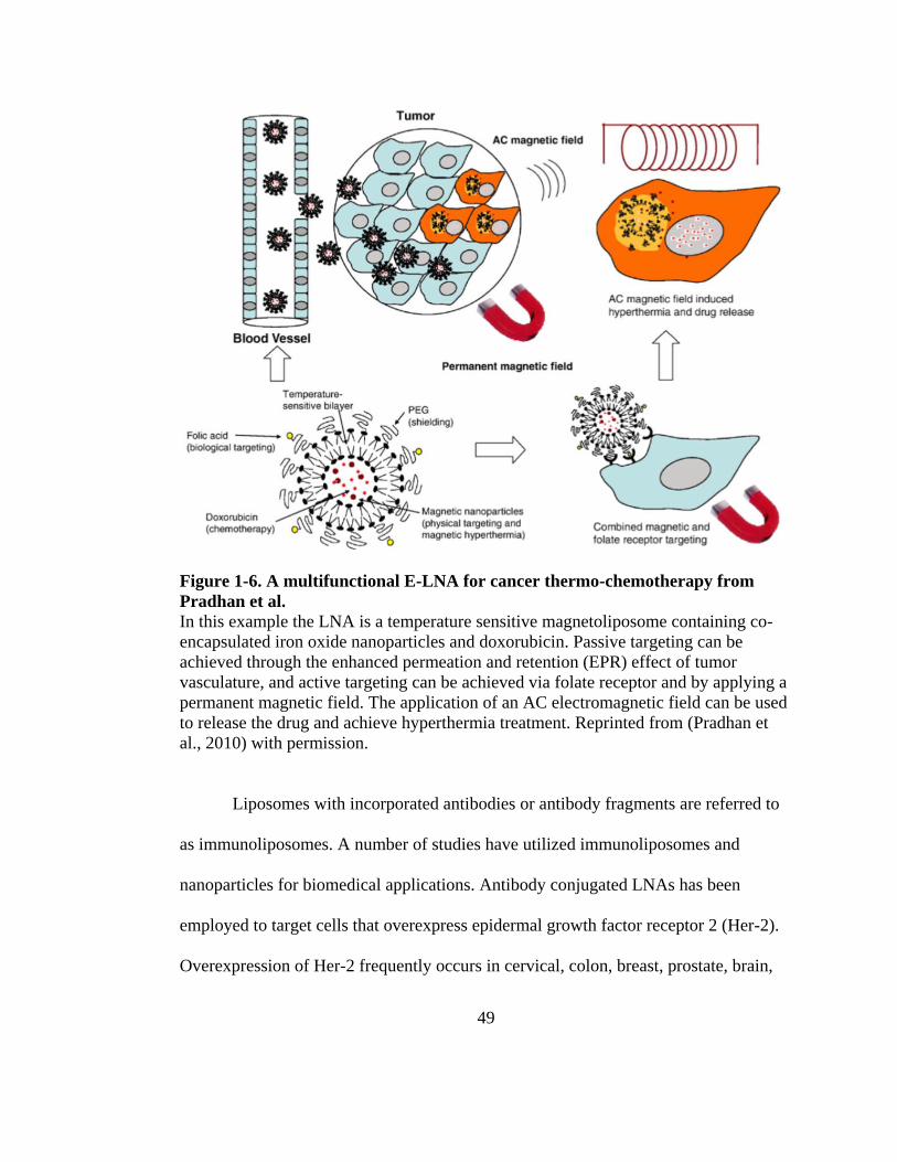
7.1 Passive Targeting ........................................................................................... 43
7.2 Active Targeting ............................................................................................. 47
7.2.1 Ligand Targeting ............................................................................................ 48
7.2.2 Magnetic Drug Targeting .............................................................................. 51
Diagnostics and Imaging ............................................................................... 53
Hyperthermia ................................................................................................. 56
In vivo and in vitro Biomedical Applications .............................................. 58
10.1 Cellular Uptake and Drug Delivery ............................................................. 58
10.2 Drug Delivery and Hyperthermia ................................................................ 59
Conclusion and Future Outlook ................................................................... 60
References ....................................................................................................... 61
CHAPTER 2 ............................................................................................................... 84
Stimuli-Responsive Liposome-Nanoparticle Assemblies ........................................ 84
Abstract ........................................................................................................... 85
1.1 Introduction: ................................................................................................... 85
1.2 Areas Covered: ............................................................................................... 85
1.3 Expert Opinion: .............................................................................................. 85
Introduction .................................................................................................... 87
2.1 Liposomes ....................................................................................................... 92
2.2 Gold and iron oxide nanoparticles ............................................................... 95
2.3 Nanoparticle-mediated hyperthermia .......................................................... 97
LNA Formation and Structure ..................................................................... 98
Page 13
xi
3.1 Core encapsulation ....................................................................................... 102
3.2 Bilayer embedment ...................................................................................... 105
3.3 Surface decoration and complexation ........................................................ 107
LNA Controlled Release .............................................................................. 109
4.1 Gold nanoparticles and photothermal effects............................................ 110
4.2 Iron oxide nanoparticles and alternating magnetic field effects .............. 111
LNA Performance In vitro and In vivo ....................................................... 113
5.1 Cellular uptake and drug delivery ............................................................. 113
5.2 Drug delivery and hyperthermia ................................................................ 114
Expert Opinion ............................................................................................. 116
References ..................................................................................................... 119
CHAPTER 3 ............................................................................................................. 134
Local Heating in Magnetite Nanoparticle-Liposome Dispersions via Fluorescence
Anisotropy ..................................................................................................... 134
Abstract ......................................................................................................... 135
Introduction .................................................................................................. 136
Materials and methods ................................................................................ 139
3.1 Chemicals ...................................................................................................... 139
3.2 Preparation of Fe3O4 nanoparticle-liposome dispersions ......................... 139
3.3 Cryogenic transmission electron microscopy (cryo-TEM) ...................... 140
3.4 RF heating ..................................................................................................... 141
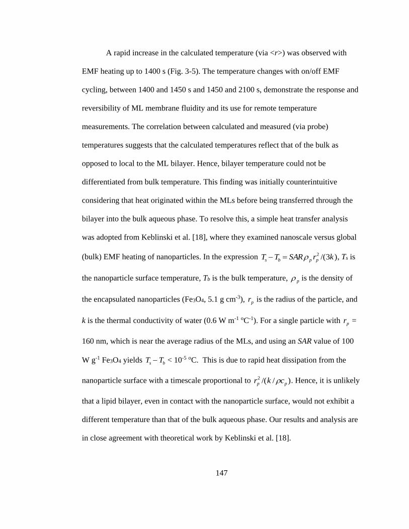
Results and discussion ................................................................................. 142
4.1 Nanoparticle RF-heating ............................................................................. 142
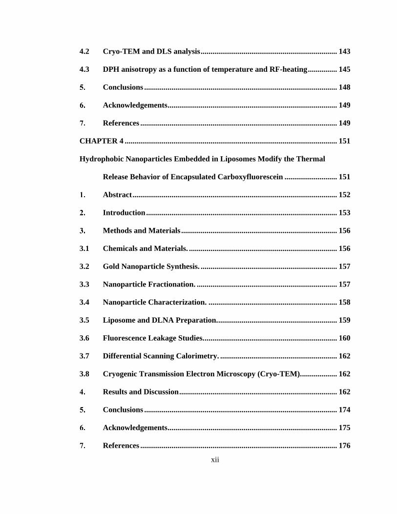
Page 14
xii
4.2 Cryo-TEM and DLS analysis ...................................................................... 143
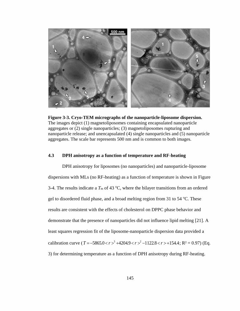
4.3 DPH anisotropy as a function of temperature and RF-heating ............... 145
Conclusions ................................................................................................... 148
Acknowledgements ....................................................................................... 149
References ..................................................................................................... 149
CHAPTER 4 ............................................................................................................. 151
Hydrophobic Nanoparticles Embedded in Liposomes Modify the Thermal
Release Behavior of Encapsulated Carboxyfluorescein ........................... 151
Abstract ......................................................................................................... 152
Introduction .................................................................................................. 153
Methods and Materials ................................................................................ 156
3.1 Chemicals and Materials. ............................................................................ 156
3.2 Gold Nanoparticle Synthesis. ...................................................................... 157
3.3 Nanoparticle Fractionation. ........................................................................ 157
3.4 Nanoparticle Characterization. .................................................................. 158
3.5 Liposome and DLNA Preparation. ............................................................. 159
3.6 Fluorescence Leakage Studies. .................................................................... 160
3.7 Differential Scanning Calorimetry. ............................................................ 162
3.8 Cryogenic Transmission Electron Microscopy (Cryo-TEM). .................. 162
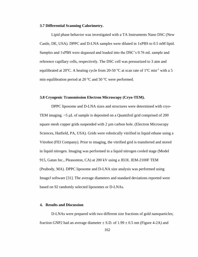
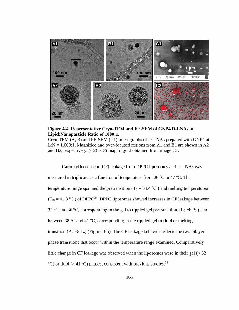
Results and Discussion ................................................................................. 162
Conclusions ................................................................................................... 174
Acknowledgements ....................................................................................... 175
References ..................................................................................................... 176
Page 15
xiii
CHAPTER 5 ............................................................................................................. 182
MRI Relaxivity and siRNA Binding Capacity of Lipid-Coated Magnetic
Nanoparticles Controlled by Polyethylene Glycol Confirmation ............ 182
Abstract ......................................................................................................... 183
Supporting Information .............................................................................. 202
Chemicals and Materials ............................................................................. 203
Experimental Section ................................................................................... 204
4.1 Lipid Coated Magnetic Nanoparticle (LMNP) Formation ...................... 204
4.2 Cryogenic Transmission Electron Microscopy (Cryo-TEM) ................... 205
4.3 Dynamic Light Scattering (DLS) ................................................................ 205
4.4 Heating with Alternating Current Electromagnetic Field (AC EMF)
Operating at Radio Frequency (RF) .......................................................... 206
Small Interfering RNA (siRNA) Experiments ........................................... 207
5.1 siRNA Binding .............................................................................................. 207
5.2 siRNA Release with Temperature .............................................................. 207
5.3 siRNA RF Release ........................................................................................ 208
Magnetic Resonance Imaging (MRI) ......................................................... 209
6.1 MRI Sample Preparation ............................................................................ 209
6.2 MRI Methods for r2 Relaxation .................................................................. 209
6.3 Acknowledgements ....................................................................................... 210
References ..................................................................................................... 210
Page 16
xiv
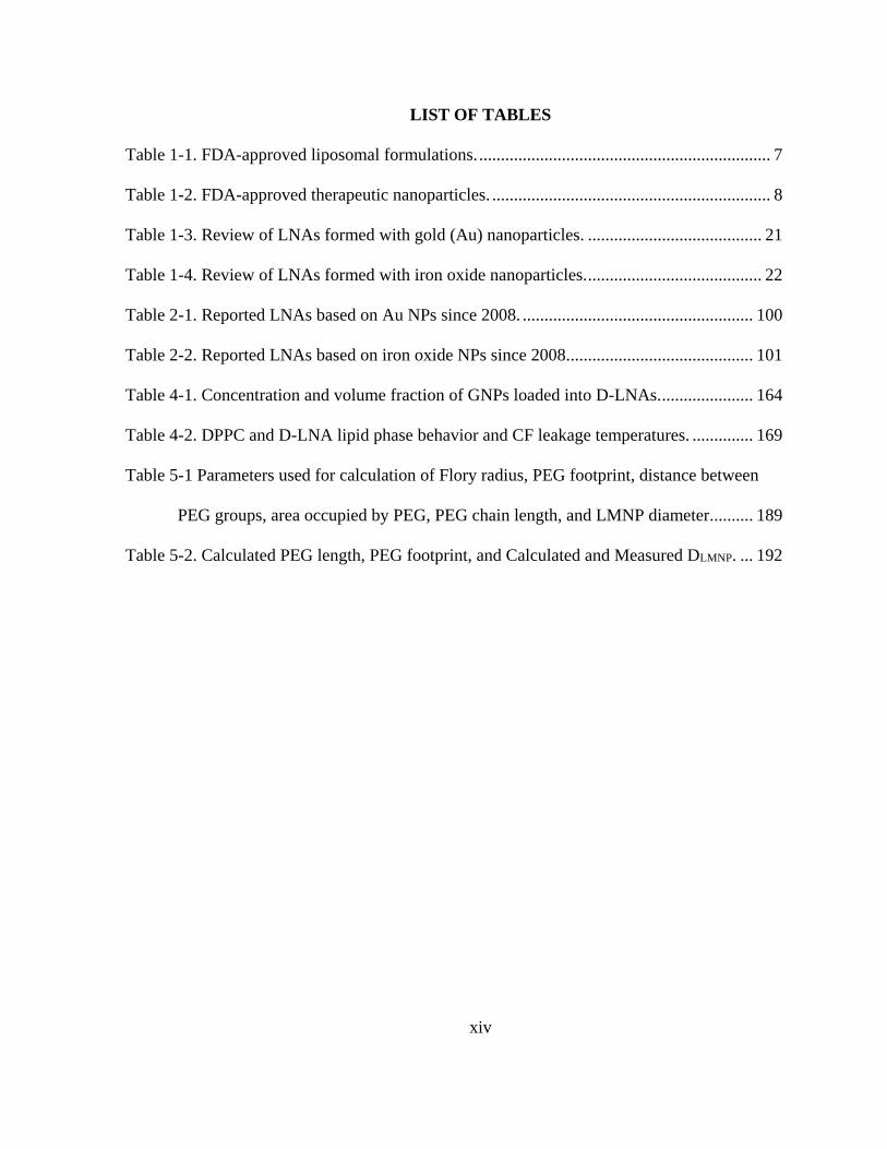
LIST OF TABLES
Table 1-1. FDA-approved liposomal formulations. ................................................................... 7
Table 1-2. FDA-approved therapeutic nanoparticles. ................................................................ 8
Table 1-3. Review of LNAs formed with gold (Au) nanoparticles. ........................................ 21
Table 1-4. Review of LNAs formed with iron oxide nanoparticles. ........................................ 22
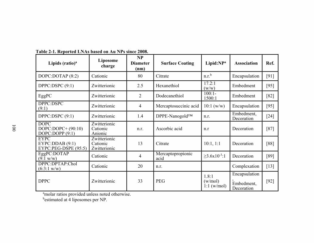
Table 2-1. Reported LNAs based on Au NPs since 2008. ..................................................... 100
Table 2-2. Reported LNAs based on iron oxide NPs since 2008. .......................................... 101
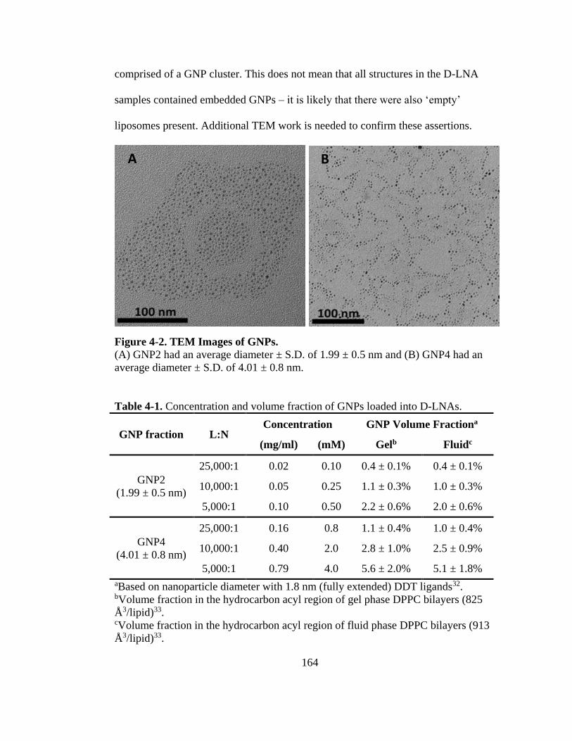
Table 4-1. Concentration and volume fraction of GNPs loaded into D-LNAs. ..................... 164
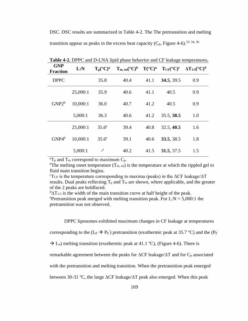
Table 4-2. DPPC and D-LNA lipid phase behavior and CF leakage temperatures. .............. 169
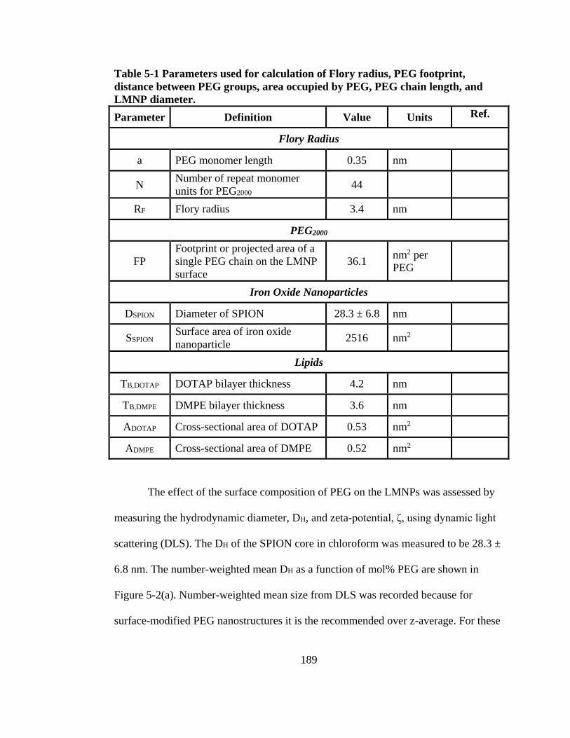
Table 5-1 Parameters used for calculation of Flory radius, PEG footprint, distance between
PEG groups, area occupied by PEG, PEG chain length, and LMNP diameter. ......... 189
Table 5-2. Calculated PEG length, PEG footprint, and Calculated and Measured DLMNP. ... 192
Page 17
xv
LIST OF FIGURES
Figure 1-1. Schematic and cryogenic transmission electron micrograph of
dipalmitoylphosphatidylcholine liposomes (DPPC). ................................................... 10
Figure 1-2. Schematics and cryogenic transmission electron micrograph of liposome-
nanoparticle assemblies. ............................................................................................... 19
Figure 1-4. Changes in bilayer decoration mechanism of D-LNAs with increasing
nanoparticle diameter. .................................................................................................. 32
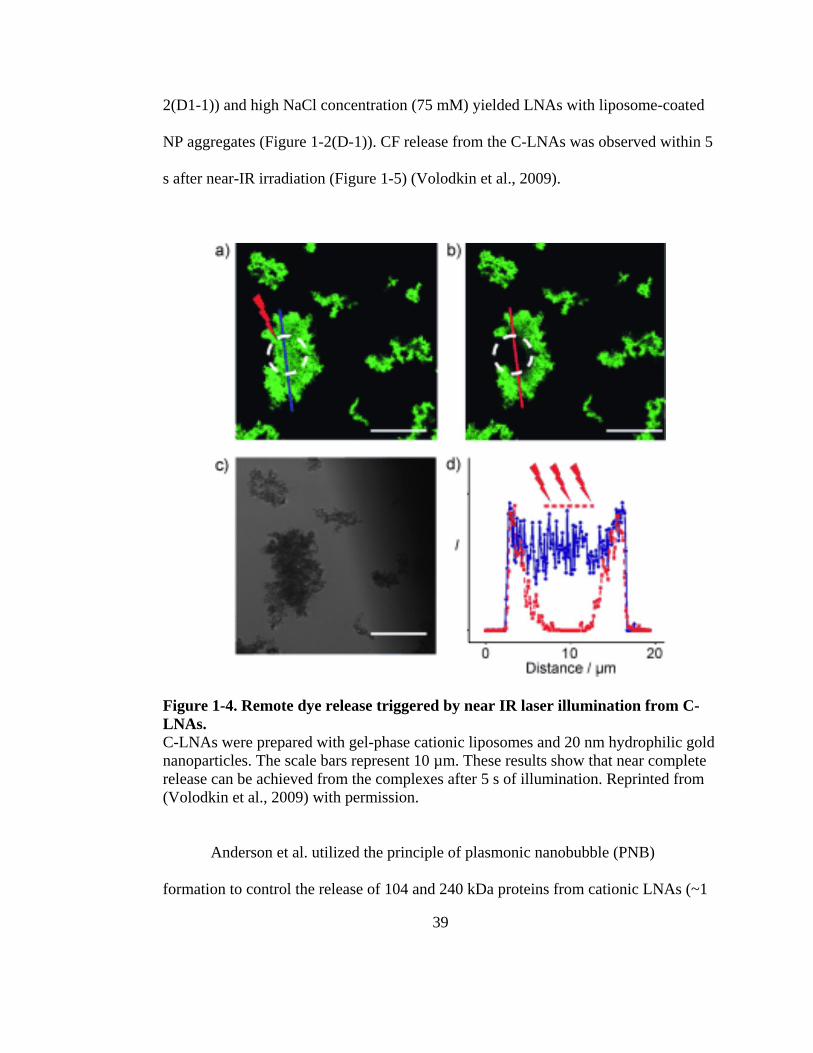
Figure 1-5. Remote dye release triggered by near IR laser illumination from C-LNAs. ......... 39
Figure 1-6. Proposed ‘plasmonic nanobubble’ release mechanism of encapsulated molecules
from E-LNAs prepared with gold nanoparticles. ......................................................... 40
Figure 1-7. A multifunctional E-LNA for cancer thermo-chemotherapy from Pradhan et al. 49
Figure 1-8. Combined imaging and doxorubicin release from D-LNAs prepared with
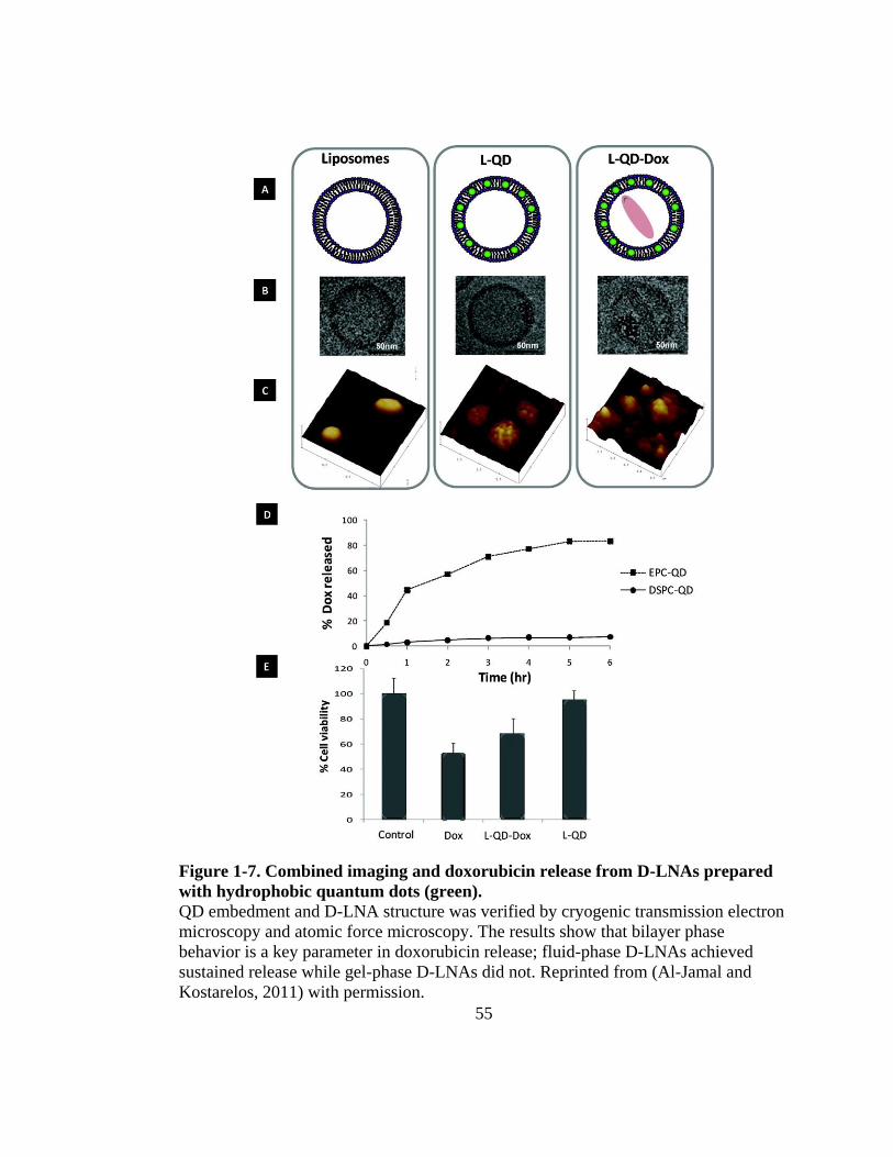
hydrophobic quantum dots (green). ............................................................................. 55
Figure 2-1. Schematics and TEM micrographs of liposome-nanoparticle assemblies. ........... 91
Figure 2-2. Conceptualization of a multifunctional liposome-nanoparticle assembly. ........... 92
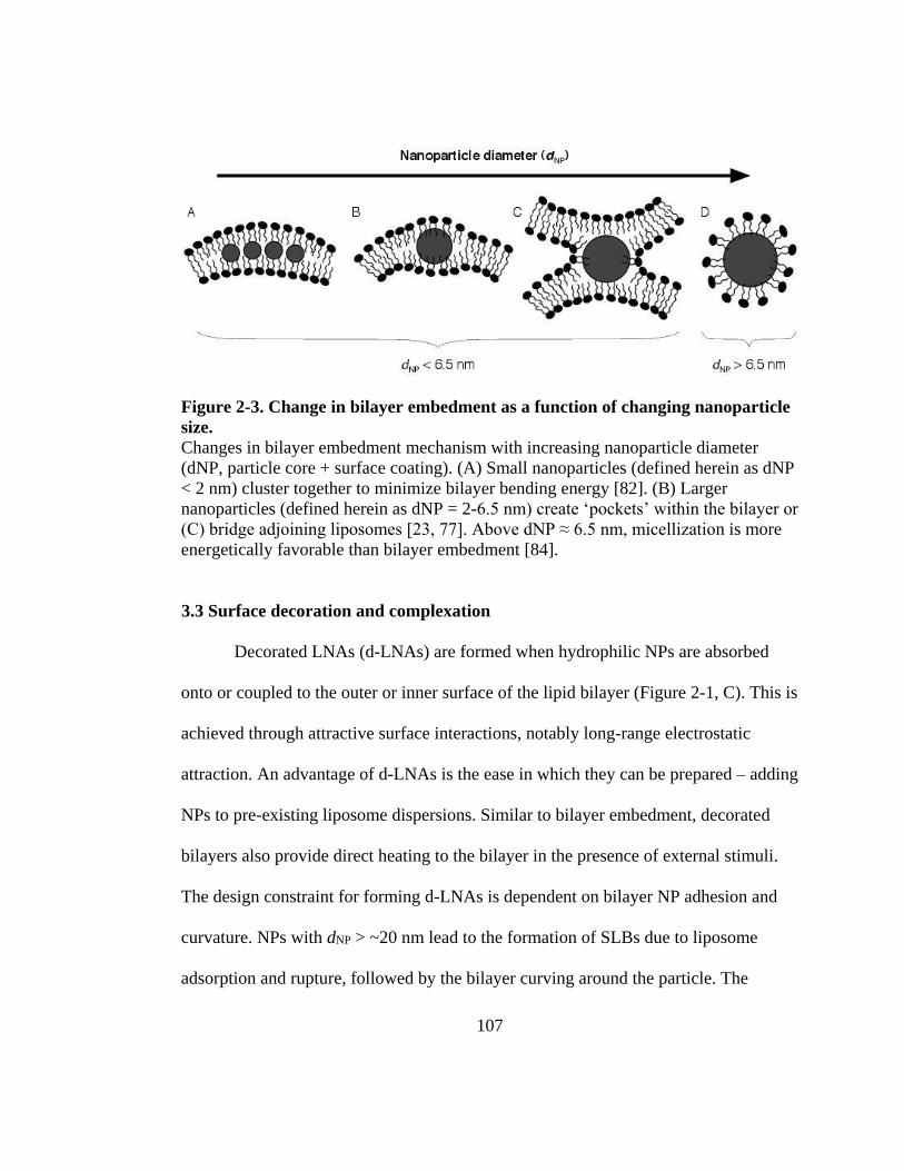
Figure 2-3. Change in bilayer embedment as a function of changing nanoparticle size. ...... 107
Figure 2-4. Controlled release from a decorated liposome-nanoparticle assembly under the
influence of an alternating current electromagnetic field at radio frequency. ........... 113
Figure 2-5. Nanoparticle heating controlled release mechanisms from liposome-nanoparticle
assemblies. ................................................................................................................. 118
Figure 3-1. Magnetoliposome structures................................................................................ 137
Figure 3-2. Specific absorption rate (SAR) of the Fe3O4 nanoparticles as a function of RF field
strength (Hf, in legend) and nanoparticle mass fraction (in water). ........................... 143
Page 18
xvi
Figure 3-3. Cryo-TEM micrographs of the nanoparticle-liposome dispersion. ..................... 145
Figure 3-4. Melting transition determined by DPH anisotropy. ............................................ 146
Figure 3-5. DPH anisotropy and bilayer calculated temperature with RF heating. ............... 148
Figure 4-1. Schematics of D-LNA structures loaded with GNP2 and GNP4. ....................... 156
Figure 4-2. TEM Images of GNPs. ........................................................................................ 164
Figure 4-3. Representative Cryo-TEM micrographs. ............................................................ 165
Figure 4-4. Representative Cryo-TEM and FE-SEM of GNP4 D-LNAs at Lipid:Nanoparticle
Ratio of 1000:1. .......................................................................................................... 166
Figure 4-5. Percentage of carboxyfluorecein (CF) leakage as a function of temperature. .... 168
Figure 4-6. Change in CF leakage over change in time and excess heating capacity as a
function of temperature. ............................................................................................. 171
Figure 5-1. Schematic of LMNP. ........................................................................................... 187
Figure 5-2. (a) LMNP Measured Hydrodynamic Diameter and ζ, (b) Predicted PEG Length
and Polymer Conformation, (c) Schematic and cryogenic transmission electron
microscope images of LMNPs. .................................................................................. 188
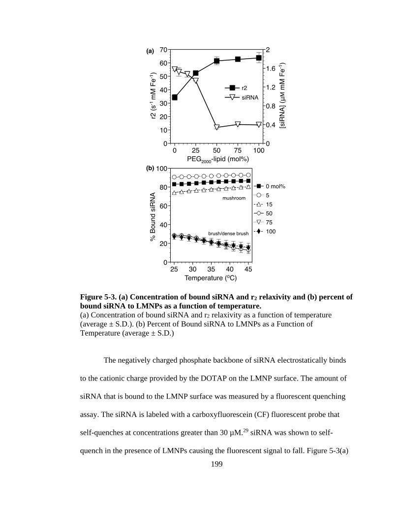
Figure 5-3. (a) Concentration of bound siRNA and r2 relaxivity and (b) percent of bound
siRNA to LMNPs as a function of temperature. ........................................................ 199
Figure 5-4. (a) Change in Bulk Sample Temperature over RF Heating Time and (b) Percent
siRNA Release over RF Heating Time. ..................................................................... 202
Page 19
1
CHAPTER 1
Liposome-Nanoparticle Assemblies
Matthew R. Preiss1, Anju Gupta2, and Geoffrey D. Bothun1†
Published in: Bionanotechnology: Biological Self-Assembly and its Applications,
B.H.A. Rehm, eds. (Norfolk, UK: Caister Academic Press), pp. 265-297 (2013).
1University of Rhode Island,
Department of Chemical Engineering,
Kingston, RI, USA
2Rochester Institute of Technology,
Department of Chemical Engineering,
Rochester, NY, 14623
†Correspondence: Geoffrey D Bothun
Department of Chemical Engineering
University of Rhode Island
205 Crawford Hall, 16 Greenhouse Road,
Kingston, RI, 02881, USA
Phone: +1-401-874-9518
Geoffrey D. Bothun Email: [email protected]
Matthew R. Preiss Email: [email protected]
Page 20
2
Abstract
Liposome-nanoparticle assemblies (LNAs) combine the demonstrated potential
of clinically approved nanoparticles and liposomes to achieve multiple therapeutic and
diagnostic objectives. Efficient and effective biomedical application requires
assemblies to be stable, biocompatible, and bioavailable, while enhancing the
properties of encapsulates. LNAs have been demonstrated to be very effective for in
vivo and in vitro providing targeting and stimuli-responsive delivery of therapeutic and
imaging agents. The ability to design LNAs with nanoparticle encapsulation, bilayer-
decoration, and surface coupling provides a variety of different structures and
functions. While the potential of LNAs has been demonstrated, future investigation
into the interaction between the lipid bilayer and nanoparticles is necessary to
understand and develop LNAs for clinical applications. This section will discuss the
current state of liposome-nanoparticle assembly design, characterization, and
applications of liposome-nanoparticle assemblies.
Page 21
3
Introduction
Only about 11% of new promising therapeutic compounds in clinical
development are eventually approved. Nearly 70% of drug failures are attributed to
poor pharmacokinetics, efficacy, toxicology, clinical safety, and formulation (Kola
and Landis, 2004; Leeson and Davis, 2004). High drug attrition rates are the major
cause of the recent decline in breakthrough drugs and the rise in costs of new drug
therapies. Developments in nanotechnology have demonstrated potential for
overcoming the issues related to drug pharmacokinetics and pharmacodynamics.
Targeted and controlled delivery of therapeutic agents directly to targeted tissues can
be achieved, improving efficacy, lowering the necessary dose, and reducing adverse
effects. Nobel Laureate Paul Ehrlich’s dream of a “magic bullet” to fight disease may
be realized through controlled and targeted nanoscale therapeutics (Koo et al., 2005).
In 2004, the National Cancer Institute launched the Alliance for
Nanotechnology in Cancer (Alliance). The Alliance’s goal is development of
nanotechnology-based cancer treatments and imaging. Specifically, the Alliance is
emphasizing the development of drug delivery that targets tumor cells, tumor’s
microenvironment, and metastatic, recurrent, and drug resistant cancers with
nanotherapeutic delivery systems, theranostics, contrast agents, and complexes
capable of providing multiple therapies (National Cancer Institute). The design of such
multifunctional constructs is inherently complex as it requires combining different
molecular, colloidal, and/or particulate agents. Furthermore, the construct must be
stable, resistant to protein and immune system absorption, and capable of targeting.
Page 22
4
Novel liposome-nanoparticle assemblies (LNAs) provide a biologically
inspired route for designing multifunctional targeted therapeutics and imaging. The
LNA structure is inspired by the early development of magnetoliposomes (liposomes
with magnetic nanoparticles encapsulated in the aqueous core). Recent literature has
referred to LNAs as “liposome-nanoparticle hybrids” (Al-Jamal and Kostarelos,
2007). LNAs are liposome structures in which nanoparticles (NPs) are encapsulated in
the aqueous core, embedded in the lipid bilayer, or coupled to the bilayer surface.
Liposomes are a well-established vehicle for the administration of therapeutic and
diagnostic agents (Bangham and Horne, 1964; Bangham et al., 1965; Gregoriadis,
1973; Papahadjopoulos and Ohki, 1969). As a biocompatible carrier, liposomes
provide a stable means for the transportation and protection of hydrophilic and/or
hydrophobic molecules. Nanoparticles are nanoscale moieties that have been
demonstrated to be effective transportation vehicles, contrast agents, and agents
responsive to external stimuli (such as electromagnetic fields and light). LNAs
combine the advantageous properties of liposomes with functional nanoparticles to
create a multifunctional therapeutic and diagnostic construct (Zhang et al., 2008).
LNAs have several advantages when utilized for drug delivery, hyperthermia,
imaging, and diagnostic applications. LNAs are able to delivery hydrophobic and/or
hydrophilic molecules and NPs (Zhang et al., 2009). The liposome can be modified to
protect encapsulated agents from biomolecule absorption and functionalized for
targeting. LNAs can also be used to concentrate encapsulates, increasing the efficiency
of delivery. Also, the strategies for processing, stabilizing, and targeting liposomes are
Page 23
5
well established (Immordino et al., 2006). NPs can be magnetically guided for in vivo
targeting and provide a mechanism for stimuli-responsive triggering. Surface-bound
NPs also enhance the colloidal stability of LNAs and bilayer-embedded NPs can
reduce spontaneous leakage (Chen et al., 2010; Paasonen et al., 2007b; Yu et al., 2007;
Zhang and Granick, 2006). LNAs harness the intrinsic advantages of a liposomal
carrier, enhancing stability, bioavailability, and biocompatibility, and adds the imaging
and/or responsive functionality of a NP (Zhang et al., 2008).
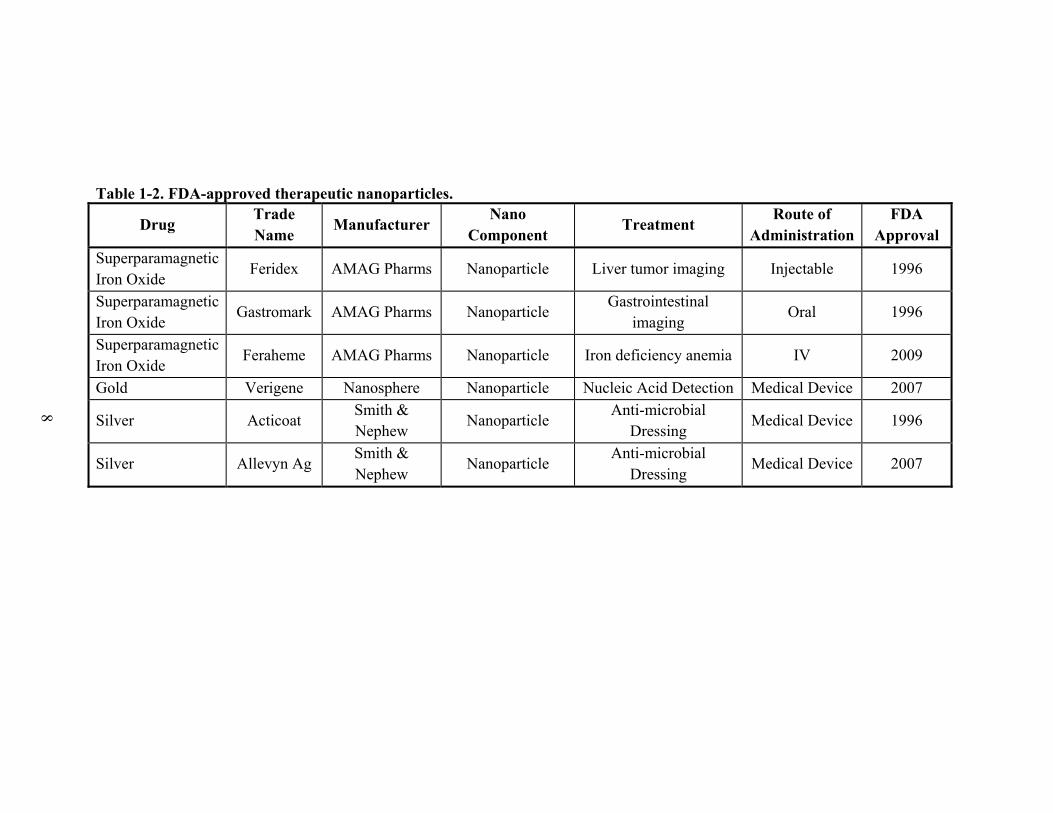
Drug delivering liposomes and nanoparticles have both been approved
separately for clinical use by the U.S. Food and Drug Administration (FDA), see
Table 1-1 and Table 1-2, respectively. These approved therapies represent the first-
generation of development for nano-scale therapeutics and diagnostics. Combining
liposomes and nanoparticles, to form multimodal LNAs, is the natural evolution for
these technologies. This section will focus on a review of LNA design and structure,
characterization techniques, and biomedical applications, such as controlled drug
release, imaging, and hyperthermia, expanding on our group’s review on stimuli-
responsive LNAs (Preiss and Bothun, 2011). Recent reviews focusing on the
therapeutic and diagnostic applications of liposomes are provided in references (Goyal
et al., 2005; Immordino et al., 2006; Kshirsagar et al., 2005; Maurer et al., 2001;
Mulder et al., 2006; Puri et al., 2009; Samad et al., 2007; Torchilin, 2005) and NPs are
provided in references (Corchero and Villaverde, 2009; Emerich and Thanos, 2006;
Fukumori and Ichikawa, 2006; Groneberg et al., 2006; Jin and Ye, 2007; Laurent et
Page 24
6
al., 2008; McCarthy and Weissleder, 2008; Michalet et al., 2005; Polyak and
Friedman, 2009; Rotomskis et al., 2006; Wang et al., 2008).
Page 25
7
Table 1-1. FDA-approved liposomal formulations.
Drug Trade Name Manufacturer Nano
Component Treatment
Route of Administration
FDA Approval
Amphotericin B Abelcet Sigma-Tau Lipid complex Fungal infections Injectable 1995
Amphotericin B AmBisome Astellas Liposome Fungal and protozoal infections
Injectable 1997
Amphotericin B Amphotec Aldopharma USA
Lipid colloidal dispersion
Fungal infections Injectable 1996
Daunorubicin DaunoXome Galen Liposome Advanced HIV Kaposi’s sarcoma; Ovarian cancer
Injectable 1996
Cytarabine DepoCyt Pacira Pharms Liposome Malignant lymphomatous meningitis
Injectable 1999
Morphine DepoDur EKR Theraputics
Liposome Postsurgical analgesia Epidural 2004
Doxorubicin Doxil Janssen PEGylated liposomes
Metastatic ovarian cancer; AIDS-related Kaposi’s sarcoma
Injectable 1995
Verteporfin Visudyne QLT Liposome
Age-related macular degeneration, pathologic myopia, ocular histoplasmosis
Injectable 2000
Propofol Diprivan APP Pharms Liposome Anesthetic Injectable 1989
Bupivacaine Exparel Pacira Pharmaceuticals
Liposome Analgesic Injectable 2011
Page 26
8
Table 1-2. FDA-approved therapeutic nanoparticles.
Drug Trade Name
Manufacturer Nano
Component Treatment
Route of Administration
FDA Approval
Superparamagnetic Iron Oxide
Feridex AMAG Pharms Nanoparticle Liver tumor imaging Injectable 1996
Superparamagnetic Iron Oxide
Gastromark AMAG Pharms Nanoparticle Gastrointestinal
imaging Oral 1996
Superparamagnetic Iron Oxide
Feraheme AMAG Pharms Nanoparticle Iron deficiency anemia IV 2009
Gold Verigene Nanosphere Nanoparticle Nucleic Acid Detection Medical Device 2007
Silver Acticoat Smith & Nephew
Nanoparticle Anti-microbial
Dressing Medical Device 1996
Silver Allevyn Ag Smith & Nephew
Nanoparticle Anti-microbial
Dressing Medical Device 2007
Page 27
9
Liposome
Since the early work by Bangham, Papahadjopoulos, and Gregoriadis in the
1960-1970s, liposomes have become one of the most highly investigated nano-
structures. Today, liposomes are used as model biological membranes and for
therapeutic and diagnostic agent delivery (Bangham and Horne, 1964; Bangham et al.,
1965; Gregoriadis, 1973; Papahadjopoulos and Ohki, 1969). Liposomes are reliable
systemic drug delivery systems because they are non-toxic, biocompatible, capable of
prolonging bioavailability of encapsulated agents by reducing or preventing drug
degradation and enhancing solubility and stability (Al-Jamal and Kostarelos, 2007).
Liposomes, as depicted in Figure 1-1(A), are composed of self-assembled
spherical vesicles consisting of one or multiple lipid bilayers surrounding an internal
aqueous core. Bilayer thickness (lb) is ~5 nm thick (lb), composed of a hydrophobic
acyl lipid tail region (~3 nm) and a hydrophilic headgroup. Liposomes can be prepared
with zwitterionic, anionic, or cationic lipids, and the net liposome surface charge can
be adjusted by mixing different ratios of these components. From a morphological
aspect, liposomes are distinguished according to their diameter, small (<100 nm),
large (100-1000 nm, or giant (>1000 nm), and number of bilayers, single (unilamellar)
or multiple (multilamellar) (Sivashankar, 2011). Figure 1-1(B) is a cryogenic
transmission electron microscope (cryo-TEM) image of liposomes depicting the
structures that can be formed. For drug delivery and diagnostics, liposomes are
attractive because of their ability to encapsulate both hydrophilic (in the aqueous core
or bound to the liposome surface) and hydrophobic (in the lipid bilayer) molecules.
Page 28
10
Liposomes also open the therapeutic window, reducing adverse effects, by altering the
pharmacokinetic and pharmacodynamic characteristics of the encapsulated agent (Al-
Jamal and Kostarelos, 2007).
Figure 1-1. Schematic and cryogenic transmission electron micrograph of
dipalmitoylphosphatidylcholine liposomes (DPPC).
(A) Liposome schematic depicting the aqueous core, hydrophilic headgroup, and
hydrophobic tail regions and (B) a cryogenic transmission electron micrograph of
dipalmitoylphosphatidylcholine liposomes (DPPC, 10 mM) prepared in phosphate
buffered saline.
Page 29
11
FDA-approved liposomal amphotericin B formulations (Abelcet®,
AmBisome®, and Amphotec®) are good examples of the effectiveness of liposomes
for drug delivery. Amphotericin B is considered the “gold standard” for systemic
treatment of fungal infections. However, amphotericin B is hydrophobic and
nephrotoxic, limiting its stability and administered dosage. Encapsulation of
amphotericin B in the lipid bilayer reduced the concentration of amphotericin B in the
kidneys, providing similar efficacy as conventional amphotericin B while significantly
reducing adverse side-effects (Gibaldi et al., 2007; Moen et al., 2009).
Release of encapsulated molecules from liposomes is controlled by the
permeability through the lipid bilayer, which can be achieved by transbilayer diffusion
or transient pore formation triggered by bilayer disruption or phase separation. Phase
separation can be induced by ‘melting’ the liposomal bilayers – i.e. heating to a
temperature greater than the characteristic main phase transition or melting
temperature of the lipids (Tm). Below Tm the lipids are in the solid or gel phase in
which the lipids are rigid and highly organized. Above Tm the lipids are disordered in a
liquid crystalline or fluid phase. Permeability is high at the interface between gel and
fluid phases. Phase separation and bilayer permeability can be manipulated by
adjusting the lipid bilayer composition. A simple example illustrating this principle
can be made with dipalmitoylphosphatidylcholine (DPPC, Tm = 42 oC) and
dimyristoylphosphatidylcholine (DMPC, Tm = 23 oC). At a DPPC/DMPC molar ratio
of 74:26 the melting temperature occurs at physiological temperature (37 oC).
Furthermore, cholesterol is commonly incorporated into the bilayer to reduce
Page 30
12
membrane fluidity above the melting temperature. Membrane fluidity has been shown
to be affected by pH, ion concentration, and the presence of molecules (such as
nanoparticles) absorbed into the bilayer (Al-Jamal and Kostarelos, 2007; Bothun,
2008; Chen et al., 2010).
A major limitation to liposomal drug delivery is the short half-life (Zhang et
al., 2008). Within minutes, the reticuloendothelial system (RES) will eliminate the
liposomes from the blood, limiting the drug’s efficacy and ability to accumulate at
target sites (Moghimi and Szebeni, 2003). Proteins, called opsonins, recognize and
target foreign agents (such as untargeted liposomes) for elimination by the
mononuclear phagocyte system (MPS) or by hepatocyte uptake. Other proteins are
capable of lysing liposomes directly by compromising the stability of the lipid bilayer
(Ishida et al., 2002; Maurer et al., 2001; Yan et al., 2005). Liposome residence time is
dependent on liposome size, surface charge, lipid packing, bilayer composition, and
surface modifiers (Maurer et al., 2001; Samad et al., 2007). Attaching polyethylene
glycol (PEG) to the liposome, forming “stealth liposomes”, can increasing half-life to
2-24 hours and increase liposome stability (Medina et al., 2004; Moghimi and
Szebeni, 2003; Zhang et al., 2008). The first FDA approved liposomal drug
formulation (and FDA approved “nanodrug”) was Doxil® in 1995. Doxil® is
doxorubicin, the most commonly used anthracycline anticancer drug, encapsulated
within a PEGylated liposome. The elimination half-life for Doxil® is 55 hours and an
area under the plasma concentration time curve of 900μg h mL-1, compared to 0.2
hours and 4μg h mL-1 for free doxorubicin (Barenholz, 2012; Chang and Yeh, 2012).
Page 31
13
Drug delivery from liposomes can also be accomplished by cellular uptake,
which can occur by adsorption, endocytosis, fusion, and/or lipid transfer (Pagano and
Weinstein, 1978; Samad et al., 2007; Torchilin, 2005). Adsorption is the association of
liposome bilayer with cell bilayer without destroying the liposome bilayer or being
internalized by the cell. Adsorption can be specific (assisted by targeting ligands such
as antibodies) or nonspecific (controlled by intermolecular and surface forces).
Endocytosis involves the uptake of liposomes into the cell by encapsulation within
endosomes. Release of drugs to the cytoplasm can occur by membrane destabilization
of the encapsulating endosome or by delivery to lysosomes. Lysosomes have an acidic
pH and contain lysing enzymes. Drug release is accomplished when lysosome
enzymes hydrolyze the lipid bilayer releasing the drug. Lysosome drug release is only
effective when the encapsulated drugs are not susceptible to lysosome enzymes and
pH. Fusion involves the adsorption and incorporation of the liposome bilayer with the
cell membrane, releasing the payload into the cytoplasm. Finally, lipid transfer
involves the exchange of lipids between the liposome bilayer and the cell membrane
without enveloping the liposome (Samad et al., 2007; Torchilin, 2005).
Nanoparticles
Nanoparticles are nanoscale moieties having magnetic and optical properties
for use in therapeutic and imaging applications. The high surface area-to-volume ratio,
stability, functionalization, and size (1-100nm, on the order of biological
macromolecules) of nanoparticles make them particularly attractive for biomedical
Page 32
14
applications. Nanoparticles have shown to be particularly effective as a contrast agent,
a heat source, and as a targeting agent. Clinical application of nanoparticles can be
hindered by poor colloidal stability, hydrophobicity, protein absorption, immune
system uptake, and cytotoxicity. LNAs provide a carrier to take advantage of the
properties of nanoparticles for controlled release, targeted therapies, hyperthermia,
diagnostics, and imaging applications (Al-Jamal and Kostarelos, 2007; Huang et al.,
2011). A number of different inorganic nanoparticles have been used in LNAs, such as
quantum dots (Al-Jamal et al., 2008b; Bothun et al., 2009; Gopalakrishnan et al.,
2006), fullerenes (fullerenosomes) (Babincova et al., 2003, 2004; Chen and Bothun,
2009; Doi et al., 2008; Hwang and Mauzerall, 1993; Ikeda and Kikuchi, 2008; Ikeda et
al., 2009; Ikeda et al., 2005; Jeng et al., 2005; Niu and Manzerall, 1996), silver
(Bothun, 2008; Park et al., 2005), superparamagnetic iron oxide (SPIO) (Bothun and
Priess, 2011; Chen et al., 2010), and gold (Park et al., 2006). This section will discuss
several nanoparticles that have been utilized in LNA applications.
Despite their applications in drug and gene delivery and cosmetics,
cytotoxicity remains a major concern. Understanding the interactions between
nanoparticles and cell membranes is crucial to NP biomedical applications and
provides insight into their toxicity. NPs can be designed to bind on the cell surface,
adsorb within the membrane, and translocate across the cell membrane. NPs can be
exploited for novel applications by controlling the interaction between the NP and
bilayer. A common way to achieve this interaction is by modifying the surface of the
NP, specifically by adding positive or negative charges onto NP surface (N. Li, 2006;
Page 33
15
S. Legrand, 2008). Binding interaction between superparamagnetic iron oxide
particles and stem cells are being used in cell selection process (L.F. Pavon, 2008).
NPs used in drug delivery applications can be modified to avoid drug degradation by
increasing the circulation period which in turn results in cell uptake efficiency (S. Jin,
2007).
4.1 Quantum Dots
Quantum dots (QDs), 2-10nm florescent semiconductor nanocrystals, have
been demonstrated as effective imaging and diagnostics agents. QDs can provide a
highly sensitive contrast agent capable of exhibiting fluorescence that is 10-20 times
greater than conventional imaging agents, such as organic dyes and florescent
proteins. QDs are also 100 times more stable against photobleaching than organic dyes
(Chan, 1998). The optical properties of QDs can be tuned by adjusting their size and
composition. Commonly used quantum dots for biomedical applications include
cadmium selenide (CdSe), cadmium telluride (CdTe), indium phosphide (InP), and
indium arsenide (InAs) (Bharali and Mousa, 2010). Clinical application of quantum
dots is limited due to their inherent hydrophobicity and potential cytotoxicity.
Conjugation of quantum dots with liposomes have shown to be effective to overcome
these limitations (Al-Jamal and Kostarelos, 2007; Bothun et al., 2009; Dudu et al.,
2008; Smith et al., 2006; Walling et al., 2009; Weng et al., 2008).
Page 34
16
4.2 Gold Nanoparticles
Imaging and photothermal effects of gold NPs stem from their enhanced
surface plasmon resonance (SPR), where visible or near-infrared light is absorbed
causing oscillation of surface electrons (Huang et al., 2010). SPR absorbance and the
wavelength range are dependent upon nanoparticle size, core/shell configuration (e.g.
silica core/gold shell (Oldenburg et al., 1999)), and geometry. Shifts in these
properties are indicative of the degree of NP aggregation and/or molecular adsorption
on the NP surface (Li and Gu, 2010). For photothermal therapy, absorbed light energy
is converted into local heat that thermally diffuses into the surrounding medium.
Varying NP size and core/shell configuration provides a means of tuning the
frequency window for photothermal therapy. It is generally accepted that gold NP-
mediated phototherapy is attributed to heat or resulting bubble nucleation depending
on the light intensity and mode of exposure (Li and Gu, 2010). However, recent work
by Krpetic et al. at low light energies suggests that photochemical effects – the
formation of free radicals during NP irradiation – may play an important role. In
addition to photothermal heating, electromagnetic fields operating at RF can be used
to heat gold NPs (Krpetic et al., 2010). For example, Gannon et al. examined the effect
of NP concentration and RF field strength on the heating rates of 5 nm Au NPs in
water. A rate of ~74 oC min-1 was measured using an 800 W RF field at a NP
concentration of 67 μM (Gannon et al., 2008).
Page 35
17
4.3 Superparamagnetic Iron Oxide Nanoparticles
Superparamagnetic iron oxide (SPIO) NPs are 4-20nm nanoparticles typically
composed of magnetite (Fe3O4) or maghemite (γ-Fe2O3). SPIO NPs demonstrate
physical and magnetic properties, such as low toxicity and paramagnetism, making
them advantageous for in vitro and in vivo applications. Due to the nanoscale crystal
size of iron oxide, a single magnetic domain forms making the particle
superparamagnetic. The atomic magnetic dipoles of paramagnetic materials are
randomly oriented due to Brownian fluctuation in the absence of a magnetic field.
Presence of a magnetic field causes the crystals to align in the direction of the field.
After removal of the magnetic field, Brownian fluctuation will cause the random
orientation, leaving no magnetic reminisce (Thorek et al., 2006). The
superparamagnetic characteristics of SPIO NPs allow them to be used as contrast
agents for magnetic resonance imaging (MRI), targeted therapeutic agents capable of
being directed under a static magnetic field, and a heat source from when exposed to
alternating current electromagnetic fields (AC EMF) (Brezovich, 1988; Teja and Koh,
2009). SPIO NPs also have low toxicity because the iron oxide is broken down
naturally by the liver and spleen (Laurent et al., 2008; Mornet et al., 2004; Pankhurst
et al., 2003; Rivera Gil et al., 2010). The characteristics of SPIO NPs allow for the
development of multifunctional LNAs capable of simultaneous targeting, imaging,
hyperthermia and/or drug delivery.
Page 36
18
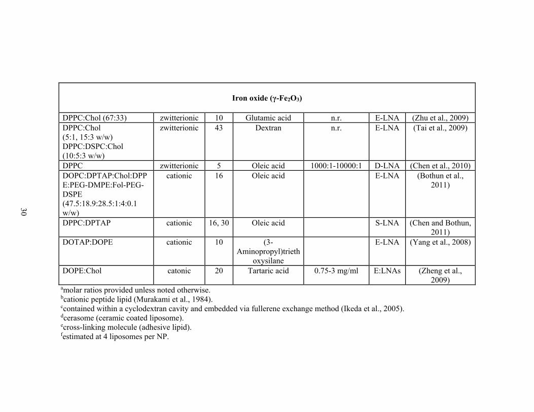
Formation, Structure, and Design Strategies
The functionality of a LNA is determined by the liposome composition,
liposome and NP surface modifiers, NPs employed, intermolecular and surface
interactions, and colloidal stability. LNA design strategies include the encapsulation of
individual or multiple NPs within the aqueous core of the liposome, embedding
hydrophobic NPs in the lipid bilayer, and binding or conjugating NPs to the liposome
surface (Figure 1-2). Table 1-3 contains a list of Au, iron oxide, and γ-iron oxide
LNAs reported in the literature since 2008. LNAs can be used to protect NPs and
encapsulated agents from the adsorption of exogenous molecules, enhancing
bioavailability and reducing the need for complex surface chemistries. Concentration
of NPs and therapeutic agents within the liposome can increase intracellular delivery,
providing greater contrast for imaging, more efficient drug delivery, and enhanced
heating capability for hyperthermia applications. Functionality can also be added by
modifying the LNA bilayer with functional lipids or surface coatings for improved
stability and providing targeting capability.
Page 37
19
Figure 1-2. Schematics and cryogenic transmission electron micrograph of
liposome-nanoparticle assemblies.
Schematic and representative transmission electron micrographs of LNAs formed by
encapsulating hydrophilic nanoparticles (E-LNAs; A, A-1; Wijaya and Hamad-
Schifferli, 2007), bilayer decorating hydrophobic nanoparticles (D-LNAs; B, B-1;
Rasch et al., 2010b), or surface coupling hydrophilic nanoparticles (S-LNAs; C C-1;
Wu et al., 2008). Surface coupling (C) can also be used to create controlled aggregates
or complexes (D1, D2; D1-1, D2-1; Volodkin et al., 2009). Structures and proportions
are not to scale. Reprinted from Rasch et al., 2010b; Volodkin et al., 2009; Wijaya and
Hamad-Schifferli, 2007; Wu et al., 2008
LNA functionality extends beyond that of a traditional liposome. Liposome
delivery requires not just creating a stable system capable of retaining cargo during
both storage and circulation, but also the ability to release encapsulates at a target site.
Efficient release can be achieved by using environmental responsive liposomes that
melt near physiological temperature or through chemical mechanisms, such as pH-
Page 38
20
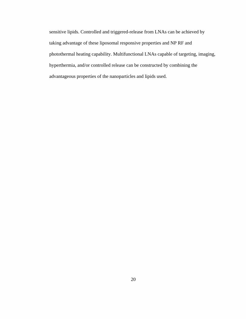
sensitive lipids. Controlled and triggered-release from LNAs can be achieved by
taking advantage of these liposomal responsive properties and NP RF and
photothermal heating capability. Multifunctional LNAs capable of targeting, imaging,
hyperthermia, and/or controlled release can be constructed by combining the
advantageous properties of the nanoparticles and lipids used.
Page 39
21
Table 1-3. Review of LNAs formed with gold (Au) nanoparticles.
Lipids (ratio)a Charge NP
diameter (nm)
NP surface coating Lipid:NPa LNA Ref
DOPC DOPC:DOPC+ (90:10) DOPC:DOPP (9:1)
Zwitterionic Cationic Anionic
n.r. Ascorbic acid n.r. S-LNA Sau et al.,
2009
DOPC:DOTAP (8:2) Cationic 80 Citrate n.r.f E-LNA Anderson et
al., 2010
DPPC:Chol (55:40) Zwitterionic 1.4 n.r. 500:1-2000:1
(DPPE-AuNP:Liposome) S-LNA
Chithrani et al., 2010
DPPC:DPTAP:Chol (6:3:1 w/w)
Cationic 20 n.r. C-LNA Volodkin et
al., 2009
DPPC:DSPC (9:1) Zwitterionic 2.5 Hexanethiol 17.2:1 (w/w) D-LNA Paasonen et al., 2010b
DPPC:DSPC (9:1) Zwitterionic 4 Mercaptosuccinic acid 10:1 (w/w) E-LNA Paasonen et al., 2010b
DPPC:DSPC (9:1) Zwitterionic 1.4 DPPE-Nanogold™ n.r. E-LNA, D-LNA
Paasonen et al., 2007a
Egg PC Cationic 10 Chitosan n.r. S-LNA Pornpattananangkul et al.,
2011
EggPC Zwitterionic 2 Dodecanethiol 100:1-1500:1 D-LNA Rasch et al.,
2010a
EggPC:DOTAP (9:1 w/w) Cationic 4 Mercaptopropionic acid ≥3.6x10⁻³:1 (mol/mol) S-LNA Pornpattananangkul et al.,
2010 EYPC EYPC:DDAB (9:1) EYPC:PEG-DSPE (95:5)
Zwitterionic Cationic
Zwitterionic 13 Citrate 10:1, 1:1 (mol/mol) S-LNA
Kojima et al., 2008b
Page 40
22
Table 1-4. Review of LNAs formed with iron oxide nanoparticles. Iron oxide (Fe3O4)
[maleimide]PEG-DSPE:FAM-DOPE (10:1 w/w)
10-14 Heptanioc acid,
acetic acid 1:1.8 (w/w) E-LNA
Larsen et al., 2008
DMPC:Chol:XLe (47.5:47.5:5) DPPC:DMPC:XLe (9.5:85.5:5)
Zwitterionic 10 Catechol ≥8.3:1 (mol/w) C-LNA Mart et al.,
2009
DMPC:DMTAP:Chol:DMPE-PEG (35:50:10:5)
Cationic n.r n.r. n.r E-LNA Dandamudi et al., 2009
DPPC:Chol:PEG-DMPE:Fol-PEG-DSPE (80:20:4.2:0.5)
Zwitterionic 10 Lauric acid n.r. E-LNA Pradhan et al., 2010
PC Zwitterionic 12.5 n.r. E-LNA
Sabate et al., 2008
PC:PE (2:1) Zwitterionic 10 n.r. n.r. E-LNA
Kikumori et al., 2008
Iron oxide (γ-Fe2O3) DOPC:DPTAP:Chol:DPPE:PEG-DMPE:Fol-PEG-DSPE (47.5:18.9:28.5:1:4:0.1 w/w)
Cationic 16 Oleic acid n.r E-LNA Bothun et al., 2011
DOPE:Chol Cationic 20 Tartaric acid 0.75-3 mg/ml E:LNAs Zheng et al.,
2009
DOTAP:DOPE Cationic 10 (3-Aminopropyl) triethoxysilane
n.r E-LNA Yang et al.,
2008
Page 41
23
DPPC Zwitterionic 5 Oleic acid 1000:1-10000:1 D-LNA Chen et al.,
2010 DPPC:Chol (5:1, 15:3 w/w) DPPC:DSPC:Chol (10:5:3 w/w)
Zwitterionic 43 Dextran n.r. E-LNA Tai et al.,
2009
DPPC:Chol (67:33) Zwitterionic 10 Glutamic acid n.r. E-LNA Zhu et al.,
2009
DPPC:DPTAP Cationic 16, 30 Oleic acid S-LNA Chen and Bothun,
2011 aMolar ratios provided unless noted otherwise, n.r. stands for not reported. bCationic peptide lipid (Murakami et al., 1984). cContained within a cyclodextran cavity and embedded via fullerene exchange method (Ikeda et al., 2005). dCerasome (ceramic coated liposome). eCross-linking molecule (adhesive lipid). fEstimated at 4 liposomes per NP.
Page 42
24
5.1 Encapsulated Liposome-Nanoparticle Assembly
Encapsulated liposome-nanoparticle assemblies (E-LNAs) are formed by
encapsulating NPs within the aqueous core of liposomes (Figure 1-2(A)). The first
investigation of LNAs was inspired by the use of liposomes as a carrier for
hydrophilic drugs. E-LNAs, by encapsulating NPs in the liposome core, force NPs to
cluster together at a high density. High density nanoparticle loading is advantageous to
hyperthermia and drug delivery because heating and drug release can be localized
preventing damage to adjacent tissues. Also, high density loading provides a strong
contrast agent for biomedical imaging (Wijaya and Hamad-Schifferli, 2007).
Magnetoliposomes (MLs), liposomes encapsulating superparamagnetic NPs,
are one of the simplest and first developed LNA configurations (De Cuyper and
Joniau, 1988; Shinkai et al., 1996). They can be prepared by encapsulating preformed
NPs in solution or by forming NPs within the liposome core, as first shown by
Papahadjopoulos in 1983 (Hong et al., 1983). E-LNAs can be prepared by thin film
hydration (TFH), double emulsion (DE) (Zheng et al., 1994), or reverse phase
evaporation (REV) (Szoka and Papahadjopoulos, 1978). Extrusion or sonication of
post-formation liposomes can be employed to control the size of E-LNAs. Supported
lipid bilayers (SLBs), NPs coated with a lipid bilayer, are formed when dcore = dNP. E-
LNA formation requires the use of colloidal stable nanoparticles with a diameter (d)
that is smaller than the inner diameter of the aqueous liposome core, dcore > dNP
(Figure 1-3(A)). The maximum theoretical number of encapsulated NPs is n ≈
0.74(Vcore/VNP; V represents the volume of the core or NP), due to the close packing of
Page 43
25
spheres and dcore >> dNP. Wijaya and Hamad-Schifferli demonstrated that it is possible
to approach this limit, demonstrating high-density encapsulation of Fe3O4 NPs (dNP =
12.5 nm) within DPPC liposomes (Figure 1-2(A-1)). With this design the available
core volume for co-encapsulating aqueous drug molecules decreases within increasing
NP concentration (Wijaya and Hamad-Schifferli, 2007). However, the ability for
embedding hydrophobic molecules within the bilayer is unaffected by NP
concentration.
The osmotic pressure differential across the lipid bilayer and the attractive or
repulsive forces between the bilayer and the NPs determine the structure of E-LNAs.
The elasticity of the bilayer determines how the LNA will deform in response to these
forces. Attractive forces can include van der Waals, hydrophobic, and electrostatic
interactions; and repulsive forces can include electrostatic, depletion, hydration, and
steric interactions. The physical stability of a liposome-NP system can be determined
by the Deryaguin-Landau-Verwey-Overbreek (DLVO) theory. The DLVO theory
balances the opposing forces to provide a total energy of interaction between the
particles. Liposome-NP systems are characterized by three types of interactions that
take place, repulsion between liposome-liposome and NP-NP and attractive forces
between liposome and NP. Electrostatic repulsion becomes significant when
nanoparticles and liposomes approach each other and their double layers begin to
interfere. Electrostatic energy curve represents the energy required to overcome the
repulsion. The maximum energy corresponds to the situation when the surfaces are
touching each other and is zero outside the double layer (Leckband, 2001).
Page 44
26
The adhering and non-adhering characteristics of nanoparticles can lead to changes in
bilayer curvature, which can impact liposome size, shape, and phase homogeneity
(Lipowsky and Dobereiner, 1998). Generally, this will occur when encapsulates are
different from molecules present outside (e.g. sugars or proteins) liposomes.
LNAs are generally formed with small non-adhering NPs because NP adhesion
to bilayers can significantly alter LNA structure and morphology. The exception to
this is LNAs formed by coating a single large NP with an adsorbed or supported lipid
bilayer. For non-adhering encapsulated particles, the bilayer can curve towards the
larger particles. As an example of a non-adhesive system, Pradhan et al. (Pradhan et
al., 2007) compared the encapsulation efficiency of 10 nm MnFe2O4 NPs coated with
lauric acid composed of egg-PC:cholesterol (at molar ratios of 1:0, 2:1, 3:2, 1:1, and
1:2) and formed by TFH and DE. In general, TFH resulted in higher encapsulation
efficiency with smaller ML diameter compared to DE due to stripping of lauric acid
during the DE process. In both cases, the observation that an Egg PC:cholesterol ratio
of 2:1 yielded the best encapsulation efficiency (70% via TFH) was attributed to
cholesterol inducing a single liquid ordered bilayer phase.
In contrast, for small adhering encapsulated particles (attractive) where dcore >>
dNP and dNP < 2lb the bilayer can curve away from the particles. For large adhering
particles, where dNP >> 2lb, the bilayer can curve around or engulf the particles. For
example, Sabate et al. examined the effect of Fe3O4 NP concentration coated with
tetramethylammonium hydroxide (58 nm hydrodynamic dNP) on the encapsulation
efficiency of extruded soybean PC MLs. The encapsulation efficiency decreased from
Page 45
27
96.6% at 1.22 g Fe3O4/mol PC to 18.5% at 119.95 g Fe3O4/mol PC. This was
attributed to electrostatic interactions (attraction) between the cationic NPs and the PC
bilayers. The size of the MLs increased from 140 to 197 nm, consistent with lower
curvature due to NP adhesion at the inner bilayer surface (Sabate et al., 2008).
Electrolytes can also effect the curvature of lipid bilayers (Lipowsky and
Dobereiner, 1998). Gomes et al. (Gomes et al., 2009) prepared polyelectrolyte-coated
MLs by encapsulating 8 nm anionic γ-Fe2O3 NPs within egg PC liposomes and then
coating with alternating poly(allylamine hydrochloride) and poly(sodium 4-
styrenesulfonate) layers. The final coating determined the surface charge (anionic PSS
or cationic PAH). The size ranged from 200-400 nm and two or more polyelectrolyte
coatings sufficiently protected the lipid bilayer from detergent-induced disruption.
Page 46
28
Lipids (ratio)a Charge DNP (nm)
NP surface coating
Lipid:NPa LNA Ref
Gold
DOPC:DOTAP (8:2) Cationic 80 Citrate n.r.f E-LNA (Anderson et al., 2010)
DPPC:DSPC (9:1) Zwitterionic 2.5 Hexanethiol 17.2:1 (w/w) D-LNA (Paasonen et al., 2010)
EggPC Zwitterionic 2 Dodecanethiol 100:1-1500:1 D-LNA (Rasch et al., 2010)
DPPC:DSPC (9:1) Zwitterionic 4 Mercaptosuccinic acid
10:1 (w/w) E-LNA (Paasonen et al., 2010)
DPPC:DSPC (9:1) Zwitterionic 1.4 DPPE-Nanogold™ n.r. E-LNA D-LNA
(Paasonen et al., 2007)
DOPC DOPC:DOPC+ (90:10) DOPC:DOPP (9:1)
Zwitterionic Cationic Anionic
n.r. Ascorbic acid n.r S-LNA (Sau et al., 2009)
EYPC EYPC:DDAB (9:1) EYPC:PEG-DSPE (95:5)
Zwitterionic Cationic
Zwitterionic
13 Citrate 10:1, 1:1 (mol/mol)
S-LNA (Kojima et al., 2008)
EggPC:DOTAP (9:1 w/w)
Cationic 4 Mercaptopropionic acid
≥3.6x10⁻³:1 (mol/mol)
S-LNA (Pornpattananangkul et al., 2010)
DPPC:DPTAP:Chol (6:3:1 w/w)
Cationic 20 n.r. C-LNA (Volodkin et al., 2009)
Page 47
29
DPPC:Chol (55:40) Zwitterionic 1.4 n.r. 500:1-2000:1 (DPPE-
AuNP:Liposome)
S-LNA (Chithrani et al., 2010)
Egg PC Cationic 10 Chitosan n.r. S-LNA (Pornpattananangkul et al., 2011)
Iron oxide (Fe3O4)
[maleimide]PEG-DSPE:FAM-DOPE (10:1 w/w)
10-14 Heptanioc acid, Acetic acid
1:1.8 (w/w) E-LNA (Larsen et al., 2008)
DMPC:Chol:XLe (47.5:47.5:5) DPPC:DMPC:XLe (9.5:85.5:5)
Zwitterionic 10 Catechol ≥8.3:1 (mol/w) C-LNA (Mart et al., 2009)
PC Zwitterionic 12.5 n.r. E-LNA (Sabate et al., 2008)
DMPC:DMTAP:Chol:DMPE-PEG (35:50:10:5)
cationic n.r n.r. n.r E-LNA (Dandamudi et al., 2009)
PC:PE (2:1) zwitterionic 10 n.r. n.r. E-LNA (Kikumori et al., 2008)
DPPC:Chol:PEG-DMPE:Fol-PEG-DSPE (80:20:4.2:0.5)
zwitterionic 10 Lauric acid n.r. E-LNA (Pradhan et al., 2010)
Page 48
30
Iron oxide (γ-Fe2O3)
DPPC:Chol (67:33) zwitterionic 10 Glutamic acid n.r. E-LNA (Zhu et al., 2009) DPPC:Chol (5:1, 15:3 w/w) DPPC:DSPC:Chol (10:5:3 w/w)
zwitterionic 43 Dextran n.r. E-LNA (Tai et al., 2009)
DPPC zwitterionic 5 Oleic acid 1000:1-10000:1 D-LNA (Chen et al., 2010) DOPC:DPTAP:Chol:DPPE:PEG-DMPE:Fol-PEG-DSPE (47.5:18.9:28.5:1:4:0.1 w/w)
cationic 16 Oleic acid E-LNA (Bothun et al., 2011)
DPPC:DPTAP cationic 16, 30 Oleic acid S-LNA (Chen and Bothun, 2011)
DOTAP:DOPE cationic 10 (3-Aminopropyl)trieth
oxysilane
E-LNA (Yang et al., 2008)
DOPE:Chol catonic 20 Tartaric acid 0.75-3 mg/ml E:LNAs (Zheng et al., 2009)
amolar ratios provided unless noted otherwise. bcationic peptide lipid (Murakami et al., 1984). ccontained within a cyclodextran cavity and embedded via fullerene exchange method (Ikeda et al., 2005). dcerasome (ceramic coated liposome). ecross-linking molecule (adhesive lipid). festimated at 4 liposomes per NP.
Page 49
31
5.2 Bilayer-Decorated Liposome-Nanoparticle Assembly
Bilayer decorated liposome-nanoparticle assemblies (D-LNA) are liposomes
with hydrophobic nanoparticles embedded in the lipid bilayer (Figure 1-2(B)). Similar
to the ability of cells to accommodate membrane proteins, liposomes can distort to
accommodate hydrophobic NPs that exceed the thickness of hydrophobic acyl region
of the bilayer (~3 nm) (Al-Jamal et al., 2008b; Bothun, 2008; Chen et al., 2010; Jang
et al., 2003). Embedded NPs can affect lipid packing, lipid phase behavior,
transbilayer permeability, and LNA structure and morphology (Binder et al., 2007;
Bothun, 2008; Bothun et al., 2009; Chen et al., 2010; Chen and Bothun, 2009; Jeng et
al., 2005; Park et al., 2005, 2006; Rasch et al., 2010b). Cryo-TEM can be used to
observe the structure and morphology of these nano-scale systems in solution (Chen
and Bothun, 2011). Atomic force microscopy has also been used to observe phase-
separated domains and monitor membrane remodeling and alteration due to the
presence and distribution of nanoparticles within the bilayer (Kirat, 2010). The
diameter of embedded nanoparticles (core and surface coating) is similar to the
thickness of the lipid bilayer (~5 nm). Theoretically, the diameter of embedded
nanoparticle must be less than 6.5 nm in order for the lipid bilayer to maintain its
structure. Hydrophobic nanoparticles with diameters greater than 6.5 nm form
micelles because they are more energetically favorable due to the high local curvature
strain on the bilayer, as described in Figure 1-4 (Ginzburg and Balijepalli, 2007; Wi et
al., 2008).
Page 50
32
Figure 1-3. Changes in bilayer decoration mechanism of D-LNAs with increasing
nanoparticle diameter.
Nanoparticle diameter, dNP, is equal to the diameter of the particle core plus two times
the surface coating. (A) Small nanoparticles (defined herein as dNP < 2 nm) can cluster
together to minimize bilayer bending energy (Rasch et al., 2010b). (B) Larger
nanoparticles (defined herein as dNP = 2-6.5 nm) can create ‘pockets’ within the
bilayer or (C) bridge adjoining liposomes (Al-Jamal et al., 2008b; Chen et al., 2010).
(D) D-LNA assembly is governed in part by the bilayer deformation energy, which
can be evaluated based on the energy penalty associated with lipid stretching (related
to the compressibility modulus) and monolayer curvature (related to bending
modulus).
The characteristics of LNAs are directly affected by embedded nanoparticles.
Embedded NPs interact with the lipid acyl tails changing the fluidity of the membrane
bilayer. Physical obstruction of the movement of lipid tails reduces the lipid bilayer
ordering. The melting temperature of LNAs with embedded silver (Bothun, 2008),
gold (Mady et al., 2011), and SPIO NPs (Chen et al., 2010) have shown to reduce the
Page 51
33
transition temperature of the bilayer with increased loading by fluorescence anisotropy
and differential scanning calorimetry. Recent FTIR measurements of embedded gold
NPs have validated the interaction of NPs and the acyl tail groups. Changes in the
frequency of CH2 stretching indicated conformational change in acyl tails of bilayers
with embedded NPs. Also, dynamic light scattering of citrate-stabilized gold NPs
embedded in DPPC liposomes demonstrated an increase in the negative charge.
Surface charge can hinder aggregation of LNAs affecting the overall stability of LNAs
(Mady et al., 2011). Therefore, changes to the membrane alter LNA properties for
delivery of therapeutic and diagnostic agents. The interactions between NPs and
liposomes still remains misunderstood. Further investigation into the effect of
nanoparticles is necessary to develop more efficient multimodal LNAs
Clustering of embedded NPs has been observed by Rasch et al. in LNAs with
dodecanethiol-coated Au (dNP = 1.6-1.8 nm) (Figure 1-4(A)) (Rasch et al., 2010b).
They showed that high NP loading with uniform distribution can be achieved in PC
liposomes via thin film hydration (with sonication and extrusion). Janus particles can
be prepared with embedded NPs clustered in approximately one half of the liposomes
via detergent loading followed by dialysis. Clustering occurs as the liposomes
minimize the energy penalty for bilayer deformation – i.e. for a given concentration of
embedded NPs the periodic bilayer bending energy needed to accommodate individual
particles is greater than that needed to accommodate nanoparticle clusters. Park et al.
(Park et al., 2006) and Chen et al. (Chen et al., 2010) have observed a similar
clustering phenomenon with stearylamine-coated 3-4 nm Au and oleic acid-coated 5
Page 52
34
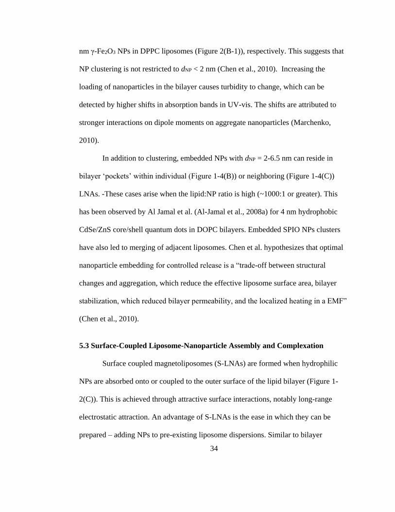
nm γ-Fe2O3 NPs in DPPC liposomes (Figure 2(B-1)), respectively. This suggests that
NP clustering is not restricted to dNP < 2 nm (Chen et al., 2010). Increasing the
loading of nanoparticles in the bilayer causes turbidity to change, which can be
detected by higher shifts in absorption bands in UV-vis. The shifts are attributed to
stronger interactions on dipole moments on aggregate nanoparticles (Marchenko,
2010).
In addition to clustering, embedded NPs with dNP = 2-6.5 nm can reside in
bilayer ‘pockets’ within individual (Figure 1-4(B)) or neighboring (Figure 1-4(C))
LNAs. -These cases arise when the lipid:NP ratio is high (~1000:1 or greater). This
has been observed by Al Jamal et al. (Al-Jamal et al., 2008a) for 4 nm hydrophobic
CdSe/ZnS core/shell quantum dots in DOPC bilayers. Embedded SPIO NPs clusters
have also led to merging of adjacent liposomes. Chen et al. hypothesizes that optimal
nanoparticle embedding for controlled release is a “trade-off between structural
changes and aggregation, which reduce the effective liposome surface area, bilayer
stabilization, which reduced bilayer permeability, and the localized heating in a EMF”
(Chen et al., 2010).
5.3 Surface-Coupled Liposome-Nanoparticle Assembly and Complexation
Surface coupled magnetoliposomes (S-LNAs) are formed when hydrophilic
NPs are absorbed onto or coupled to the outer surface of the lipid bilayer (Figure 1-
2(C)). This is achieved through attractive surface interactions, notably long-range
electrostatic attraction. An advantage of S-LNAs is the ease in which they can be
prepared – adding NPs to pre-existing liposome dispersions. Similar to bilayer
Page 53
35
embedment, decorated bilayers also provide direct heating to the bilayer in the
presence of external stimuli. The design constraint for forming S-LNAs is dependent
on bilayer NP adhesion and curvature. Recent investigations have shown that NPs
with dNP > ~20 nm lead to the formation of SLBs due to liposome adsorption and
rupture, followed by the bilayer curving around the particle (Figure 1-3(A)) (Chen and
Bothun, 2011). The critical NP diameter under which S-LNAs can be formed is dNP <
2(kb/w)1/2, where kb is the bilayer bending elasticity, which is dependent on lipid
composition and phase state, and w is the adhesion energy (Roiter et al., 2008).
The Granick group has shown that stable S-LNA dispersions can be formed
using zwitterionic liposomes with decorated cationic or anionic NPs (< 20 nm) with a
NP surface coverage above ~25% (Yu et al., 2007; Zhang and Granick, 2006). This
was achieved by electrostatic attraction. Lower surface coverage led to aggregation,
which demonstrates the need to balance the lipid:NP ratio. It was shown with
isothermal titration calorimetry that upon binding the nanoparticles could restructure
the lipid bilayer, inducing gel phases in fluid liposomes and fluid phases in gel
liposomes (Wang et al., 2008). This observation shows that, even without external
stimuli, bound NPs can induce changes in lipid phase behavior and, presumably,
permeability.
NP adhesion to the outer bilayer can affect the morphology and structure of S-
LNAs similar to E-LNAs. Cationic nanoparticle adhesion to the outer surface of
GUVs has been shown to cause pearling. The head group area of zwitterionic lipids
was increased due to the use of charged particles. Attraction of the head group and
Page 54
36
electrostatic repulsion to the cationic nanoparticles caused a mismatch of the outer and
inner curvature of liposomes. These interactions resulted in the pearling structure of
the liposome (Yu and Granick, 2009).
Sau et al. have also used electrostatic binding to prepare S-LNAs with Au NPs.
High NP surface coverage was achieved by using anionic Au NPs with physisorbed
ascorbic acid and cationic liposomes (9:1 DOPC to ethyl-DOPC; Tm = -20 oC) (Sau et
al., 2009). This high surface coverage was accompanied by NP aggregation due to the
high local concentration and (likely) to charge screening via cationic lipids between
bound particles (Kojima et al., 2008a). Binding was also achieved on zwitterionic and
anionic liposomes with decreasing coverage (and NP aggregation), respectively.
Pornpattananangkul et al. have taken this one step further and have shown that pH can
be used to control carboxyl-modified (anionic) Au NP binding to cationic liposomes
and, in turn, liposome stability. Above the pKa of the carboxyl groups the bound NPs
stabilize the S-LNAs and prevent aggregation and fusion, while below the pKa the
NPs detach and liposome fusion resumes (Pornpattananangkul et al., 2010).
Lastly, LNAs can be formed by complexation (C-LNAs) if the liposomes
surround NP aggregates (Figure 1-2(D2)) or the NPs bind to multiple liposomes and
act as “bridges” (Figure 1-2(D1)). Voldokin et al. have shown that either structure can
be formed from the same anionic Au NP-cationic liposome by manipulating
electrostatic interactions using via salt concentration. High NaCl concentration (75
mM) enhanced NP aggregation (Figure 1-2(D2-1)) and low salt concentrations
Page 55
37
inhibited it (Figure 1-2(D1-1)). In addition to non-specific physical interactions
(electrostatic), cross-linking can be used to create C-LNAs (Volodkin et al., 2009).
Mart et al. used Fe3O4 NPs coated with histidine groups to bind to and
complex zwitterionic/cholesterol liposomes containing Cu(iminodiacetate)-
functionalized lipid. The objective was to demonstrate a potential method using
histidine-Cu(IDA) binding to form C-LNAs, thereby concentrating a therapeutic and
an imaging agents at a target site. The resulting aggregates ranged from 20-100 μm in
diameter. The C-LNAs demonstrated EMF heating and controlled release of model
drugs (Mart et al., 2009).
Controlled Release
Controlled release of encapsulated payloads from LNAs can be induced by
heating of nanoparticles raising temperature of the lipid bilayer. In vivo heating of
magnetic nanoparticles has been demonstrated with external stimuli such as
alternating current electromagnetic fields, microwaves, light irradiation, and lasers
(Brazel, 2009). As discussed earlier, the bilayer permeability rises with temperature
allowing release of encapsulates. Bothun and Preiss have demonstrated local bilayer
heating due of Fe3O4 nanoparticles heated by RF causing phase transition. However,
there was negligible difference between the bulk and local bilayer temperatures.
Therefore, controlled release is likely due to both thermally-induced phase transition
and mechanical rupture of the bilayer caused by NP heating. LNA controlled release
with gold and iron oxide nanoparticle will be discussed herein (Bothun and Priess,
2011).
Page 56
38
6.1 Gold Nanoparticles and Photothermal Effects
Heating of Au NPs is caused by the SPR properties that convert absorbed light
into heat. SPR heating makes Au NPs efficient heat sources for LNA controlled
release (Sassaroli et al., 2009). Utilizing the photothermal heating of Au NPs,
Paasonen et al. (Paasonen et al., 2007b) demonstrated the ability to control the release
of calcein (622.6 MW) from Au LNAs composed of DPPC/DSPC at 9:1 (Tm = 44.9
oC) with E-, D-, and S-LNAs (Figure 1-2(A-C)). Leakage was examined with and
without UV light at a wavelength of 250 nm over 30 min at 37 oC. Without UV
exposure, spontaneous calcein release was observed for E-LNAs with encapsulated
mercaptosuccinic acid-coated NPs and D-LNAs with embedded hexanethiol-coated
NPs. This was attributed to NP-lipid interactions at the bilayer/water interface and
within the acyl tail region, respectively, which reduced bilayer integrity. With UV
exposure, direct contact between NPs and the liposomes via bilayer-embedment led to
the greatest release (~90% at 30 min). Intuitively, direct contact would improve the
local heat transfer from the NPs to the liposomal bilayers relative to encapsulation.
This led to a gel-fluid phase transition where calcein release was presumably enhanced
by diffusion at the interface between coexisting gel and fluid domains.
Volodkin et al. demonstrated the release of 5(6)-carboxyfluorescein (CF; 376.3
MW) from C-LNAs formed by the complexation of 128 nm cationic liposomes
(DPPC/DPTAP/chol, Tm ~ 40-45 oC) and 20 nm anionic Au NPs. Low NaCl
concentration (7.5 mM) yielded LNAs with NP-mediated liposome bridges (Figure 1-
Page 57
39
2(D1-1)) and high NaCl concentration (75 mM) yielded LNAs with liposome-coated
NP aggregates (Figure 1-2(D-1)). CF release from the C-LNAs was observed within 5
s after near-IR irradiation (Figure 1-5) (Volodkin et al., 2009).
Figure 1-4. Remote dye release triggered by near IR laser illumination from C-
LNAs.
C-LNAs were prepared with gel-phase cationic liposomes and 20 nm hydrophilic gold
nanoparticles. The scale bars represent 10 µm. These results show that near complete
release can be achieved from the complexes after 5 s of illumination. Reprinted from
(Volodkin et al., 2009) with permission.
Anderson et al. utilized the principle of plasmonic nanobubble (PNB)
formation to control the release of 104 and 240 kDa proteins from cationic LNAs (~1
Page 58
40
μm) containing encapsulated 80 nm anionic Au NPs (Figure 1-6). Irradiation was
achieved using a single pump laser at 532 nm over 0.5 ns. Local vapor bubble
formation led to mechanical disruption, as opposed to thermal, of the LNA bilayer and
rapid protein release. The advantage of PNB formation is heating is isolated within the
LNA triggering the immediate release of all encapsulated cargo (Anderson et al.,
2010).
Figure 1-5. Proposed ‘plasmonic nanobubble’ release mechanism of encapsulated
molecules from E-LNAs prepared with gold nanoparticles.
Pulsed laser irradiation of the nanoparticles nucleates vapor bubbles that expand and
mechanically disrupt the liposomal bilayer and trigger permeabilization. Reprinted
from (Anderson et al., 2010) with permission.
Wu et al. used hollow gold nanoshells (HGNs) encapsulated within or
decorating the surface of DPPC liposomes to trigger CF release by near-IR pulses (800
nm) via microbubble formation and collapse (Figure 1-2(C-1)). LNA release was
dependent on the proximity of the HGNs to the liposomes (decorated or tethered
Page 59
41
HGNs yielded the greatest response) and the laser power. Their results strongly
suggest that release was attributed to transient disruption or poration of the lipid
bilayer via transient bubble cavitation (Wu et al., 2008).
6.2 Iron Oxide Nanoparticles and Alternating Magnetic Fields
AC EMF operating at RF heating is due to magnetic losses being converted to
heat, typically at low frequencies between 100-400 kHz. The magnetic losses for NPs
< ~30 nm are due to Néel relaxation, arising from rapidly alternating magnetic dipole
moments, and Brownian relaxation, arising from nanoparticle rotation and viscous
losses (friction). RF heating is advantageous because it is non-invasive, easily
penetrates the body, and is physiologically acceptable for up to 1 h if the product Hf,
where H is the field amplitude (current number of coils per length) and f is the
frequency, is below 4.85105 kA/m/s (Brezovich, 1988). NP heating capability is
based on the inherent specific absorbance rate (SAR, W/g) of the nanoparticles:
SAR cp
mNP
T
t
where
cp is the average heat capacity of the sample,
mNP is the NP mass, and
T / t
is the initial heating rate of the sample. SAR values up to approximately 700 W/g can
be obtained depending on the NP size, composition, and surface coating (Mornet et al.,
2004).
Page 60
42
Tai et al. examined CF release from thermosensitive zwitterionic liposomes
containing encapsulated dextran-coated 43 nm γ-Fe2O3 NPs (Resovist™) using a high
frequency generator (6.4 kW, 750-1150 kHz) operating for 5-25 min. CF release from
DPPC:Chol (5:1) liposomes without encapsulated NPs was initiated between 35 oC
and 37 oC. In contrast, the LNA analogs exhibited initial release at 34 oC and 32 oC
with 7 and 14 mg Fe/ml, respectively. This initial release temperature was further
tuned by increasing the cholesterol content (DPPC:Chol at 15:3) and incorporating a
higher melting lipid (DSPC, Tm = 55 oC). Release was attributed to NP heating. Using
a rat model, they demonstrated that release could also be achieved in vivo (Tai et al.,
2009).
Chen et al. have recently examined the release of CF from LNAs formed with
DPPC and bilayer-embedded oleic acid-coated 5 nm γ-Fe2O3 NPs at lipid:NP ratios of
10000:1, 5000:1, and 1000:1 as a function of RF energy (1 kW; 50-250 A, 281 kHz).
Experiments were conducted at non-invasive RF energies up to 40 min and the
greatest release rate was observed at 5000:1, indicating an optimal NP loading for
triggering release. This optimum reflected a balance between NP loading and LNA
structure – high loading is needed for triggering bilayer release, but can lead to NP
aggregation and can compromise LNA structure and stability. A unique observation
was the fact that increasing NP loading reduced or eliminated spontaneous leakage by
increasing bilayer stability. CF release was attributed to bilayer disruption via local
heating and/or LNA rupture, which produced transient voids or pores. Although, these
Page 61
43
findings demonstrated that higher nanoparticle embedding does not strictly equate to
greater release controlled release during EMF heating (Chen et al., 2010).
Targeted Therapy
Optimal drug delivery and biomedical imaging involves the distribution of
drug and/or imaging agent to the diseased tissue while minimizing adverse side-effects
to healthy tissues. Adverse side-effects limit the drug dosage that may be used during
treatment, potentially requiring the dosage to be reduced, delayed, and/or
discontinued. Targeting LNAs limit adverse side-effects to healthy tissues and
enhance drug delivery and uptake by localizing drug delivery to specific target sites
(Malam et al., 2009; Minko et al., 2006).
Effective therapeutic and diagnostic treatment requires sufficient
bioavailability of LNAs to diseased cells. A major limitation of LNAs, and liposomal
drug delivery in general, is that the residence time is not long enough to enable the
non-modified liposome assemblies to accumulate at tumor sites (Zhang et al., 2008).
LNA residence time is directly related to size and surface properties (discussed herein)
(Maurer et al., 2001; Samad et al., 2007).
7.1 Passive Targeting
Passive targeting of lipid-nanoparticle assemblies is facilitated by the enhanced
permeability and retention (EPR) effect. Tumor growth requires a sufficient supply of
oxygen and nutrients. Therefore, during angiogenesis, tumors will produce an
increased amount of vascular endothelial growth factors routing new blood vessels
Page 62
44
and/or existing blood vessels directly to growing tumors. These tumor-associated
neovasculatures are typically disorganized and highly permeable allowing leakage of
larger entities, such as lipid-nanoparticle assemblies, into the tumor. The entities are
retained by the tumor because the defective lymphatic drainage prevents larger entities
from returning to the circulatory system. LNAs accumulate in the tumor providing an
effective targeting mechanism that can be utilized for drug delivery, imaging, and/or
hyperthermia (Cho et al., 2008; Corchero and Villaverde, 2009; van Vlerken and
Amiji, 2006).
In order for LNAs to accumulate within a tumor by the EPR effect, the
residence time of LNAs in the blood must be sufficient to permeate into the tumor.
Longer circulating assemblies provide a greater opportunity to reach the tumor
vascular system and enter the tumor for drug delivery and/or imaging. Nanoparticle
elimination is primarily based upon the reticuloendothelial system (RES) uptake.
LNAs (as mentioned earlier) can increase their blood residence times by reducing
clearance and absorption through PEGylation.
Passive targeting also exploits the unique environment created by tumors. As
stated above, rapidly proliferating tumor cells require sufficient oxygen and nutrients.
However, the supply of oxygen and nutrients is typically insufficient to maintain the
rapid cell growth, forcing tumor cells to breakdown sugars through glycolysis (Cho et
al., 2008; Corchero and Villaverde, 2009; Minko et al., 2006; van Vlerken and Amiji,
2006). Glycolysis, under anaerobic conditions, leads to lactic acid build-up creating an
Page 63
45
acidic environment (Pelicano et al., 2006). LNAs comprised of pH-responsive lipids
facilitating drug release in the acidic tumor environment (Cho et al., 2008).
Passive targeting can also be utilized for antibacterial applications.
Pornpattananangkul et al. (Pornpattananangkul et al., 2011) attached chitosan-coated
gold to vancomycin encapsulated Egg PC liposomes. The attached gold nanoparticles
prevented fusing and spontaneous leakage increasing stability and shelf-life.
Antibiotic release was triggered by the presence of bacterial toxins that caused pore
formation. After 24 hours of exposure, 100% of vancomycin was released from the
liposome. Vancomycin is used as a “last resort” for MRSA infections (Crossley,
2010). Such regulated LNA delivery systems reduce side effects and dosage by being
capable of releasing encapsulates at the delivery site and in response to the severity of
the infection.
Overall LNA size between 50-150 nm is optimal in order to avoid renal
elimination (nanoparticles smaller than 6 nm) and prevent uptake by the
reticuloendothelial system (nanoparticles larger than 200 nm) (Gullotti and Yeo, 2009;
Veiseh et al., 2010). LNA residence time is also dependent on surface charge. Cationic
LNAs demonstrate a much higher affinity to bind to the vasculature of tumors than to
normal healthy tissues. Tumor vessel area targeted by cationic LNAs was
approximately 5 times greater than the vessels of normal healthy tissues (Campbell et
al., 2002). Cationic LNA uptake has been shown to be almost10 times higher than
similar anionic and neutral LNAs (Ito et al., 2005). The preferential binding can be
exploited for targeted hyperthermia, drug delivery, and/or imaging.
Page 64
46
Anionic and neutral entities and bodies larger than 200 nm, upon entering the
blood stream, tend to absorb antibodies, called opsonins, activating the
reticuloendothelial system and inducing mononuclear phagocyte system (MPS) or by
hepatocyte uptake and elimination (Ishida et al., 2002; Maurer et al., 2001; Yan et al.,
2005). Incorporation of polyethylene glycol (PEG), a neutral hydrophilic polyether,
onto the surface of LNAs is very effective for preventing opsonization and increasing
the residence time. PEG has proven to be one of the most effective surface
modifications and is probably the most used for producing long-circulating entities.
For example, Doxil and Myocet in Table 1-1 is a PEG-liposome encapsulated
formulation of doxorubicin. Myocet and Doxin have an elimination half-life of 2.5 h
and 55 h, respectively, whereas free doxorubicin has elimination half-life of 0.2 h
(Danhier et al., 2010). Other surface modifiers and ligands, such as polysaccharides,
dysopsonins, dextran, etc., have been incorporated into the bilayer to increase
liposome stability, residence time, and enabling targeting capability (Zhang et al.,
2008).
The final passive targeting method is topical delivery. Topical delivery is
localized drug administration directly to a tumor by injection or surgery. This
approach bypasses drugs administration through the circulatory system. By avoiding
the circulatory system, adverse side-effects can be limited to the administration site
(Minko et al., 2006).
Page 65
47
7.2 Active Targeting
Active targeting involves incorporating a targeting ligand, most often
antibodies, antibody fragments, vitamins, glycolipids, or peptides (Jørgensen and
Nielsen, 2009), onto the surface of LNAs. In order to be effective, the targeting ligand
must specifically bind to a receptor that is exclusive to tumors or is overexpressed by
tumors compared to normal healthy tissue. Active targeting can be split into two
categories: cancer cell targeting and tumoral endothelium targeting. Cancer cell
targeting promotes tumor internalization of LNAs and intracellular delivery of
therapeutic agents. The most common receptors for cancer cell targeting are
transferrin, folate receptor, cell surface glycoproteins, and the epidermal growth
factor. Tumoral endothelium targeting restricts the blood supply available to tumor
cells by attacking angiogenic blood vessels. The most common entities subject to
tumoral endothelium targeting are vascular endothelial growth factors, vascular
endothelial growth factors receptors (VEGFR-1 and VEGFR-1), αvβ3 integrin,
vascular cell adhesion molecule-1, and matrix metalloproteinases (Danhier et al.,
2010).
Active targeting can also be accomplished with magnetic drug targeting
(MDT). MDT utilizes a static magnetic field to concentration of LNAs at selected
delivery sites. LNAs with encapsulated or embedded magnetic or superparamagnetic
NPs can be forced to aggregate at sites with applied magnetic fields. MDT allows
LNAs to be localized at an identified disease site for hyperthermia and drug delivery,
minimizing the effect to adjacent tissues.
Page 66
48
7.2.1 Ligand Targeting
Active targeting of tumors by LNAs has been accomplished by targeting folate
receptor (FR). Tumor cells express FRs almost 100 times normal cells. Also, the FR
expression rate is proportional to the how advanced the cancer is. Therefore, FR
targeting can be an effective method for identifying and treating tumors (Lu and Low,
2002). FR targeting has been demonstrated both in vitro and in vivo. Folate targeted
liposomes have been used with nanoparticles for tumor imaging, drug delivery, and
hyperthermia (Kamaly et al., 2009; Pradhan et al., 2010).
FR targeted MLs have demonstrated selective drug delivery in vitro.
PEGylated MLs incorporating FR targeting-lipids were constructed with co-
encapsulated doxorubicin and maghemite. Human cervical cancer cell (HeLa), which
overexpress FR, bound to the cell surface and uptake from HeLa cells was observed,
whereas the control showed no surface binding or uptake. FR targeting was combined
with AC EMF at RF nanoparticle heating to induce doxorubicin release. Doxorubicin
release under EMF treatment for 2 h was three times greater than without EMF
(Bothun et al., 2011).
Pradhan et al. developed multifunctional thermosensitive PEGylated
magnetoliposomes with a folic acid ligand capable of binding to FR. The
magnetoliposomes were encapsulated with doxorubicin and iron oxide nanoparticles
(Figure 1-7). Folate receptor mediated uptake of magnetoliposomes into tumor cells
was demonstrated. In vitro hyperthermia and controlled release of encapsulated
doxorubicin was achieved by subjecting cell lines to EMF (Pradhan et al., 2010).
Page 67
49
Figure 1-6. A multifunctional E-LNA for cancer thermo-chemotherapy from
Pradhan et al.
In this example the LNA is a temperature sensitive magnetoliposome containing co-
encapsulated iron oxide nanoparticles and doxorubicin. Passive targeting can be
achieved through the enhanced permeation and retention (EPR) effect of tumor
vasculature, and active targeting can be achieved via folate receptor and by applying a
permanent magnetic field. The application of an AC electromagnetic field can be used
to release the drug and achieve hyperthermia treatment. Reprinted from (Pradhan et
al., 2010) with permission.
Liposomes with incorporated antibodies or antibody fragments are referred to
as immunoliposomes. A number of studies have utilized immunoliposomes and
nanoparticles for biomedical applications. Antibody conjugated LNAs has been
employed to target cells that overexpress epidermal growth factor receptor 2 (Her-2).
Overexpression of Her-2 frequently occurs in cervical, colon, breast, prostate, brain,
Page 68
50
bladder, and lung cancers because it allows cells to grow more rapidly. Current cancer
treatments tend to lack specificity administered causing adjacent tissues to be
damaged. Her-2 targeted treatment may encounter similar adverse effects to adjacent
tissues because Her-2 is a naturally occurring protein. Kullberg et al. developed a two-
component drug delivery system to ensure that treatment would be concentrated to
cells that overexpress Her-2. The two-component system requires more than one LNA
to bind to a tumor cell in order to release the encapsulated drugs. The two-component
system could also have two or more sets of LNAs that encapsulate different substances
that are only effective when they are both present. The specificity of the two-
component system is equal to the difference of tumor and healthy tissue Her-2
expression squared. Therefore, the two-component system could potentially deliver
3600 times more drugs to glioblastomas (capable of a 60-fold overexpression) than
normal cells. The two-fold system could hypothetically be expanded to three or more
components increasing the specificity exponentially (Kullberg et al., 2005, 2009). Her-
2 antibodies have also been utilized to construct targeted magnetoliposomes for
hyperthermia treatment. The magnetoliposomes demonstrated localized hyperthermia
to Her-2 overexpressing tumors under AMF (Kikumori et al., 2008).
Cationic MLs incorporating transferrin receptor targeting antibodies (anti-
transferrin receptor single-chain antibody fragment or TfRscFv) have been developed
by Yang et al.. SPIO NPs were encapsulated into DOTAP-DOPE liposomes with
TfRscFv incorporated onto the bilayer surface. In vitro breast and pancreatic uptake of
SPIO liposomes with TfRscFv antibodies was at least ~2.5 times and 11 times greater
Page 69
51
than non-targeting SPIO liposomes and free SPIO NPs, respectively (Yang et al.,
2008).
7.2.2 Magnetic Drug Targeting
Accumulation with MDT is dependent on the properties of the encapsulated
magnetic nanoparticles, tumor depth, blood flow, vasculature, drug binding, and
concentration. MDT is also highly dependent on the external magnetic field.
Penetration depth of magnets has been shown to be on the millimeter scale making
MDT only effective for superficial tissues. Although, neodymium iron boron magnets
have demonstrated magnetic field depths of up to 15 cm on SPIOs (Neuberger et al.,
2005). Interest in MDT has declined in recent years due to lack of effectiveness and
issues such as embolization (Sun et al., 2008).
The feasibility of magnetic targeting of with magnetoliposomes in vitro and in
vivo with external magnet has been demonstrated (Fortin-Ripoche et al., 2006). MLs
(E-LNAs) encapsulating 7.5 nm maghemite have been used as effective MRI contrast
agents in vivo. MDT delivery of MLs had a MRI negative enhancement of -52%
compared to -7% for without MDT. Enhancement was maximized after 3 hours and
after 24 hours maghemite only remained in the targeted tumor (Fortin-Ripoche et al.,
2006). Rivière et al. also demonstrated that real-time in vivo imaging and drug
delivery of LNAs with a neodymium magnet to the brain vasculature of mice was
achievable (Riviere et al., 2007).
Page 70
52
Magnetic targeting has also been applied to gene delivery. Transfection of
pDNA was shown to be greater under the influence of a static magnetic field. After 10
min of magnetic induction, was 2.8 and 1.9 times greater for MLs (E-LNAs) with 3.0
mg/ml and 1.5 mg/ml magnetite concentration, respectively. Also, transfection at 0.75
mg/ml magnetite concentration was 2.6 fold greater with 30 min of magnetic induction
than without (Zheng et al., 2009).
Dandamudi et al. utilized MDT to deliver vinblastine-loaded MLs. After
application of an external magnet for 2 h, PEGylated cationic liposomes encapsulating
magnetite showed enhanced delivery of vinblastine (a chemotherapeutic drug). Tumor
uptake with the magnet was ~16% as compared to ~5% with no magnet. MDT of
vinblastine showed a significant showed an antitumor effect versus the control,
reducing tumor nodules and hindering the growth of the tumor vasculature
(Dandamudi et al., 2009).
Some studies incorporated MDT with other targeting techniques. Pradhan et al.
examined the effect of MDT on human epidermoid carcinoma (KB) and human
cervical carcinoma (HeLa) cell uptake of doxorubicin encapsulated liposomes with
and without folate receptor targeting (Figure 1-7). Uptake was 2-fold higher for KB
and 4-fold higher for HeLa with MDT than without. Also, the combination of MDT
and folate receptor targeting uptake was 8-fold greater in KB and 42-fold greater in
HeLa than without either active targeting techniques (Pradhan et al., 2010). These
results suggest that further research into MDT as for supplementary targeting may be
beneficial.
Page 71
53
Diagnostics and Imaging
Magnetic resonance imaging is a noninvasive imaging technique that can be
enhanced by the use of LNAs with superparamagnetic nanoparticles, such as iron
oxide. These particles are capable of being manipulated by magnetic fields allowing
them to be used as a contrast agent form magnetic resonance imaging (MRI). MRI is
based upon the nuclear magnetic resonance of hydrogen protons of water in the body.
A strong magnetic field (B0) is applied to the body causing these hydrogen protons
align with the magnetic field. The body is then exposed to a radio frequency (RF)
pulse, transverse to B0, perturbing proton alignment with B0. Relaxation, or the
realignment of protons with B0 after the RF pulse, releases energy absorbed during the
RF pulse. Two separate relaxations are monitored; longitudinal relaxation, or T1-
recovery, and transverse relaxation, or T2-decay. T1-recovery (also referred to as
“spin-lattice” relaxation) is the release of energy to adjacent tissue as hydrogen
realigns with B0. T2-decay (also referred to as “spin-spin” relaxation) is the energy
released from the exchange of energy between separate protons as nuclei goes from
spinning in phase with each other (due to the RF pulse) to out of phase. 2D Fourier
transform is applied to the relaxation data to generate an image. Differences in density
and physical properties of tissues are related to the relaxation time and therefore can
be distinguished in MRI images. Magnetic nanoparticles and LNAs can shorten the
relaxation times of surrounding tissue and therefore be used as a contrast agent. A
compound’s effect on the relaxation of surrounding tissues, and therein its
effectiveness on contrast agent, is called relaxivity. Relaxivity is defined as R1 = 1/T1
Page 72
54
and R2=1/T2 (Mornet et al., 2004; Sun et al., 2008). Enhancement of relaxivity by
LNA contrast agents is based on the concentration and inherent relaxivity of
nanoparticles delivered (Fattahi et al., 2011).
MRI has also provided to be a very useful tool for observing and understanding
interstitial drug delivery. Ponce et al. measured doxorubicin concentration by
measuring the change in T1 relaxation of the co-encapsulated contrast agent. Spatial
and temporal distribution profiles of doxorubicin and manganese from temperature-
sensitive liposomes during hyperthermia is crucial to understanding tumor-drug
interaction (Ponce et al., 2007). Similar real time observations will provide
pharmacodynamic and pharmacokinetic information to optimize drug efficacy from
LNAs.
QDs have also been utilized for to measure and observe the cellular delivery.
Bothun et al. demonstrated the ability to deliver different types of QDs. Hydrophobic
and hydrophilic QDs were co-encapsulated in the bilayer and aqueous core of a
cationic liposome. Uptake by human hepatocellular carcinoma cells demonstrated
multi-fluorescent imaging (Bothun et al., 2009). Subsequent work by Al-Jamal et al.
demonstrated the release characteristics of D-LNAs containing QDs (for imaging) and
encapsulated doxorubicin from gel (DSPC-QD) and fluid phase (EPC-QD) LNA
bilayers. Their results showed that fluid phase D-LNAs were able to achieve 80%
release over 6 h and that the cytotoxicity of doxorubicin was reduced through D-LNA
encapsulation. The QD D-LNAs without doxorubicin exhibited minimal cytotoxicity
(Figure 1-8) (Al-Jamal and Kostarelos, 2011; Tian et al., 2011).
Page 73
55
Figure 1-7. Combined imaging and doxorubicin release from D-LNAs prepared
with hydrophobic quantum dots (green).
QD embedment and D-LNA structure was verified by cryogenic transmission electron
microscopy and atomic force microscopy. The results show that bilayer phase
behavior is a key parameter in doxorubicin release; fluid-phase D-LNAs achieved
sustained release while gel-phase D-LNAs did not. Reprinted from (Al-Jamal and
Kostarelos, 2011) with permission.
Page 74
56
Hyperthermia
Hyperthermia therapy to treat cancer dates back to Ancient Egyptian medical
texts from about 3000 BC (Bolomey et al., 1995). Hyperthermia involves heating the
malignant tissues to temperatures between ~40-45°C. During hyperthermia, heat
denatures intracellular proteins inducing death by necrosis or apoptosis. Hyperthermia
has also demonstrated the ability to make tumor cells more vulnerable to therapies.
Therefore, when hyperthermia is used, it is frequently used in conjunction with
chemotherapy and radiotherapy. While the side-effects of hyperthermia are far less
severe than chemotherapy and radiotherapy, there is a tendency to damage adjacent
tissues. Use of hyperthermia has been tempered in recent years because of difficulty in
applying heat to deeper tumors and delivering targeted heating (Hsu and Su, 2008; Ito
et al., 2004; Pradhan et al., 2010). Development of new therapies may reenergize the
field as an effective treatment for malignant tumors. Local hyperthermia has been
demonstrated with NPs capable of photothermal and RF heating treatment of
malignant tissues (Hildebrandt et al., 2002). Targeting LNAs potentially can provide a
local heat source for both hyperthermia and drug release without adversely effecting
adjacent tissue (Hsu and Su, 2008; Huang et al., 2011; Ito et al., 2004; Smith et al.,
2006).
NPs capable of RF or photothermal heating have been used for local
hyperthermia treatment of malignant tissues, which involves heating the tissues to
temperatures between ~40-45 °C (Hildebrandt et al., 2002). During hyperthermia, heat
Page 75
57
denatures intracellular proteins inducing death by necrosis or apoptosis. Hyperthermia
has also been shown to make tumor cells more vulnerable to therapies, such as
chemotherapy and radiotherapy, therefore it can be used in conjunction with these
therapies (Hsu and Su, 2008; Ito et al., 2004; Pradhan et al., 2010). The use of
conventional hyperthermia (i.e. without NPs) has been tempered in recent years
because of difficulty in applying heat to deeper tumors and delivering targeted heating.
This difficulty may be addressed by targeting NPs to malignant cells and tissues.
Heat transfer within tissues via NP heating can be described by a modified
Pennes’ bio-heat transfer model (Pennes, 1948)
tcp, t
T
t (ktT ) bcp,b b(T Tb )Qm QNP
where
t is the tissue density and
cp, t is the tissue heat capacity. The first term on the
right hand side (RHS) of the equation describes the conductive heat transfer (
k t is the
tissue thermal conductivity) and the second term describes the convective heat transfer
(
b is the blood density,
cp,b is the blood heat capacity,
b is the blood perfusion rate,
and
Tb is the blood temperature).
Qm is the rate of heat generated metabolically and
QNP is the rate of heat generated from the power dissipation by the NPs, which
accounts for the concentration of NPs (e.g.
QNP represented as SAR). This equation
represents the case where the temperature profile in a tissue mass (macro-scale) can be
determined as a function of NP concentration and applied field strength (e.g. laser or
RF). It has been shown theoretically that sufficient iron oxide NP heating can be
Page 76
58
achieved (> 42 oC) at low blood perfusion rates to achieve tissue-level hyperthermia
(Rast and Harrison, 2010). For cellular-level heating (nano- or micro-scale),
convective heat transfer due to blood transfusion and
Qm are eliminated from the
equation, yielding the expression reported by Keblinski et al. (Keblinski et al., 2006)
for RF NP heating and Xu et al.(Xu et al., 2007) for Fe3O4 NP hyperthermia in vitro.
In vivo and in vitro Biomedical Applications
10.1 Cellular Uptake and Drug Delivery
Chithrani et al. prepared anionic Au decorated liposomes with 105 nm dh by
incorporating Au-conjugated DPPE (DPPE-Nanogold; 1.4 nm Au particles) into
preformed DPPC/cholesterol liposomes. Incorporation of DPPE-Nanogold was
confirmed by TEM and EDS. Liposome uptake by HeLa cells in vitro was
independent of the DPPE-Nanogold concentration (2000:1, 1000:1, and 500:1 Au NPs
per liposome). This key discovery suggests that the presence of DPPE-Nanogold does
not influence cell uptake and that high nanoparticle loadings can be achieved in such
LNAs without compromising internalization. Examining the intracellular fate revealed
that the Au-liposomes were present in lysosomes and accumulated near the nuclear
membrane after incubating for 45 min (Chithrani et al., 2010).
Paasonen et al. examined uptake and light-induced calcein release of b-LNAs
prepared hexanethiol-coated Au NPs (extension of (Paasonen et al., 2007b)). The
LNAs were composed of DSPC:DPPC (9:1 molar ratio) and were multilamellar with
sizes ranging from 250-370 nm. In vitro studies with human retinal pigment epithelial
cell line (ARPE-19) showed that the b-LNAs were internalized by endocytosis and
Page 77
59
localized in endosomes. Exposure to UV light at 400 mW/cm2 for 300+ s led to
calcien release from D -LNAs, but not from liposomes that did not contain embedded
nanoparticles (Paasonen et al., 2010a).
10.2 Drug Delivery and Hyperthermia
Pradhan et al. recently prepared folate (Fol) ligated magnetoliposomes (E-
LNAs) via thin film hydration that contained co-encapsulated 60 nm iron oxide NPs
and doxorubicin in PBS (Figure 1-8). The lipid components included DPPC, Chol,
PEG2000-DSPE, and Fol-PEG2000-DSPE and the liposomes exhibited a melting
temperature near 41 oC. The liposomes were 361 nm in diameter and polydispersed
(0.289), with a nanoparticle encapsulation efficiency of 24% (low, presumably
attributed to the use of the thin film hydration method). The liposomes exhibited
temperature-dependent doxorubicin release (>2-fold increase from 37 oC to 43 oC).
Folate-receptor targeted uptake was demonstrated in HeLa and KB cell cultures, and
reductions in cell viability were attributed to a synergistic effect of doxorubicin and
hyperthermia treatment achieved in the presence of an EMF. While not mentioned
specifically, we speculate that liposomal heating by the nanoparticles may have aided
doxorubicin release (Pradhan et al., 2010).
Babincova et al. prepared DPPC/PEG2000-DSPE LNAs via REV that contained
both dextran-coated Fe3O4 and C60 fullerene (referred to as magnetofullerenosomes) to
achieve magnetic targeting and photodynamic therapy (PDT), respectively. Bis(di-
isobutyloctadecylsiloxy)-2,3-naphthalocyanato silicon (isoBO-SiNc), a
photosensitizing agent, was also encapsulated. LNA performance for treating B16
Page 78
60
pigmented melanoma was examined in vivo using a female mouse model. Without
magnetic targeting, C60-mediated PDT followed by isoBO-SiNc-mediated PDT led to
a significant reduction in tumor growth over 19 days relative to no treatment or
isoBO-SiNc-mediated PDT alone. When magnetic targeting was applied (0.32 T
magnet placed on the tumor surface for 24 h) there was negligible tumor growth over
the same duration (Babinec et al., 2005).
Conclusion and Future Outlook
Liposomes and NPs are both well-established therapeutic and diagnostic
agents. As both have been approved for clinical use, the next stage of development is
to combine these two systems. LNAs combine the therapeutic advantages of these two
nanotechnology systems creating a unique opportunity for achieving multi-functional
therapeutic objectives. The liposomes can act to concentrate small NPs and shield
them from the immune system. In turn, the NPs can be used to initiate and control
drug release when exposed to external stimuli. Recent work demonstrates that there is
a range of options for the design of LNAs to provide desired structures and functions.
LNAs have been demonstrated to enhance the qualities of encapsulated payloads, by
providing a system capable of targeting, responding to external stimuli, and
concentrating encapsulates. Also, LNAs are capable of both therapeutic and diagnostic
functions.
Nanotechnology is a burgeoning new field, providing solutions to problems
that were once considered unsolvable. The multi-functional quality of LNAs makes
them a very exciting development in the field of nanotheranostics. However, the
Page 79
61
design and use of LNAs is still in its infancy. Further investigation of the interactions
between nanoparticles and the lipid bilayer is necessary to fully understand the
formation, structure, and stability of LNAs. For biomedical applications, LNA
bioavailability and toxicity need to be studied to grasp the full potential as multimodal
nanotechnology therapeutics and diagnostics.
References
Al-Jamal, W.T., Al-Jamal, K.T., Bomans, P.H., Frederik, P.M., and Kostarelos, K.
(2008a). Functionalized-quantum-dot-liposome hybrids as multimodal
nanoparticles for cancer. Small 4, 1406-1415.
Al-Jamal, W.T., Al-Jamal, K.T., Tian, B., Lacerda, L., Bomans, P.H., Frederik, P.M.,
and Kostarelos, K. (2008b). Lipid-quantum dot bilayer vesicles enhance tumor
cell uptake and retention in vitro and in vivo. Acs Nano 2, 408-418.
Al-Jamal, W.T., and Kostarelos, K. (2007). Liposome–nanoparticle hybrids for
multimodal diagnostic and therapeutic applications. Nanomedicine 2, 85-98.
Al-Jamal, W.T., and Kostarelos, K. (2011). Liposomes: From a Clinically Established
Drug Delivery System to a Nanoparticle Platform for Theranostic
Nanomedicine. Accounts of Chemical Research 44, 1094-1104.
Anderson, L.J.E., Hansen, E., Lukianova-Hleb, E.Y., Hafner, J.H., and Lapotko, D.O.
(2010). Optically guided controlled release from liposomes with tunable
plasmonic nanobubbles. Journal of Controlled Release 144, 151-158.
Page 80
62
Babincova, M., Sourivong, P., Leszczynska, D., and Babinec, P. (2003).
Fullerenosomes: Design of a novel nanomaterial for laser controlled topical
drug release. Physica Medica 19, 213-216.
Babincova, M., Sourivong, P., Leszczynska, D., and Babinec, P. (2004).
Photodynamic therapy of pigmented melanoma B16 using sterically stabilized
fullerenosomes. Laser Phys Lett 1, 476-478.
Babinec, P., Babincova, M., Sourivong, P., and Leszczynska, D. (2005). Efficient
treatment of pigmented B16 melanoma using photosensitized long-circulating
magnetofullerenosomes. J Magn Magn Mater 293, 394-397.
Bangham, A.D., and Horne, R.W. (1964). Negative staining of phospholipids and their
structural modification by surface active agents as observed in the electron
microscope. J Mol Biol 8, 660-668.
Bangham, A.D., Standish, M.M., and Watkins, J.C. (1965). Diffusion of univalent
ions across the lamellae of swollen phospholipids. J Mol Biol 13, 238-252.
Barenholz, Y. (2012). Doxil® — The first FDA-approved nano-drug: Lessons
learned. Journal of Controlled Release.
Bharali, D.J., and Mousa, S.A. (2010). Emerging nanomedicines for early cancer
detection and improved treatment: current perspective and future promise.
Pharmacol Ther 128, 324-335.
Binder, W.H., Sachsenhofer, R., Farnik, D., and Blaas, D. (2007). Guiding the location
of nanoparticles into vesicular structures: a morphological study. Phys Chem
Chem Phys 9, 6435-6441.
Page 81
63
Bolomey, J.C., Seegenschmiedt, M.H., Fessenden, P., and Vernon, C.C. (1995).
Thermoradiotherapy and thermochemotherapy (Berlin ; New York, Springer).
Bothun, G.D. (2008). Hydrophobic silver nanoparticles trapped in lipid bilayers: Size
distribution, bilayer phase behavior, and optical properties. Journal of
Nanobiotechnology 6, 13.
Bothun, G.D., Lelis, A., Chen, Y., Scully, K., Anderson, L.E., and Stoner, M.A.
(2011). Multicomponent folate-targeted magnetoliposomes: design,
characterization, and cellular uptake. Nanomedicine: Nanotechnology, Biology
and Medicine 7, 797-805.
Bothun, G.D., and Priess, M.R. (2011). Bilayer heating in magnetite nanoparticle–
liposome dispersions via fluorescence anisotropy. J Coll Inter Sci in press.
Bothun, G.D., Rabideau, A.E., and Stoner, M.A. (2009). Hepatoma cell uptake of
cationic multifluorescent quantum dot liposomes. J Phys Chem B 113, 7725-
7728.
Brazel, C.S. (2009). Magnetothermally-responsive nanomaterials: combining
magnetic nanostructures and thermally-sensitive polymers for triggered drug
release. Pharm Res 26, 644-656.
Brezovich, I.A. (1988). Low frequency hyperthermia. Med Phys Monograph 16, 82-
111.
Campbell, R.B., Fukumura, D., Brown, E.B., Mazzola, L.M., Izumi, Y., Jain, R.K.,
Torchilin, V.P., and Munn, L.L. (2002). Cationic charge determines the
Page 82
64
distribution of liposomes between the vascular and extravascular
compartments of tumors. Cancer Res 62, 6831-6836.
Chan, W.C. (1998). Quantum Dot Bioconjugates for Ultrasensitive Nonisotopic
Detection. Science 281, 2016-2018.
Chang, H.I., and Yeh, M.K. (2012). Clinical development of liposome-based drugs:
formulation, characterization, and therapeutic efficacy. Int J Nanomedicine 7,
49-60.
Chen, Y., and Bothun, G.D. (2011). Cationic Gel-Phase Liposomes with “Decorated”
Anionic SPIO Nanoparticles: Morphology, Colloidal, and Bilayer Properties.
Langmuir 27, 8645-8652.
Chen, Y.J., Bose, A., and Bothun, G.D. (2010). Controlled Release from Bilayer-
Decorated Magnetoliposomes via Electromagnetic Heating. Acs Nano 4, 3215-
3221.
Chen, Y.J., and Bothun, G.D. (2009). Lipid-Assisted Formation and Dispersion of
Aqueous and Bilayer-Embedded Nano-C60. Langmuir 25, 4875-4879.
Chithrani, D.B., Dunne, M., Stewart, J., Allen, C., and Jaffray, D.A. (2010). Cellular
uptake and transport of gold nanoparticles incorporated in a liposomal carrier'.
Nanomed-Nanotechnol 6, 161-169.
Cho, K., Wang, X., Nie, S., Chen, Z.G., and Shin, D.M. (2008). Therapeutic
nanoparticles for drug delivery in cancer. Clin Cancer Res 14, 1310-1316.
Corchero, J.L., and Villaverde, A. (2009). Biomedical applications of distally
controlled magnetic nanoparticles. Trends Biotechnol 27, 468-476.
Page 83
65
Crossley, K.B. (2010). Staphylococci in human disease, 2nd edn (Chichester, West
Sussex ; Hoboken, NJ, Wiley-Blackwell).
Dandamudi, S., Patil, V., Fowle, W., Khaw, B.-A., and Campbell, R.B. (2009).
External magnet improves antitumor effect of vinblastine and the suppression
of metastasis. Cancer Science 100, 1537-1543.
Danhier, F., Feron, O., and Préat, V. (2010). To exploit the tumor microenvironment:
Passive and active tumor targeting of nanocarriers for anti-cancer drug
delivery. Journal of Controlled Release 148, 135-146.
De Cuyper, M., and Joniau, M. (1988). Magnetoliposomes: Formation and structural
characterization. Eur Biophys J 15, 311-319.
Doi, Y., Ikeda, A., Akiyama, M., Nagano, M., Shigematsu, T., Ogawa, T., Takeya, T.,
and Nagasaki, T. (2008). Intracellular uptake and photodynamic activity of
water-soluble [60]- and [70]fullerenes incorporated in liposomes. Chem--Eur J
14, 8892-8897.
Dudu, V., Ramcharan, M., Gilchrist, M.L., Holland, E.C., and Vazquez, M. (2008).
Liposome delivery of quantum dots to the cytosol of live cells. J Nanosci
Nanotechnol 8, 2293-2300.
Emerich, D.F., and Thanos, C.G. (2006). The pinpoint promise of nanoparticle-based
drug delivery and molecular diagnosis. Biomol Eng 23, 171-184.
Fattahi, H., Laurent, S., Liu, F., Arsalani, N., Elst, L.V., and Muller, R.N. (2011).
Magnetoliposomes as multimodal contrast agents for molecular imaging and
cancer nanotheragnostics. Nanomedicine 6, 529-544.
Page 84
66
Fortin-Ripoche, J.P., Martina, M.S., Gazeau, F., Menager, C., Wilhelm, C., Bacri,
J.C., Lesieur, S., and Clement, O. (2006). Magnetic Targeting of
Magnetoliposomes to Solid Tumors with MR Imaging Monitoring in Mice:
Feasibility. Radiology 239, 415-424.
Fukumori, Y., and Ichikawa, H. (2006). Nanoparticles for cancer therapy and
diagnosis. Advanced Powder Technology 17, 1-28.
Gannon, C.J., Patra, C.R., Bhattacharya, R., Mukherjee, P., and Curley, S.A. (2008).
Intracellular gold nanoparticles enhance non-invasive radiofrequency thermal
destruction of human gastrointestinal cancer cells. J Nanobiotechnology 6, 2.
Gibaldi, M., Lee, M., Desai, A., and American Society of Health-System Pharmacists.
(2007). Gibaldi's drug delivery systems in pharmaceutical care (Bethesda, Md.,
American Society of Health-System Pharmacists).
Ginzburg, V.V., and Balijepalli, S. (2007). Modeling the thermodynamics of the
interaction of nanoparticles with cell membranes. Nano Lett 7, 3716-3722.
Gomes, J.F.P.S., Rank, A., Kronenberger, A., Fritz, J., Winterhalter, M., and Ramaye,
Y. (2009). Polyelectrolyte-coated unilamellar nanometer-sized magnetic
liposomes. Langmuir 25, 6793-6799.
Gopalakrishnan, G., Danelon, C., Izewska, P., Prummer, M., Bolinger, P.-Y.,
Geissbühler, I., Demurtas, D., Dubochet, J., and Vogel, H. (2006).
Multifunctional Lipid/Quantum Dot Hybrid Nanocontainers for Controlled
Targeting of Live Cells. Angewandte Chemie International Edition 45, 5478-
5483.
Page 85
67
Goyal, P., Goyal, K., Vijaya Kumar, S.G., Singh, A., Katare, O.P., and Mishra, D.N.
(2005). Liposomal drug delivery systems--clinical applications. Acta Pharm
55, 1-25.
Gregoriadis, G. (1973). Drug entrapment in liposomes. FEBS Lett 36, 292-296.
Groneberg, D.A., Giersig, M., Welte, T., and Pison, U. (2006). Nanoparticle-based
diagnosis and therapy. Curr Drug Targets 7, 643-648.
Gullotti, E., and Yeo, Y. (2009). Extracellularly Activated Nanocarriers: A New
Paradigm of Tumor Targeted Drug Delivery. Molecular Pharmaceutics 6,
1041-1051.
Hildebrandt, B., Wust, P., Ahlers, O., Dieing, A., Sreenivasa, G., Kerner, T., Felix, R.,
and Riess, H. (2002). The cellular and molecular basis of hyperthermia. Crit
Rev Oncol Hematol 43, 33-56.
Hong, K., Friend, D.S., Glabe, C.G., and Papahadjopoulos, D. (1983). Liposomes
containing colloidal gold are a useful probe of liposome-cell interactions.
Biochim Biophys Acta 732, 320-323.
Hsu, M.H., and Su, Y.C. (2008). Iron-oxide embedded solid lipid nanoparticles for
magnetically controlled heating and drug delivery. Biomed Microdevices 10,
785-793.
Huang, H.-C., Barua, S., Sharma, G., Dey, S.K., and Rege, K. (2011). Inorganic
nanoparticles for cancer imaging and therapy. Journal of Controlled Release
155, 344-357.
Page 86
68
Huang, X., El-Sayed, I.H., and El-Sayed, M.A. (2010). Applications of gold nanorods
for cancer imaging and photothermal therapy. Methods Mol Biol 624, 343-357.
Hwang, K.C., and Mauzerall, D. (1993). Photoinduced Electron-Transport across a
Lipid Bilayer Mediated by C70. Nature 361, 138-140.
Ikeda, A., and Kikuchi, J.-I. (2008). Fullerene C70-containing liposome, method for
producing the same, and use of the same (National University Corporation
Nara Institute of Science and Technology, Japan .), pp. 47pp.
Ikeda, A., Nagano, M., Akiyama, M., Matsumoto, M., Ito, S., Mukai, M., Hashizume,
M., Kikuchi, J.-i., Katagiri, K., Ogawa, T., et al. (2009). Photodynamic activity
of C70 caged within surface-cross-linked liposomes. Chem--Asian J 4, 199-
205.
Ikeda, A., Sato, T., Kitamura, K., Nishiguchi, K., Sasaki, Y., Kikuchi, J., Ogawa, T.,
Yogo, K., and Takeya, T. (2005). Efficient photocleavage of DNA utilising
water-soluble lipid membrane-incorporated [60]fullerenes prepared using a
[60]fullerene exchange method. Org Biomol Chem 3, 2907-2909.
Immordino, M.L., Dosio, F., and Cattel, L. (2006). Stealth liposomes: review of the
basic science, rationale, and clinical applications, existing and potential. Int J
Nanomed 1, 297-315.
Ishida, T., Harashima, H., and Kiwada, H. (2002). Liposome clearance. Biosci Rep 22,
197-224.
Ito, A., Kuga, Y., Honda, H., Kikkawa, H., Horiuchi, A., Watanabe, Y., and
Kobayashi, T. (2004). Magnetite nanoparticle-loaded anti-HER2
Page 87
69
immunoliposomes for combination of antibody therapy with hyperthermia.
Cancer Letters 212, 167-175.
Ito, A., Shinkai, M., Honda, H., and Kobayashi, T. (2005). Medical application of
functionalized magnetic nanoparticles. J Biosci Bioeng 100, 1-11.
Jang, H., Pell, L.E., Korgel, B.A., and English, D.S. (2003). Photoluminescence
quenching of silicon nanoparticles in phospholipid vesicle bilayers. J
Photochem Photobiol A 158, 111-117.
Jeng, U.-S., Hsu, C.-H., Lin, T.-L., Wu, C.-M., Chen, H.-L., Tai, L.-A., and Hwang,
K.-C. (2005). Dispersion of fullerenes in phospholipid bilayers and the
subsequent changes in the host bilayers. Physica B 357, 193-198.
Jin, S., and Ye, K. (2007). Nanoparticle-mediated drug delivery and gene therapy.
Biotechnol Prog 23, 32-41.
Jørgensen, L., and Nielsen, H.M. (2009). Delivery technologies for
biopharmaceuticals : peptides, proteins, nucleic acids and vaccines (Chichester,
U.K., Wiley).
Kamaly, N., Kalber, T., Thanou, M., Bell, J.D., and Miller, A.D. (2009). Folate
receptor targeted bimodal liposomes for tumor magnetic resonance imaging.
Bioconjug Chem 20, 648-655.
Keblinski, P., Cahill, D.G., Bodapati, A., Sullivan, C.R., and Taton, T.A. (2006).
Limits of localized heating by electromagnetically excited nanoparticles. J
Appl Phys 100, -.
Page 88
70
Kikumori, T., Kobayashi, T., Sawaki, M., and Imai, T. (2008). Anti-cancer effect of
hyperthermia on breast cancer by magnetite nanoparticle-loaded anti-HER2
immunoliposomes. Breast Cancer Research and Treatment 113, 435-441.
Kirat, K.E., Morandat,S., Dufrene, Y.F. (2010). Nanoscale analysis of supported lipid
bilayers using atomic force microscopy. Biochimica et Biophysica Acta 1798,
750-765.
Kojima, C., Hirano, Y., Yuba, E., Harada, A., and Kono, K. (2008a). Preparation and
characterization of complexes of liposomes with gold nanoparticles. Coll Surf
B 66, 246-252.
Kojima, C., Hirano, Y., Yuba, E., Harada, A., and Kono, K. (2008b). Preparation and
characterization of complexes of liposomes with gold nanoparticles. Colloids
and Surfaces B-Biointerfaces 66, 246-252.
Kola, I., and Landis, J. (2004). Opinion: Can the pharmaceutical industry reduce
attrition rates? Nature Reviews Drug Discovery 3, 711-716.
Koo, O.M., Rubinstein, I., and Onyuksel, H. (2005). Role of nanotechnology in
targeted drug delivery and imaging: a concise review. Nanomedicine:
Nanotechnology, Biology and Medicine 1, 193-212.
Krpetic, Z., Nativo, P., See, V., Prior, I.A., Brust, M., and Volk, M. (2010). Inflicting
Controlled Nonthermal Damage to Subcellular Structures by Laser-Activated
Gold Nanoparticles. Nano Letters 10, 4549-4554.
Page 89
71
Kshirsagar, N.A., Pandya, S.K., Kirodian, G.B., and Sanath, S. (2005). Liposomal
drug delivery system from laboratory to clinic. J Postgrad Med 51 Suppl 1, S5-
15.
Kullberg, M., Mann, K., and Owens, J.L. (2005). Improved drug delivery to cancer
cells: a method using magnetoliposomes that target epidermal growth factor
receptors. Med Hypotheses 64, 468-470.
Kullberg, M., Mann, K., and Owens, J.L. (2009). A two-component drug delivery
system using Her-2-targeting thermosensitive liposomes. J Drug Target 17, 98-
107.
L.F. Pavon, L.F.G., L.C. Marti, E. Amaro, C.A. Moreira, M.I. Camargo-Mathias, O.K.
Okamoto (2008). Ultrastructural characterization of CD133(+) stem cells29
bound to superparamagnetic nanoparticles: possible biotechnological
applications. Journal of Microscopy-Oxford 231, 374-383.
Larsen, B.A., Haag, M.A., Serkova, N.J., Shroyer, K.R., and Stoldt, C.R. (2008).
Controlled aggregation of superparamagnetic iron oxide nanoparticles for the
development of molecular magnetic resonance imaging probes.
Nanotechnology 19, -.
Laurent, S., Forge, D., Port, M., Roch, A., Robic, C., Vander Elst, L., and Muller,
R.N. (2008). Magnetic iron oxide nanoparticles: synthesis, stabilization,
vectorization, physicochemical characterizations, and biological applications.
Chem Rev 108, 2064-2110.
Page 90
72
Leckband, D., Israelachvili,J. (2001). Intermolecular forces in biology. Quarterly
Reviews of Biophysics 34, 105–267.
Leeson, P.D., and Davis, A.M. (2004). Time-Related Differences in the Physical
Property Profiles of Oral Drugs. Journal of Medicinal Chemistry 47, 6338-
6348.
Li, J.L., and Gu, M. (2010). Gold-Nanoparticle-Enhanced Cancer Photothermal
Therapy. Ieee J Sel Top Quant 16, 989-996.
Lipowsky, R., and Dobereiner, H.G. (1998). Vesicles in contact with nanoparticles
and colloids. Europhys Lett 43, 219-225.
Lu, Y., and Low, P.S. (2002). Folate-mediated delivery of macromolecular anticancer
therapeutic agents. Advanced Drug Delivery Reviews 54, 675-693.
Mady, M.M., Fathy, M.M., Youssef, T., and Khalil, W.M. (2011). Biophysical
characterization of gold nanoparticles-loaded liposomes. Physica Medica.
Malam, Y., Loizidou, M., and Seifalian, A.M. (2009). Liposomes and nanoparticles:
nanosized vehicles for drug delivery in cancer. Trends Pharmacol Sci 30, 592-
599.
Marchenko, I., Yashchenok,A., German,S., Inozemtseva,O., Gorin,D., Bukreeva,T.,
Mohwald, H., Skirtach,A. (2010). Polyelecrolytes: Influence of evaporative
self-assembly of particle and assembly of multilayres with polymers,
nanoparticle, and carnon nanotubes. Polymers 2, 690-708.
Mart, R.J., Liem, K.P., and Webb, S.J. (2009). Creating Functional Vesicle
Assemblies from Vesicles and Nanoparticles. Pharm Res-Dord 26, 1701-1710.
Page 91
73
Maurer, N., Fenske, D.B., and Cullis, P.R. (2001). Developments in liposomal drug
delivery systems. Expert Opin Biol Ther 1, 923-947.
McCarthy, J., and Weissleder, R. (2008). Multifunctional magnetic nanoparticles for
targeted imaging and therapy☆. Advanced Drug Delivery Reviews 60, 1241-
1251.
Medina, O.P., Zhu, Y., and Kairemo, K. (2004). Targeted liposomal drug delivery in
cancer. Curr Pharm Des 10, 2981-2989.
Michalet, X., Pinaud, F.F., Bentolila, L.A., Tsay, J.M., Doose, S., Li, J.J., Sundaresan,
G., Wu, A.M., Gambhir, S.S., and Weiss, S. (2005). Quantum dots for live
cells, in vivo imaging, and diagnostics. Science 307, 538-544.
Minko, T., Pakunlu, R.I., Wang, Y., Khandare, J.J., and Saad, M. (2006). New
generation of liposomal drugs for cancer. Anticancer Agents Med Chem 6,
537-552.
Moen, M.D., Lyseng-Williamson, K.A., and Scott, L.J. (2009). Liposomal
amphotericin B: a review of its use as empirical therapy in febrile neutropenia
and in the treatment of invasive fungal infections. Drugs 69, 361-392.
Moghimi, S.M., and Szebeni, J. (2003). Stealth liposomes and long circulating
nanoparticles: critical issues in pharmacokinetics, opsonization and protein-
binding properties. Prog Lipid Res 42, 463-478.
Mornet, S., Vasseur, S., Grasset, F., and Duguet, E. (2004). Magnetic nanoparticle
design for medical diagnosis and therapy. J Mater Chem 14, 2161-2175.
Page 92
74
Mulder, W.J., Strijkers, G.J., van Tilborg, G.A., Griffioen, A.W., and Nicolay, K.
(2006). Lipid-based nanoparticles for contrast-enhanced MRI and molecular
imaging. NMR Biomed 19, 142-164.
Murakami, Y., Nakano, A., Yoshimatsu, A., Uchitomi, K., and Matsuda, Y. (1984).
Characterization of Molecular Aggregates of Peptide Amphiphiles and
Kinetics of Dynamic Processes Performed by Single-Walled Vesicles. Journal
Of The American Chemical Society 106, 3613-3623.
N. Li, D.S.K., Y. Lvov, W. Liebenberg, L.R. Tiedt, M.M. De Villiers, (2006).
Nanoparticle multilayers: Surface modification of photosensitive drug
microparticles for increased stability and in vitro bioavailability. Journal of
Nanoscience and Nanotechnology 6, 3252-3260.
National Cancer Institute. National Cancer Institute, 2005.
Neuberger, T., Schöpf, B., Hofmann, H., Hofmann, M., and von Rechenberg, B.
(2005). Superparamagnetic nanoparticles for biomedical applications:
Possibilities and limitations of a new drug delivery system. Journal of
Magnetism and Magnetic Materials 293, 483-496.
Niu, S.F., and Manzerall, D. (1996). Fast and efficient charge transport across a lipid
bilayer is electronically mediated by C-70 fullerene aggregates. Journal Of The
American Chemical Society 118, 5791-5795.
Oldenburg, S.J., Jackson, J.B., Westcott, S.L., and Halas, N.J. (1999). Infrared
extinction properties of gold nanoshells. Appl Phys Lett 75, 2897-2899.
Page 93
75
Paasonen, L., Laaksonen, T., Johans, C., Yliperttula, M., Kontturi, K., and Urth, A.
(2007a). Gold nanoparticles enable selective light-induced contents release
from liposomes. Journal of Controlled Release 122, 86-93.
Paasonen, L., Laaksonen, T., Johans, C., Yliperttula, M., Kontturi, K., and Urth, A.
(2007b). Gold nanoparticles enable selective light-induced contents release
from liposomes. J Control Release 122, 86-93.
Paasonen, L., Sipila, T., Subrizi, A., Laurinmaki, P., Butcher, S.J., Rappolt, M.,
Yaghmur, A., Urtti, A., and Yliperttula, M. (2010a). Gold-embedded
photosensitive liposomes for drug delivery: Triggering mechanism and
intracellular release. J Control Release 147, 136-143.
Paasonen, L., Sipila, T., Subrizi, A., Laurinmaki, P., Butcher, S.J., Rappolt, M.,
Yaghmur, A., Urtti, A., and Yliperttula, M. (2010b). Gold-embedded
photosensitive liposomes for drug delivery: Triggering mechanism and
intracellular release. Journal of Controlled Release 147, 136-143.
Pagano, R.E., and Weinstein, J.N. (1978). Interactions of Liposomes with
Mammalian-Cells. Annu Rev Biophys Bio 7, 435-468.
Pankhurst, Q.A., Connolly, J., Jones, S.K., and Dobson, J. (2003). Applications of
magnetic nanoparticles in biomedicine. J Phys D 36, 167-181.
Papahadjopoulos, D., and Ohki, S. (1969). Stability of asymmetric phospholipid
membranes. Science 164, 1075-1077.
Page 94
76
Park, S.-H., Oh, S.-G., Mun, J.-Y., and Han, S.-S. (2005). Effects of silver
nanoparticles on the fluidity of bilayer in phospholipid liposome. Coll Surf B
44, 117-122.
Park, S.-H., Oh, S.-G., Mun, J.-Y., and Han, S.-S. (2006). Loading of gold
nanoparticles inside the DPPC bilayers of liposome and their effects on
membrane fluidities. Coll Surf B 48, 112-118.
Pelicano, H., Martin, D.S., Xu, R.H., and Huang, P. (2006). Glycolysis inhibition for
anticancer treatment. Oncogene 25, 4633-4646.
Pennes, H.H. (1948). Analysis of tissue and arterial temperature in the resting human
forearm. J Appl Physiol 1, 93-122.
Polyak, B., and Friedman, G. (2009). Magnetic targeting for site-specific drug
delivery: applications and clinical potential. Expert Opin Drug Deliv 6, 53-70.
Ponce, A.M., Viglianti, B.L., Yu, D., Yarmolenko, P.S., Michelich, C.R., Woo, J.,
Bally, M.B., and Dewhirst, M.W. (2007). Magnetic Resonance Imaging of
Temperature-Sensitive Liposome Release: Drug Dose Painting and Antitumor
Effects. JNCI Journal of the National Cancer Institute 99, 53-63.
Pornpattananangkul, D., Olson, S., Aryal, S., Sartor, M., Huang, C.M., Vecchio, K.,
and Zhang, L. (2010). Stimuli-Responsive Liposome Fusion Mediated by Gold
Nanoparticles. Acs Nano 4, 1935-1942.
Pornpattananangkul, D., Zhang, L., Olson, S., Aryal, S., Obonyo, M., Vecchio, K.,
Huang, C.-M., and Zhang, L. (2011). Bacterial Toxin-Triggered Drug Release
Page 95
77
from Gold Nanoparticle-Stabilized Liposomes for the Treatment of Bacterial
Infection. Journal of the American Chemical Society 133, 4132-4139.
Pradhan, P., Giri, J., Banerjee, R., Bellage, J., and Bahadur, D. (2007). Preparation and
characterization of manganese ferrite-based magnetic liposomes for
hyperthermia treatment of cancer. J Magn Magn Mater 311, 208-215.
Pradhan, P., Giri, J., Rieken, F., Koch, C., Mykhaylyk, O., Döblinger, M., Banerjee,
R., Bahadur, D., and Plank, C. (2010). Targeted temperature sensitive
magnetic liposomes for thermo-chemotherapy. Journal of Controlled Release
142, 108-121.
Preiss, M.R., and Bothun, G.D. (2011). Stimuli-responsive liposome-nanoparticle
assemblies. Expert Opinion on Drug Delivery 8, 1025-1040.
Puri, A., Loomis, K., Smith, B., Lee, J.H., Yavlovich, A., Heldman, E., and
Blumenthal, R. (2009). Lipid-based nanoparticles as pharmaceutical drug
carriers: from concepts to clinic. Crit Rev Ther Drug Carrier Syst 26, 523-580.
Rasch, M.R., Rossinyol, E., Hueso, J.L., Goodfellow, B.W., Arbiol, J., and Korgel,
B.A. (2010a). Hydrophobic Gold Nanoparticle Self-Assembly with
Phosphatidylcholine Lipid: Membrane-Loaded and Janus Vesicles. Nano
Letters 10, 3733-3739.
Rasch, M.R., Rossinyol, E., Hueso, J.L., Goodfellow, B.W., Arbiol, J., and Korgel,
B.A. (2010b). Hydrophobic Gold Nanoparticle Self-Assembly with
Phosphatidylcholine Lipid: Membrane-Loaded and Janus Vesicles. Nano Lett
10, 3733-3739.
Page 96
78
Rast, L., and Harrison, J.G. (2010). Computational modeling of electromagnetically
induced heating of magnetic manoparticle materials for hyperthermic cancer
treatment. PIERS Online 6, 690-694.
Rivera Gil, P., Hühn, D., del Mercato, L.L., Sasse, D., and Parak, W.J. (2010).
Nanopharmacy: Inorganic nanoscale devices as vectors and active compounds.
Pharmacological Research 62, 115-125.
Riviere, C., Martina, M.S., Tomita, Y., Wilhelm, C., Tran Dinh, A., Menager, C.,
Pinard, E., Lesieur, S., Gazeau, F., and Seylaz, J. (2007). Magnetic targeting of
nanometric magnetic fluid loaded liposomes to specific brain intravascular
areas: a dynamic imaging study in mice. Radiology 244, 439-448.
Roiter, Y., Ornatska, M., Rammohan, A.R., Balakrishnan, J., Heine, D.R., and Minko,
S. (2008). Interaction of Nanoparticles with Lipid Membrane. Nano Letters 8,
941-944.
Rotomskis, R., Streckyte, G., and Karabanovas, V. (2006). [Nanoparticles in
diagnostics and therapy: towards nanomedicine]. Medicina (Kaunas) 42, 542-
558.
S. Jin, K.Y. (2007). Nanoparticle-mediated drug delivery and gene therapy.
Biotechnology Progress 23, 32-41.
S. Legrand, A.C., L. Kind, E.C. Constable, C.E. Housecroft, L. Landmann, P. Banse,
U. Pielesa, A. Wirth-Heller (2008). Controlling silica nanoparticle properties
for biomedical applications through surface modification. New Journal of
Chemistry 32, 588-593.
Page 97
79
Sabate, R., Barnadas-Rodriguez, R., Callejas-Fernandez, J., Hidalgo-Alvarez, R., and
Estelrich, J. (2008). Preparation and characterization of extruded
magnetoliposomes. Int J Pharm 347, 156-162.
Samad, A., Sultana, Y., and Aqil, M. (2007). Liposomal drug delivery systems: an
update review. Curr Drug Deliv 4, 297-305.
Sassaroli, E., Li, K.C.P., and O'Neill, B.E. (2009). Numerical investigation of heating
of a gold nanoparticle and the surrounding microenvironment by nanosecond
laser pulses for nanomedicine applications. Physics in Medicine and Biology
54, 5541-5560.
Sau, T.P., Urban, A.S., Dondapati, S.K., Fedoruk, M., Horton, M.R., Rogach, A.L.,
Stefani, F.D., Radler, J.O., and Feldmann, J. (2009). Controlling loading and
optical properties of gold nanoparticles on liposome membranes. Coll Surf A
342, 92-96.
Shinkai, M., Yanase, M., Honda, H., Wakabayashi, T., Yoshida, J., and Kobayashi, T.
(1996). Intracellular hyperthermia for cancer using magnetite cationic
liposomes: In vitro study. Jpn J Cancer Res 87, 1179-1183.
Sivashankar, M., Katyayani, T. (2011). Liposomes-the future of formulations.
International Journal of research pharmacy and chemistry 1, 259-267.
Smith, A.M., Dave, S., Nie, S., True, L., and Gao, X. (2006). Multicolor quantum dots
for molecular diagnostics of cancer. Expert Review of Molecular Diagnostics
6, 231-244.
Page 98
80
Sun, C., Lee, J.S., and Zhang, M. (2008). Magnetic nanoparticles in MR imaging and
drug delivery. Adv Drug Deliv Rev 60, 1252-1265.
Szoka, F., Jr., and Papahadjopoulos, D. (1978). Procedure for preparation of liposomes
with large internal aqueous space and high capture by reverse-phase
evaporation. Proc Natl Acad Sci USA 75, 4194-4198.
Tai, L.A., Tsai, P.J., Wang, Y.C., Wang, Y.J., Lo, L.W., and Yang, C.S. (2009).
Thermosensitive liposomes entrapping iron oxide nanoparticles for
controllable drug release. Nanotechnology 20, -.
Teja, A.S., and Koh, P.-Y. (2009). Synthesis, properties, and applications of magnetic
iron oxide nanoparticles. Progress in Crystal Growth and Characterization of
Materials 55, 22-45.
Thorek, D.L.J., Chen, A.K., Czupryna, J., and Tsourkas, A. (2006).
Superparamagnetic Iron Oxide Nanoparticle Probes for Molecular Imaging.
Annals of Biomedical Engineering 34, 23-38.
Tian, B., Al-Jamal, W.T., Al-Jamal, K.T., and Kostarelos, K. (2011). Doxorubicin-
loaded lipid-quantum dot hybrids: Surface topography and release properties.
Int J Pharm 416, 443-447.
Torchilin, V.P. (2005). Recent advances with liposomes as pharmaceutical carriers.
Nat Rev Drug Discov 4, 145-160.
van Vlerken, L.E., and Amiji, M.M. (2006). Multi-functional polymeric nanoparticles
for tumour-targeted drug delivery. Expert Opin Drug Deliv 3, 205-216.
Page 99
81
Veiseh, O., Gunn, J.W., and Zhang, M. (2010). Design and fabrication of magnetic
nanoparticles for targeted drug delivery and imaging. Advanced Drug Delivery
Reviews 62, 284-304.
Volodkin, D.V., Skirtach, A.G., and Mohwald, H. (2009). Near-IR Remote Release
from Assemblies of Liposomes and Nanoparticles. Angew Chem Int Ed 48,
1807-1809.
Walling, M.A., Novak, J.A., and Shepard, J.R.E. (2009). Quantum Dots for Live Cell
and In Vivo Imaging. International Journal of Molecular Sciences 10, 441-491.
Wang, B., Zhang, L.F., Bae, S.C., and Granick, S. (2008). Nanoparticle-induced
surface reconstruction of phospholipid membranes. Proc Nat Acad Sci USA
105, 18171-18175.
Weng, K.C., Noble, C.O., Papahadjopoulos-Sternberg, B., Chen, F.F., Drummond,
D.C., Kirpotin, D.B., Wang, D., Hom, Y.K., Hann, B., and Park, J.W. (2008).
Targeted tumor cell internalization and imaging of multifunctional quantum
dot-conjugated immunoliposomes in vitro and in vivo. Nano Lett 8, 2851-
2857.
Wi, H.S., Lee, K., and Pak, H.K. (2008). Interfacial energy consideration in the
organization of a quantum dot-lipid mixed system. J Phys Cond Mat 20, 1-6.
Wijaya, A., and Hamad-Schifferli, K. (2007). High-Density Encapsulation of
Fe3O4Nanoparticles in Lipid Vesicles. Langmuir 23, 9546-9550.
Page 100
82
Wu, G.H., Milkhailovsky, A., Khant, H.A., Fu, C., Chiu, W., and Zasadzinski, J.A.
(2008). Remotely triggered liposome release by near-infrared light absorption
via hollow gold nanoshells. J Am Chem Soc 130, 8175-8177.
Xu, R.Z., Zhang, Y., Ma, M., Xia, J.G., Liu, J.W., Guo, Q.Z., and Gu, N. (2007).
Measurement of specific absorption rate and thermal simulation for arterial
embolization hyperthermia in the Maghemite-Gelled model. Ieee T Magn 43,
1078-1085.
Yan, X., Scherphof, G.L., and Kamps, J.A.A.M. (2005). Liposome Opsonization.
Journal of Liposome Research 15, 109-139.
Yang, C., Rait, A., Pirollo, K.F., Dagata, J.A., Farkas, N., and Chang, E.H. (2008).
Nanoimmunoliposome delivery of superparamagnetic iron oxide markedly
enhances targeting and uptake in human cancer cells in vitro and in vivo.
Nanomedicine: Nanotechnology, Biology and Medicine 4, 318-329.
Yu, Y., Anthony, S.M., Zhang, L., Bae, S.C., and Granick, S. (2007). Cationic
nanoparticles stabilize zwitterionic liposomes better than anionic ones. J Phys
Chem C 111, 8233-8236.
Yu, Y., and Granick, S. (2009). Pearling of Lipid Vesicles Induced by Nanoparticles.
Journal of the American Chemical Society 131, 14158-14159.
Zhang, L., Gu, F.X., Chan, J.M., Wang, A.Z., Langer, R.S., and Farokhzad, O.C.
(2008). Nanoparticles in medicine: therapeutic applications and developments.
Clin Pharmacol Ther 83, 761-769.
Page 101
83
Zhang, L.F., and Granick, S. (2006). How to stabilize phospholipid liposomes (using
nanoparticles). Nano Lett 6, 694-698.
Zhang, S.L., Li, J., Lykotrafitis, G., Bao, G., and Suresh, S. (2009). Size-Dependent
Endocytosis of Nanoparticles. Adv Mater 21, 419-+.
Zheng, S., Zheng, Y., Beissinger, R.L., and Fresco, R. (1994). Microencapsulation of
Hemoglobin in Liposomes Using a Double Emulsion, Film Dehydration
Rehydration Approach. Bba-Biomembranes 1196, 123-130.
Zheng, X., Lu, J., Deng, L., Xiong, Y., and Chen, J. (2009). Preparation and
characterization of magnetic cationic liposome in gene delivery. Int J Pharm
366, 211-217.
Zhu, L., Huo, Z.L., Wang, L.L., Tong, X., Xiao, Y., and Ni, K.Y. (2009). Targeted
delivery of methotrexate to skeletal muscular tissue by thermosensitive
magnetoliposomes. International Journal of Pharmaceutics 370, 136-143.
Page 102
84
CHAPTER 2
Stimuli-Responsive Liposome-Nanoparticle Assemblies
Matthew R Preiss1 and Geoffrey D Bothun1†
Published in: Expert Opinion on Drug Delivery 8(8) 1025-1040 (2011).
1University of Rhode Island, Department of Chemical Engineering,
Rhode Island Consortium for Nanoscience and Nanotechnology,
Kingston, RI, USA
†Author for correspondence: Geoffrey D Bothun
Department of Chemical Engineering
University of Rhode Island
205 Crawford Hall, 16 Greenhouse Road,
Kingston, RI, 02881, USA
Phone: +1-401-874-9518
Email: [email protected]
Page 103
85
Abstract
1.1 Introduction:
Nanoscale assemblies are needed that achieve multiple therapeutic objectives
including cellular targeting, imaging, diagnostics, and drug delivery. These must
exhibit high stability, bioavailability, and biocompatibility, while maintaining or
enhancing the inherent activity of the therapeutic cargo. Liposome-nanoparticle
assemblies (LNAs) combine the demonstrated potential of liposome-based therapies
with functional nanoparticles. Specifically, LNAs can be used to concentrate and
shield the nanoparticles and, in turn, stimuli-responsive nanoparticles that respond to
external fields can be used to control liposomal release. The ability to design LNAs
via nanoparticle encapsulation, decoration, or bilayer-embedment offers a range of
structures with different structures and functions.
1.2 Areas Covered:
The current state of research and understanding of the design, characterization,
and performance of LNAs. Brief reviews are provided for liposomes and nanoparticles
for therapeutic application, followed by a discussion of the opportunities and
challenges associated with combining the two in a single assembly to achieve
controlled release via light or radiofrequency stimuli.
1.3 Expert Opinion:
LNAs offer a unique opportunity to combine the therapeutic properties of
liposomes and nanoparticles. Liposomes act to concentrate small nanoparticles and
Page 104
86
shield nanoparticles from the immune system while, the nanoparticle can be used to
initiate and control drug release when exposed to external stimuli. These properties
provide a platform to achieve nanoparticle-controlled liposomal release. LNA design
and application is still in its infancy. Research concentrating on the relationships
between LNA structure, function, and performance is essential for future clinical use
of LNAs.
Keywords: liposome, nanoparticle, photothermal, electromagnetic, controlled release
Page 105
87
Article Highlights.
Review of recently reported liposome-nanoparticle assemblies (LNAs) designed
for stimuli-responsive controlled-release. Radio frequency-triggered
magnetoliposomes are a classic example of a stimuli-responsive LNA. In recent
years, there has been an increased interest in LNA designs utilizing light-
responsive nanoparticles, such as gold nanoparticles.
Three distinct strategies are used to design LNAs: nanoparticle encapsulation,
bilayer-embedment, or surface decoration. With each design, nanoparticle-
liposome interactions must be considered as they play a role in LNA structure and
stability.
Intuitively, LNA release in the presence of an electromagnetic field is enhanced
when the nanoparticle is closely associated with the bilayer. Examples are
presented from the literature for iron oxide and gold nanoparticles.
LNA release is commonly attributed to local nanoparticle heating; however,
mechanically-induced release may be more plausible.
This box summarizes key points contained in the article.
Introduction
A significant challenge faced today in drug discovery is that many promising
therapeutics have poor pharmacological properties, making them unsuitable for use in
their native forms [1]. Some estimate that greater than 95% of new drug candidates
Page 106
88
fail to have the pharmacokinetic properties needed to be an effective treatment [2].
Improving pharmacokinetics requires chemically modifying the drug, for instance to
make it water soluble, or physically modifying it by mixing or encapsulating it within
a suitable matrix. Disconnect between drug discovery and drug delivery is one of the
biggest reasons for the decline in breakthrough drugs in recent years [1]. New
nanotechnology-based drug delivery systems have shown great potential for
overcoming obstacles related to poor pharmacokinetics by providing a mechanism for
controlling the delivering of low drug dosages to specific tissues or cells [3, 4].
Targeted and controlled delivery can reduce the adverse effects of systemic delivery
and off-target affects. The dream of Nobel Laureate Paul Ehrlich’s “magic bullet”
may be within reached through controlled and targeted nanoscale therapeutics.
In 2005 the National Cancer Institute provided a vision for nanotechnology-
based cancer treatment that combines targeted delivery with imaging, diagnostics, and
the ability to provide multiple therapies within a single nanoscale construct [5]. The
design of such a multifunctional construct is inherently complex as it requires
combining different molecular, colloidal, and/or particulate agents that, for example,
may have different degrees of hydrophobicity or thermal instability. Furthermore, the
construct must be colloidally stable, resist protein adsorption and immune system
recognition, and achieve cellular targeting in its native form (i.e. without ‘losing’
components or cargo during circulation).
Liposome-nanoparticle assemblies (LNAs) represent a promising route for
designing multifunctional therapeutic constructs. They draw inspiration from
Page 107
89
magnetoliposomes (liposomes containing encapsulated magnetic nanoparticles [6-11])
and have also been referred to in recent literature as liposome-nanoparticle hybrids or
liposome-nanoparticle complexes (Lip-NPs) [12, 13]. LNAs consist of liposomes that
contain nanoparticles encapsulated in the aqueous core, embedded in the lipid bilayer,
or bound (decorated) onto the surface (Figure 2-1). While liposomes and
nanoparticles have both been approved separately for clinical use, the research and
development of LNAs is still relatively new. Liposomes are attractive for drug
delivery and biomedical imaging because they are biocompatible carriers capable of
protecting and transporting hydrophobic and/or hydrophilic therapeutic molecules.
Nanoparticles (NPs, up to 100 nm) have also been shown to be effective transporters,
contrast agents, and agents capable of providing in vivo heating when subjected to
external stimuli such as alternating current electromagnetic fields (EMFs; typically at
radiofrequencies, RF) or light [14-19]. LNAs can incorporate the intrinsic properties
of liposomes and NPs, providing novel multifunctional therapeutic and diagnostic
vehicles. This concept was depicted by Pradhan et al. [20] for folate receptor and
magnetically targeted magnetoliposomes capable of combined drug delivery and
hyperthermia (Figure 2-2). Principle advantages of LNAs include the following:
Delivery of hydrophobic and hydrophilic molecules and NPs, including small
NPs (< 25 nm) that are less prone to endocytic uptake due to the high curvature
energy required for a membrane to ‘wrap’ around the particle [21].
Page 108
90
Strategies for processing, stabilizing, and targeting liposomes are well
established [22].
NPs can be magnetically guided for targeting in vivo and provide a triggering
mechanism for controlled release (not discussed in detail herein).
Surface-bound NPs can also enhance the colloidal stability of LNAs and
bilayer-embedded NPs can reduce spontaneous leakage [23-26].
The objective of this article is to provide a detailed review of LNA design and
structure with an emphasis on recent work that utilize photothermal (via gold NPs) or
RF heating (via iron oxide NPs) to achieve hyperthermia treatment, controlled drug
release, or combined hyperthermia and drug release. LNAs containing carbon
fullerenes such as C60 of C70 (i.e. fullerenosomes, see references [27-36]) are
promising therapeutic structures and provide insight into LNA design, but will not be
discussed herein. Likewise, NPs containing supported lipid bilayer coatings are also
quite promising, but will not be discussed (see, for example [7, 37-39]). A discussion
of reported LNA performance in vitro and in vivo will be provided. This compliments
a review of “liposome-nanoparticle hybrids” by Al Jamal and Kostarelos in 2007 [12].
Recent reviews focusing on liposomes or NPs for therapeutic application, which are
discussed only briefly herein, are provided in references [22, 40-43] and [4, 15-17, 44-
48], respectively. An expert opinion is provided that focuses on the need for more
complete design principles, additional characterization of LNA structure and stability,
and the validity of local heating.
Page 109
91
Figure 2-1. Schematics and TEM micrographs of liposome-nanoparticle
assemblies.
Schematic and TEM micrographs of LNAs formed by encapsulated hydrophilic
nanoparticles with an aqueous liposome core (A; A-1 [70]), embedding hydrophobic
nanoparticles within a liposome bilayer (B; B-1 [82], B-2 [23]), or binding hydrophilic
nanoparticles to a liposome surface (C; C-1 [92]). Surface decoration (C) can also be
used to create controlled aggregates or complexes (D; D1-1, D2-1 [13]). Structures
and proportions are not to scale. Reprinted from [13, 23, 70, 82, 92] with permission.
Page 110
92
Figure 2-2. Conceptualization of a multifunctional liposome-nanoparticle
assembly.
The concept of a multifunctional LNA (a temperature sensitive magnetoliposome
containing co-encapsulated iron oxide nanoparticles and doxorubicin) for cancer
thermo-chemotherapy from Pradhan et al. [20]. Passive targeting can be achieved
through the enhanced permeation and retention (EPR) effect of tumor vasculature, and
active targeting can be achieved via folate receptor and by applying a permanent
magnetic field. The application of an AC electromagnetic field can be used to release
the drug and achieve hyperthermia treatment. Reprinted from [20] with permission.
2.1 Liposomes
Since the pioneering work by Bangham and Papahadjopoulos in the 1960s [49-
51], liposomes have become a well-established platform for administering therapeutic
and imaging agents. In 1973, Gregoriadis reported the potential of liposome-aided
Page 111
93
drug delivery and started what would become a burgeoning new field of liposomes as
nanoscale delivery vehicles [52]. Since then, liposomes have become one of the most
reliable systemic drug delivery systems, particularly because of their biocompatibility
and ability to reduce or prevent drug degradation and toxicity.
Liposomes are self-assembled spherical vesicles consisting of one
(unilamellar) or multiple (multilamellar) lipid bilayers surrounding an internal aqueous
core. Bilayer thickness (lb) is ~5 nm thick (lb), of which 3 nm is the acyl lipid tail
region. Liposomes can be prepared with zwitterionic, anionic, or cationic lipids, and
the net liposome surface charge can be adjusted by mixing different ratios of these
components. Lipids with headgroup-conjugated polyethylene glycol (PEG) and
ligands can be used to improve liposome stability, increase blood circulation times,
and for cellular targeting is achieved using [22, 41]. For drug delivery and diagnostics,
liposomes are attractive because of their ability to encapsulate both hydrophilic (in the
aqueous core or bound to the liposome surface) and hydrophobic (in the lipid bilayer)
molecules. This enhances the solubility and stability of these molecules and prolongs
their bioavailability.
Release of encapsulated molecules from liposomes is controlled by the
permeability through the lipid bilayer, which can be achieved by transbilayer diffusion
or transient pore formation triggered by bilayer disruption or phase separation. Phase
separation can be induced by ‘melting’ the liposomal bilayers – i.e. heating to a
temperature greater than the characteristic main phase transition or melting
temperature of the lipids (Tm). Below Tm the lipids are in the solid or gel phase in
Page 112
94
which the lipids are rigid and highly organized. Above Tm the lipids are disordered in
a liquid crystalline or fluid phase. Permeability is high at the interface between gel
and fluid phases. Phase separation and bilayer permeability can be manipulated by
adjusting the lipid bilayer composition. A simple example illustrating this principle
can be made with dipalmitoylphosphatidylcholine (DPPC, Tm = 42 oC) and
dimyristoylphosphatidylcholine (DMPC, Tm = 23 oC). At a DPPC/DMPC molar ratio
of 74:26 the melting temperature occurs at physiological temperature (37 oC).
Furthermore, cholesterol is commonly incorporated into the bilayer to reduce
membrane fluidity above the melting temperature. Membrane fluidity is affected by,
for example, pH, ion concentration, and the presence of molecules absorbed into the
bilayer.
Drug delivery from liposomes is accomplished by cellular uptake, which can
occur by adsorption, endocytosis, fusion, and/or lipid transfer [41, 42, 53]. Adsorption
is the association of liposome bilayer with cell bilayer without destroying the liposome
bilayer or being internalized by the cell. Adsorption can be specific (assisted by
targeting ligands such as antibodies) or nonspecific (controlled by intermolecular and
surface forces). Endocytosis involves the uptake of liposomes into the cell by
encapsulation within endosomes. Release of drugs to the cytoplasm can occurs by
membrane destabilization of the encapsulating endosome or by delivery to lysosomes.
Lysosomes have an acidic pH and contain lysing enzymes. Drug release is
accomplished when lysosome enzymes hydrolyze the lipid bilayer releasing the drug.
Lysosome drug release is only effective when the encapsulated drugs are not
Page 113
95
susceptible to lysosome enzymes and pH. Fusion involves the adsorption and
incorporation of the liposome bilayer with the cell membrane, releasing the payload
into the cytoplasm. Finally, lipid transfer involves the exchange of lipids between the
liposome bilayer and the cell membrane without enveloping the liposome [41, 42].
2.2 Gold and iron oxide nanoparticles
Imaging and photothermal effects of gold NPs stem from their enhanced
surface plasmon resonance (SPR), where visible or near-infrared light is absorbed
causing oscillation of surface electrons [54]. SPR absorbance and the wavelength
range are dependent upon nanoparticle size, core/shell configuration (e.g. silica
core/gold shell [55]), and geometry. Shifts in these properties are indicative of the
degree of NP aggregation and/or molecular adsorption on the NP surface [19]. For
photothermal therapy, absorbed light energy is converted into local heat that thermally
diffuses into the surrounding medium. Varying NP size and core/shell configuration
provides a means of tuning the frequency window for photothermal therapy. It is
generally accepted that gold NP-mediated phototherapy is attributed to heat or
resulting bubble nucleation depending on the light intensity and mode of exposure
[19]. However, recent work by Krpetic et al. [56] at low light energies suggests that
photochemical effects – the formation of free radicals during NP irradiation – may
play an important role. In addition to photothermal heating, electromagnetic fields
operating at RF can also be used to heat gold NPs. For example, Gannon et al. [57]
examined the effect of NP concentration and RF field strength on the heating rates of 5
Page 114
96
nm Au NPs in water. A rate of ~74 oC min-1 was measured using an 800 W RF field at
a NP concentration of 67 µM.
The magnetic properties of iron oxide NPs, notably single domain
superparamagnetic magnetite (γ-Fe2O3) or maghemite (Fe3O4), can also be exploited
for imaging and therapy. They act as contrast agents for MR imaging, can be directed
by static magnetic fields (magnetic drug delivery), and can be heated by RF
(hyperthermia) [16, 18, 58]. RF heating is due to magnetic losses being converted to
heat, typically at low frequencies between 100-400 kHz. The magnetic losses for NPs
< ~30 nm are due to Néel relaxation, arising from rapidly alternating magnetic dipole
moments, and Brownian relaxation, arising from nanoparticle rotation and viscous
losses (friction). RF heating is advantageous because it is non-invasive, easily
penetrates the body, and is physiologically acceptable for up to 1 h if the product Hf,
where H is the field amplitude (current number of coils per length) and f is the
frequency, is below 4.85105 kA m-1 s-1 [59]. NP heating capability is based on the
inherent specific absorbance rate (SAR, W g-1) of the nanoparticles
SAR cp
mNP
T
t (1)
where
cp is the average heat capacity of the sample,
mNP is the NP mass, and
T /t
is the initial heating rate of the sample. SAR values up to approximately 700 W g-1 can
be obtained depending on the NP size, composition, and surface coating [18].
Page 115
97
2.3 Nanoparticle-mediated hyperthermia
NPs capable of RF or photothermal heating have been used for local
hyperthermia treatment of malignant tissues, which involves heating the tissues to
temperatures between ~40-45 °C [60]. During hyperthermia, heat denatures
intracellular proteins inducing death by necrosis or apoptosis. Hyperthermia has also
been shown to make tumor cells more vulnerable to therapies, such as chemotherapy
and radiotherapy, therefore it can be used in conjunction with these therapies [20, 61,
62]. The use of conventional hyperthermia (i.e. without NPs) has been tempered in
recent years because of difficulty in applying heat to deeper tumors and delivering
targeted heating. This difficulty may be addressed by targeting NPs to malignant cells
and tissues.
Heat transfer within tissues via NP heating can be described by a modified
Pennes’ bio-heat transfer model [63]
tcp, t
T
t (ktT)bcp,bb(T Tb)Qm QNP
(2)
where
t is the tissue density and
cp, t is the tissue heat capacity. The first term on the
right hand side (RHS) of the equation describes the conductive heat transfer (
kt is the
tissue thermal conductivity) and the second term describes the convective heat transfer
(
b is the blood density,
cp,b is the blood heat capacity,
b is the blood perfusion rate,
and
Tb is the blood temperature).
Qm is the rate of heat generated metabolically and
QNP is the rate of heat generated from the power dissipation by the NPs, which
accounts for the concentration of NPs (e.g.
QNP represented as SAR). Equation 2
Page 116
98
represents the case where the temperature profile in a tissue mass (macro-scale) can be
determined as a function of NP concentration and applied field strength (e.g. laser or
RF). It has been shown theoretically that sufficient iron oxide NP heating can be
achieved (> 42 oC) at low blood perfusion rates to achieve tissue-level hyperthermia
[64]. For cellular-level heating (nano- or micro-scale), convective heat transfer due to
blood transfusion and
Qm are eliminated from equation 2, yielding the expression
reported by Keblinski et al. [65] for RF NP heating and Xu et al. [66] for Fe3O4 NP
hyperthermia in vitro.
LNA Formation and Structure
LNA design strategies include the encapsulation of individual or multiple NPs
within the aqueous core of the liposome, embedding hydrophobic NPs in the lipid
bilayer, and binding or conjugating NPs to the liposome surface (Figure 2-1). Tables
2-1 and 2-2 contains a list of Au and iron oxide LNAs reported in the literature since
2008. For a given design strategy, the functionality of a LNA is determined by the
liposome composition, the type of NPs employed, the intermolecular and surface
interactions between the lipid bilayer and NP, and (as in all cases) the colloidal
stability. LNAs can be used to concentrate NPs and shield them from the adsorption of
exogenous molecules. Concentrating the NPs can increase the degree of intracellular
delivery, which is critical, for example, in imaging and hyperthermia applications. In
turn, shielding the NPs from the adsorption of biomolecules can enhance their
bioavailability and reduce the need for more complex NP surface chemistries. The
Page 117
99
caveat here, which is germane to all LNA configurations, is that the liposome itself
must contain functional lipids or surface coatings for stabilization and, when needed,
targeting [12].
In addition to serving as a vehicle for NP delivery and being multifunctional,
LNAs can be used to overcome design challenges of ‘conventional’ liposomes. With
respect to delivery, the main challenge includes creating an assembly that is stable and
retains its cargo during both storage and circulation, but is capable of releasing its
cargo in vivo at a target site (i.e. stable until it needs to become unstable). This
challenge has been addressed by using lipid mixtures that melt near physiological
temperature or through chemical mechanisms such as pH-sensitive lipids; which has
ultimately reduced the number of viable lipid molecules that can be used. In contrast,
LNAs can utilize physical triggers, predominantly NP heating, to control the onset and
duration over which a molecule is released. While lipid composition plays an active
role in determining the release profile from LNAs, the lipids themselves would not
provide the release trigger. This could greatly expand the range of lipids amenable to
liposomal release [12].
Finally, LNAs can potentially be used to deliver high concentrations of NPs
capable of RF or photothermal heating for local hyperthermia. Targeted LNA
administration can be achieved through known liposomal-based mechanisms (e.g.
targeting lipids) and may provide a local heat source for both hyperthermia and drug
release without adversely effecting adjacent tissue.
Page 118
100
Table 2-1. Reported LNAs based on Au NPs since 2008.
Lipids (ratio)a Liposome
charge
NP Diameter
(nm) Surface Coating Lipid:NPa Association Ref.
DOPC:DOTAP (8:2) Cationic 80 Citrate n.r.b Encapsulation [91]
DPPC:DSPC (9:1) Zwitterionic 2.5 Hexanethiol 17.2:1 (w/w) Embedment [95]
EggPC Zwitterionic 2 Dodecanethiol 100:1-1500:1 Embedment [82]
DPPC:DSPC (9:1) Zwitterionic 4 Mercaptosuccinic acid 10:1 (w/w) Encapsulation [95]
DPPC:DSPC (9:1) Zwitterionic 1.4 DPPE-Nanogold™ n.r. Embedment,Decoration [24]
DOPC DOPC:DOPC+ (90:10) DOPC:DOPP (9:1)
ZwitterionicCationic Anionic
n.r. Ascorbic acid n.r Decoration [87]
EYPC EYPC:DDAB (9:1) EYPC:PEG-DSPE (95:5)
ZwitterionicCationic Zwitterionic
13 Citrate 10:1, 1:1 Decoration [88]
EggPC:DOTAP (9:1 w/w) Cationic 4 Mercaptopropionic
acid ≥3.6x10⁻³:1 Decoration [89]
DPPC:DPTAP:Chol (6:3:1 w/w) Cationic 20 n.r. Complexation [13]
DPPC Zwitterionic 33 PEG 1.8:1 (w/mol) 1:1 (w/mol)
Encapsulation, Embedment, Decoration
[92]
amolar ratios provided unless noted otherwise. bestimated at 4 liposomes per NP.
Page 119
101
Table 2-2. Reported LNAs based on iron oxide NPs since 2008.
Lipids (ratio)a Liposome
charge
NP diameter
(nm)
Surface coating
Lipid:NPa Association Ref.
Magnetite (Fe3O4)
DPPC:Chol (75:25) Zwitterionic 12.5 n.d. 0.8:1 Encapsulation [98] [maleimide]PEG-DSPE:FAM-DOPE (10:1 w/w)
10-14 Heptanioc acid, acetic acid
1:1.8 (w/w) Encapsulation [99]
DMPC:Chol:XLb (47.5:47.5:5) DPPC:DMPC:XLb (9.5:85.5:5)
Zwitterionic 10 Catechol ≥8.3:1 (mol/w) Complexation [90]
Maghemite (g-Fe2O3)
DPPC:Chol (67:33) Zwitterionic 10 Glutamic acid n.r. Encapsulation [100]DPPC:Chol (5:1, 15:3 w/w) DPPC:DSPC:Chol (10:5:3 w/w)
Zwitterionic 43 Dextran n.r. Encapsulation [93]
DPPC Zwitterionic 5 Oleic acid 1000:1-10000:1
Embedment [23]
amolar ratios provided unless noted otherwise. bcross-linking molecule (adhesive lipid).
Page 120
102
3.1 Core encapsulation
Encapsulating inorganic NPs within the aqueous core of liposomes is one of
the simplest and earliest developed LNA configurations (e.g. magnetoliposomes or
MLs [7, 9]). They can be prepared by encapsulating preformed NPs in solution or by
forming NPs within the liposome core as first shown by Papahadjopoulos in 1983
[67]. The later approach will not be discussed herein. Encapsualted LNAs (e-LNAs)
can be prepared by thin film hydration (TFH), double emulsion (DE) [68], or reverse
phase evaporation (REV) [69]. Prior to removing unencapsulated NPs or diluting,
post-formation liposome processing such as membrane extrusion or sonication can be
employed. The obvious design constraints are that the nanoparticles must be colloidal
stable during LNA formation and that their diameter (d) must be less than that of the
aqueous liposome core. When dcore = dNP these structures are referred to as supported
lipid bilayers (SLBs; i.e. NPs containing a lipid bilayer coating). Based on close
packing of spheres and dcore >> dNP, the maximum theoretical number of encapsulated
NPs is n ≈ 0.74(Vcore/VNP) where V represents the volume of the core or NP. Wijaya
and Hamad-Schifferli [70] have shown that it is possible to approach this limit,
demonstrating high-density encapsulation of Fe3O4 NPs (dNP = 12.5 nm) within DPPC
liposomes (Figure 2-1, A-1). With this design the available core volume for co-
encapsulating aqueous drug molecules decreases within increasing NP concentration.
However, the ability for embedding hydrophobic molecules within the bilayer is
unaffected by NP concentration.
Page 121
103
The structure of e-LNAs is dependent on the osmotic pressure differential
across the lipid bilayer, and the attractive or repulsive forces between the bilayer and
the NPs. The elasticity of the bilayer determines how the LNA will deform in response
to these forces. Attractive forces can include van der Waals, hydrophobic, and
electrostatic interactions; and repulsive forces can include electrostatic, depletion,
hydration, and steric interactions. As classically described by Lipowsky and
Döbereiner [71], adhering and non-adhering nanoparticles can lead to changes in
bilayer curvature, which can impact liposome size, shape, and phase homogeneity.
This occurs when different particles are present within (i.e. encapsulated NPs) and
outside (e.g. sugars or proteins) liposomes. For non-adhering encapsulated particles,
the bilayer can curve towards the larger particles. In contrast, for small adhering
encapsulated particles (attractive) where dcore >> dNP and dNP < 2lb the bilayer can
curve away from the particles. For large adhering particles where dNP >> 2lb the
bilayer can curve around or engulf the particles. Given that NP adhesion to bilayers
can significantly alter LNA structure and morphology, LNAs with encapsulated NPs
are generally formed with small non-adhering NPs. The exception to this is LNAs
formed by coating a single large NP with an adsorbed or supported lipid bilayer (not
discussed herein).
Pradhan et al. [72] compared the encapsulation efficiency of 10 nm Fe3O4 NPs
coated with lauric acid in LNAs (or more specifically MLs) composed of egg
PC:cholesterol (1:0 to 1:2, molar ratio) and formed by THF and DE. This represents a
non-adhesive system. In general higher encapsulation efficiency was achieved by THF
Page 122
104
compared to DE. This was attributed to NP aggregation due to lauric acid stripping
from the nanoparticle surfaces during the DE process. In both cases, the observation
that an Egg PC:cholesterol ratio of 2:1 yielded the best encapsulation efficiency (70%
via THF) was attributed to cholesterol inducing a single liquid ordered bilayer phase.
Changes in liposome size upon encapsulation were not reported.
In similar work, Sabate et al. [73] examined the effect of Fe3O4 NP
concentration coated with tetramethylammonium hydroxide (58 nm hydrodynamic
dNP) on the encapsulation efficiency of extruded soybean PC MLs. This represents an
adhesive system. The encapsulation efficiency decreased from 96.6% at 1.22 g
Fe3O4/mol PC to 18.5% at 119.95 g Fe3O4/mol PC. This was attributed to electrostatic
interactions (attraction) between the cationic NPs and the PC bilayers. The size of the
MLs increased from 140 to 197 nm, consistent with lower curvature due to NP
adhesion at the inner bilayer surface.
Gomes et al. [74] prepared polyelectrolyte-coated MLs by encapsulating 8 nm
anionic γ-Fe2O3 NPs within egg PC liposomes and then coating with alternating
poly(allylamine hydrochloride) and poly(sodium 4-styrenesulfonate) layers. The final
coating determined the surface charge (anionic PSS or cationic PAH). The size ranged
from 200-400 nm and the polyelectrolyte coating stabilized them against detergent-
induced leakage, which is caused by membrane disruption or solubilization.
Page 123
105
3.2 Bilayer embedment
Embedding NPs into the bilayer requires that the NPs be hydrophobic and have
diameters comparable to or smaller than the thickness of the lipid bilayer (~5 nm;
Figure 2-1, B). LNAs formed by bilayer embedment (b-LNAs) can be advantageous as
many nanoparticles are inherently hydrophobic or synthesized in organic solvents (e.g.
in reverse microemulsions where the surfactant is the initial surface coating) before
undergoing surface modification for aqueous environments. Similar to the ability of
cells to accommodate membrane proteins, liposomes can distort to accommodate
hydrophobic NPs that exceed the thickness of hydrophobic acyl region of the bilayer
(~3 nm) [23, 75-77]. As with proteins, embedded NPs can affect lipid packing, lipid
phase behavior, transbilayer permeability, and LNA structure and morphology [23, 28,
34, 76, 78-82]. A unique aspect of b-LNAs (as well as surface decorated LNAs) is that
the NPs can provide direct localized heating to the bilayer in the presence of external
stimuli to trigger release [23, 24].
It is intuitive that the size of a NP (core + surface coating) and its
concentration, or more specifically the lipid:NP ratio, will influence how the lipid
bilayer distorts to accommodate it and the resulting LNA structure (Figure 2-3).
Theoretical studies by Ginzburg and Balijepalli [83] and Wi et al. [84] suggest that the
maximum size of a NP (dNP) that can be incorporated into a LNA while maintaining a
lipid bilayer structure is ca. 6.5 nm (Figure 2-3, A and B). Above this size micellar
structures are more energetically favorable due to high local curvature strain within
the bilayer [84]. Experimental verification of this critical size and, furthermore, the
Page 124
106
general size effects of NPs on embedment mechanism and LNA structure are more
elusive.
Clustering of embedded NPs has been observed by Rasch et al. [82] in LNAs
with dodecanethiol-coated Au (dNP = 1.6-1.8 nm) (Figure 2-1, B-1; Figure 2-3, A).
They showed that high NP loading with uniform distribution can be achieved in PC
liposomes via thin film hydration (with sonication and extrusion). Janus particles can
be prepared with embedded NPs clustered in approximately one half of the liposomes
via detergent loading followed by dialysis. Clustering occurs as the liposomes
minimize the energy penalty for bilayer deformation – i.e. for a given concentration of
embedded NPs the periodic bilayer bending energy needed to accommodate individual
particles is greater than that needed to accommodate nanoparticle clusters. Park et al.
[80] and Chen et al. [23] have observed a similar clustering phenomenon with
stearylamine-coated 3-4 nm Au and oleic acid-coated 5 nm γ-Fe2O3 NPs in DPPC
liposomes (Figure 2-1, B-2), respectively. This suggests that NP clustering is not
restricted to dNP < 2 nm [23].
In addition to clustering, embedded NPs with dNP = 2-6.5 nm can reside in
bilayer ‘pockets’ within individual (Figure 2-3B) or neighboring (Figure 2-3, C)
LNAs. These cases arise when the lipid:NP ratio is high (~1000:1 or greater). This has
been observed by Al Jamal et al. [85] for 4 nm hydrophobic CdSe/ZnS core/shell
quantum dots in DOPC bilayers.
Page 125
107
Figure 2-3. Change in bilayer embedment as a function of changing nanoparticle
size.
Changes in bilayer embedment mechanism with increasing nanoparticle diameter
(dNP, particle core + surface coating). (A) Small nanoparticles (defined herein as dNP
< 2 nm) cluster together to minimize bilayer bending energy [82]. (B) Larger
nanoparticles (defined herein as dNP = 2-6.5 nm) create ‘pockets’ within the bilayer or
(C) bridge adjoining liposomes [23, 77]. Above dNP ≈ 6.5 nm, micellization is more
energetically favorable than bilayer embedment [84].
3.3 Surface decoration and complexation
Decorated LNAs (d-LNAs) are formed when hydrophilic NPs are absorbed
onto or coupled to the outer or inner surface of the lipid bilayer (Figure 2-1, C). This is
achieved through attractive surface interactions, notably long-range electrostatic
attraction. An advantage of d-LNAs is the ease in which they can be prepared – adding
NPs to pre-existing liposome dispersions. Similar to bilayer embedment, decorated
bilayers also provide direct heating to the bilayer in the presence of external stimuli.
The design constraint for forming d-LNAs is dependent on bilayer NP adhesion and
curvature. NPs with dNP > ~20 nm lead to the formation of SLBs due to liposome
adsorption and rupture, followed by the bilayer curving around the particle. The
Page 126
108
critical NP diameter under which d-LNAs can be formed is dNP < 2(kb/w)1/2, where kb
is the bilayer bending elasticity, which is dependent on lipid composition and phase
state, and w is the adhesion energy.
The Granick group has shown that stable d-LNA dispersions can be formed
using zwitterionic liposomes with decorated cationic or anionic NPs (< 20 nm) with a
NP surface coverage above ~25% [25, 26]. This was achieved by electrostatic
attraction. Lower surface coverage led to aggregation, which demonstrates the need to
balance the lipid:NP ratio. It was shown that upon binding the nanoparticles could
restructure the lipid bilayer, inducing gel phases in fluid liposomes and fluid phases in
gel liposomes [86]. This observation shows that, even without external stimuli, bound
NPs can induce changes in lipid phase behavior and, presumably, permeability.
Sau et al. [87] have also used electrostatic binding to prepare d-LNAs with Au
NPs. High NP surface coverage was achieved by using anionic Au NPs with
physisorbed ascorbic acid and cationic liposomes (9:1 DOPC to ethyl-DOPC; Tm = -
20 oC). This high surface coverage was accompanied by NP aggregation due to the
high local concentration and (likely) to charge screening via cationic lipids between
bound particles (similar to Kojima et al. [88]). Binding was also achieved on
zwitterionic and anionic liposomes with decreasing coverage (and NP aggregation),
respectively. Pornpattananangkul et al. [89] have taken this one step further and have
shown that pH can be used to control carboxyl-modified (anionic) Au NP binding to
cationic liposomes and, in turn, liposome stability. Above the pKa of the carboxyl
Page 127
109
groups the bound NPs stabilize the d-LNAs and prevent aggregation and fusion, while
below the pKa the NPs detach and liposome fusion resumes.
Last, LNAs can be formed by complexation (c-LNAs) if the liposomes
surround NP aggregates (Figure 2-1, D1 and D2) or the NPs bind to multiple
liposomes and act as “bridges” (Figure 2-1, D2). Voldokin et al. [13] have shown that
either structure can be formed from the same anionic Au NP-cationic liposome by
manipulating electrostatic interactions using via salt concentration. High NaCl
concentration (75 mM) enhanced NP aggregation (Figure 2-1, D1-1) and low salt
concentrations inhibited it (Figure 2-1, D2-1). In addition to non-specific physical
interactions (electrostatic), cross-linking can be used to create c-LNAs. Mart et al. [90]
used Fe3O4 NPs coated with histidine groups to bind to and complex
zwitterionic/cholesterol liposomes containing Cu(iminodiacetate)-functionalized lipid.
The objective was to demonstrate a potential method using histidine-Cu(IDA) binding
to form c-LNAs, thereby concentrating a therapeutic and an imaging agents at a target
site. The resulting aggregates ranged from 20-100 µm in diameter.
LNA Controlled Release
This section reviews recent work on gold or iron oxide NP-mediated release
from LNAs. Articles that apply these principles in vitro or in vivo are presented in
section 4.
Page 128
110
4.1 Gold nanoparticles and photothermal effects
Utilizing the photothermal heating of Au NPs, Paasonen et al. [24]
demonstrated the ability to control the release of calcein (622.6 MW) from Au LNAs
composed of DPPC/DSPC at 9:1 (Tm = 44.9 oC) with e-, d-, and b-LNAs (Figure 2-1,
A-C). Leakage was examined with and without UV light at a wavelength of 250 nm
over 30 min at 37 oC. Without UV exposure, spontaneous calcein release was
observed for e-LNAs with encapsulated mercaptosuccinic acid-coated NPs and b-
LNAs with embedded hexanethiol-coated NPs. This was attributed to NP-lipid
interactions at the bilayer/water interface and within the acyl tail region, respectively,
which reduced bilayer integrity. With UV exposure, direct contact between NPs and
the liposomes via bilayer-embedment led to the greatest release (~90% at 30 min).
Intuitively, direct contact would improve the local heat transfer from the NPs to the
liposomal bilayers relative to encapsulation. This led to a gel-fluid phase transition
where calcein release was presumably enhanced by diffusion at the interface between
coexisting gel and fluid domains.
Volodkin et al. [13] demonstrated the release of 5(6)-carboxyfluorescein (CF;
376.3 MW) from LNAs formed by the complexation of 128 nm cationic liposomes
(DPPC/DPTAP/chol, Tm ~ 40-45 oC) and 20 nm anionic Au NPs. Low NaCl
concentration (7.5 mM) yielded LNAs with NP-mediated liposome bridges (Figure 2-
1, D2-1; type I) and high NaCl concentration (75 mM) yielded LNAs with liposome-
coated NP aggregates (Figure 2-1, D1-1; type II). The type II LNAs were ~5 µm. CF
release from type II LNAs was observed within 5 s after near-IR irradiation.
Page 129
111
Anderson et al. [91] utilized the principle of plasmonic nanobubble (PNB)
formation to control the release of 104 and 240 kDa proteins from cationic LNAs (~1
µm) containing encapsulated 80 nm anionic Au NPs. Irradiation was achieved using a
single pump laser at 532 nm over 0.5 ns. Local vapor bubble formation led to
mechanical disruption, as opposed to thermal, of the LNA bilayer and rapid protein
release. The advantage of PNB formation is heating is isolated within the LNA
triggering the immediate release of all encapsulated cargo.
Wu et al. [92] used hollow gold nanoshells (HGNs) encapsulated within or
decorating the surface of DPPC liposomes to trigger CF release by near-IR pulses (800
nm) via microbubble formation and collapse (Figure 2-1, C-1). LNA release was
dependent on the proximity of the HGNs to the liposomes (decorated or tethered
HGNs yielded the greatest response) and the laser power. Their results strongly
suggest that release was attributed to transient disruption or poration of the lipid
bilayer via transient bubble cavitation.
4.2 Iron oxide nanoparticles and alternating magnetic field effects
Tai et al. [93] examined CF release from thermosensitive zwitterionic
liposomes containing encapsulated dextran-coated 43 nm γ-Fe2O3 NPs (Resovist™)
using a high frequency generator (6.4 kW, 750-1150 kHz) operating for 5-25 min. CF
release from DPPC:Chol (5:1) liposomes without encapsulated NPs was initiated
between 35 oC and 37 oC. In contrast, the LNA analogs exhibited initial release at 34
oC and 32 oC with 7 and 14 mg Fe/ml, respectively. This initial release temperature
Page 130
112
was further tuned by increasing the cholesterol content (DPPC:Chol at 15:3) and
incorporating a higher melting lipid (DSPC, Tm = 55 oC). Release was attributed to NP
heating. Using a rat model, they demonstrated that release could also be achieved in
vivo.
Chen et al. [23] have recently examined the release of CF from LNAs formed
with DPPC and bilayer-embedded oleic acid-coated 5 nm γ-Fe2O3 NPs at lipid:NP
ratios of 10000:1, 5000:1, and 1000:1 as a function of RF energy (1 kW; 50-250 A,
281 kHz) (Figure 2-4). Experiments were conducted at non-invasive RF energies near
or below 4.85105 kA m-1 s-1 for 0-40 min [59]. The greatest release rate was observed
at 5000:1, indicating an optimal NP loading for triggering release. This optimum
reflected a balance between NP loading and LNA structure – high loading is needed
for triggering bilayer release, but can lead to NP aggregation and can compromise
LNA structure and stability. A unique observation was the fact that increasing NP
loading reduced or eliminated spontaneous leakage by increasing bilayer stability. CF
release was attributed to bilayer disruption via local heating and/or LNA rupture,
which produced transient voids or pores.
Page 131
113
Figure 2-4. Controlled release from a decorated liposome-nanoparticle assembly
under the influence of an alternating current electromagnetic field at radio
frequency.
Controlled release of carboxyfluorescein (CF), a model encapsulated drug molecule,
from magnetoliposomes consisting of 5 nm hydrophobic iron oxide NPs embedded
within the bilayers of DPPC liposomes. (A) Release is shown with (red) and without
(black) RF heating at 4.85105 kA m-1 s-1 as a function of RF exposure. Images (B)
and (C) show bare DPPC liposomes and the magnetoliposomes, respectively. The
initial and total leakage is shown as (D) a function of iron oxide concentration and (E)
the lipid to NP ratio (L/N), respectively. (F) Total CF leakage after 40 min of RF
exposure could be varied with line current. Reprinted from [23] with permission.
LNA Performance In vitro and In vivo
5.1 Cellular uptake and drug delivery
Chithrani et al. [94] prepared anionic Au decorated liposomes with 105 nm dh
by incorporating Au-conjugated DPPE (DPPE-Nanogold; 1.4 nm Au particles) into
Page 132
114
preformed DPPC/cholesterol liposomes. Incorporation of DPPE-Nanogold was
confirmed by TEM and EDS. Liposome uptake by HeLa cells in vitro was
independent of the DPPE-Nanogold concentration (2000:1, 1000:1, and 500:1 Au NPs
per liposome). This key discovery suggests that the presence of DPPE-Nanogold does
not influence cell uptake and that high nanoparticle loadings can be achieved in such
LNAs without compromising internalization. Examining the intracellular fate revealed
that the Au-liposomes were present in lysosomes and accumulated near the nuclear
membrane after incubating for 45 min.
Paasonen et al. [95] examined uptake and light-induced calcein release of b-
LNAs prepared hexanethiol-coated Au NPs (extension of [24]). The LNAs were
composed of DSPC:DPPC (9:1 molar ratio) and were multilamellar with sizes ranging
from 250-370 nm. In vitro studies with human retinal pigment epithelial cell line
(ARPE-19) showed that the b-LNAs were internalized by endocytosis and localized in
endosomes. Exposure to UV light at 400 mW/cm2 for 300+ s led to calcien release
from b-LNAs, but not from liposomes that did not contain embedded nanoparticles.
5.2 Drug delivery and hyperthermia
Pradhan et al. [20] recently prepared folate (Fol) ligated magnetoliposomes (e-LNAs)
via thin film hydration that contained co-encapsulated 60 nm iron oxide NPs and
doxorubicin (DOX) in PBS. A proposed schematic of the e-LNA and the therapeutic
concept is shown in Figure 2-2. The lipid components included DPPC, Chol, PEG2000-
DSPE, and Fol-PEG2000-DSPE and the liposomes exhibited a melting temperature near
Page 133
115
41 oC. The liposomes were 361 nm in diameter and polydispersed (0.289), with a
nanoparticle encapsulation efficiency of 24% (low, presumably attributed to the use of
the thin film hydration method). The liposomes exhibited temperature-dependent
DOX release (>2-fold increase from 37 oC to 43 oC). Folate-receptor targeted uptake
was demonstrated in HeLa and KB cell cultures, and reductions in cell viability were
attributed to a synergistic effect of DOX and hyperthermia treatment achieved in the
presence of an AC electromagnetic field. While not mentioned specifically, we
speculate that liposomal heating by the nanoparticles may have aided DOX release.
Babincova et al. [96] prepared DPPC/PEG2000-DSPE LNAs via REV that
contained both dextran-coated Fe3O4 and C60 fullerene (referred to as
magnetofullerenosomes) to achieve magnetic targeting and photodynamic therapy
(PDT), respectively. Bis(di-isobutyloctadecylsiloxy)-2,3-naphthalocyanato silicon
(isoBO-SiNc), a photosensitizing agent, was also encapsulated. LNA performance for
treating B16 pigmented melanoma was examined in vivo using a female mouse model.
Without magnetic targeting, C60-mediated PDT followed by isoBO-SiNc-mediated
PDT led to a significant reduction in tumor growth over 19 days relative to no
treatment or isoBO-SiNc-mediated PDT alone. When magnetic targeting was applied
(0.32 T magnet placed on the tumor surface for 24 h) there was negligible tumor
growth over the same duration.
Page 134
116
Expert Opinion
Liposomes or NPs for therapeutic application are well established and have
been approved by the FDA for clinical use. The combination of these two systems in
hybrid structures represents a unique opportunity for achieving multiple therapeutic
objectives. The liposomes can act to concentrate small NPs and shield them from the
immune system. In turn, the NPs can be used to initiate and control drug release when
exposed to external stimuli. However, the design and use of LNAs is still in its
infancy. This is apparent from the literature where, for example, NP loading is
expressed in multiple ways (e.g. total concentration; lipid:NP molar or mass ratios;
liposome:NP surface area ratio). Further studies in the design and use of LNAs are
needed addressing the following questions:
How do physical and chemical interactions between NPs and the lipid bilayer
affect LNA formation, structure, and stability? Encapsulated, decorated, and
embedded NPs can affect lipid ordering (ordering or disordering are possible) and
alter bilayer phase behavior. Ordering or phase state affects the permeability and
stability of LNAs. These effects will be dependent upon the physical and chemical
properties of the liposome, the NP, and the dispersing phase. Studies are needed
that demonstrate the relationship between NP loading; liposome morphology,
structure, and stability; and bilayer permeability for different LNA designs.
Is the concept of local or nanoscale heating valid and under what conditions?
LNAs designed for controlled release usually report a local or nanoscale heating
mechanism where the NPs transfer heat to the LNA and causes a phase changes
Page 135
117
within the bilayer that trigger bilayer permeabilization. From this rationale, NPs in
close proximity to bilayer, such as the case for embedment or decoration, would
have the greatest effect on controlled release. However, other release mechanisms
as a result of NP stimulation, working in tandem with bilayer heating, may be
utilized (Figure 2-5). This statement is based in part on work by Keblinski et al.
[65], who have shown that the theoretical temperature difference between a
nanoparticle surface and the bulk phase due to EMF heating (light or RF) is almost
negligible. This was further verified experimentally by Gupta et al. [97] and
Bothun and Preiss [98] for Fe3O4 nanoparticles heated by RF. The fact that little
difference between nanoscale and bulk heating is observed is due to the negligible
heat transfer resistance in nanoscale films and rapid heat dissipation from the NP
surface. Hence, NP heating does indeed occur and can trigger LNA release, but the
heating is not localized to the LNA and no measurable difference can be observed
with the bulk phase. It is unlikely that a lipid bilayer, even in contact with the
nanoparticle surface, would exhibit a different temperature than that of the
surrounding aqueous phase.
Page 136
118
Figure 2-5. Nanoparticle heating controlled release mechanisms from liposome-
nanoparticle assemblies.
Controlled release mechanisms of LNAs. (A) Thermally-induced phase transitions due
to NP heating where release is achieved through high diffusivity at the interface
between gel and fluid lipid phases. (B) Mechanically-induced release where the
bilayer is ‘broken’ due to bubble formation/rupture as a result of NP heating. (C)
Mechanically-induced release where the bilayer is ‘broken’ due to NP rotation or
oscillation at or within a LNA bilayer.
What is LNA toxicity and how does the design strategy affect? It is unclear if LNA
toxicity will stem from the liposomes or the NPs, or if a synergistic effect will
occur. Toxicity, which is important from a clinical perspective, will depend on the
LNA design and associated colloidal stability. For example, toxicity could be
‘low’ if an LNA can retain its NP agent until it reaches a target site. This could be
the case for bound NPs formed by embedment or encapsulation. However, ‘higher’
toxicity could occur if the NPs are released during circulation (i.e. toxicity of
liposomes + NPs). Release of encapsulated NPs could be attributed to liposome
fusion or bilayer disintegration, release of embedded NPs may occur due to bilayer
solubilization by surface-active agents, and release of decorated NPs may occur
due to charge screening or competitive binding.
Page 137
119
What clinical challenges exist to LNA-based therapeutic assemblies? As with
toxicity, it is unclear if LNAs present unique clinical challenges beyond those
reported for liposomes or nanoparticles [3, 4, 22]. These challenges include
achieving biocompatibility, bioavailability, and cellular targeting and uptake. LNA
structure, function, and stability will clearly impact how these challenges are
addressed.
Declaration of interest
This work was supported by grants from the National Science Foundation (CBET-
0931875) and the NASA Rhode Island Space Grant Consortium.
References
Papers of special note have been highlighted as either of interest (*) or of considerable
interest (**) to readers.
1. Koo OM, Rubinstein I, Onyuksel H. Role of nanotechnology in targeted drug
delivery and imaging: a concise review. Nanomedicine 2005 Sep;1(3):193-212.
2. Brayden DJ. Controlled release technologies for drug delivery. Drug Discov
Today 2003 Nov 1;8(21):976-8.
3. Petros RA, DeSimone JM. Strategies in the design of nanoparticles for
therapeutic applications. Nat Rev Drug Discov 2010 Aug;9(8):615-27.
**A very nice recent review of nanoparticle design for therapeutic applications.
4. Phillips MA, Gran ML, Peppas NA. Targeted nanodelivery of drugs and
diagnostics. Nano Today 2010 Apr;5(2):143-59.
Page 138
120
5. National Cancer Institute, 2005. [cited; Available from: www.nano.cancer.gov]
6. Babincova M, Cicmanec P, Altanerova V, Altaner C, Babinec P. AC-magnetic
field controlled drug release from magnetoliposomes: design of a method for site-
specific chemotherapy. Bioelectrochemistry 2002 Jan;55(1-2):17-19.
**To our knowledge this is the first report on magnetoliposomes.
7. De Cuyper M, Joniau M. Magnetoliposomes: Formation and structural
characterization. Eur Biophys J 1988;15:311-19.
8. Kullberg M, Mann K, Owens JL. Improved drug delivery to cancer cells: a
method using magnetoliposomes that target epidermal growth factor receptors. Med
Hypotheses 2005;64(3):468-70.
9. Shinkai M, Yanase M, Honda H, Wakabayashi T, Yoshida J, Kobayashi T.
Intracellular hyperthermia for cancer using magnetite cationic liposomes: In vitro
study. Jpn J Cancer Res 1996;87:1179-83.
10. Soenen SJH, Hodenius M, De Cuyper M. Magnetoliposomes: versatile
innovative nanocolloids for use in biotechnology and biomedicine. Nanomed 2009
Feb;4(2):177-91.
*A thorough review of magnetoliposomes.
11. Viroonchatapan E, Sato H, Ueno M, Adachi I, Tazawa K, Horikoshi I. Release
of 5-fluorouracil from thermosensitive magnetoliposomes induced by an
electromagnetic field. J Control Release 1997 Jun 2;46(3):263-71.
12. Al-Jamal WT, Kostarelos K. Liposome-nanoparticle hybrids for multimodal
diagnostic and therapeutic applications. Nanomed 2007 Feb;2(1):85-98.
Page 139
121
**An excellent review of liposome-nanoparticle hybrids (or LNAs as referred to
herein).
13. Volodkin DV, Skirtach AG, Mohwald H. Near-IR Remote Release from
Assemblies of Liposomes and Nanoparticles. Angew Chem Int Ed 2009;48(10):1807-
09.
14. Drbohlavova J, Adam V, Kizek R, Hubalek J. Quantum Dots -
Characterization, Preparation and Usage in Biological Systems. Int J Mol Sci 2009
Feb;10(2):656-73.
15. Fukumori Y, Ichikawa H. Nanoparticles for cancer therapy and diagnosis.
Advanced Powder Technology 2006;17(1):1-28.
16. Laurent S, Forge D, Port M, Roch A, Robic C, Elst LV, et al. Magnetic iron
oxide nanoparticles: Synthesis, stabilization, vectorization, physicochemical
characterizations, and biological applications. Chem Rev 2008 Jun;108(6):2064-110.
17. Rotomskis R, Streckyte G, Karabanovas V. Nanoparticles in diagnostics and
therapy: towards nanomedicine. Medicina 2006;42(7):542-58.
18. Mornet S, Vasseur S, Grasset F, Duguet E. Magnetic nanoparticle design for
medical diagnosis and therapy. J Mater Chem 2004;14:2161-75.
19. Li JL, Gu M. Gold-Nanoparticle-Enhanced Cancer Photothermal Therapy. Ieee
J Sel Top Quant 2010 Jul-Aug;16(4):989-96.
20. Pradhan P, Giri J, Rieken F, Koch C, Mykhaylyk O, Doblinger M, et al.
Targeted temperature sensitive magnetic liposomes for thermo-chemotherapy. J
Control Release 2010 Feb 25;142(1):108-21.
Page 140
122
*Demonstrates potential synergistic effect of combined hyperthermia and drug release.
21. Zhang SL, Li J, Lykotrafitis G, Bao G, Suresh S. Size-Dependent Endocytosis
of Nanoparticles. Adv Mater 2009 Jan 26;21(4):419-+.
22. Immordino ML, Dosio F, Cattel L. Stealth liposomes: review of the basic
science, rationale, and clinical applications, existing and potential. Int J Nanomed
2006;1(3):297-315.
23. Chen YJ, Bose A, Bothun GD. Controlled Release from Bilayer-Decorated
Magnetoliposomes via Electromagnetic Heating. ACS Nano 2010 Jun;4(6):3215-21.
24. Paasonen L, Laaksonen T, Johans C, Yliperttula M, Kontturi K, Urth A. Gold
nanoparticles enable selective light-induced contents release from liposomes. J
Control Release 2007 Sep 11;122(1):86-93.
*Comparitive study of photothermal release with different LNA designs.
25. Yu Y, Anthony SM, Zhang L, Bae SC, Granick S. Cationic nanoparticles
stabilize zwitterionic liposomes better than anionic ones. J Phys Chem C
2007;111:8233-36.
26. Zhang LF, Granick S. How to stabilize phospholipid liposomes (using
nanoparticles). Nano Lett 2006 Apr;6(4):694-98.
*Demonstrates how nanoparticle decoration can stabilize liposome dispersions.
27. Babincova M, Sourivong P, Leszczynska D, Babinec P. Fullerenosomes:
Design of a novel nanomaterial for laser controlled topical drug release. Physica
Medica 2003 Jul-Sep;19(3):213-16.
Page 141
123
28. Chen YJ, Bothun GD. Lipid-Assisted Formation and Dispersion of Aqueous
and Bilayer-Embedded Nano-C60. Langmuir 2009 May 5;25(9):4875-79.
29. Doi Y, Ikeda A, Akiyama M, Nagano M, Shigematsu T, Ogawa T, et al.
Intracellular uptake and photodynamic activity of water-soluble [60]- and
[70]fullerenes incorporated in liposomes. Chem--Eur J 2008;14(Copyright (C) 2010
American Chemical Society (ACS). All Rights Reserved.):8892-97.
30. Hwang KC, Mauzerall D. Photoinduced Electron-Transport across a Lipid
Bilayer Mediated by C70. Nature 1993 Jan 14;361(6408):138-40.
31. Ikeda A, Kikuchi J-I, inventors; National University Corporation Nara Institute
of Science and Technology, Japan . assignee. Fullerene C70-containing liposome,
method for producing the same, and use of the same. 2008.
32. Ikeda A, Nagano M, Akiyama M, Matsumoto M, Ito S, Mukai M, et al.
Photodynamic activity of C70 caged within surface-cross-linked liposomes. Chem--
Asian J 2009;4(Copyright (C) 2010 American Chemical Society (ACS). All Rights
Reserved.):199-205.
33. Ikeda A, Sato T, Kitamura K, Nishiguchi K, Sasaki Y, Kikuchi J, et al.
Efficient photocleavage of DNA utilising water-soluble lipid membrane-incorporated
[60]fullerenes prepared using a [60]fullerene exchange method. Org Biomol Chem
2005 Aug 21;3(16):2907-09.
34. Jeng U-S, Hsu C-H, Lin T-L, Wu C-M, Chen H-L, Tai L-A, et al. Dispersion
of fullerenes in phospholipid bilayers and the subsequent changes in the host bilayers.
Physica B 2005;357:193-98.
Page 142
124
35. Niu SF, Manzerall D. Fast and efficient charge transport across a lipid bilayer
is electronically mediated by C-70 fullerene aggregates. Journal Of The American
Chemical Society 1996 Jun 19;118(24):5791-95.
36. Babincova M, Sourivong P, Leszczynska D, Babinec P. Photodynamic therapy
of pigmented melanoma B16 using sterically stabilized fullerenosomes. Laser Phys
Lett 2004 Sep;1(9):476-78.
37. Savarala S, Ahmed S, Ilies MA, Wunder SL. Formation and Colloidal Stability
of DMPC Supported Lipid Bilayers on SiO2 Nanobeads. Langmuir 2010 Jul
20;26(14):12081-88.
38. Troutier AL, Ladaviere C. An overview of lipid membrane supported by
colloidal particles. Adv Colloid Interfac 2007 May 31;133(1):1-21.
39. Zhang LX, Sun XP, Song YH, Jiang X, Dong SJ, Wang EA.
Didodecyldimethylammonium bromide lipid bilayer-protected gold nanoparticles:
Synthesis, characterization, and self-assembly. Langmuir 2006 Mar 14;22(6):2838-43.
40. Kshirsagar NA, Pandya SK, Kirodian GB, Sanath S. Liposomal drug delivery
system from laboratory to clinic. J Postgrad Med 2005;51 Suppl 1:S5-15.
41. Samad A, Sultana Y, Aqil M. Liposomal drug delivery systems: an update
review. Curr Drug Deliv 2007 Oct;4(4):297-305.
42. Torchilin VP. Recent advances with liposomes as pharmaceutical carriers. Nat
Rev Drug Discov 2005 Feb;4(2):145-60.
Page 143
125
43. Mulder WJM, Strijkers GJ, van Tilborg GAF, Griffioen AW, Nicolay K.
Lipid-based nanoparticles for contrast-enhanced MRI and molecular imaging. Nmr
Biomed 2006 Feb;19(1):142-64.
44. Emerich DF, Thanos CG. The pinpoint promise of nanoparticle-based drug
delivery and molecular diagnosis. Biomol Eng 2006;23:171-84.
45. Groneberg DA, Giersig M, Welte T, Pison U. Nanoparticle-based diagnosis
and therapy. Current Drug Targets 2006 Jun;7(6):643-48.
46. Jin S, Ye K. Nanoparticle-mediated drug delivery and gene therapy.
Biotechnol Prog 2007;23:32-41.
47. Michalet X, Pinaud FF, Bentolila LA, Tsay JM, Doose S, Li JJ, et al. Quantum
dots for live cells, in vivo imaging, and diagnostics. Science 2005 Jan
28;307(5709):538-44.
48. Babincova M, Altanerova V, Altaner C, Bergemann C, Babinec P. In vitro
analysis of cisplatin functionalized magnetic nanoparticles in combined cancer
chemotherapy and electromagnetic hyperthermia. IEE T Nanobiosci 2008
Mar;7(1):15-19.
49. Bangham AD, Standish MM, Watkins JC. Diffusion of univalent ions across
the lamellae of swollen phospholipids. J Mol Biol 1965;13:238-52.
50. Bangham AD, Horne RW. Negative staining of phospholipids and their
structural modification by surface active agents as observed in the electron
microscope. J Mol Biol 1964;8:660-68.
Page 144
126
51. Papahadjopoulos D, Ohki S. Stability of asymmetric phospholipid membranes.
Science 1969 May 30;164(883):1075-7.
52. Gregoriadis G. Drug entrapment in liposomes. FEBS Lett 1973 Nov
1;36(3):292-6.
53. Pagano RE, Weinstein JN. Interactions of Liposomes with Mammalian-Cells.
Annu Rev Biophys Bio 1978;7:435-68.
54. Huang X, El-Sayed IH, El-Sayed MA. Applications of gold nanorods for
cancer imaging and photothermal therapy. Methods Mol Biol 2010;624:343-57.
55. Oldenburg SJ, Jackson JB, Westcott SL, Halas NJ. Infrared extinction
properties of gold nanoshells. Appl Phys Lett 1999 Nov 8;75(19):2897-99.
56. Krpetic Z, Nativo P, See V, Prior IA, Brust M, Volk M. Inflicting Controlled
Nonthermal Damage to Subcellular Structures by Laser-Activated Gold Nanoparticles.
Nano Letters 2010 Nov;10(11):4549-54.
57. Gannon CJ, Patra CR, Bhattacharya R, Mukherjee P, Curley SA. Intracellular
gold nanoparticles enhance non-invasive radiofrequency thermal destruction of human
gastrointestinal cancer cells. J Nanobiotechnology 2008;6:2.
58. Pankhurst QA, Connolly J, Jones SK, Dobson J. Applications of magnetic
nanoparticles in biomedicine. J Phys D 2003;36:167-81.
59. Brezovich IA. Low frequency hyperthermia. Med Phys Monograph
1988;16:82-111.
Page 145
127
60. Hildebrandt B, Wust P, Ahlers O, Dieing A, Sreenivasa G, Kerner T, et al. The
cellular and molecular basis of hyperthermia. Crit Rev Oncol Hematol 2002
Jul;43(1):33-56.
61. Hsu MH, Su YC. Iron-oxide embedded solid lipid nanoparticles for
magnetically controlled heating and drug delivery. Biomed Microdevices 2008
Dec;10(6):785-93.
62. Ito A, Kuga Y, Honda H, Kikkawa H, Horiuchi A, Watanabe Y, et al.
Magnetite nanoparticle-loaded anti-HER2 immunoliposomes for combination of
antibody therapy with hyperthermia. Cancer Lett 2004 Aug 30;212(2):167-75.
63. Pennes HH. Analysis of tissue and arterial temperature in the resting human
forearm. J Appl Physiol 1948;1:93-122.
64. Rast L, Harrison JG. Computational modeling of electromagnetically induced
heating of magnetic manoparticle materials for hyperthermic cancer treatment. PIERS
Online 2010;6(7):690-94.
65. Keblinski P, Cahill DG, Bodapati A, Sullivan CR, Taton TA. Limits of
localized heating by electromagnetically excited nanoparticles. J Appl Phys 2006 Sep
1;100(5):-.
*One of the first studies challenging the concept of nanoscale heating by nanoparticles
using electromagnetic fields.
66. Xu RZ, Zhang Y, Ma M, Xia JG, Liu JW, Guo QZ, et al. Measurement of
specific absorption rate and thermal simulation for arterial embolization hyperthermia
in the Maghemite-Gelled model. Ieee T Magn 2007 Mar;43(3):1078-85.
Page 146
128
67. Hong K, Friend DS, Glabe CG, Papahadjopoulos D. Liposomes containing
colloidal gold are a useful probe of liposome-cell interactions. Biochim Biophys Acta
1983 Jul 13;732(1):320-3.
68. Zheng S, Zheng Y, Beissinger RL, Fresco R. Microencapsulation of
Hemoglobin in Liposomes Using a Double Emulsion, Film Dehydration Rehydration
Approach. Biochimica Et Biophysica Acta-Biomembranes 1994 Dec 30;1196(2):123-
30.
69. Szoka F, Jr., Papahadjopoulos D. Procedure for preparation of liposomes with
large internal aqueous space and high capture by reverse-phase evaporation. Proc Natl
Acad Sci USA 1978 Sep;75(9):4194-8.
70. Wijaya A, Hamad-Schifferli K. High-density encapsulation of Fe3O4
nanoparticles in lipid vesicles. Langmuir 2007;23(19):9546-50.
*Demonstrates high-density encapsulation in magnetoliposomes.
71. Lipowsky R, Dobereiner HG. Vesicles in contact with nanoparticles and
colloids. Europhys Lett 1998 Jul 15;43(2):219-25.
72. Pradhan P, Giri J, Banerjee R, Bellage J, Bahadur D. Preparation and
characterization of manganese ferrite-based magnetic liposomes for hyperthermia
treatment of cancer. J Magn Magn Mater 2007;311:208-15.
73. Sabate R, Barnadas-Rodriguez R, Callejas-Fernandez J, Hidalgo-Alvarez R,
Estelrich J. Preparation and characterization of extruded magnetoliposomes.
International Journal of Pharmaceutics 2008 Jan 22;347(1-2):156-62.
Page 147
129
74. Gomes JFPS, Rank A, Kronenberger A, Fritz J, Winterhalter M, Ramaye Y.
Polyelectrolyte-coated unilamellar nanometer-sized magnetic liposomes. Langmuir
2009 Jun 16;25(12):6793-9.
75. Jang H, Pell LE, Korgel BA, English DS. Photoluminescence quenching of
silicon nanoparticles in phospholipid vesicle bilayers. J Photochem Photobiol A
2003;158:111-17.
*Describes the critical nanoparticle size for bilayer embedment (see also references
69, 76, and 77).
76. Bothun GD. Hydrophobic silver nanoparticles trapped in lipid bilayers: Size
distribution, bilayer phase behavior, and optical properties. J Nanobiotechnology
2008;6:13.
77. Al-Jamal WT, Al-Jamal KT, Tian B, Lacerda L, Bomans PH, Frederik PM, et
al. Lipid-quantum dot bilayer vesicles enhance tumor cell uptake and retention in vitro
and in vivo. ACS Nano 2008;2(3):408-18.
78. Bothun GD, Rabideau AE, Stoner MA. Hepatoma Cell Uptake of Cationic
Multifluorescent Quantum Dot Liposomes. J Phys Chem B 2009 Jun 4;113(22):7725-
28.
79. Park S-H, Oh S-G, Mun J-Y, Han S-S. Effects of silver nanoparticles on the
fluidity of bilayer in phospholipid liposome. Coll Surf B 2005;44:117-22.
80. Park S-H, Oh S-G, Mun J-Y, Han S-S. Loading of gold nanoparticles inside
the DPPC bilayers of liposome and their effects on membrane fluidities. Coll Surf B
2006;48:112-18.
Page 148
130
81. Binder WH, Sachsenhofer R, Farnik D, Blaas D. Guiding the location of
nanoparticles into vesicular structures: a morphological study. Phys Chem Chem Phys
2007;9(48):6435-41.
82. Rasch MR, Rossinyol E, Hueso JL, Goodfellow BW, Arbiol J, Korgel BA.
Hydrophobic Gold Nanoparticle Self-Assembly with Phosphatidylcholine Lipid:
Membrane-Loaded and Janus Vesicles. Nano Lett 2010 Sep;10(9):3733-39.
**Depicts how embedded Au nanoparticles cluster in LNAs and how LNA
preparation yields different structures.
83. Ginzburg VV, Balijepalli S. Modeling the thermodynamics of the interaction
of nanoparticles with cell membranes. Nano Lett 2007;7(12):3716-22.
84. Wi HS, Lee K, Pak HK. Interfacial energy consideration in the organization of
a quantum dot-lipid mixed system. J Phys Cond Mat 2008 Dec 10;20(49):1-6.
85. Al-Jamal WT, Al-Jamal KT, Bomans PH, Frederik PM, Kostarelos K.
Functionalized-quantum-dot-liposome hybrids as multimodal nanoparticles for cancer.
Small 2008 Sep;4(9):1406-15.
86. Wang B, Zhang LF, Bae SC, Granick S. Nanoparticle-induced surface
reconstruction of phospholipid membranes. Proc Nat Acad Sci USA 2008 Nov
25;105(47):18171-75.
*A key study that demonstrates how nanoparticle binding to liposomes can induce
phase separation.
Page 149
131
87. Sau TP, Urban AS, Dondapati SK, Fedoruk M, Horton MR, Rogach AL, et al.
Controlling loading and optical properties of gold nanoparticles on liposome
membranes. Coll Surf A 2009;342:92-96.
*Compares Au nanoparticle binding as a function of liposome surface charge.
88. Kojima C, Hirano Y, Yuba E, Harada A, Kono K. Preparation and
characterization of complexes of liposomes with gold nanoparticles. Coll Surf B 2008
Oct 15;66(2):246-52.
89. Pornpattananangkul D, Olson S, Aryal S, Sartor M, Huang CM, Vecchio K, et
al. Stimuli-Responsive Liposome Fusion Mediated by Gold Nanoparticles. Acs Nano
2010 Apr;4(4):1935-42.
90. Mart RJ, Liem KP, Webb SJ. Creating Functional Vesicle Assemblies from
Vesicles and Nanoparticles. Pharmaceutical Research 2009 Jul;26(7):1701-10.
91. Anderson LJE, Hansen E, Lukianova-Hleb EY, Hafner JH, Lapotko DO.
Optically guided controlled release from liposomes with tunable plasmonic
nanobubbles. Journal of Controlled Release 2010 Jun 1;144(2):151-58.
92. Wu GH, Milkhailovsky A, Khant HA, Fu C, Chiu W, Zasadzinski JA.
Remotely triggered liposome release by near-infrared light absorption via hollow gold
nanoshells. J Am Chem Soc 2008 Jul 2;130(26):8175-77.
**Demonstrates ability to use encapsulated or decorated hollow Au nanoshells for
laser-triggered release.
Page 150
132
93. Tai LA, Tsai PJ, Wang YC, Wang YJ, Lo LW, Yang CS. Thermosensitive
liposomes entrapping iron oxide nanoparticles for controllable drug release.
Nanotechnology 2009 Apr 1;20(13):-.
94. Chithrani DB, Dunne M, Stewart J, Allen C, Jaffray DA. Cellular uptake and
transport of gold nanoparticles incorporated in a liposomal carrier'. Nanomed-
Nanotechnol 2010 Feb;6(1):161-69.
95. Paasonen L, Sipila T, Subrizi A, Laurinmaki P, Butcher SJ, Rappolt M, et al.
Gold-embedded photosensitive liposomes for drug delivery: Triggering mechanism
and intracellular release. J Control Release 2010 Oct 1;147(1):136-43.
96. Babinec P, Babincova M, Sourivong P, Leszczynska D. Efficient treatment of
pigmented B16 melanoma using photosensitized long-circulating
magnetofullerenosomes. J Magn Magn Mater 2005 May 1;293(1):394-97.
97. Gupta A, Kane RS, Borca-Tasciuc DA. Local temperature measurement in the
vicinity of electromagnetically heated magnetite and gold nanoparticles. J Appl Phys
2010 Sep 15;108(6):-.
98. Bothun GD, Preiss MR. Bilayer heating in magnetite nanoparticle–liposome
dispersions via fluorescence anisotropy. J Coll Inter Sci 2011;in press.
99. Larsen BA, Haag MA, Serkova NJ, Shroyer KR, Stoldt CR. Controlled
aggregation of superparamagnetic iron oxide nanoparticles for the development of
molecular magnetic resonance imaging probes. Nanotechnology 2008 Jul 2;19(26):-.
Page 151
133
100. Zhu L, Huo ZL, Wang LL, Tong X, Xiao Y, Ni KY. Targeted delivery of
methotrexate to skeletal muscular tissue by thermosensitive magnetoliposomes.
International Journal of Pharmaceutics 2009 Mar 31;370(1-2):136-43.
Page 152
134
CHAPTER 3
Local Heating in Magnetite Nanoparticle-Liposome Dispersions via Fluorescence
Anisotropy
Geoffrey D. Bothun1† and Matthew R. Preiss1
Published in: Journal of Colloid and Interface Science 357(1) 70-74 (2011).
1University of Rhode Island, Department of Chemical Engineering,
Kingston, RI, USA
†Author for correspondence: Geoffrey D Bothun
Department of Chemical Engineering
University of Rhode Island
205 Crawford Hall, 16 Greenhouse Road,
Kingston, RI, 02881, USA
Phone: +1-401-874-9518
Email: [email protected]
Page 153
135
Abstract
Temperature measurements have been made within magnetite (Fe3O4)
nanoparticle-liposome dispersions subjected to electromagnetic field at radiofrequency
(RF) heating based on the fluorescence anisotropy of diphenylhexatriene (DPH)
embedded within the bilayer. Incorporating cholesterol within
dipalmitoylphosphatidylcholine (DPPC) bilayers broadened the anisotropy window
associated with lipid melting. Cryogenic transmission electron microscopy showed
that the dispersions contained magnetoliposomes with nanoparticle aggregates at both
low and high encapsulation densities. RF heating results demonstrated the ability to
measure the temperature of the ML bilayer with on/off RF cycles using DPH
anisotropy. These measurements reflected the temperature of the bulk aqueous phase,
which is consistent with previous work showing rapid heat dissipation from a
nanoparticle surface during RF heating and a negligible difference between surface
and bulk temperature.
Keywords: Magnetoliposome; Magnetic nanoparticle; Electromagnetic heating;
Hyperthermia; Lipid bilayer
Page 154
136
Introduction
Magnetoliposomes (MLs) consist of iron oxide nanoparticles encapsulated
within lipid bilayer vesicles or liposomes [1-4]. Two common magnetoliposome
structures formed by encapsulating hydrophilic magnetic nanoparticles are shown in
Figure 3-1. The lipid bilayer coating provides a biocompatible means of dispersing the
nanoparticles in aqueous milieu. In addition, the liposome structure provides a means
of delivering hydrophobic (within the bilayer) or hydrophilic (within the aqueous core)
molecules co-encapsulated with the nanoparticles. ML delivery to target sites in vivo
can be achieved physically, by guiding the MLs using magnets placed on a body’s
exterior [5], as well as chemically using cationic lipids or ligand-conjugated lipids
with selectivity for specific cellular receptors [5, 6]. Pradhan et al, for example,
recently showed that the inclusion of a folate-conjugated lipid can be used to target
MLs to cancer cells via folate receptor (FR) binding and FR-mediated endocytosis [7].
A unique property of MLs is the ability to heat the encapsulated magnetic
nanoparticles, typically iron oxide (maghemite, -Fe2O3 or magnetite, Fe3O4), using
external alternating current electromagnetic fields (EMFs) operating at
radiofrequencies (RFs) [6, 8]. With this mode of heating, magnetic losses are
converted to energy. The magnetic losses are attributed to Neel relaxation, which is
due to rapidly alternating magnetic dipole moments, and Brownian relaxation, which
is due to nanoparticle rotation (i.e. friction losses) [9]. ML heating using
physiologically compatible or benign RF strengths can provide local hyperthermia
treatment [10] or combined hyperthermia and drug delivery [7]. This is attributed to
radiofrequencies easily penetrating the body and being non-invasive for up to 1 hour if
Page 155
137
the product Hf, where H is the field amplitude (current number of coils per length)
and f is the frequency, is below 4.85105 kA m-1 s-1 [11]. With respect to drug
delivery, initiating and controlling drug release from MLs has been attributed to the
ability to manipulate the phase behavior and diffusivity of the lipid bilayer [7, 12-14].
Combining the ability to guide MLs using magnets and trigger release with RFs can
overcome a major challenge of ‘conventional’ liposomes for drug delivery – obtaining
high liposome accumulation at a target site and achieving a desired controlled release
profile [15].
Figure 3-1. Magnetoliposome structures.
Magnetoliposome structures consisting of (top) a single nanoparticle encapsulated
within a supported lipid bilayer and (bottom) multiple nanoparticles encapsulated
within a liposome.
Page 156
138
Challenges to ML-based hyperthermia and drug release include measuring
changes in temperature without the use of invasive probes and at the site of interest –
i.e. local temperatures in the vicinity of the nanoparticles [8]. With respect to
hyperthermia, the local temperature in vivo is important in cancer treatment where
elevated temperatures must be maintained for a period of time to cause cell death (e.g.
42 oC or a ΔT of 5 oC from physiological temperature for >30 min) [16]. In this case
questions arise concerning what minimum cellular nanoparticle (or ML) loading is
needed to achieve sufficient heating, as well as the ability to selectively heat the
cancer cells or tumor mass and not damage healthy cells or tissues [9]. In turn, for
drug release applications, the heat delivered to or generated within the MLs may be
used to control transbilayer release by raising the bilayer to its melting temperature [1,
12, 17]. However, there is some controversy associated with this statement. Keblinski
et al. [18] have shown that the theoretical temperature difference between a
nanoparticle surface and the bulk phase due to EMF heating is almost negligible. This
was further verified by Gupta et al. [19] for Fe3O4 nanoparticles heated by RFs. In this
work quantum dots were mixed or covalently anchored to the nanoparticles as
temperature probes to compare bulk verse local temperature.
In this work we describe an in situ spectrofluorometric-based approach to
determine the effect of RF heating on the temperature of liposomal bilayers in a Fe3O4
nanoparticle-liposome dispersion containing MLs. The approach is based on the
anisotropy, <r>, of a lipid bilayer probe molecule, diphenylhexatriene (DPH), and the
proportionality among anisotropy, bilayer viscosity, and lipid phase state, which
depend on temperature [20]. Nanoparticle-liposome dispersions were prepared with
Page 157
139
dipalmitoylphosphatidylcholine (DPPC) and contained 25 mol% cholesterol and 0.2
mol% DPH. While cholesterol is known to stabilize liposomes, in this work it was
added specifically to broaden the main phase transition or melting temperature region
[21, 22]. This provided a large heating window for anisotropy measurements.
Calculated (via <r>) and measured temperatures were compared to differentiate
between local and bulk heating relative to MLs as a function of time during on/off RF
cycles.
Materials and methods
3.1 Chemicals
Dipalmitoylphosphatidylcholine (DPPC) and cholesterol were purchased from
Avanti Polar Lipids, and diphenylhexatriene (DPH) from Sigma Chemical Company.
The aqueous Fe3O4 nanoparticle solution was purchased from Ferrotec GmbH (EMG
705 ferrofluid, 3.9 vol%). Previous work has shown that in this solution the particles
are well dispersed with an average diameter of 12.5 3.4 nm [23]. Sterile deionized
water was obtained from a Millipore Direct-3Q purification system. All materials were
used as received with the exception of dilution.
3.2 Preparation of Fe3O4 nanoparticle-liposome dispersions
Samples were prepared by reverse phase evaporation [24] (Buchi Rotavapor R-
215, Zurich, Switzerland), similar to the procedure by Wijaya and Hamad-Schifferli
[23]. The following components were added to a 25 ml round bottom flask: 320 µl of
10 mM DPPC in chloroform, 80 µl of 10 mM cholesterol in chloroform, 40 µl of 0.2
mM DPH in tetrahydrofuran, and 40 µl of ferrofluid. To this, 4 ml of DI water and 16
Page 158
140
ml of chloroform were added and an emulsion was formed by bath sonication for 5
min at 50 °C, which is above the DPPC melting temperature (Tm) of 42 oC [25]. The
sample was cooled at room temperature for 30 min. The solvent phase was then
removed by rotary evaporation at 25 °C and 100 RPM under 300 mbar for 30 min
followed by 200 mbar for 2 h. The final dispersions contained 0.8 mM DPPC and 1
mM Fe3O4, which was equivalent to approximately 0.23 mg ml-1, equivalent to 0.23%
mass fraction in water and 287.5 g Fe3O4 mol-1 DPPC.
Nanoparticle encapsulation efficiency was estimated using a centrifugation
procedure employed by Pradhan et al. [10]. Briefly, 0.9 wt% NaCl was added to the
samples (in DI water) at a volume ratio of 1:1. This led to the aggregation of
unencapsulated nanoparticles via charge screening. The solution was then centrifuged
at 1000 g for 10 min at 4 oC. The supernatant containing MLs and (if present) empty
liposomes was removed and the precipitate was resuspended in DI water, digested in
nitric acid, and analyzed by inductively coupled plasma mass spectrometry (ICP-MS,
Thermo Electron X Series, Waltham, MA). Encapsulation was calculated based on the
57Fe isotope.
3.3 Cryogenic transmission electron microscopy (cryo-TEM)
The nanoparticle-liposome dispersions were prepared for cryo-TEM at 25 oC
using a Vitrobot (FEI Company). A quantifoil grid with 2 µm carbon holes on 200
square mesh copper (Electron Microscopy Sciences, Hatfield, PA) was immersed in
the sample, blotted to reduce film thickness, and vitrified in liquid ethane. The sample
was transferred to liquid nitrogen for storage and imaging, which was performed in a
Page 159
141
cooled stage (Model 915, Gatan Inc., Pleasonton, CA) at 200 kV using a JEOL JEM-
2100F TEM (Peabody, MA). Size analysis was performed using ImageJ software [26].
The average size reported was based on all liposomes observed in the cryo-TEM
micrographs.
3.4 RF heating
RF heating was performed using a 1 kW Hotshot™ (Ameritherm Inc., Scottsville,
NY) operating from 50 to 250 A and 291 to 287 kHz, respectively. The specific
absorption rates (SAR) of the nanoparticles were measured using a 3-turn copper coil
with a 4.5 cm outer diameter. To determine SAR values, sample temperatures were
measured as a function of nanoparticle concentration and electromagnetic field
strength, Hf, which was varied between 1×10⁵ and 5×10⁵ kA m-1 s-1. SAR (W g-1)
values were calculated from the initial slope of adiabatic temperature rise of the
samples,
T /t SAR mNP /(c p mV ) (Eq. 1), where
mNP and
mV are the mass of the
nanoparticles and sample volume, respectively, and
c p cp, N (1)cp,F is the
average heat capacity based on the nanoparticle volume fraction in the sample,
.
For in situ fluorescence anisotropy measurements, the 3-turn coil was placed
around the cuvette holder within the sample compartment of the spectrophotometer.
The coil was separated slightly to allow for the excitation and emission light paths.
The sample was diluted 7-fold (0.11 mM DPPC, 0.03 mg Fe3O4 ml-1), placed within a
quartz cuvette (10 mm path length), and subjected to RF-heating cycles as a function
of time at 5×10⁵ kA m-1 s-1 (250 A and 281 kHz), which was the maximum field
strength used for SAR. The bulk solution temperature was measured intermittently
Page 160
142
using a temperature probe. Steady-state DPH anisotropy within the DPPC bilayer was
determined with at ex = 350 nm and em = 452 nm using the expression <r> = (IVV -
IVH)/(IVV + GIVH) (Eq. 2) where I represents the fluorescence emission intensity, V and
H represent the vertical and horizontal orientation of the excitation and emission
polarizers, and G = IHV/IHH accounts for the sensitivity of the instrument towards
vertically and horizontally polarized light. The excitation and emission slit widths
were set at 10 nm.
Results and discussion
4.1 Nanoparticle RF-heating
SAR values were determined as a function of RF field strength and
nanoparticle mass fraction. The SAR increased with increasing field strength and
decreased slightly with increasing nanoparticle mass fraction (Figure 3-2). Given that
SAR values are normalized for nanoparticle concentration, the decrease with
increasing nanoparticle mass fraction may reflect greater heat loss from the sample
tubes to the surroundings prior to temperature measurements.
Page 161
143
Figure 3-2. Specific absorption rate (SAR) of the Fe3O4 nanoparticles as a
function of RF field strength (Hf, in legend) and nanoparticle mass fraction (in
water).
4.2 Cryo-TEM and DLS analysis
Cryo-TEM micrographs of the nanoparticle-liposome dispersion taken one
week after preparation are shown in Figure 3-3. Unilamellar liposomes with and
without smaller encapsulated unilamellar liposomes were observed. The liposomes
had an average diameter of 317 192 nm and exhibited significant polydispersity
(based on all structures observed). Polydispersity can be attributed to the fact that no
post-formation processing was performed, and was not a concern as DPH anisotropy is
weakly dependent on liposome size (excluding high curvature effects in small
liposomes). Based on all the liposomal structures shown in Figure 3-3, 30% contained
Page 162
144
encapsulated nanoparticles (Figure 3-3, arrowed region 2) or nanoparticle aggregates
(Figure 3-3, arrowed region 1) indicative of MLs. MLs were also observed in ruptured
or rupturing states with nanoparticle aggregates being released into the bulk aqueous
phase (Figure 3-3, arrowed region 3).
It should be noted that the percentage of liposomes containing nanoparticles or
nanoparticle aggregates is not equivalent to encapsulation efficiency. An
encapsulation efficiency of 67% was measured one day after sample preparation based
on ICP-MS analysis of precipitate and supernatant fractions after centrifugation [10].
Comparing cryo-TEM and encapsulation efficiency results suggests that most of the
nanoparticles were encapsulated as aggregates within a small population of liposomes
and/or that encapsulated nanoparticles were released due to ML rupture. Nanoparticle
release from MLs has been observed by Wijaya and Hamad-Schifferli for high-density
nanoparticle-loaded vesicles (HNLVs) prepared using DPPC and the same ferrofluid
[23]. In their study the samples were analyzed 30 min after preparation and release
was attributed to incomplete closure, which reduces encapsulation efficiency. In the
present work, cryo-TEM was conducted one week after ML formation (stored at 25
oC) and rupture was still clearly evident.
Page 163
145
Figure 3-3. Cryo-TEM micrographs of the nanoparticle-liposome dispersion.
The images depict (1) magnetoliposomes containing encapsulated nanoparticle
aggregates or (2) single nanoparticles; (3) magnetoliposomes rupturing and
nanoparticle release; and unencapsulated (4) single nanoparticles and (5) nanoparticle
aggregates. The scale bar represents 500 nm and is common to both images.
4.3 DPH anisotropy as a function of temperature and RF-heating
DPH anisotropy for liposomes (no nanoparticles) and nanoparticle-liposome
dispersions with MLs (no RF-heating) as a function of temperature is shown in Figure
3-4. The results indicate a Tm of 43 oC, where the bilayer transitions from an ordered
gel to disordered fluid phase, and a broad melting region from 31 to 54 oC. These
results are consistent with the effects of cholesterol on DPPC phase behavior and
demonstrate that the presence of nanoparticles did not influence lipid melting [21]. A
least squares regression fit of the liposome-nanoparticle dispersion data provided a
calibration curve (
T 5865.0 r 3 4204.9 r 2 1122.8 r 154.4; R² = 0.97) (Eq.
3) for determining temperature as a function of DPH anisotropy during RF-heating.
Page 164
146
Figure 3-4. Melting transition determined by DPH anisotropy.
The reduction in DPH anisotropy with increasing temperature reflects an increase in
bilayer fluidity, or decrease in microviscosity, as the bilayers transition from a gel to a
fluid phase.
DPH anisotropy was measured in situ during RF-heating (5×10⁵ kA m-1 s-1) as
a function of time. Heating was initiated at time zero and cycled between off and on.
The T versus <r> calibration curve (Eq. 3) was used to calculate temperature. Prior to
conducting the experiment, a cuvette containing water was heated by RF for 1 h. The
temperature rise from this test was from 25 to 26 oC, which demonstrates that heating
of the cuvette, cuvette holder, and base within the spectrophotometer sample
compartment was minimal and its contribution to sample heating was negligible.
Page 165
147
A rapid increase in the calculated temperature (via <r>) was observed with
EMF heating up to 1400 s (Fig. 3-5). The temperature changes with on/off EMF
cycling, between 1400 and 1450 s and 1450 and 2100 s, demonstrate the response and
reversibility of ML membrane fluidity and its use for remote temperature
measurements. The correlation between calculated and measured (via probe)
temperatures suggests that the calculated temperatures reflect that of the bulk as
opposed to local to the ML bilayer. Hence, bilayer temperature could not be
differentiated from bulk temperature. This finding was initially counterintuitive
considering that heat originated within the MLs before being transferred through the
bilayer into the bulk aqueous phase. To resolve this, a simple heat transfer analysis
was adopted from Keblinski et al. [18], where they examined nanoscale versus global
(bulk) EMF heating of nanoparticles. In the expression
Ts Tb SAR p rp
2 /(3k ), Ts is
the nanoparticle surface temperature, Tb is the bulk temperature,
p is the density of
the encapsulated nanoparticles (Fe3O4, 5.1 g cm-3),
rp is the radius of the particle, and
k is the thermal conductivity of water (0.6 W m-1 oC-1). For a single particle with
rp =
160 nm, which is near the average radius of the MLs, and using an SAR value of 100
W g-1 Fe3O4 yields
Ts Tb < 10-5 oC. This is due to rapid heat dissipation from the
nanoparticle surface with a timescale proportional to
rp
2 /(k /cp ). Hence, it is unlikely
that a lipid bilayer, even in contact with the nanoparticle surface, would not exhibit a
different temperature than that of the bulk aqueous phase. Our results and analysis are
in close agreement with theoretical work by Keblinski et al. [18].
Page 166
148
Figure 3-5. DPH anisotropy and bilayer calculated temperature with RF heating.
DPH anisotropy (<r>, black squares) was measured at a RF field strength of 5×10⁵ kA
m-1 s-1. The field was cycled randomly between off and on. Calculated temperature
(red triangles) was determined from anisotropy using Eq. 1. The results were fitted by
a fourth order spline fit with an exponential correlation function, and the fitted line is
intended to guide the eye and depict general trends. Temperature measurements of the
bulk phase (red squares) are shown for comparison.
Conclusions
The fluorescence-based approach provides a means of measuring the lipid bilayer
temperature in nanoparticle-liposome dispersions subjected to EMF heating. While the
intent of this work was to provide proof-of-concept using Fe3O4 nanoparticles, the
technique is applicable to other inorganic nanoparticles provided they do not interfere
with the spectroscopic measurements. Based on the experimental results and a simple
heat transfer analysis, the temperature of the bilayer in the MLs employed was equal
Page 167
149
to that of the bulk aqueous phase. This was due to rapid heat dissipation from the
nanoparticle surface where the difference between the surface and bulk temperatures
was negligible. We emphasis that in this work the MLs were not subjected to post-
formation processing, such as sonication, extrusion, or purification. Hence, the MLs
were heterogeneous and contained unencapsulated iron oxide nanoparticles and
nanoparticle aggregates. However, based on the heat transfer analysis and inferred by
previous theoretical work [18], liposomal heating is not dependent on the ratio of
encapsulated to unencapsulated nanoparticles, but rather on the total nanoparticle
concentration within the dispersion. This suggests that nanoparticle-lipid interactions.
Acknowledgements
This work was supported by grants from the National Science Foundation (CBET-
0931875) and the NASA Rhode Island Space Grant Consortium. We are grateful to
Dr. Yanjing Chen for his assistance with cryo-TEM imaging and Julia Roder-Hanna
for her assistance with sample preparation.
References
[1] M. Babincova, Ceska. Slov. Farm. 48 (1999) 27–29.
[2] M. De Cuyper, M. Joniau, Eur. Biophys. J. 15 (1988) 311–319.
[3] S.J.H. Soenen, M. Hodenius, M. De Cuyper, Nanomedicine 4 (2) (2009) 177–191.
[4] R. Sabate et al., Int. J. Pharm. 347 (1–2) (2008) 156–162.
[5] S. Dandamudi, R.B. Campbell, BBA-Biomembranes 1768 (3) (2007) 427–438.
[6] M. Shinkai et al., Jpn. J. Cancer Res. 87 (1996) 1179–1183.
Page 168
150
[7] P. Pradhan et al., J. Controlled Release 142 (1) (2010) 108–121.
[8] G. Kong, M.W. Dewhirst, Int. J. Hyperthermia 15 (5) (1999) 345–370.
[9] S. Mornet et al., J. Mater. Chem. 14 (2004) 2161–2175.
[10] P. Pradhan et al., J. Magn. Magn. Mater. 311 (2007) 208–215.
[11] I. Brezovich, Med. Phys. Monograph. 16 (1988) 82–111.
[12] E. Viroonchatapan et al., J. Control. Release 46 (3) (1997) 263–271.
[13] M. Babincova et al., Bioelectrochemistry 55 (1–2) (2002) 17–19.
[14] M. Babincova, P. Babinec, Cell Mol. Biol. Lett. 2 (1997) 3–7.
[15] M.L. Immordino, F. Dosio, L. Cattel, Int. J. Nanomed. 1 (3) (2006) 297–315.
[16] Q.A. Pankhurst et al., J. Phys. D 36 (2003) 167–181.
[17] L.A. Tai et al., Nanotechnology 20 (13) (2009).
[18] P. Keblinski et al., J. Appl. Phys. 100 (5) (2006).
[19] A. Gupta, R.S. Kane, D.A. Borca-Tasciuc, J. Appl. Phys. 108 (6) (2010).
[20] Lakowicz JR. Fluorescence anisotropy, in: J.R. Lakowicz (Ed.), Principles of
Fluorescence Spectroscopy, 2nd ed, ed., 1999, Kluwer Academic, New York.
[21] C. Bernsdorff, R. Winter, J. Phys. Chem. B 107 (38) (2003) 10658–10664.
[22] A. Samad, Y. Sultana, M. Aqil, Curr. Drug Deliv. 4 (4) (2007) 297–305.
[23] A. Wijaya, K. Hamad-Schifferli, Langmuir 23 (19) (2007) 9546–9550.
[24] F. Szoka Jr., D. Papahadjopoulos, Proc. Natl. Acad. Sci. U. S. A. 75 (9) (1978)
4194–4198.
[25] R. Koynova, M. Caffrey, Biochim. Biophys. Acta 1376 (1998) 91–145.
[26] M.D. Abramoff, P.J. Magelhaes, S.J. Ram, Biophotonics Int. 11 (7) (2004).
Page 169
151
CHAPTER 4
Hydrophobic Nanoparticles Embedded in Liposomes Modify the Thermal
Release Behavior of Encapsulated Carboxyfluorescein
Matthew Ryan Preiss1, Christopher Kitchens2, Ashley Hart2, and Geoffrey Bothun1†
In preparation for: ACS Nano
1University of Rhode Island, Department of Chemical Engineering,
Kingston, RI, USA
2Department of Chemical and Biomolecular Engineering, Clemson University, 130
Earle Hall, Clemson, SC, USA
†Author for correspondence: Geoffrey D Bothun
Department of Chemical Engineering
University of Rhode Island
205 Crawford Hall, 16 Greenhouse Road,
Kingston, RI, 02881, USA
Phone: +1-401-874-9518
Email: [email protected]
Page 170
152
Abstract
Understanding the effect of embedded nanoparticles on the characteristics and
behavior of lipid bilayers is critical to the development of lipid-nanoparticle
assemblies (LNAs) for biomedical applications. Decorated LNAs (D-LNAs) were
formed by embedding 2 nm (GNP2) and 4 nm (GNP4) dodecanethiol-capped gold
nanoparticles into DPPC liposomes at lipid to nanoparticle ratios (L:N) of 25,000:1,
10,000:1, and 5,000:1. The effect of nanoparticle size and concentration on D-LNA
structure was investigated by cryogenic transmission electron microscopy, and lipid
bilayer permeability and phase behavior were investigated based on the leakage of a
model drug, carboxyfluorescein, and by differential scanning calorimetry,
respectively. Studying leakage and phase behavior provides information on the
interaction of nanoparticles on the temperature-dependent stability and permeability of
liposomes. The presence of bilayer nanoparticles caused changes in the lipid bilayer
release and phase behavior compared to pure lipid controls at very low nanoparticle to
bilayer volume fractions (0.3%-4.6%). Also, the smaller nanoparticles (GNP2)
affected the bilayer differently than the nanoparticles were closer to the bilayer
thickness (GNP4). By altering the bilayer phase behavior, embedded gold
nanoparticles changed the bilayer permeability in a temperature-dependent fashion.
Page 171
153
Introduction
Liposomes are well-established biocompatible carriers capable of protecting,
transporting, and delivering hydrophobic cargo (in the bilayer) and/or hydrophilic
cargo (in the aqueous core) for biomedical applications.1, 2 Liposomal drug delivery
systems can increase therapeutic effectiveness, increase stability, target diseased sites,
and control release while reducing overall toxicity and side effects 3. As of 2013, the
U.S. FDA has approved 13 lipid-based products for clinical use to treat cancers (such
as breast, ovarian, Kaposi's sarcoma, and acute lymphoblastic leukemia), meningitis,
and other ailments. They have also been approved as an anesthetic, to treat fungal
infections, and for menopausal therapy. Clinical trials of lipid-based therapies are
expanding especially for treatment of cancers, including colorectal, gastric, pancreatic,
colon, lung, and liver cancers 4, 5.
One approach to controlling liposomal delivery and imparting additional
functionality is through the addition of nanoparticles to form lipid-nanoparticle
assemblies (LNAs) 1-3. LNAs generally refer to liposome structures with embedded,
encapsulated or adhered nanoparticles. For more extensive background on the
formation, applications, and types of LNAs see reviews by Al-Jamal and Kostarelos 3
and Bothun et al.1, 2 Decorated LNAs (D-LNAs) formed with hydrophobic
nanoparticles embedded in the acyl tail region of the lipid bilayer represent a unique
hybrid structure where the nanoparticles add functionality and can be used to control
liposomal release. Nanoparticles used to form D-LNAs must be similar in size to the
thickness of the lipid bilayer (4-5 nm) in order to embed. Theoretically, the lipid
bilayer can distort to accommodate nanoparticles up to 6.5 nm 6, 7. Stable D-LNAs
Page 172
154
have been formed with iron oxide 8, 9, quantum dots (CdSe and ZnS) 10-14, gold 14-18,
silver 19, 20, silicon 21, C60 fullerenes 22, 23, and cobalt ferrite24 nanoparticles giving them
targeting, controlled release, diagnostic, and stimuli-responsive capabilities.
Recent D-LNA research has focused on forming and characterizing D-LNA
structures or developing D-LNAs with new functionalities. Structural D-LNA research
has mainly focused on increasing nanoparticle concentrations, incorporating larger
nanostructures and/or nanoparticle aggregates, and nanoparticle localization and
mobility within the bilayers 8, 15, 16, 25-28. Functional D-LNA research has led to the
development of stimuli-responsive, targeting, imaging, diagnostic, therapeutic, and/or
controlled release capabilities for biomedical applications 8-11, 13, 18. However, few
studies have been conducted to determine the effects of embedded nanoparticles on
lipid phase behavior, notably the pretransition (Tp) and melting (Tm) temperatures, and
the associated thermal release behavior. Furthermore, studies that have been reported
suggest that nanoparticle size and concentration can lead to stabilizing or destabilizing
effects based on lipid ordering and phase behavior. These properties are important
when designing D-LNAs for drug delivery applications; and a deeper understanding of
the relationship between D-LNA structure, phase behavior, and release properties
could be used to design D-LNAs a priori for specific applications.
Oh et al. have shown that the membrane fluidity above Tm of
Dipalmitoylphosphatidylcholine (DPPC) increased with increased loading of 4 nm
nanoparticles, consistent with liposome destablization17, 19. Similarly, Bothun et al.
have observed that increasing the concentration of 5.7 nm nanoparticles reduced the
Tp, Tm, and lipid ordering of DPPC D-LNAs 20. In contrast, liposome stabilization was
Page 173
155
observed by Chen et. al. where embedded 5 nm nanoparticles suppressed leakage by
stabilizing the bilayer based on increases in Tm with increasing nanoparticle loading8.
This behavior was also observed by Von White et al. where 3.9 nm gold nanoparticles
and excess stabilizing ligand could significantly increase Tm at high nanoparticle
loadings 15.
The aim of this research is to investigate the effect of hydrophobic nanoparticle
size and concentration on the structure, leakage, and phase behavior of D-LNAs.
DPPC (1,2-dipalmitoyl-sn-glycero-3-phosphocholine) bilayers were loaded with
dodecanthiol-coated gold nanoparticles (GNPs) of different sizes and lipid-to-
nanoparticle (L:N) ratios. D-LNAs were loaded with 2 nm (GNP2) and 4 nm (GNP4)
gold nanoparticles. Figure 4-1 depicts the likely D-LNA structures formed from DPPC
and GNP2 and GNP4, and the structure of DPPC and associated thermal phase
behavior. Figure 4-1 also depicts nanoparticle clusters in the bilayer of a D-LNA. This
study was possible by fractionating the AuNPs into discrete size fractions using a
high-pressure carbon dioxide-based antisolvent process. The interaction between the
nanoparticles and the lipid bilayer was investigated with cryogenic transmission
electron microscopy (cryo-TEM), fluorescent leakage of a model drug
(carboxyfluorescein or CF), and differential scanning calorimetry (DSC). Our results
show that GNPs influence the phase behavior of DPPC liposomes, which in turn
modifies the thermal CF release behavior.
Page 174
156
Figure 4-1. Schematics of D-LNA structures loaded with GNP2 and GNP4.
(A) GNP2 are assimilated without much bilayer deformation because GNP2 are
smaller than the thickness of a DPPC lipid bilayer (4-5 nm).6 (B) GNP4 cause the lipid
bilayer to thicken around the nanoparticle.15 (B) Structure and (C) phase behavior of
DPPC with associated pretransition (Tp) and main or melting transition (Tm)
temperatures.
Methods and Materials
3.1 Chemicals and Materials.
Dipalmitoylphosphatidylcholine (DPPC) in chloroform (25 mg/mL) was
purchased from Avanti Polar Lipids (Alabaster, AL). 5,6-Carboxyfluorescein (CF) and
Triton X-100 were purchased from Sigma Aldrich (St. Louis, MO). Phosphate
buffered saline 1X solution (PBS) was purchased from Fisher Scientific (Suwanee,
GA). Sterile deionized ultrafiltered (DI) water at 18.2 mΩ was used from a Millipore
Direct-Q3 UV purification system (Billerica, MA).
gel phase (Lβ)
rippled-gel phase (Pβ')
fluid phase (Lα)
Tp ≈ 34 oC
Tm ≈ 41 oC
DPPC
(R = C15H29)A1 B C
A2
Page 175
157
3.2 Gold Nanoparticle Synthesis.
Dodecanethiol-stabilized gold nanoparticles (DDT-GNPs) were synthesized
via an arrested precipitation method previous described for silver29, then modified for
gold.16 In short, 330 mg of gold chloride trihydrate (Acros Organics, 99%) was
dissolved in 20 mL of DI water and 6 g of tetraoctylammonium bromide, TOAB,
(Chem-Impex Int’l Inc, 99.35%) was dissolved in 80 mL of chloroform (Alfa Aesar,
HPLC grade 99.5%). The two solutions were then combined and stirred vigorously for
1 hour until the chloroform phase become an orange/red color. The aqueous phase was
discarded and 600 µL of dodecanethiol, DDT, (Tokyo Chemical Industry, >95%) was
added to the gold/chloroform solution and stirred for 30 minutes. 20 mL of 0.5 M
sodium borohydride was then added to the gold/DDT/chloroform mixture and stirred
for 12 hours. The aqueous phase was then discarded and the nanoparticle solution was
then washed with methanol (Burdick & Jackson, HPLC grade) to remove any excess
DDT and TOAB and resuspended in neat toluene (BDH, 99.5%).
3.3 Nanoparticle Fractionation.
The nanoparticle synthesis process results in a polydisperse sample comprised of
nanoparticles with diameters ranging from 1-10 nm. For monodisperse nanoparticle
samples large (diameters of 4 nm) or small (diameters less than 3 nm) nanoparticles,
nanoparticle fractionation using a solvent/antisolvent centrifugation method (as
described in Korgel et. al.16) was used to separate the nanoparticles by size. During the
fractionation process, the largest nanoparticles precipitate out during centrifugation
with the supernatant comprised of the smaller nanoparticles. Fractionations are
Page 176
158
performed with different volume percentages of antisolvent (methanol). The first
fractionation is performed with 40% antisolvent or 40%v/v methanol. The
nanoparticle solution is mixed with the 40% antisolvent and vortexed. The
methanol/Au NP mixture is centrifuged at 14500 rpm for 10 minutes. The precipitate
(with nanoparticles ~4-10 nm in diameter) is resuspended in toluene or chloroform.
Large and small nanoparticles remain in the supernatant. The supernatant is
transferred to a new centrifuge tube and the process is repeated for the second
fractionation at 70% antisolvent. The precipitate, consisting of large nanoparticles (4
nm nanoparticles), is resuspended in toluene or chloroform. Small nanoparticles
(nanoparticle diameters less than 3 nm) remain in the supernatant, which is retained
for the final fractionation with 90%v/v methanol. The solvent/antisolvent mixture is
removed by rotary evaporation and then is resuspended in toluene or chloroform.
3.4 Nanoparticle Characterization.
Transmission electron microscopy (TEM) was used to determine the size distribution
of each nanoparticle fraction. After fractionation and redispersement in neat solvent, a
drop of the nanoparticle solution was placed on a 400 mesh Formvar/carbon coated
copper grid (Electron Microscopy Sciences) and dried in air. The TEM measurements
were conducted on a Hitachi 7600 instrument operating at an acceleration voltage of
120 kV. The size distributions were analyzed using ImageJ software. For GNP2, size
was determined by manually measuring 50 random particles. For GNP4, size was
determined by the automatic measurement of 500–1000 particles.
Page 177
159
Dynamic Light Scattering (DLS) was also used to study the size of each
nanoparticle fraction. Nanoparticle solutions were placed in clear, scratch free
scintillation vials for DLS measurement with a Wyatt Technologies Dawn Heleos II
with the QELS option. First, a nanoparticle free solvent, in this case toluene, was
analyzed to subtract the scattering of the solvent from the scattering pattern of the
nanoparticles. Each fraction was analyzed separately and the nanoparticle free solvent
was analyzed again after each fraction to establish a baseline. The sample time for
each fraction and background measurement was 5 minutes. It is important to note that
the size obtained by DLS also includes the solvent-ligand saturation layer around the
nanoparticle and therefore will differ from TEM size results.
3.5 Liposome and DLNA Preparation.
10 mM DPPC control liposomes and 10 mM DPPC DLNAs with lipid
molecule to nanoparticle (L:N) ratios of 25000:1, 10000:1, and 5000:1 were formed
by thin film hydration. Lipids and GNPs in chloroform were mixed in a 25 mL round
bottom flask. Chloroform was removed by a BÜCHI Rotavapor R-215 rotoary
evaporation (Flawil, Switzerland) at 50oC (greater than the melting temperature of
DPPC) at 300 mbar for 30 min and then 50 mbar for 10 min. To evaporate residual
chloroform forming a thin film, the flask was placed in a vacuum at greater than 25 in
Hg overnight. For release studies, the thin film was rehydrated with 1X PBS
containing 50 mM CF in the flask. The flask was sonicated in a 40 kHz Branson
Ultrasonics 1510 ultrasonic cleaner (Danbury, CT, USA) a 50oC bath for 60 min.
while rotating. All liposomes and D-LNA samples had broad diameter ranges typically
Page 178
160
between 40-125 nm. Overall, the largest and the smallest liposome and D-LNA
structures observed were 266 nm and 39 nm in diameter, respectively.
Unencapsulated CF was removed by a modified dry size exclusion
chromatography [30]. Chromatography columns were prepared by loading 0.5g of
Sephadex G-50 (medium) mixed in PBS. Columns were centrifuged in a Thermo
Fisher Scientific Heraeus Megafuge 16R (Waltham, MA, USA) at 1000 XG for 3 min.
removing the PBS and leaving a chromatography column packed with Sephadex. The
liposome or DLNA sample with unencapsulated CF is added to the dry Sephadex
column and centrifuged for 100 XG for 10 min. then 1000 XG for 3 min. removing the
unencapsulated CF. The sample that flows through the Sephadex column is collected
(repeated 2 times).
3.6 Fluorescence Leakage Studies.
CF leakage experiments were conducted within a PerkinElmer LS 55 fluorescence
spectrometer with a PTP 1 Peltier Temperature Programmer for temperature control
(Waltham, MA, USA). Fluorescence measurements were taken at excitation and
emission wavelengths of 492 and 517 nm, respectively, a 5 nm excitation and
emission slit width, and an integration time of 10 sec. 2 µL of DPPC control or D-
LNA was added to 3 mL of 1X PBS in a quartz cuvette constantly mixing with a
magnetic stir bar. CF intensity readings of the samples were taken as a function of
temperature between 25 oC and 46 oC at increments of 1 oC. Each temperature was
maintained for 5 min. After the temperature readings, 10 µL of 2% Triton X-100 was
Page 179
161
added to the cuvette and the cuvette was sonicated to lyse the liposomes or DLNAs.
The percentage of CF leakage was calculated from the following equation:
𝐶𝐹 𝐿𝑒𝑎𝑘𝑎𝑔𝑒 % = 100 ×𝐼(𝑇) − 𝐼0
𝐼𝑓 − 𝐼0
where I(T) is the CF intensity at temperature T, I0 is the initial CF intensity, and If is
the CF intensity after lysing. All leakage studies were performed in triplicate.
CF intensity readings were also taken as a function of time at constant temperature, 25
°C and 45 °C, corresponding to the DPPC liposomes being in the gel phase and fluid
phase, respectively. 2 µL of DPPC control or D-LNA was added to 3 mL of 1X PBS
in a quartz cuvette constantly mixing with a magnetic stir bar. A baseline CF intensity,
I0, was recorded for 5 min. The temperature was changed and CF intensity was
integrated and recorded every 10 sec. for 30 min., I(t). After 30 min, 10 µL of 2%
Triton X-100 was added to the cuvette to lyse the DPPC liposomes or D-LNAs.
Intensity was recorded for another 15 min to determine the intensity of all the CF in
the sample, If. Using these terms, percent CF leakage was calculated with the
following equation:
𝐶𝐹 𝐿𝑒𝑎𝑘𝑎𝑔𝑒 % = 100 ×𝐼(𝑡) − 𝐼0
𝐼𝑓 − 𝐼0
For all CF quenching experiments, the quenching was not affected by the gold
nanoparticles because the CF quenching was normalized and they reside in the lipid
bilayer preventing it from affecting CF quenching.
Page 180
162
3.7 Differential Scanning Calorimetry.
Lipid phase behavior was investigated with a TA Instruments Nano DSC (New
Castle, DE, USA). DPPC and D-LNA samples were diluted in 1xPBS to 0.5 mM lipid.
Samples and 1xPBS were degassed and loaded into the DSC’s 0.76 mL sample and
reference capillary cells, respectively. The DSC cell was pressurized to 3 atm and
equilibrated at 20oC. A heating cycle from 20-50 oC at scan rate of 1oC min-1 with a 5
min equilibration period at 20 oC and 50 oC were performed.
3.8 Cryogenic Transmission Electron Microscopy (Cryo-TEM).
DPPC liposome and D-LNA sizes and structures were determined with cryo-
TEM imaging. ~5 µL of sample is deposited on a Quantifoil grid comprised of 200
square mesh copper grids suspended with 2 µm carbon hole. (Electron Microscopy
Sciences, Hatfield, PA, USA). Grids were robotically vitrified in liquid ethane using a
Vitrobot (FEI Company). Prior to imaging, the vitrified grid is transferred and stored
in liquid nitrogen. Imaging was performed in a liquid nitrogen cooled stage (Model
915, Gatan Inc., Pleasonton, CA) at 200 kV using a JEOL JEM-2100F TEM
(Peabody, MA). DPPC liposome and D-LNA size analysis was performed using
ImageJ software [31]. The average diameters and standard deviations reported were
based on 92 randomly selected liposomes or D-LNAs.
Results and Discussion
D-LNAs were prepared with two different size fractions of gold nanoparticles;
fraction GNP2 had an average diameter ± S.D. of 1.99 ± 0.5 nm (Figure 4-2A) and
Page 181
163
fraction GNP4 had an average diameter ± S.D. of 4.01 ± 0.8 nm (Figure 4-2B). The
nanoparticles were loaded into the liposomal bilayers at lipid:nanoparticle (L:N) ratios
of 25,000:1, 10,000:1, and 5,000:1. GNP concentrations and calculated volume
fractions within the D-LNAs are provided in Table 4-1.
DPPC liposomes and D-LNAs were characterized by cryo-TEM (Figure 4-3).
DPPC liposomes were primarily spherical unilamellar structures with an average
diameter of 101 ± 45 nm (Figure 4-3A) D-LNAs formed with GNP2 and GNP4 had
average diameters of 56 ± 21 nm (Figure 4-3B, C) and 69 ± 36 nm (Figures 4-3D, E,
F1, and F2), respectively. In addition to reducing the liposome diameter, GNP loading
led to less spherical structures with faceted edges and thicker bilayers. GNP4 yielded
liposomes containing nanoparticle clusters within the bilayers; this likely also
occurred for GNP2 based on previous work, however, we could not confirm this. It
should be noted that direct evidence of nanoparticle loading was difficult to obtain at
the high L:N ratios used. As demonstrated by recent D-LNA studies, nanoparticle
loading can be more easily observed at significantly lower L:N ratios or higher
nanoparticle concentrations.9, 10, 16, 22, 25, 26, 28 Furthermore, the structures were
heterogeneous, with some D-LNAs containing GNP clusters that are clearly visible
(Figure 4-3D, F1-F2) and others were GNPs were not directly observed. There were
also ‘darker’ D-LNAs that suggest they were loaded with GNPs, but the individual
GNPs could not be resolved. We believe that the difficulty in identifying bilayer-
embedded GNPs stems in part from the limitations experienced during cryo-TEM
imaging. This is depicted in Figure 4-3F1-F2 where a D-LNA with a ‘dark spot’ is
observed, but only when the TEM is over-focused do we see that this spot is
Page 182
164
comprised of a GNP cluster. This does not mean that all structures in the D-LNA
samples contained embedded GNPs – it is likely that there were also ‘empty’
liposomes present. Additional TEM work is needed to confirm these assertions.
Figure 4-2. TEM Images of GNPs.
(A) GNP2 had an average diameter ± S.D. of 1.99 ± 0.5 nm and (B) GNP4 had an
average diameter ± S.D. of 4.01 ± 0.8 nm.
Table 4-1. Concentration and volume fraction of GNPs loaded into D-LNAs.
GNP fraction L:N Concentration GNP Volume Fractiona
(mg/ml) (mM) Gelb Fluidc
GNP2
(1.99 ± 0.5 nm)
25,000:1 0.02 0.10 0.4 ± 0.1% 0.4 ± 0.1%
10,000:1 0.05 0.25 1.1 ± 0.3% 1.0 ± 0.3%
5,000:1 0.10 0.50 2.2 ± 0.6% 2.0 ± 0.6%
GNP4
(4.01 ± 0.8 nm)
25,000:1 0.16 0.8 1.1 ± 0.4% 1.0 ± 0.4%
10,000:1 0.40 2.0 2.8 ± 1.0% 2.5 ± 0.9%
5,000:1 0.79 4.0 5.6 ± 2.0% 5.1 ± 1.8%
aBased on nanoparticle diameter with 1.8 nm (fully extended) DDT ligands32. bVolume fraction in the hydrocarbon acyl region of gel phase DPPC bilayers (825
Å3/lipid)33. cVolume fraction in the hydrocarbon acyl region of fluid phase DPPC bilayers (913
Å3/lipid)33.
A
B
Page 183
165
Figure 4-3. Representative Cryo-TEM micrographs.
(A) DPPC liposomes, (B-C) D-LNAs prepared with GNP2, and (D-F) D-LNAs
prepared with GNP4. The L:N ratio is shown on each micrograph. (F1-F2) are
micrographs of the same region with different focus, which was able to identify the
individual GNPs that comprised the GNP aggregate contained within the D-LNA
bilayer. White arrows identify the location of embedded nanoparticles that can be
viewed directly.
Additional analyses were performed on D-LNAs prepared with GNP4 at a
higher L:N ratio of 1,000:1 to confirm that GNPs were loaded into the bilayers. CF
leakage was not examined at this higher L:N ratio. D-LNAs loaded with GNPs were
easily visible by cryo-TEM analysis (Figure 4-4A1, B1). Magnified images of D-
LNAs with slight over-focusing revealed that the GNPs were disordered within the
bilayers (Figure 4-4A2, B2). FE-SEM analysis with EDS confirmed that the structures
were loaded with GNPs (Figure 4-4C1, C2). In this case gold is concentrated where D-
LNAs are observed.
Page 184
166
Figure 4-4. Representative Cryo-TEM and FE-SEM of GNP4 D-LNAs at
Lipid:Nanoparticle Ratio of 1000:1.
Cryo-TEM (A, B) and FE-SEM (C1) micrographs of D-LNAs prepared with GNP4 at
L:N = 1,000:1. Magnified and over-focused regions from A1 and B1 are shown in A2
and B2, respectively. (C2) EDS map of gold obtained from image C1.
Carboxyfluoroscein (CF) leakage from DPPC liposomes and D-LNAs was
measured in triplicate as a function of temperature from 26 oC to 47 oC. This
temperature range spanned the pretransition (Tp = 34.4 oC ) and melting temperatures
(Tm = 41.3 oC ) of DPPC34. DPPC liposomes showed increases in CF leakage between
32 oC and 36 oC, corresponding to the gel to rippled gel pretransition, (Lβ Pβ’), and
between 38 oC and 41 oC, corresponding to the rippled gel to fluid or melting
transition (Pβ’ Lα) (Figure 4-5). The CF leakage behavior reflects the two bilayer
phase transitions that occur within the temperature range examined. Comparatively
little change in CF leakage was observed when the liposomes were in their gel (< 32
oC) or fluid (> 41 oC) phases, consistent with previous studies.35
100 nm
20 nm
100 nm
20 nm
A2
A1 B1
B2
1 μm
C1
C2
Page 185
167
CF leakage from D-LNAs prepared with GNP2 exhibited a ‘temperature lag’
where leakage shifted to higher temperatures and the total amount of CF leakage at 47
°C reduced by roughly 50% when compared to DPPC (Figure 4-5A). CF leakage
increased between 35 oC and 42 oC, after which a modest linear increase in CF leakage
was observed above 41 oC (< 5%, Figure 4-5A). There was no clear trend between the
CF leakage and GNP2 loading (L:N).
D-LNAs prepared with GNP4 exhibited different CF leakage behavior
compared to GNP2. At the two lower GNP loadings (L:N = 25,000:1 and 10,000:1),
increases in CF leakage were observed between 34 oC and 36 oC, and between 39 oC
and 42 oC (Figure 4-5B). The ‘temperature lag’ for CF leakage was reduced with the
larger GNPs (compared to GNP2) and the leakage behavior reflected the phase
transitions that were observed for DPPC, though the absolute leakage was not as large.
At L:N = 5,000:1 there was no ‘temperature lag’ and CF leakage increased linearly
from 26 oC to 40 oC, with no additional leakage above 43 oC. Based on the CF leakage
behavior, GNP2 had a greater effect on stabilizing the bilayer and reducing bilayer
permeability compared to GNP4. This is consistent with the ability of liposome
bilayers to accommodate smaller nanoparticles.
Page 186
168
Figure 4-5. Percentage of carboxyfluorecein (CF) leakage as a function of
temperature.
DPPC liposomes and D-LNAs prepared with nanoparticle fractions (A) GNP2 and (B)
GNP4. The symbols and error bars correspond to the average ± S.D.
Results from CF leakage suggest that the leakage behavior is dependent upon
bilayer phase behavior and the degree to which this changes in the presence of the
GNPs. To examine this more closely, the derivative of CF leakage as a function of
temperature was calculated numerically based on ΔCF leakage/ΔT to determine the
temperatures associated within maximum and minimum changes in CF leakage, and to
directly compare changes in CF leakage with D-LNA phase behavior determined by
Page 187
169
DSC. DSC results are summarized in Table 4-2. The The pretransition and melting
transition appear as peaks in the excess heat capacity (Cp, Figure 4-6).33, 34, 36
Table 4-2. DPPC and D-LNA lipid phase behavior and CF leakage temperatures.
GNP
Fraction L:N Tp(°C)a Tm, on(°C)b T(°C)a TCF(°C)c ΔT1/2(°C)d
DPPC 35.8 40.4 41.1 34.5, 39.5 0.9
GNP2b
25,000:1 35.9 40.6 41.1 40.5 0.9
10,000:1 36.0 40.7 41.2 40.5 0.9
5,000:1 36.3 40.6 41.2 35.5, 38.5 1.0
GNP4b
25,000:1 35.6e 39.4 40.8 32.5, 40.5 1.6
10,000:1 35.6e 39.1 40.6 33.5, 38.5 1.8
5,000:1 -e 40.2 41.5 31.5, 37.5 1.5
aTp and Tm correspond to maximum Cp. bThe melting onset temperature (Tm, on) is the temperature at which the rippled gel to
fluid main transition begins. cTCF is the temperature corresponding to maxima (peaks) in the ΔCF leakage/ΔT
results. Dual peaks reflecting Tp and Tm are shown, where applicable, and the greater
of the 2 peaks are boldfaced. dΔT1/2 is the width of the main transition curve at half height of the peak. ePretransition peak merged with melting transition peak. For L:N = 5,000:1 the
pretransition was not observed.
DPPC liposomes exhibited maximum changes in CF leakage at temperatures
corresponding to the (Lβ Pβ’) pretransition (exothermic peak at 35.7 oC) and the (Pβ
’
Lα) melting transition (exothermic peak at 41.1 oC), (Figure 4-6). There is
remarkable agreement between the peaks for ΔCF leakage/ΔT and for Cp associated
with the pretransition and melting transition. When the pretransition peak emerged
between 30-31 oC, the large ΔCF leakage/ΔT peak also emerged. When this peak
Page 188
170
plateaued between 37-38 oC the ΔCF leakage/ΔT peak also plateaued. This same trend
was found for the melting transition. Gel phase lipids are ordered with fully extended
acyl tails in the trans conformation, whereas fluid phase lipids are disordered in the
gauche conformation and exhibit a thinner bilayer.33 The rippled gel phase, while not
completely understood, is defined as a gel phase lipid bilayer with periodic domains of
fluid phase lipids. The bilayer ripples are caused by the differences in gel and fluid
phase bilayer thickness and hydration.36, 37 Changes in CF leakage are greatest during
these transitions because they represent co-existing phase domains within the bilayers
where transient leakage is high at the interface between the domains. Our results show
that the change in CF leakage is greatest during the pretransition where Lβ Pβ’
phases coexist.
Page 189
171
Figure 4-6. Change in CF leakage over change in time and excess heating
capacity as a function of temperature.
Change in CF leakage over the change in temperature (ΔCF leakage/ΔT, □) and excess
heat capacity (Cp, solid line, -) for DPPC (A), GNP2 DLNAs (B1, C1, and D1), and
GNP4 DLNAs (B2, C2, and D2) at different lipid to nanoparticle ratios (L:N). For
ΔCF leakage/ΔT, The vertical dashed lines correspond to the pretransition (Tp) and
melting (Tm) temperatures for DPPC liposomes. For the ΔCF leakage/ΔT graphs, the
symbols and error bars correspond to the average ± S.D.
0
10
20
30
40
50
0.00
0.02
0.04
0.06
0.08
0.10
25 30 35 40 45
Cp (
KJ/m
ol
oC
)
ΔC
F L
ea
ka
ge
/ΔT
Temperature (oC)
Large NPs 25,000:1
0
10
20
30
40
50
0.00
0.02
0.04
0.06
0.08
0.10
25 30 35 40 45
Cp (
KJ/m
ol
oC
)
ΔC
F L
eaka
ge
/ΔT
Temperature (oC)
Large NPs 10,000:1
0
10
20
30
40
50
0.00
0.02
0.04
0.06
0.08
0.10
25 30 35 40 45
Cp
(K
J/m
ol
oC
)
ΔC
F L
ea
ka
ge/Δ
T
Temperature (oC)
Large NPs 5,000:1
B2
C2
D2
L:N = 25,000:1
L:N = 10,000:1
L:N = 5,000:1
0
10
20
30
40
50
60
70
80
0.00
0.02
0.04
0.06
0.08
0.10
25 30 35 40 45
Cp (
KJ/m
ol
oC
)
ΔC
F L
eakag
e/Δ
T
Temperature (oC)
DPPC DPPC
0
10
20
30
40
50
60
70
80
0.00
0.02
0.04
0.06
0.08
0.10
25 30 35 40 45
Cp (
KJ/m
ol
oC
)
ΔC
F L
ea
ka
ge
/ΔT
Temperature (oC)
L:N = 25,000:1
0
10
20
30
40
50
60
70
80
0.00
0.02
0.04
0.06
0.08
0.10
25 30 35 40 45
Cp (
KJ/m
ol
oC
)
ΔC
F L
eaka
ge
/ΔT
Temperature (oC)
L:N = 10,000:1
0
10
20
30
40
50
60
70
80
0.00
0.02
0.04
0.06
0.08
0.10
25 30 35 40 45
Cp (
KJ/m
ol
oC
)
ΔC
F L
ea
ka
ge/Δ
T
Temperature (oC)
Small AuNPs 5,000:1 L:N = 5,000:1
A
B1
C1
D1
Page 190
172
D-LNAs formed with GNP2 do not exhibit pretransition or melting
temperatures that are significantly different than that of DPPC based on the position of
the peaks (Figure 4-6B1, C1, and D1, Table 4-2). However, the changes in CF leakage
measured for the D-LNAs are considerably different. For DPPC, the greatest change
in CF leakage occurred during the pretransition. In comparison, the change in CF
leakage for the D-LNAs corresponding to the pretransition was much smaller, and the
change in CF leakage corresponding to melting was larger. The effects of GNP
loading on the bilayer phase transitions and associated CF leakage is more pronounced
when D-LNAs were prepared with GNP4, though the behavior is different than that
for GNP2. In this case, changes in CF leakage are observed at temperatures
corresponding to the pretransition despite the fact that increasing GNP4 loading
reduced or eliminated this transition (Figure 6B2, C2, and D2). The change in CF
leakage was shifted to lower temperatures and at the highest GNP4 loading (L:N =
5,000:1) significant CF leakage occurs between 26 oC and 38 oC.
GNP4 loading also had a much larger effect on the interaction of bilayer lipids
during main phase transition based on ΔT1/2 (Table 4-2). ΔT1/2 is the width of the main
transition curve at half the height of the peak. This parameter relates to the
cooperativity of neighboring lipids when undergoing the rippled gel to fluid phase
transition 38. For a first-order lipid phase transition, the peak theoretically should be
infinitely sharp because all the lipids would undergo the phase transition as a single
unit. Broadening of the main transition peak is caused by the occurrence of lipids in
Page 191
173
multiple melting domains and phase states39, 40 For GNP2, only at L:N = 5,000:1 can
a change in ΔT1/2 be observed. Loading of GNP4 showed a 68-93% change in ΔT1/2.
Analyzing the CF leakage results in Figures 4-5 and 4-6 suggests that the
GNPs influence the thermal release behavior (bilayer permeability) by hindering or
altering the bilayer phase behavior. Cryo-TEM results further show that the GNPs
alter the D-LNA size and shape, relative to DPPC liposomes, which would also
influence the release behavior (i.e. CF leakage represents a mass flowrate after time t,
which is proportional to permeability × surface area). This depicts an exciting yet
complicated relationship between bilayer permeability and D-LNA structure, where
GNP size and loading could be used to control the release behavior in a temperature-
dependent way. What is most striking is that the changes in CF leakage and lipid
phase behavior occurred at such low volume fractions of GNPs within the bilayer
(Table 4-1). Multiple mechanisms could be at work including (i) changes in lipid
phase behavior and stabilization or destabilization of lipid phase domains during phase
transitions (inferred by this and previous work);8, 15, 20, 41, 42 (ii) changes in bilayer
structure and mechanics such as thickness and elasticity, respectively;9, 15-17, 19; and
(iii) changes in liposome size and structure, (also inferred by this and previous work)
and the formation of non-bilayer structures.8 These mechanisms are interrelated and
likely occur simultaneously, necessitating additional work to determine the how these
mechanisms contribute to the release behavior.
Additional insight can be gained by considering molecules that partition into
lipid bilayers and are known to influence lipid organization, which in turn influences
inter-lipid interactions that drive phase transitions and the kinetics associated with
Page 192
174
these transitions. The characteristic time of a pretransition has been reported as 5 ± 2
min.43 With a DSC scan rate of 1 oC min-1, results for DPPC show that the
pretransition peak present over a 3 oC temperature range is consistent with this
characteristic time. In addition to GNPs disrupting lipid organization, either locally or
globally within the bilayer, the GNPs would also exhibit low translational diffusion
times within the bilayers that would impact the kinetics associated with lipid re-
organization. This was apparent during the D-LNA pretransitions, where they were
suppressed or merged with the melting transition at higher temperatures, and the
greatest for GNP3-4 which occupy more space within the bilayer and are less mobile
than GNP2. Given that CF leakage is greatest at the interface between phase domains,
the net effect of inhibiting or delaying a phase transition would be lower CF leakage or
an apparent temperature lag.
Conclusions
As shown for the GNP D-LNAs, the permeability and phase behavior of
liposomes can be manipulated by the size and concentration of embedded
nanoparticles at low volume fractions within the bilayers. A lipid bilayer is
approximately 4-5 nm thick, and nanoparticles that were smaller than the bilayer
thickness (GNP2) affected the bilayer differently than nanoparticles that were closer to
the bilayer thickness (GNP4). GNP2 caused the pretransition and main transition to
merge while maintaining similar phase transition temperatures observed for DPPC
liposomes. Unlike DPPC liposomes, which exhibited the greatest leakage at the
pretransition temperature, the merger of pretransition and main transition caused by
Page 193
175
GNP2 corresponded to the merger of the thermal leakage curves associated with these
transitions. These smaller nanoparticles demonstrated the ability to reduce
spontaneous leakage at lower temperatures by providing greater bilayer stability. In
contrast, GNP4 suppressed the pretransition while broadening the main transition, and
maximum leakage was observed near the pretransition temperature or at lower
temperatures. These findings demonstrate that liposome leakage and stability can be
manipulated by the size of embedded nanoparticles even at low loadings. Thus,
embedded nanoparticles can be used to engineer D-LNAs with desired release
characteristics for therapeutic applications. The nanoparticles also influence the
thermal leakage behavior, allowing temperature to be used as a triggered release
mechanism. Understanding how nanoparticles effect the behavior of lipid bilayers is
important to the development of new multifunctional drug delivery vehicles from lipid
nanoparticle assemblies.
Acknowledgements
This material is based upon work supported by the National Science
Foundation under grant numbers CBET-1055652 and CBET-1337061. We gratefully
acknowledge Yanjing Chen and Richard Kingsley for their assistance with cryo-TEM,
Everett Crisman for his assistance with FE-SEM, and Arijit Bose, Yanina Breakiron,
and Fiaz Mohammed for their assistance, advice, and support on this research. DLS,
FE-SEM, and TEM facilities were provided through the RI Consortium for
Nanoscience and Nanotechnology.
Page 194
176
References
1. Preiss, M. R.; Bothun, G. D. Stimuli-responsive liposome-nanoparticle
assemblies. Expert opinion on drug delivery 2011, 8, 1025-40.
2. Preiss, M. R.; Gupta, A.; Bothun, G. D. Liposome–Nanoparticle Assemblies.
Bionanotechnology: Biological Self-assembly and Its Applications 2013, 273.
3. Al-Jamal, W. T.; Kostarelos, K. Liposome-nanoparticle hybrids for multimodal
diagnostic and therapeutic applications. Nanomedicine 2007, 2, 85-98.
4. Allen, T. M.; Cullis, P. R. Liposomal drug delivery systems: from concept to
clinical applications. Advanced drug delivery reviews 2013, 65, 36-48.
5. Chang, H. I.; Yeh, M. K. Clinical development of liposome-based drugs:
formulation, characterization, and therapeutic efficacy. International journal of
nanomedicine 2012, 7, 49-60.
6. Wi, H. S.; Lee, K.; Pak, H. K. Interfacial energy consideration in the
organization of a quantum dot–lipid mixed system. Journal of Physics:
Condensed Matter 2008, 20, 494211.
7. Ginzburg, V. V.; Balijepalli, S. Modeling the Thermodynamics of the Interaction
of Nanoparticles with Cell Membranes. Nano Letters 2007, 7, 3716-3722.
8. Chen, Y.; Bose, A.; Bothun, G. D. Controlled release from bilayer-decorated
magnetoliposomes via electromagnetic heating. ACS nano 2010, 4, 3215-21.
9. Amstad, E.; Kohlbrecher, J.; Muller, E.; Schweizer, T.; Textor, M.; Reimhult, E.
Triggered Release from Liposomes through Magnetic Actuation of Iron Oxide
Nanoparticle Containing Membranes. Nano Letters 2011, 11, 1664-1670.
Page 195
177
10. Gopalakrishnan, G.; Danelon, C.; Izewska, P.; Prummer, M.; Bolinger, P.-Y.;
Geissbühler, I.; Demurtas, D.; Dubochet, J.; Vogel, H. Multifunctional
Lipid/Quantum Dot Hybrid Nanocontainers for Controlled Targeting of Live
Cells. Angewandte Chemie International Edition 2006, 45, 5478-5483.
11. Bothun, G. D.; Rabideau, A. E.; Stoner, M. A. Hepatoma Cell Uptake of
Cationic Multifluorescent Quantum Dot Liposomes. The Journal of Physical
Chemistry B 2009, 113, 7725-7728.
12. Al-Jamal, W. T.; Al-Jamal, K. T.; Bomans, P. H.; Frederik, P. M.; Kostarelos, K.
Functionalized-Quantum-Dot-Liposome Hybrids as Multimodal Nanoparticles
for Cancer. Small 2008, 4, 1406-1415.
13. Al-Jamal, W. T.; Al-Jamal, K. T.; Tian, B.; Lacerda, L.; Bomans, P. H.;
Frederik, P. M.; Kostarelos, K. Lipid−Quantum Dot Bilayer Vesicles Enhance
Tumor Cell Uptake and Retentionin Vitroandin Vivo. ACS nano 2008, 2, 408-
418.
14. Binder, W. H.; Sachsenhofer, R.; Farnik, D.; Blaas, D. Guiding the location of
nanoparticles into vesicular structures: a morphological study. Physical
chemistry chemical physics : PCCP 2007, 9, 6435-41.
15. Von White, G., 2nd; Chen, Y.; Roder-Hanna, J.; Bothun, G. D.; Kitchens, C. L.
Structural and thermal analysis of lipid vesicles encapsulating hydrophobic gold
nanoparticles. ACS nano 2012, 6, 4678-85.
16. Rasch, M. R.; Rossinyol, E.; Hueso, J. L.; Goodfellow, B. W.; Arbiol, J.; Korgel,
B. A. Hydrophobic Gold Nanoparticle Self-Assembly with Phosphatidylcholine
Page 196
178
Lipid: Membrane-Loaded and Janus Vesicles. Nano Letters 2010, 10, 3733-
3739.
17. Park, S. H.; Oh, S. G.; Mun, J. Y.; Han, S. S. Loading of gold nanoparticles
inside the DPPC bilayers of liposome and their effects on membrane fluidities.
Colloids and surfaces. B, Biointerfaces 2006, 48, 112-8.
18. Paasonen, L.; Sipila, T.; Subrizi, A.; Laurinmaki, P.; Butcher, S. J.; Rappolt, M.;
Yaghmur, A.; Urtti, A.; Yliperttula, M. Gold-embedded photosensitive
liposomes for drug delivery: triggering mechanism and intracellular release.
Journal of controlled release : official journal of the Controlled Release Society
2010, 147, 136-43.
19. Park, S. H.; Oh, S. G.; Mun, J. Y.; Han, S. S. Effects of silver nanoparticles on
the fluidity of bilayer in phospholipid liposome. Colloids and surfaces. B,
Biointerfaces 2005, 44, 117-22.
20. Bothun, G. D. Hydrophobic silver nanoparticles trapped in lipid bilayers: Size
distribution, bilayer phase behavior, and optical properties. Journal of
nanobiotechnology 2008, 6, 13.
21. Jang, H.; Pell, L. E.; Korgel, B. A.; English, D. S. Photoluminescence quenching
of silicon nanoparticles in phospholipid vesicle bilayers. Journal of
Photochemistry and Photobiology A: Chemistry 2003, 158, 111-117.
22. Chen, Y.; Bothun, G. D. Lipid-assisted formation and dispersion of aqueous and
bilayer-embedded nano-C60. Langmuir : the ACS journal of surfaces and
colloids 2009, 25, 4875-9.
Page 197
179
23. Jeng, U. S.; Hsu, C.-H.; Lin, T.-L.; Wu, C.-M.; Chen, H.-L.; Tai, L.-A.; Hwang,
K.-C. Dispersion of fullerenes in phospholipid bilayers and the subsequent phase
changes in the host bilayers. Physica B: Condensed Matter 2005, 357, 193-198.
24. Santhosh, P. B.; Penič, S.; Genova, J.; Iglič, A.; Kralj-Iglič, V.; Ulrih, N. P. A
study on the interaction of nanoparticles with lipid membranes and their
influence on membrane fluidity. Journal of Physics: Conference Series 2012,
398, 012034.
25. Bonnaud, C.; Monnier, C. A.; Demurtas, D.; Jud, C.; Vanhecke, D.; Montet, X.;
Hovius, R.; Lattuada, M.; Rothen-Rutishauser, B.; Petri-Fink, A. Insertion of
Nanoparticle Clusters into Vesicle Bilayers. ACS nano 2014, 8, 3451-3460.
26. Rasch, M. R.; Yu, Y.; Bosoy, C.; Goodfellow, B. W.; Korgel, B. A. Chloroform-
Enhanced Incorporation of Hydrophobic Gold Nanocrystals into
Dioleoylphosphatidylcholine (DOPC) Vesicle Membranes. Langmuir : the ACS
journal of surfaces and colloids 2012, 28, 12971-12981.
27. Lee, H.-Y.; Shin, S. H. R.; Abezgauz, L. L.; Lewis, S. A.; Chirsan, A. M.;
Danino, D. D.; Bishop, K. J. M. Integration of Gold Nanoparticles into Bilayer
Structures via Adaptive Surface Chemistry. Journal of the American Chemical
Society 2013, 135, 5950-5953.
28. Rasch, M. R.; Bosoy, C. A.; Yu, Y.; Korgel, B. A. Chains, Sheets, and Droplets:
Assemblies of Hydrophobic Gold Nanocrystals with Saturated
Phosphatidylcholine Lipid and Squalene. Langmuir : the ACS journal of surfaces
and colloids 2012, 28, 15160-15167.
Page 198
180
29. Brust, M.; Walker, M.; Bethell, D.; Schiffrin, D. J.; Whyman, R. Synthesis of
thiol-derivatised gold nanoparticles in a two-phase Liquid?Liquid system.
Journal of the Chemical Society, Chemical Communications 1994, 801.
30. Torchilin, V. P.; Weissig, V. Liposomes : a practical approach. 2nd ed.; Oxford
University Press: Oxford ; New York, 2003; p xxiii, 396 p.
31. Abramoff, M.; Magalhaes, P.; Ram, S. Image Processing with ImageJ.
Biophotonics International 2004, 36-42.
32. Park, Y.-K.; Park, S. Directing Close-Packing of Midnanosized Gold
Nanoparticles at a Water/Hexane Interface. Chemistry of Materials 2008, 20,
2388-2393.
33. Nagle, J. F.; Tristram-Nagle, S. Structure of lipid bilayers. Biochimica et
Biophysica Acta (BBA) - Reviews on Biomembranes 2000, 1469, 159-195.
34. Koynova, R.; Caffrey, M. Phases and phase transitions of the
phosphatidylcholines. Biochimica et biophysica acta 1998, 1376, 91-145.
35. Hashizaki, K.; Taguchi, H.; Sakai, H.; Abe, M.; Saito, Y.; Ogawa, N.
Carboxyfluorescein leakage from poly (ethylene glycol)-grafted liposomes
induced by the interaction with serum. Chemical and pharmaceutical bulletin
2006, 54, 80-84.
36. Heimburg, T. A model for the lipid pretransition: coupling of ripple formation
with the chain-melting transition. Biophysical journal 2000, 78, 1154-65.
37. Tristram-Nagle, S.; Nagle, J. F. Lipid bilayers: thermodynamics, structure,
fluctuations, and interactions. Chemistry and physics of lipids 2004, 127, 3-14.
Page 199
181
38. Benesch, M. G.; Mannock, D. A.; Lewis, R. N.; McElhaney, R. N. A
calorimetric and spectroscopic comparison of the effects of lathosterol and
cholesterol on the thermotropic phase behavior and organization of
dipalmitoylphosphatidylcholine bilayer membranes. Biochemistry 2011, 50,
9982-97.
39. Stillwell, W. Chapter 9 - Basic Membrane Properties of the Fluid Mosaic Model.
In An Introduction to Biological Membranes, Elsevier: San Diego, 2013; pp 131-
174.
40. Demetzos, C. Differential Scanning Calorimetry (DSC): a tool to study the
thermal behavior of lipid bilayers and liposomal stability. Journal of liposome
research 2008, 18, 159-73.
41. Qiu, D.; An, X. Q.; Chen, Z. Y.; Ma, X. Y. Microstructure study of liposomes
decorated by hydrophobic magnetic nanoparticles. Chemistry and physics of
lipids 2012, 165, 563-570.
42. Lin, X.; Gu, N. Surface properties of encapsulating hydrophobic nanoparticles
regulate the main phase transition temperature of lipid bilayers: A simulation
study. Nano Research 2014, 7, 1195-1204.
43. Lentz, B. R.; Freire, E.; Biltonen, R. L. Fluorescence and calorimetric studies of
phase transitions in phosphatidylcholine multilayers: kinetics of the
pretransition. Biochemistry 1978, 17, 4475-4480.
Page 200
182
CHAPTER 5
MRI Relaxivity and siRNA Binding Capacity of Lipid-Coated Magnetic
Nanoparticles Controlled by Polyethylene Glycol Confirmation
Matthew Ryan Preiss1, Eily Cournoyer1, Kayla Belanger1, Edward Walsh2,
Niall Howlett3, and Geoffrey Bothun1*
In Preparation for: Nano Letters
1Department of Chemical Engineering, University of Rhode Island, 16 Greenhouse
Road, Kingston, RI 02881
2Department of Neuroscience, Department of Diagnostic Imaging, Institute for Brain
Science, Institute for Molecular and Nanoscale Innovation, Associate Director for
MRI Physics, Brown University, Sidney E. Frank Hall, 185 Meeting Street,
Providence, RI 02912
3Department of Cell and Molecular Biology, University of Rhode Island, 379 CBLS
Building, Kingston, RI 02881
†Author for correspondence: Geoffrey D Bothun
Department of Chemical Engineering
University of Rhode Island
205 Crawford Hall, 16 Greenhouse Road,
Kingston, RI, 02881, USA
Phone: +1-401-874-9518
Email: [email protected]
Page 201
183
Abstract
The development of multifunctional bionanotheranostic devices are the
pinnacle of biomedical research and represent a major step towards the development
of personalized medicine. Here, lipid coated magnetic nanoparticles (LMNPs) were
created by coating iron oxide nanoparticles with a PEG (2000 MW) and cationic lipid
using a dual solvent exchange (DSE) method. LMNPs were created with 0-100 mol%
PEG and their structure, zeta potential (ζ), MRI r2 relaxivity, binding and release
siRNA, and heating when subjected to an electromagnetic field (EMF). Based on size
and ζ, it was confirmed that the DSE method could control the surface composition of
lipids. PEG surface density and conformation was shown to have an effect on the
characteristics of LMNPs. MRI r2 relaxivity could be controlled by the surface
composition of PEG. The greater the PEG surface composition the greater the r2
relaxivity. LMNPs were also shown to be able to bind siRNA. Release of siRNA
could be triggered by temperature or when subjected to EMF. The LMNPs developed
are bionanotheranostic devices combining the enhancement of MRI r2 relaxivities,
triggered siRNA release, and hyperthermia.
Page 202
184
Bionanotheranostics (BNT) involves the application of biology and
nanotechnology to develop platforms with theranostic (dual therapeutic and diagnostic
functions) capabilities. Superparamagnetic iron oxide nanoparticles (SPIONSs) are
well established for biomedical applications and provide an attractive platform for
designing such multifunctional BNTs. The magnetic properties of SPIONs allow them
to be used as an MRI contrast enhancement agent and for magnetic targeting. Also,
SPIONs generate heat when subjected to an external alternating magnetic field, which
can be used for hyperthermia and for triggered release from the surface.1 In addition to
providing colloidal stability, minimizing clearance in vivo, and achieving cellular
targeting, the ability to control the surface composition and chemistry can impart the
SPION with additional capabilities, such as drug and oligonucleotide delivery where
these molecules can be incorporated within or adsorbed to the coating.1
Polyethylene glycol (PEG) is a hydrophilic biocompatible polymer commonly
used to improve the blood circulation half-life, surface hydrophilicity, and reduce
cytotoxicity of nanoparticles.2-5 PEG is the most heavily studied surface modifying
polymer for improving the efficiency and effectiveness of therapeutics. PEG has the
ability to make a particle “stealth”, improving its pharmacokinetics by reducing
mononuclear phagocyte (immune) system uptake.6 However, there is no consensus as
to what the optimum PEG coverage-density, conformation, and molecular weight to
prevent necessary to prevent uptake by the immune system.3, 7, 8 PEG has been used
for numerous clinical applications, including drug delivery, therapeutic targeting,
reducing toxicity, preventing uptake by the immune system, and improving circulation
in the blood by preventing clearance. PEG has also been used to in conjunction with
Page 203
185
SPIONs for the development BNTs. PEG and SPIONs have been particularly
interesting in the development of diagnostics, demonstrating the ability to enhance the
MRI signals. SPION MRI signal is heavily influenced by both the thickness and the
chemical composition of the SPION surface coating and its effect on the diffusivity of
water. The SPION magnetic moment influence the magnetic dipole relaxation of water
held in close proximity to the SPION surface by hydrophilic coatings, such as PEG.
Relaxivity increases because PEG influences neighboring water molecules by surface
exclusion, hindering diffusion, and hydrogen bond immobilization.9, 10 Therefore,
functionalizing with polyethylene glycol (PEG) is an obvious route to achieve the
hydration shell needed for negative MRI contrast agent. Nanoparticle PEGylation has
also been shown to inhibit protein adsorption and recognition by the immune system,
improving the pharmacokinetics and cytotoxicity of nanoparticles.3-5 This work
provides insight into selecting PEGylation strategies to optimize negative contrast for
MRI imaging.
This research extends the concept of surface coating engineering of SPIONs to
lipid-coated magnetic nanoparticles (LMNPs) with different compositions of cationic
and PEG-lipids designed for combined MRI contrast enhancement, hyperthermia, and
delivery of small interfering ribonucleic acid (siRNA). Cationic DOTAP (1,2-
dioleoyl-3-trimethylammonium-propane) lipid electrostatically interacts with the
negatively charged phosphate backbone of nucleic acids, enabling siRNA to be bound
to the surface of the LMNPs.5, 11-13 LMNPs were formed with surface lipids
compositions of methoxy-terminated PEG2000 anchored to a DMPE lipid (1,2-
dimyristoyl-sn-glycero-3-phosphoethanolamine-N-[methoxy(polyethylene glycol)]
Page 204
186
(2000 MW)) and the balance of the lipid surface composition comprised of DOTAP.
Hereinafter, LMNP samples will be identified by the surface lipid mol% of PEG2000.
We systematically examined the effect of the surface PEG concentration on
physicochemical properties of LMNPs, the transverse magnetic relaxivity (r2), and the
siRNA binding capacity. These properties are directly related to PEG-lipid
concentration and PEG conformation on the LMNP surface. Furthermore, we present
results demonstrating that PEG conformation also influences the release of siRNA due
to temperature or in the presence of an alternating electromagnetic field where heat is
provided by the SPIONs.
Monodispersed LMNPs were formed using a dual solvent exchange (DSE)
method to control the lipid surface composition.14 DSE induces the adsorption of
lipids onto the hydrophobic SPION surface by changing the polarity of the suspending
solvents rendering water soluble LMNPs. LMNPs were formed by coating
hydrophobic (oleic acid-coated) SPIONs with cationic DOTAP and/or PEG-DMPE
(DMPE modified with a PEG-terminated headgroup). DSE involves initially mixing
SPIONs and lipids at the desired composition in chloroform. In the first solvent
exchange, chloroform is replaced with dimethyl sulfoxide (DMSO), a solvent miscible
in both chloroform and water, by rotary evaporation. This exchange induces the
hydrophobic tail of DOTAP and PEG-DMPE lipids to assemble on the hydrophobic
SPION surface. The second solvent exchange involves replacing DMSO with water by
ultracentrifugation, further adhering the lipids to the SPION surface, rendering water
soluble LMNPs.14
Page 205
187
Figure 5-1. Schematic of LMNP.
LMNP consisting of an iron oxide core, a hydrophobic oleic acid coating (red), a lipid
monolayer (green), and PEG layer (blue).
LMNPs are comprised of 4 layers: (1) iron oxide core, (2) hydrophobic oleic
acid coating, (3) lipid monolayer, and (4) PEG layer (Figure 5-1). A SPION core is
coated with oleic acid making it hydrophobic. A lipid monolayer forms on the surface
by hydrophobic interaction between the lipid tails of DOTAP and PEG-DMPE and
oleic acid. The hydrophilic DOTAP and PEG-DMPE lipid heads faces away from the
surface making the LMNPs water soluble. Finally, PEG chains attached to the DMPE
lipid head extend away from the monolayer forming a water permeable layer.
Page 206
188
Figure 5-2. (a) LMNP Measured Hydrodynamic Diameter and ζ, (b) Predicted
PEG Length and Polymer Conformation, (c) Schematic and cryogenic
transmission electron microscope images of LMNPs.
(a) Hydrodynamic diameter and ζ versus LMNP surface mol% of PEG2000 (average ±
S.D.) (b) Theoretical Flory radius, PEG2000 length, and PEG2000 conformation as a
function LMNP surface mol% of PEG2000. (c) Schematic and cryogenic transmission
electron microscope (Cryo-TEM) images of LMNPs with 0, 15, and 50 mol% PEG2000
surface concentration.
0
10
20
30
40
50
60
-60
-40
-20
0
20
40
60
80
DH (
nm
, ∆
)
PEG2000-lipid (mol%)
(m
V, ▼
)
0 25 50 75 1002
3
4
5
6
7
8
PE
G len
gth
(n
m)
PEG2000-lipid (mol%)
brush
D < RF
dense brush
L < 2RF
mushroom
D > RF
RF
L
(a)
(b)
100 nm
20 nm
+
Fe3O4
Fe3O4
(c)
0 mol%
15 mol%
50 mol%
10 nm
Page 207
189
Table 5-1 Parameters used for calculation of Flory radius, PEG footprint,
distance between PEG groups, area occupied by PEG, PEG chain length, and
LMNP diameter.
Parameter Definition Value Units Ref.
Flory Radius
a PEG monomer length 0.35 nm
N Number of repeat monomer
units for PEG2000 44
RF Flory radius 3.4 nm
PEG2000
FP
Footprint or projected area of a
single PEG chain on the LMNP
surface
36.1 nm2 per
PEG
Iron Oxide Nanoparticles
DSPION Diameter of SPION 28.3 ± 6.8 nm
SSPION Surface area of iron oxide
nanoparticle 2516 nm2
Lipids
TB,DOTAP DOTAP bilayer thickness 4.2 nm
TB,DMPE DMPE bilayer thickness 3.6 nm
ADOTAP Cross-sectional area of DOTAP 0.53 nm2
ADMPE Cross-sectional area of DMPE 0.52 nm2
The effect of the surface composition of PEG on the LMNPs was assessed by
measuring the hydrodynamic diameter, DH, and zeta-potential, ζ, using dynamic light
scattering (DLS). The DH of the SPION core in chloroform was measured to be 28.3 ±
6.8 nm. The number-weighted mean DH as a function of mol% PEG are shown in
Figure 5-2(a). Number-weighted mean size from DLS was recorded because for
surface-modified PEG nanostructures it is the recommended over z-average. For these
Page 208
190
structures, values calculated based on number distribution have been shown to be
more comparable to other sizing techniques, specifically TEM.3
When PEG lipids are added, the length of PEG (and the LMNP’s size) is
determined by the structural conformation of the PEG polymer chain on the
nanoparticle surface. PEG most commonly adopts the mushroom, brush, or dense
brush conformations on a nanoparticle surface at a liquid/solid interface in a good
solvent. However, if the PEG chain can interact with the nanoparticle surface, it can
take on rarer conformations, such as pancake or loop (discussed below).
PEG’s conformation is determined can be determined by 3 parameters: (1) the
Flory radius which is the mean radius of the hemispherical volume occupied by an
unperturbed random coil PEG polymer chain (RF), (2) the distance between PEG
chains (D), and (3) the PEG chain length (L)2, 3. The parameters used for these
calculations art in Table 5-1. The Flory radius, RF, is calculated as:
𝑅𝐹 = 𝑎𝑁3 5⁄ = 3.4 𝑛𝑚 (1)
where a is the PEG monomer length and N is the number of repeat monomer units
(parameters for calculations are in Table 5-1).3 The mean distance between PEG
groups, D, is calculated with the following equation:
𝐷 = 2√𝐴𝑃𝐸𝐺
𝜋 (2)
Page 209
191
where APEG is the area that a PEG chain occupies. APEG is calculated with the
following:
𝐴𝑃𝐸𝐺 =𝑆𝑁𝑃
𝑁𝑃𝐸𝐺 (3)
where SNP is the surface area of the iron oxide nanoparticle (2516 nm2) and NPEG is the
number of PEG on each nanoparticle. NPEG is calculated based upon the mol% PEG2000
and the cross-sectional area of the acyl lipid tails, ALIPID, of DOTAP and DMPE (the
PEG anchor lipid).3
PEG takes on the mushroom conformation at low PEG density, D > RF and RF
≥ L.2, 15 In the mushroom regime, PEG chains have a random coil conformation with
no interaction with adjacent PEG chains. When D ≈ RF, individual PEG chains begin
to take on the brush conformation. The brush conformation occurs when PEG chains
are close enough to have some interaction that disrupting the ability of PEG chains to
retain their mushroom conformation.15 PEG is in the brush conformation when RF < D
< 2RF. The dense brush conformation occurs at very high PEG densities when there is
an increased amount of inter-chain interactions and steric repulsion forcing the PEG
chains further away from the nanoparticle surface. The dense brush conformation
occurs when D < RF and L < 2RF.3, 15 PEG2000 should be in the mushroom
conformation below 9 mol% PEG, in the brush conformation from 9 to 70 mol% PEG,
and in the dense brush conformation at greater than 70 mol% PEG (Figure 5-2(b))
Page 210
192
Table 5-2. Calculated PEG length, PEG footprint, and Calculated and Measured
DLMNP.
mol% PEG L (nm) FP (nm2) DLMNP (nm)
Calculated Measured
0 0 - 32.5 31.7 ± 12.0
5 3.4 36.1 38.7 29.9 ± 11.1
15 4.3 57.6 40.5 36.8 ± 10.8
25 5.1 81.1 42.1 41.2 ± 12.0
50 6.4 129.1 44.7 39.2 ± 12.0
75 7.3 169.7 46.6 37.3 ± 11.0
100 8.1 206.2 48.1 39.1 ± 10.8
The size of the LMNPs was calculated with the following equation:
𝐷𝐿𝑀𝑁𝑃 = 2𝐿 + 𝐷𝑁𝑃 + 𝑇𝐵 (4)
where L is the length of the PEG chain, DNP is the diameter of the iron oxide
nanoparticle core (28.3 ± 6.8 nm), and TB is the thickness of the lipid bilayer. TB for
DOTAP and DMPE (the anchor lipid for PEG) is 4.2 nm16 and 3.6 nm17, respectively.
The calculated L, FP, and DLMNP and the measured DLMNP are recorded in Table 5-2.
At 0 mol% PEG, the LMNPs are completely covered with DOTAP. Therefore,
L = 0 nm and TB was the thickness of DOTAP. The calculated and measured DLMNP
were 32.5 nm and 31.7 ± 12 nm, respectively.
PEG2000 should take on the mushroom conformation below 9 mol% PEG.
When PEG2000 is in the mushroom conformation, LMushroom = RF = 3.4 nm.3 PEG2000
should take on the mushroom conformation below 9 mol% PEG. Only the 5 mol%
PEG LMNP sample should be in the mushroom conformation. The calculated DLMNP =
38.7 nm. The calculated size does fall within the standard deviation of the measured
size at DH 29.9 ± 11.1 nm.
Page 211
193
The measured size may be smaller than expected because the PEG chains are
methoxy-terminated giving them an anionic charge. When polymer chains are not
repelled from the nanoparticle surface, a special “pancake” conformation has been
observed.18 PEG lies along its own length on the nanoparticle surface. The PEG length
in the pancake conformation, Lpancake, is calculated as:
𝐿𝑝𝑎𝑛𝑐𝑎𝑘𝑒 =
1
√𝑁
(5)
where, N is the number of repeat monomer units (44).3, 18, 19 For PEG2000, Lpancake is 0.2
nm. The PEG2000 lipid anchor is DMPE which has a bilayer thickness of 3.6 nm.17
Therefore, the calculated size 5 mol% PEG LMNPs is 32.2 nm, which is close to the
measured DH and well within the standard deviation. However, if the PEG chain is
lying on the nanoparticle surface, the chain may be forcing other lipids to lie parallel
to the surface. We do not have a way to account for this but it may be the cause of the
smaller than calculated LMNPs.
PEG can also form a loop conformation when the unattached end of the
polymer chain can absorb to the surface. The methoxy-terminated PEG lipid used here
provides an anionic charge at the end of the PEG chain that could potentially attach to
the cationic DOTAP surface forming a loop conformation. However, little research
has been done on the behavior and characteristics of this conformation and no
predictive equations to calculate LPEG were available. Despite, this could account for
the overestimation of DLMNP.
Page 212
194
PEG chains in the brush conformation should start to appear on LMNPs with
greater than 9 mol% PEG. Brush layer thickness, Lbrush, is calculated as:
𝐿𝑏𝑟𝑢𝑠ℎ =𝑁𝑎5 3⁄
𝐷2 3⁄ (6)
At 15 mol% PEG, Lbrush = 4.3 nm and DH = 40.5 nm. Like 5 mol% PEG, the predicted
DH is larger than the measured DH = 36.8 ± 10.8 nm, but well within the standard
deviation. The measured DH then plateaus for all samples ≥ 25 mol% PEG around an
average of 39.2 nm, through the brush (9-70 mol% PEG) and the dense brush (>70
mol% PEG) conformations. The size plateau that spans both the brush and dense brush
conformations is likely due to the structural similarities in the brush and dense brush
regimes.
In the predicted brush regime, 25-50 mol% PEG have calculated DH of 42.1
nm and 44.7 nm, respectively, and measured DH are 41.2 ± 12.0 nm and 39.2 ± 12.0
nm. In the dense brush regime, 75, and 100 mol% PEG LMNPs have calculated DH of
46.6 nm, and 48.1 nm, respectively, and the measured DH are 37.3 ± 11.0, and 39.1 ±
10.8, respectively. The calculated PEG length and LMNP diameter consistently
overestimated the measured length and LMNP diameter. Despite, the calculated
parameters were relatively accurate being within the measured standard deviation for
all samples.
The size of the LMNP was approximately constant from 25-100 mol% PEG.
This size plateau is likely due to the anionic methoxy-terminated PEG forcing it
towards the LMNP surface and restricting the PEG chain from extending away from
the cationic surface charge. The lack of size change is unlikely to be caused by the
Page 213
195
LMNP surface being saturated with PEG because the ζ continues to change. The
changing ζ suggests that the PEG concentration does not remain constant. Also, the
size changes until the PEG conformation transitions from mushroom-to-brush
suggesting that this behavior is related to the conformation and/or structure of the
LMNP surface.
The overestimation of DH is likely due to the fact that the assumptions under
which this system relies does not perfectly match with the LMNP system. The system
used to calculate LMNP size assumes that the PEG chains are fixed to the lipid head
of the PEG lipid, the lipids form a rigid fixed monolayer, the lipids extend away from
the nanoparticle surface at full height, and the PEG chain does not interact with the
nanoparticle surface, the PEG does not interdigitate into the LMNP lipid monolayer,
and the PEG chains do not interact other than being sterically repelled. The accuracy
of the above calculations relies most heavily on determining PEG surface coverage,
which is affected by the above assumptions. PEG surface coverage, whether calculated
as APEG, measured as the surface chain coverage PEG/nm2 (σ), determined from the
PEG footprint (FP), or by other means, defines the conformation of surface PEG and
therein how it occupies the surface. The overestimation of L is likely due to the
assumption that PEG chains remain fixed and their surface anchor point cannot move
with the PEG chain. Despite, the calculated sizes were all within the measured
standard deviation and all the calculations were greater than the measured size.
Also, the calculation of APEG, and therefore L, are based on the assumption that
the DSE method coats the LMNPs with the lipid ratio as designed. While the ζ results
and of other researchers with this method 9, 14, 20, appear to validate this assumption,
Page 214
196
the DSE method has not been used for coating with lipids having significantly
different structures. For example, Tong et. al.9 only tested the ability to control the
surface ratios of DSPE-PEG2000 with different functional groups (NH2, COOH,
maleimide, and non-functionalized). It remains unclear how effectively the DSE
method can control the surface composition of significantly different structures, such
as DOTAP and DMPE-mPEG2000 that have significantly different characteristics
including critical micelle concentration and the presence of a linked polymer chain.
The issue is further complicated by the ability of PEG2000 to screen the cationic
DOTAP lipids in close proximity to the nanoparticle surface potentially skewing the ζ
results. Despite these issues, the purpose of choosing PEG was to see if it could
protect siRNA attached to the DOTAP close to the nanoparticle surface, similar to
PEG’s ability to protect structures from the immune system, and further functionalize
iron oxide nanoparticles for multifunctional bionanotheranostics. The overestimation
of LMNP size was likely due to an assumption that caused there to be an error in the
APEG parameter.
The ζ is a measure of the surface charge of LMNPs.(Figure 5-2(a)). LMNPs
coated exclusively with DOTAP displayed the greatest ζ = 58.5 ± 7.8 mV, as
expected. The ζ drops linearly between 5-25 mol% PEG2000 from 37.0-31.2 mV as
PEG transitions from the mushroom-to-brush conformation. From 25-75 mol% PEG,
in the brush regime, the ζ drops steeply from 31.2 to -22.2 mV. This drop is likely due
to the rise in the anionic/cationic charge ratio and electrostatic screening from the PEG
chains that extend away from the LMNP surface. The ζ plateaus at about -22 mV in
the dense brush regime (75-100 mol% PEG) when the anionic PEG completely masks
Page 215
197
the cationic lipid. The ζ is the electrostatic potential at the interface between the shear
plane (where oppositely charged ions are bound) and the bulk solution. PEG can
influence the ζ by displacing counter-ions, shifting the shear plane, and/or providing
additional charge to the surface.21 The ζ changes with the distance away from the
nanoparticle surface according to the following equation:
ln 𝜁(𝐿) = ln 𝐴 − 𝜅L (7)
where L is the position of the shear plans, A is the surface potential, and κ is the
inverse Debye length.3, 22 Therefore, as the PEG surface density and/or distance away
from the surface of the charged particle increases, the LMNP ζ decreases.23-26
Surface charge can be tuned by changing the LMNP lipid surface composition,
as can be seen in Figure 5-2(a). The recorded ζ confirms that the lipid surface
composition can be manipulated with the DSE method. Other groups have similarly
demonstrated that the ζ of a nanoparticle can be manipulated by changing the
concentration of differently charged surface lipids.27 The ability to control surface
charge with DSE is important to making LMNPs capable of MRI contrast
enhancement and siRNA delivery.
Cryo-TEM images of LMNPs with 0, 15, and 50 mol% PEG-lipid are
displayed in Figure 5-2(c). No structures can be seen on the surface of the DOTAP (0
mol% PEG) LMNPs. In contrast, globule-like PEG structures can be seen in the 15
and 50 mol% images extending from the LMNP surface. The 50 mol% images show a
higher density of PEG structures than 15 mol%. PEG brush conformations are
Page 216
198
observed for 50 mol% consistent with previous cryo-TEM observations for PEG
brushes on block copolymer micelles.28
The ability to control the surface composition with the DSE method allows the
benefits of PEGylation to be applied to SPIONs to form multifunctional LMNPs.
Along with PEG’s ability to improve circulation half-life, biodistribution, efficacy,
and safety, it also has been shown to improve the r2 relaxivity allowing it to be used as
a negative MRI contrast agent. Increasing concentrations of surface PEG has been
show to increase the r2 relaxivitiy by thickening the hydration shell around the
LMNPs.9, 10 The effect of surface PEG concentration on r2 relaxivity of LMNPs is
displayed in Figure 5-3(a).
Page 217
199
Figure 5-3. (a) Concentration of bound siRNA and r2 relaxivity and (b) percent of
bound siRNA to LMNPs as a function of temperature.
(a) Concentration of bound siRNA and r2 relaxivity as a function of temperature
(average ± S.D.). (b) Percent of Bound siRNA to LMNPs as a Function of
Temperature (average ± S.D.)
The negatively charged phosphate backbone of siRNA electrostatically binds
to the cationic charge provided by the DOTAP on the LMNP surface. The amount of
siRNA that is bound to the LMNP surface was measured by a fluorescent quenching
assay. The siRNA is labeled with a carboxyfluorescein (CF) fluorescent probe that
self-quenches at concentrations greater than 30 µM.29 siRNA was shown to self-
quench in the presence of LMNPs causing the fluorescent signal to fall. Figure 5-3(a)
Page 218
200
displays the effect of different siRNA concentrations on LMNPs. The trend in the
amount of bound siRNA is likely caused by both the LMNPs being more cationic at
lower mol% PEG and the structural conformation of PEG. At lower PEG
concentrations, the ζ is more cationic and there are less PEG chains to obstruct siRNA
from bindng to the LMNP surface. Therefore, more siRNA could attach to the surface
at lower mol% PEG, but it attachment does not strictly follow ζ.
The amount of siRNA bound to the LMNP surface was dependent on PEG
conformation and temperature, as shown in Figure 5-3(b). In the mushroom
conformation (0-15 mol% PEG), more siRNA was able to bind to the LMNP, due to
the hightened ζ. PEG becomes more hydrophobic with increasing temperature causing
the PEG to collapse and potentially interdigitate with the lipid monolayer.30 Therefore,
in the mushroom conformation, as the temperature rises more cationic charges may be
available for siRNA to bind to the LMNP due to the change in PEG as temperature
increases. For the brush conformation (>15 mol% PEG), the percent of bound siRNA
also decreases with increasing temperature. Under these conditions, the temperature
causes the PEG to become more hydrophobic, expelling siRNA from the surface.
These results indicate that LMNPs can be used for the controlled release of siRNA.
LMNPs also can generate heat when subjected to alternating current
electromagnetic field operating at radio frequency (RF), allowing them to be used for
hyperthermia and/or triggered siRNA release. RF heating was performed using a 1 kW
Hotshot™ set to 300 A (actual output ~227-235A and 362 kHz) for 30 min. The
change in temperature from the recorded room temperature was recorded and is
Page 219
201
displayed in Figure 4(a). The heating capacity of nanoparticles in an RF is measured
by the specific absorption rate (SAR):
𝑆𝐴𝑅 =∆𝑇
∆𝑡
𝑐𝑝𝑚𝑉
𝑚𝑆𝑃𝐼𝑂𝑁 (8)
where cp is the average heat capacity of the sample, in this instance water (C = 4.18
J/g K), mV is the mass of the sample volume, and mSPION is the mass of the SPIONs. ∆𝑇
∆𝑡
was calculated based on the initial slope of the heating curve. SAR for 15 and 50
mol% PEG LMNPs were subjected were 52.8 W g-1 and 11.2 W g-1, respectively. This
ability to heat when subjected to RF
The percent of siRNA released from the LMNP surface when subjected to RF
heating is shown in Figure 5-4(b). The heating generated by the LMNPs caused the
siRNA to release from the surface. At 50 mol% PEG, the LMNPs release siRNA when
subjected to RF heating. At 15 mol% PEG, the LMNPs cause more siRNA to bind to
the surface when subjected to RF heating, causing the percent of siRNA released to be
negative. However, surprisingly, after ~23 min percent siRNA released begins to
increase. Currently, it is unclear what mechanism causes this to happen.
In conclusion, LMNPs was created capable of being used as an MRI contrast
agent and a temperature triggered siRNA delivery vehicle. LMNPs were coated with
different compositions of cationic DOTAP and a methoxy-terminated PEG2000
anchored to a DMPE lipid. The surface composition of LMNPs were controlled by a
DSE method. LMNPs were shown to be multifunctional capable of increased MRI
contrast and siRNA delivery. The MRI r2 relaxivity increased with increasing surface
Page 220
202
PEG concentration. LMNPs also demonstrated the ability to bind and release siRNA.
Release of siRNA was shown to be triggered by temperature and RF heating.
Figure 5-4. (a) Change in Bulk Sample Temperature over RF Heating Time and
(b) Percent siRNA Release over RF Heating Time.
(a) Change in temperature from baseline temperature (~26 °C) over 30 min. when
subjected to RF heating set to 300 A (actual output ~226.8-235.4 A) and 362 kHz. (b)
Percent in bound siRNA over 30 min when subjected to RF heating set to 300 A
(actual output ~226.8-235.4 A) and 362 kHz.
Supporting Information
Chemicals and Materials and Experimental Sections.
Page 221
203
The Experimental Section contains the following sections: Lipid Coated
Magnetic Nanoparticle (LMNP) Formation, Lipid Coated Magnetic Nanoparticle
(LMNP) Formation, Dynamic Light Scattering (DLS), Heating with Alternating
Current Electromagnetic Field (AC EMF) at Radio Frequency (RF), Small Interfering
RNA (siRNA) Experiments, and Magnetic Resonance Imaging (MRI).
The Small Interfering RNA (siRNA) Experiments contains the following
subsections: siRNA Binding, siRNA Release with Temperature, and siRNA RF
Release.
The Magnetic Resonance Imaging (MRI) section contains the following
subsections: MRI Sample Preparation and MRI Methods for r2 Relaxation.
Chemicals and Materials
1,2-stearoyl-3-trimethylammonium-propane (DOTAP; 25 mg/mL), 1,2-
dipalmitoyl-sn-glycero-3-phosphoethanolamine-N-(lissamine rhodamine B sulfonyl)
(DMPE-PEG2000;25 mg/mL), and 1,2-dipalmitoyl-sn-glycero-3-phosphoethanolamine-
N-(lissamine rhodamine B sulfonyl) (rhodamine;1 mg/mL) lipids were purchased from
Avanti Polar Lipids (Alabaster, AL USA). Oleic acid coated 30 nm superparamagnetic
iron oxide nanoparticles were purc)hased from Ocean NanoTech (San Diego, CA
USA). DMSO, agarose, 10X Tris-borate-EDTA (TBE) buffer, fetal bovine serum
(FBS), Opti-MEM Reduced Serum Media, Dulbecco's Modified Eagle Medium
(DMEM), and Vivaspin 2 100kDa MWCO Sample Concentrators from GE Health
were purchased from Fisher Scientific (Suwanee, GA USA). FAM-labeled Negative
Control siRNA (21 base pairs) was purchased from Shanghai GenePharma Co., Ltd.
Page 222
204
(Shanghai, China). Opti-MEM, DMEM, and FBS were purchased from Sterile
deionized ultrafiltered (DI) water at 18.2 mΩ was used from a Millipore Direct-Q3
UV purification system (Billerica, MA USA).
Experimental Section
4.1 Lipid Coated Magnetic Nanoparticle (LMNP) Formation
LMNPs were formed with a weight ratio of 32/1 lipid/Fe with a dual solvent
exchange method derived from Tong, et. al.14 LMNPs with mole ratios of 100/0, 95/5,
85/15, 75/25, 50/50, 25/75, and 0/100 DOTAP/PEG and nanoparticles in chloroform
were mixed together at desired concentrations in a round bottom flask. For rhodamine
labeled LMNPs, rhodamine lipid was also added at a concentration of 0.6 mol% of
total lipids. DMSO, equal to 4 times the volume of chloroform, was then added to the
flask in 50 µL aliquots while sonicating in a 40 kHz Branson Ultrasonics 1510
ultrasonic cleaner (Danbury, CT USA). The flask was them placed in a New
Brunswick Scientific Excella E1 Platform Shaker (Enfield, CT USA) at a low-medium
setting for 30 min. Chloroform was removed by a BÜCHI Rotavapor R-215 rotoary
evaporation (Flawil, Switzerland) at room temperature and 50 RPM at 200 mbar for
35 minutes and then 50 mbar for 30 min. 2-3 mL of DI water (at least equal to the
volume of DMSO) was them added at ~1 mL per 5-6 min. The sample was then
placed in Vivaspin 2 Sample Concentrator tubes and centrifuged in a Thermo
Scientific Megafuge 16R (Asheville, NC USA) at 20°C and 7000g for 30 min. 1 mL
of DI water was then added to each tube using pipette mixing was used to remove any
LMNPs from the membrane and then centrifuged again. This is repeated 2 more times
Page 223
205
before suspending the LMNPs in the 1-2 mL of DI water. To remove excess lipids and
micelles, the samples were centrifuged at 18516g for 15 min. at room temperature.
The supernatant was removed and discarded. The precipitate was resuspended in the
desired volume of DI Water and redispersed by pipette mixing and vortexing.
4.2 Cryogenic Transmission Electron Microscopy (Cryo-TEM)
Iron oxide nanoparticles and LMNPs were examined with cryo-TEM imaging.
~5 µL of sample is deposited on a Quantifoil grid comprised of 200 square mesh
copper grids suspended with 2 µm carbon hole. (Electron Microscopy Sciences,
Hatfield, PA, USA). Grids were robotically vitrified in liquid ethane using a Vitrobot
(FEI Company, Hillsboro, OR USA). Prior to imaging, the vitrified grid is transferred
and stored in liquid nitrogen. Imaging was performed in a liquid nitrogen cooled stage
(Model 915, Gatan Inc., Pleasonton, CA USA) at 200 kV using a JEOL JEM-2100F
TEM (Peabody, MA USA).
4.3 Dynamic Light Scattering (DLS)
Hydrodynamic radius and zeta potential was investigated using a Malvern
Zetasizer Nano ZS (Worcestershire, United Kingdom) equipped with a backscattering
detector angle of 173° and a 4 mW, 633 nm He–Ne laser. Hydrodynamicdiameters
(dh) were measured using optical grade polystyrene cuvettes. Zeta potential was
determined by combined Doppler electrophoretic velocimetry and phase analysis light
scattering using folded capillary cells.
Page 224
206
4.4 Heating with Alternating Current Electromagnetic Field (AC EMF)
Operating at Radio Frequency (RF)
RF heating was performed using a 1 kW Hotshot™ (Ameritherm Inc.,
Scottsville, NY) set to 300 A (actual output ~227-235A and 362 kHz) with a 3 turn
induction heating coil with a 3 cm outer diameter. A 3 mL plastic test tube was
suspended in the center of the induction heating coils. 1 mL of 1 mg/mL of LMNPs
were dispersed into the centrifuge tube. Temperature was taken with a LUXTRON
ONE (LumaSense Technologies, Santa Clara, CA) 200 µm all-silica with PFA Teflon
jacket (RF immune) fiber optic probe. A hole was made at the center of the test tube
cap to all the temperature probe to be placed at about the middle of the sample, while
keeping the sample sealed. Temperature readings were recorded every 0.25 sec. with
HyperTerminal. Sample temperature recording was then started. The sample was left
at room temperature for 2 min. to determine a baseline temperature. The sample was
then subjected to RF heating for 30 min. The specific absorption rates (SAR) of the
nanoparticles were measured using a 3-turn copper coil with a 3 cm cm outer
diameter. To determine SAR values, sample temperatures were measured from the
initial slope of adiabatic temperature rise of the samples:
𝑆𝐴𝑅 =𝑐��𝑚𝑣
𝑚𝑁𝑃
∆𝑇
∆𝑡
Page 225
207
Small Interfering RNA (siRNA) Experiments
5.1 siRNA Binding
Fluorescently labeled siRNA enabled LMNP binding to be investigated with a
fluorescent quenching assay. Fluorescent measurements were taken with a
PerkinElmer Model LS 55 fluorescence spectrometer (Waltham, MA USA) with an
excitation and emissions slit width of 10 nm, a 1% attenuation filter, and excitation
and emission wavelength of 494 nm and 516 nm, respectively. Opti-MEM was added
to a quartz cuvette and the intensity, IOptiMEM, was recorded. siRNA was then added
and the intensity, IsiRNA/OptiMEM, was recorded. Finally, LMNPs were added and the
intensity, I(t), was integrated over 10 sec. intervals and recorded over 15 min. The
percent of bound siRNA was calculated with the following equation:
𝐵𝑜𝑢𝑛𝑑 𝑠𝑖𝑅𝑁𝐴 % = (𝐼𝑠𝑖𝑅𝑁𝐴/𝑂𝑝𝑡𝑖𝑀𝐸𝑀 − 𝐼(𝑡))
(𝐼𝑠𝑖𝑅𝑁𝐴/𝑂𝑝𝑡𝑖𝑀𝐸𝑀 − 𝐼𝑂𝑝𝑡𝑖𝑀𝐸𝑀)
To ensure that quenching was due to LMNPs only, samples were then transferred to
centrifuge tubes and centrifuged at 6000g for 15 min. The supernatant was then
transferred to a quartz cuvette and 3 scans were taken.
5.2 siRNA Release with Temperature
Intensity readings were taken similar to above. Opti-MEM was added to a
quartz cuvette and the intensity, IOptiMEM, was recorded. siRNA was then added and the
intensity, IsiRNA/OptiMEM, was recording. LMNPs were then added and intensity readings
Page 226
208
were recorded between 25°-45°C. Temperature was ramped at 2°C and after holding
the temperature for 5 min. the intensity, I(T), was recorded. The percent of bound
siRNA was calculated with the following equation:
𝐵𝑜𝑢𝑛𝑑 𝑠𝑖𝑅𝑁𝐴 % = (𝐼𝑠𝑖𝑅𝑁𝐴/𝑂𝑝𝑡𝑖𝑀𝐸𝑀 − 𝐼(𝑇))
(𝐼𝑠𝑖𝑅𝑁𝐴/𝑂𝑝𝑡𝑖𝑀𝐸𝑀 − 𝐼𝑂𝑝𝑡𝑖𝑀𝐸𝑀)
5.3 siRNA RF Release
siRNA RF release experiments were conducted with a copper heating coil (3
turns at 4.5 cm mean diameter) around a custom-designed polycarbonate cuvette
holder and base of the fluorescent spectrometer. The amount of bound siRNA before
RF heating was calculated with the following equation:
𝐵𝑜𝑢𝑛𝑑 𝑠𝑖𝑅𝑁𝐴 % = (𝐼𝑠𝑖𝑅𝑁𝐴/𝑂𝑝𝑡𝑖𝑀𝐸𝑀 − 𝐼𝐿𝑀𝑁𝑃/𝑠𝑖𝑅𝑁𝐴/𝑂𝑝𝑡𝑖𝑀𝐸𝑀)
(𝐼𝑠𝑖𝑅𝑁𝐴/𝑂𝑝𝑡𝑖𝑀𝐸𝑀 − 𝐼𝑂𝑝𝑡𝑖𝑀𝐸𝑀)
Opti-MEM was added to a quartz microcuvette and the intensity was taken for 2 min.
The intensity, IOptiMEM, was the average of the last 30 secs. Then, 2.0 µL of siRNA
(16.67 µM) was added and the intensity taken for 2 min. The intensity, IsiRNA/OptiMEM,
was the average of the last 30 sec. Lastly, 41.2 µL of LMNP (1.25x10¹² LMNP/mL)
was added and the intensity taken for 2 min. The intensity, ILMNP/siRNA/OptiMEM, was the
average of the last 30 sec. The microcuvette was subjected to RF heating for 10 min.
RF heating was conducted within a 1 kW Hotshot (Ameritherm Inc., Scottsville, NY
USA) operating at a setting of 300 A and 270-281 kHz.
Page 227
209
Magnetic Resonance Imaging (MRI)
6.1 MRI Sample Preparation
LMNPs were suspended in 1% agarose at concentrations of 1, 10, and 20
µg/mL Fe3O4 in 4 mL plastic sample tubes. 1% agarose gels were prepared by mixing
agarose with 10X TBE buffer (diluted to 1X) in a flask on a hot plate set to keep the
gel at 80°C until the agarose is completely dissolved. The gel and LMNP sample was
then pipetted into a sample holder, vortexed, and stored at 4°-8°C until imaged.
6.2 MRI Methods for r2 Relaxation
Samples were imaged using a Siemens Prisma 3T scanner. The sample holder
was placed in a 64 channel head receive array. LMNP samples in 4ml vials were
scanned using spin echo (r2). Cross section images of the vials were obtained with
voxel size of 0.78mm and slice thickness of 4mm. Repetition time was 2400ms for all
sequences. For the spin echo acquisition 24 echoes were collected over the range of 9
– 216ms (9ms step). The inversion recovery data were taken with inversion times of
100, 200, 300, 400, 600, 1000, and 1500ms. Relaxation time constants were
determined using a nonlinear least squares fit for pixel intensity vs echo time for r2.
Three-parameter nonlinear least squares fitting routines (M0, T1,2. DC offset) were
used for r2 to take into account through-slice dephasing effects31. Relaxivity was
calculated as a linear fit of relaxation rates to iron concentration.
Page 228
210
6.3 Acknowledgements
This research was sponsored by CBET-1337061. We gratefully acknowledge Dr.
Yanjing Chen, Dr. Everett Crisman, Dr. Samantha Meenach, Yaser Kashcooli, Andrea
Richard Kingsley, T. Ryan Lynn, Karissa Paquin, Alexandra Tsoras, Andrea
Villarroel, and Elizabeth Vuono for their assistance, advice, and support on this
project.
References
1. Rudolf, H.; Silvio, D.; Robert, M.; Matthias, Z. Journal of Physics: Condensed
Matter 2006, 18, (38), S2919.
2. Perry, J. L.; Reuter, K. G.; Kai, M. P.; Herlihy, K. P.; Jones, S. W.; Luft, J. C.;
Napier, M.; Bear, J. E.; DeSimone, J. M. Nano letters 2012, 12, (10), 5304-10.
3. Rabanel, J. M.; Hildgen, P.; Banquy, X. Journal of controlled release : official
journal of the Controlled Release Society 2014, 185, 71-87.
4. Moghimi, S. M.; Szebeni, J. Progress in lipid research 2003, 42, (6), 463-78.
5. Kanasty, R.; Dorkin, J. R.; Vegas, A.; Anderson, D. Nature materials 2013,
12, (11), 967-77.
6. Immordino, M. L.; Dosio, F.; Cattel, L. International Journal of Nanomedicine
2006, 1, (3), 297-315.
7. Vonarbourg, A.; Passirani, C.; Saulnier, P.; Benoit, J.-P. Biomaterials 2006,
27, (24), 4356-4373.
8. Owens Iii, D. E.; Peppas, N. A. International Journal of Pharmaceutics 2006,
307, (1), 93-102.
Page 229
211
9. Tong, S.; Hou, S.; Zheng, Z.; Zhou, J.; Bao, G. Nano letters 2010, 10, (11),
4607-13.
10. LaConte, L. E.; Nitin, N.; Zurkiya, O.; Caruntu, D.; O'Connor, C. J.; Hu, X.;
Bao, G. Journal of magnetic resonance imaging : JMRI 2007, 26, (6), 1634-
41.
11. Whitehead, K. A.; Langer, R.; Anderson, D. G. Nature reviews. Drug
discovery 2009, 8, (2), 129-38.
12. Kanasty, R. L.; Whitehead, K. A.; Vegas, A. J.; Anderson, D. G. Molecular
therapy : the journal of the American Society of Gene Therapy 2012, 20, (3),
513-24.
13. Zhang, S.; Zhao, B.; Jiang, H.; Wang, B.; Ma, B. Journal of controlled release
: official journal of the Controlled Release Society 2007, 123, (1), 1-10.
14. Tong, S.; Hou, S.; Ren, B.; Zheng, Z.; Bao, G. Nano letters 2011, 11, (9),
3720-6.
15. Damodaran, V. B.; Fee, C. J.; Ruckh, T.; Popat, K. C. Langmuir 2010, 26,
(10), 7299-7306.
16. Rädler, J. O.; Koltover, I.; Jamieson, A.; Salditt, T.; Safinya, C. R. Langmuir
1998, 14, (15), 4272-4283.
17. Ohki, S., Molecular Mechanisms of Membrane Fusion. Springer US: 2012.
18. Sukhishvili, S. A.; Chen, Y.; Muller, J. D.; Gratton, E.; Schweizer, K. S.;
Granick, S. Nature 2000, 406, (6792), 146-146.
19. Sukhishvili, S. A.; Chen, Y.; Müller, J. D.; Gratton, E.; Schweizer, K. S.;
Granick, S. Macromolecules 2002, 35, (5), 1776-1784.
Page 230
212
20. Ma, Y.; Tong, S.; Bao, G.; Gao, C.; Dai, Z. Biomaterials 2013, 34, (31), 7706-
7714.
21. Ostolska, I.; Wiśniewska, M. Colloid and Polymer Science 2014, 292, (10),
2453-2464.
22. Sadzuka, Y.; Nakade, A.; Hirama, R.; Miyagishima, A.; Nozawa, Y.; Hirota,
S.; Sonobe, T. International Journal of Pharmaceutics 2002, 238, (1–2), 171-
180.
23. Meng, F.; Engbers, G. H.; Feijen, J. Journal of Biomedical Materials Research
Part A 2004, 70, (1), 49-58.
24. Rahme, K.; Chen, L.; Hobbs, R. G.; Morris, M. A.; O'Driscoll, C.; Holmes, J.
D. RSC Advances 2013, 3, (17), 6085-6094.
25. Gref, R.; Miralles, G.; Dellacherie, E. Polymer international 1999, 48, (4),
251-256.
26. Stolnik, S.; Dunn, S. E.; Garnett, M. C.; Davies, M. C.; Coombes, A. G.;
Taylor, D.; Irving, M.; Purkiss, S.; Tadros, T. F.; Davis, S. S. Pharmaceutical
research 1994, 11, (12), 1800-1808.
27. Xu, F.; Reiser, M.; Yu, X.; Gummuluru, S.; Wetzler, L.; Reinhard, B. M. ACS
nano 2016, 10, (1), 1189-200.
28. Fenyves, R.; Schmutz, M.; Horner, I. J.; Bright, F. V.; Rzayev, J. Journal of
the American Chemical Society 2014, 136, (21), 7762-70.
29. Torchilin, V. P.; Weissig, V., Liposomes : a practical approach. 2nd ed.;
Oxford University Press: Oxford ; New York, 2003; p xxiii, 396 p.
Page 231
213
30. Branca, C.; Magazù, S.; Maisano, G.; Migliardo, F.; Migliardo, P.; Romeo, G.
The Journal of Physical Chemistry B 2002, 106, (39), 10272-10276.
31. Fernández-Seara, M. A.; Wehrli, F. W. Magnetic Resonance in Medicine 2000,
44, (3), 358-366.