14 Lipid Metabolism in Neurodegenerative Diseases Lynette Lim, Guanghou Shui, and Markus R. Wenk 14.1 Introduction Neurodegeneration is a broad term for diseases that encompass progressive loss of neuronal structure and function, eventually leading to cell death. The most com- mon types of neurodegenerative diseases are Alzheimer’s (AD), Parkinson’s (PD), and Huntington’s (HD). A different brain region is affected in each of these dis- eases resulting in distinct clinical symptoms. Genetically, with the exception of HD, most incidents of neurodegenerative diseases have unknown etiology. In both PD and AD, genetically linked mutations represent only about 5–20% of the total cases. Yet, these rare genetic mutations have led to many insights into the under- lying molecular events preceding disease. Interestingly, despite the strikingly differ- ent clinical features of PD, HD, and AD, at the cellular and molecular levels, these diseases share many similarities, including the synaptic dysfunction [1], axonal transport deficit [2], deranged calcium signaling [3] and mitochondrial functions [4], and amyloidogenic protein self-aggregation [5], all of which precede the actual loss of neurons. Lipids account for about 50% of the brain’s dry weight. One of their main func- tions has been attributed to electrical insulation in myelin [6]. However, it is now clear that lipids play many other roles in brain function, including modulation of cellular calcium (Ca 2þ ) signaling and other signal transduction cascades and target- ing of proteins to membranes [7–10]. Thus, alterations in the levels and distribu- tion of various classes of lipids could, therefore, influence biological membrane properties such as membrane fluidity, the clustering of certain receptors, and so on. Ultimately, this could lead to changes in synaptic fidelity [11]. Thus, it is not too surprising that alterations in lipid metabolism have been linked to various neuro- degenerative diseases. In the context of AD, which is characterized by a progressive loss of memory and cognition, alterations in both sterol and glycerophospholipid (GPL) metabolism have been reported. Brains from AD patients are marked with senile plaques of Lipidomics, First Edition. Edited by Kim Ekroos. # 2012 Wiley-VCH Verlag GmbH & Co. KGaA. Published 2012 by Wiley-VCH Verlag GmbH & Co. KGaA. j269
Transcript
14Lipid Metabolism in Neurodegenerative DiseasesLynette Lim, Guanghou Shui, and Markus R. Wenk
14.1Introduction
Neurodegeneration is a broad term for diseases that encompass progressive loss ofneuronal structure and function, eventually leading to cell death. The most com-mon types of neurodegenerative diseases are Alzheimer’s (AD), Parkinson’s (PD),and Huntington’s (HD). A different brain region is affected in each of these dis-eases resulting in distinct clinical symptoms. Genetically, with the exception ofHD, most incidents of neurodegenerative diseases have unknown etiology. In bothPD and AD, genetically linked mutations represent only about 5–20% of the totalcases. Yet, these rare genetic mutations have led to many insights into the under-lying molecular events preceding disease. Interestingly, despite the strikingly differ-ent clinical features of PD, HD, and AD, at the cellular and molecular levels, thesediseases share many similarities, including the synaptic dysfunction [1], axonaltransport deficit [2], deranged calcium signaling [3] and mitochondrial functions [4],and amyloidogenic protein self-aggregation [5], all of which precede the actual lossof neurons.Lipids account for about 50% of the brain’s dry weight. One of their main func-
tions has been attributed to electrical insulation in myelin [6]. However, it is nowclear that lipids play many other roles in brain function, including modulation ofcellular calcium (Ca2þ) signaling and other signal transduction cascades and target-ing of proteins to membranes [7–10]. Thus, alterations in the levels and distribu-tion of various classes of lipids could, therefore, influence biological membraneproperties such as membrane fluidity, the clustering of certain receptors, and soon. Ultimately, this could lead to changes in synaptic fidelity [11]. Thus, it is not toosurprising that alterations in lipid metabolism have been linked to various neuro-degenerative diseases.In the context of AD, which is characterized by a progressive loss of memory and
cognition, alterations in both sterol and glycerophospholipid (GPL) metabolismhave been reported. Brains from AD patients are marked with senile plaques of
Lipidomics, First Edition. Edited by Kim Ekroos.# 2012 Wiley-VCH Verlag GmbH & Co. KGaA. Published 2012 by Wiley-VCH Verlag GmbH & Co. KGaA.
j269
beta-amyloid peptides (Ab), neurofibrillary tangles (NFT), and lipid aggregates.Loss of synaptic function precedes neuronal death. Interestingly, the loss of perti-nent membrane lipid composition and architecture appears to be an early meta-bolic event along with the loss of synapses and the formation of lipid–proteinaggregates/plaques.PD, on the other hand, is classified as a movement disorder and marked by the
selective degeneration of dopaminergic neurons in the nigrostriatal pathway[12–14]. Compared to AD, apparent links of (aberrant) lipid metabolism with onsetof PD are less obvious. In clinical studies, low-density lipoprotein cholesterol(LDL-C), associated with higher risk of PD [13, 14], and higher serum levels of totalcholesterol are associated with a significantly decreased risk of Parkinson’s disease[15]. In the brains of post-mortem PD patients, elevated levels of polyunsaturatedfatty acids (PUFA) were detected [16], suggesting misregulation of lipid could playa role in the disease.Synaptic defects, misregulation of autophagy or mitophagy, protein misfolding,
and formation of protein–lipid aggregates appear to be common events in all threediseases mentioned above. In HD, the mutated trinucleotide expansion of the HTTgene (mHTT) can self-aggregate to form a nuclear inclusion. In PD, Lewy bodiesare formed by the protein a-synuclein. Beta-amyloid plaques are formed by self-association of Ab. More intriguingly, the monomeric form of all three peptides,mHTT, Ab, and a-synuclein, is intrinsically unstructured, whereby the secondarystructures are unlikely to spontaneously fold into well-organized globular struc-tures [17]. In the case of both Ab and a-synuclein, the influence of different typesof lipids has been shown to accelerate or stabilize the formation of fibrils like beta-sheets [18–25].Thus, could (subtle) changes in specific lipid classes or particular lipid species
either directly or in concert with other changes and factors contribute to braindefects in mitochondrial dysfunction and synaptic transmission? Do alterations incellular lipids promote aggregation of structures associated with neurodegenerativediseases?In this chapter, we will first briefly highlight some of the major lipids found in
brain and provide an overview of two general mass spectrometry-based (“targeted”and “nontargeted”) approaches for their detection. We will discuss how misregula-tion in lipid metabolism could contribute to neurological disease (AD and PD). Wehope to bring in some new perspectives on current challenges for lipidomics inthese contexts. Emphasis will be placed on how complementary observations (inparticular, genetics) could be used in combined approaches.
14.1.1Brain Lipids
GPL, sphingolipids, and sterols comprise the bulk of lipid mass in the brain and inproportions comparable to those found in mammalian cell membranes. This distri-bution varies somewhat between nerve cell bodies (gray matter), axons (white
270j 14 Lipid Metabolism in Neurodegenerative Diseases
matter), and myelin (Figure 14.1), in particular with respect to sphingomyelin thatis enriched in myelin [26].Free cholesterol (FC, i.e., nonesterified) is an essential structural component of
the plasma membrane of a cell. In humans, the average level of free cholesterol inthe central nervous system (CNS) is higher than in any other tissue. The cholesterolrequired for growth and various CNS functions comes from de novo synthesisrather than from the diet and transport via the blood and blood–brain barrier. Regu-lation of the sterol excretory process is very important to maintain cholesterolhomeostasis when the rate of synthesis exceeds the need for new structural sterols.GPL are key components of cellular membranes and the major classes
in brain include phosphatidylcholine (PC), phosphatidylethanolamine (PE),
O
O OO X
O
OH
NH
O
O Z
Hydrogen DAG
Head group LipidX: P (Phosphate) PA
P-choline PCP-ethanolamine PEP-serine PSP-inositol PI
Cho
P-choline SM
Z: Hydrogen Cer
Fatty acyl TAG
P-glycerol PG
Glucose GluCer
R: HydrogenCEFatty acyl
Galactose
Sulfated galactoside
GalCer
STLactose LacCer
P-PG CL
aNeu5Ac(2-3)bDGalp(1-4)bDGlcp(1-1)
GM3
Cer-1-PP (Phosphate)
Sulfate CSOxysterolsOther modifications
O
OH
NH2Y
Y: Hydrogen SphS1PP (Phosphate)
P-ethanolamine PE-Cer
Prenol lipids Quinones Examples Isoprenoids,
etc
OR
O
O
O
O
9Coenzymes Q10
Mass spectrometry
ESI-ESI+/-ESI-/+ESI-ESI-ESI-ESI-
ESI-/+ESI+
ESI-/+ESI-ESI-/+
ESI-/+ESI+/-
ESI+/-ESI+ESI-ESI-
APCI+ESI+; APCI+
ESI-APCI+; GCMS
ESI+; APCI+ESI+; APCI+
APCI+
Figure 14.1 Molecular structures of some of the lipids discussed in this chapter.
14.1 Introduction j271
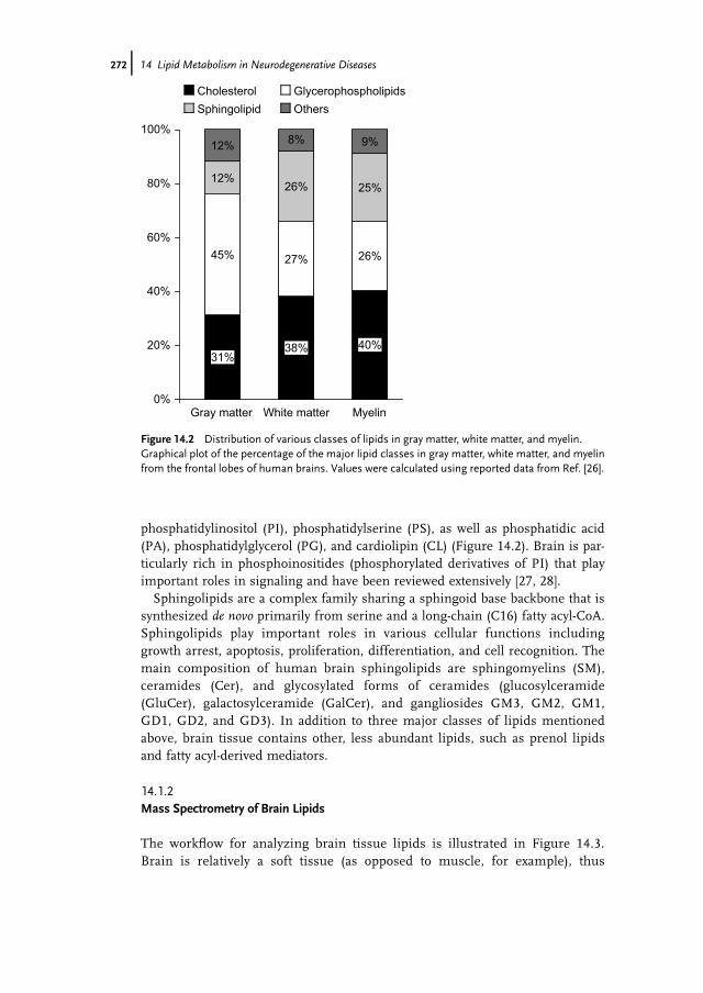
phosphatidylinositol (PI), phosphatidylserine (PS), as well as phosphatidic acid(PA), phosphatidylglycerol (PG), and cardiolipin (CL) (Figure 14.2). Brain is par-ticularly rich in phosphoinositides (phosphorylated derivatives of PI) that playimportant roles in signaling and have been reviewed extensively [27, 28].Sphingolipids are a complex family sharing a sphingoid base backbone that is
synthesized de novo primarily from serine and a long-chain (C16) fatty acyl-CoA.Sphingolipids play important roles in various cellular functions includinggrowth arrest, apoptosis, proliferation, differentiation, and cell recognition. Themain composition of human brain sphingolipids are sphingomyelins (SM),ceramides (Cer), and glycosylated forms of ceramides (glucosylceramide(GluCer), galactosylceramide (GalCer), and gangliosides GM3, GM2, GM1,GD1, GD2, and GD3). In addition to three major classes of lipids mentionedabove, brain tissue contains other, less abundant lipids, such as prenol lipidsand fatty acyl-derived mediators.
14.1.2Mass Spectrometry of Brain Lipids
The workflow for analyzing brain tissue lipids is illustrated in Figure 14.3.Brain is relatively a soft tissue (as opposed to muscle, for example), thus
Figure 14.2 Distribution of various classes of lipids in gray matter, white matter, and myelin.Graphical plot of the percentage of the major lipid classes in gray matter, white matter, and myelinfrom the frontal lobes of human brains. Values were calculated using reported data from Ref. [26].
272j 14 Lipid Metabolism in Neurodegenerative Diseases
simplifying homogenization that is either done in frozen form (powder forma-tion under liquid nitrogen or, less optimal, on ice). Selection of a robust andefficient extraction protocol for quantitative recovery of lipids from the tissue isin any case the crucial (and often ignored) first step. Most widely used extrac-tion protocols, which are either based on or modified from Bligh and Dyer orFolch methods, have proven to be reliable for qualitative lipid analysis and com-parative studies. Typically, crude lipid extracts are directly introduced into amass spectrometer without (shotgun lipidomics) or with prior chromatographicseparation (LC-MS). Shotgun approaches have the advantage of simple experi-mental setup and high capacity (high-throughput analysis). Coupling
Biochemicalcomposition
Lipid extraction
rela
tive
abun
danc
e
elution time
Mass spectrometers with or without chromatography
rela
tive
abun
danc
e
mass/charge (m/z)
single-stage MS-survey profile (untargeted)-SIM-accurate mass mesurement
rela
tive
abun
danc
e
mass/charge (m/z)
Tandem MS-product ions (structure of lipids)-MRM (targeted)-precursor ions analysis-NL
Data/statisticalanalysis
Pathway analysis
Brain tissue
Figure 14.3 Generalized workflow for mass spectrometric measurement of lipids in extractsfrom brain.
14.1 Introduction j273
chromatography to MS significantly reduces ion suppression and enables detec-tion of minor lipid species.Lipids are preferably detected using mass spectrometers with soft ionization
techniques such as electrospray ionization (ESI) and atmospheric pressurechemical ionization (APCI). Most phospholipid and sphingolipids are well ion-ized in ESI modes, while cholesterol and coenzyme Q9 are well ionized inAPCI mode. ESI/MS has been widely used to measure lipids in various applica-tions. While many cellular phospholipids (PS, PA, PG, PI, and PE) are effec-tively ionized and detected by ESI-MS in the negative ionization mode, PC andSM are more sensitively detected in positive ionization mode. Both phospholi-pids and sphingolipids are effectively ionized and detected by ESI-MS in thenegative ionization mode, with or without a chemical modifier or signalenhancer. Thus, negative ESI/MS could be directly used to obtain full-scan MSspectra of the crude lipid extracts for nontargeted lipidomics. For instance, anMS scan ranging from 400 to 1200 amu will include various polar lipids such aslysophospholipids, phospholipids (PC, PE, PI, PS, PG, and PA), and sphingoli-pids. Furthermore, other abundant sphingolipids such as sulfatides and GM3are measured in negative ESI mode. Minor neutral lipid species, such as TAGand cholesteryl ester species, can be monitored as adduct ions in positive ESImode [29, 30].Present lipidomic strategies can be classified into two broad categories
according to the nature of the MS data collected, (1) nontargeted approachesand (2) full MS scan mode, although alternative naming according to theparticular single or tandem MS operation is equally valid and often moreprecise in describing the technical details of the approaches taken (Figure14.3). For simplicity, we shall use “nontargeted” for single-stage MS and“targeted” for tandem MS based on multiple reaction monitoring, MRM,approaches. Nontargeted lipidomic approaches aim to cover the lipidome asbroadly as possible, while targeted approaches focus only on lipid species ofinterest. Nontargeted approaches are important tools in discovering novellipids or unexpected lipid metabolites between paired sample sets. Targetedapproaches, on the other hand, are usually more specific, sensitive, and conferrelative ease in terms of data processing. Inevitably, loss of information dueto the restrictions imposed by the targeting lists of lipids to be measured isa limitation.The selection of a suitable combination of mass analyzers (quadrupole, time of
flight, ion trap, Orbitrap) represents the next critical step in order to ensure aneffective analytical scheme for both qualitative investigation (i.e., mass detectionbased on m/z values) and quantitative (i.e., measuring the intensity of an ion ofinterest) analyses. In the case of nontargeted lipidomic approaches, a high-resolu-tion (TOF or Orbitrap) mass analyzer is desirable to provide accurate mass data(m/z values) at sufficiently high spectral resolution. In targeted lipidomicapproaches, quadrupole mass analyzers are popular due to the relative ease of oper-ation (e.g., both MRM and SIM, precursor ion, and neutral loss scans fall into thiscategory to a certain degree).
274j 14 Lipid Metabolism in Neurodegenerative Diseases
14.2Alzheimer’s Disease
AD, first described by the German psychiatrist Alois Alzheimer in 1906, is now oneof the most common forms of incurable neurodegenerative diseases. In a shortpaper, Alzheimer identified three main features in the brain of Mrs Deter, who suf-fered from advanced dementia in her 50s and died at the age of 56. These mainfeatures are (1) striking changes in neurofibrils (better known as neurofibrillarytangles, NFT), (2) plaques or foci visible without staining (beta-amyloid plaques),and (3) lipoid inclusion granules in glia [31–33] (Table 14.1). Since then researchershave made substantial progress toward biochemical understanding of these immu-nohistological features.
Table 14.1 Summary of lipids that have been implicated in Alzheimer’s disease.
Lipid name Commonabbreviation
Role in disease References
Arachidonic acid AA AA is increased upon Abproduction and elevated AMPARand excitotoxcity
[34, 35]
Cholesterol andcholesteryl-ester
FC and CE In AD models, hyperactive ACAT1led to increased CE; ACAT1inhibitor are protective in ADmouse models
[36–41]
Docosahexanoicacid/neuroprotectin D1
DHA/NPD1 DHA and NPD1 are survivalfactors in Ab-induced toxicity. Theypromote neurite outgrowth andreduce Ab-42 formation
[42–45]
Diacylglycerol DAG DAG increases a-secretase activity,thereby reducing Ab formation
[46]
Phosphatidylinositol-4,5-bisphosphate
PI(4,5)P2 Reduction of PI(4,5)P2 is observedin AD models due to hyperactivePLC activity; increase in PI(4,5)P2is protective in AD model
[27, 47, 48]
Plasmalogenphosphatidylethanolamine
pPE Decreased levels of pPE wereobserved in gray matter of ADbrain. pPE may act as a buffer foroxidative stress. AGPS, theenzyme, which catalyzes theformation of pPE, can bemodulated by APP
[49–51]
Phosphatidylcholines PC Decreased levels of PC wereobserved in AD brain
[52]
Sulfatides Depleted levels of sulfatide in brainand CSF of AD patients. Sulfatidelevels are regulated by ApoE alleles
[53–55]
14.2 Alzheimer’s Disease j275
For example, the “changes in neurofibrils” are now considered hyperphosphory-lation of the microtubule-associated protein tau. The “foci” or “plaque” are now“b-amyloid plaques” composed of beta-amyloid peptides of 36–43 amino acids. Theprecursor protein APP is cleaved by either beta (BACE1) or gamma-secretase (con-sisting of presenilin (PS1, nicastrin, APH-1, and PEN-2) to generate these beta-amyloid peptides of varying length. Various components of this pathway, such asAPP and PS1, have been identified to cause early-onset Alzheimer’s disease, possi-bly due to the changes in APP cleavage [56–58].
14.2.1Cholesterol and Cholesterol Esters
However, the third feature, that is, the “lipoid inclusion,” was largely ignored untilthe early 1990s when gene-mapping studies identified carriers of the apo-lipoprotein (APOE) e4 allele with increased risk for AD [59, 60]. Interestingly, theAPOE e2 allele appears to be associated with lowered risk of the disease [60]. Inaddition, APOE e4 allele is the only known major genetic risk factor that accountsfor 95% of both sporadic and familial form of late-onset AD [61]. In the nervoussystem, APOE is produced by astrocytes, which allows for the transport of choles-terol into neurons. While APOE is particularly important in the brain, other apoli-poproteins are involved in sterol/lipid transport in the body periphery. Althoughcorrelations between hypercholesterolemia in mid-life as a risk factor for AD havebeen identified in epidemiological studies [62, 63], the precise details of howperipheral sterol/lipid levels affect the brain remain poorly understood.APP and Ab are found to be associated with cholesterol-rich microdomains.
More specifically, Puglielli et al. showed that elevation of cholesteryl-esters (CE) butnot free cholesterol results in increased Ab production [36, 37]. There are (at least)two major interchangeable pools of cellular cholesterol, (1) FC in membranes and(2) CE in cytoplasmic lipid droplets. Acetyl-coenzyme A acetyltransferase (ACAT)regulates this dynamic equilibrium, and is thus likely to be an important functionin the brain that does not rely on bulk fat storage in the form of lipid droplets.Inhibition of ACAT that leads to reduced levels of CEs also lowers Ab generation
[36, 38–40, 64]. Furthermore, in fibroblasts from AD patients ACAT-1 mRNA levelsare increased significantly. Recent genetic evidence associates a single nucleotidepolymorphism (SNP, codon 405 isoleucine to valine (V405)) of the cholesterol estertransfer protein (CETP) with the lowered risk of dementia [65, 66]. Similar to whatwas first noted by Alzheimer, ultrastructural studies on autopsied brain tissue fromAlzheimer’s disease patients using immunocytochemistry with an antibody thatrecognizes amyloid-beta peptides revealed cytosolic clusters of lipid droplets inimmunopositive areas and there are cytosolic granules [67]. One possibility is thatsterols could modulate processing and/or accumulation of amyloid beta.Supporting this view, cognitive defects seen in mice expressing the human form
of the Swedish APP mutant (hAPPsw) are ameliorated in ACTA1 gene ablatedbackground (ACAT1�/�, [41]. In addition, ACAT1 inhibitor CP-113818 [68] andCI-1011 [69] showed some effectiveness in preclinical models. ACAT1�/� animals
276j 14 Lipid Metabolism in Neurodegenerative Diseases
also exhibit an increase in 24-hydroxycholesterol in the endoplasmic reticulum anddecreased rate of sterol synthesis in the brain [41], thus an overall effect on choles-terol metabolism. Indeed, it has been widely proposed to test the effects of statinsas potential therapeutics for AD treatment. Statins, which inhibit HMG-CoA reduc-tase, have been used in clinical trials for AD with inconsistent results within theprospective cohorts. However, it remains to be seen how effectively statins actuallyget transported to the brain and influence sterol biosynthesis in this organ.
14.2.2Sulfatides
Sulfatides, a class of sulfated galactosphingolipids, are found in high abundance inthe CNS. They are synthesized by oligodendrocytes and misregulation in their dis-tribution has been shown in AD brains. One of the first evidence of aberrant sphin-golipid metabolism in AD was the observation that within degenerating neuronsfrom both the cortex and the hippocampus of AD cases, there were high levelsof immunoreactivity for a monoclonal antibody raised against gangliosides,A2B5 [70]. It was later shown that A2B5 also binds to sulfatides [71].Using an unbiased lipidomics approach, Han et al. found that at very early stages
of AD, sulfatides are substantially and specifically depleted both in brains and incerebrospinal fluid (CSF) of individuals with AD [54, 55]. To identify the mecha-nism(s) of sulfatide loss concurrent with AD onset, the same group analyzed thesulfatide content in the cortex and hippocampus from animals either lacking ApoEor expressing the human ApoE4 allele. Interestingly, they found that the ApoE4-expressing mice had approximately 60% less sulfatides than those found in wild-type mice of the same age [53]. In contrast, the sulfatide levels in hippocampus andcortex of ApoE knockout mice were, respectively, 61 and 114% higher than in wild-type mice, suggesting that ApoE and the ApoE alleles play important role in regulat-ing sulfatide levels. A separate study of whole-brain extracts frommice with variousApoE knock-in alleles did not find major changes in lipids [30], which may beattributed to masking effects (whole brain versus selected regions). Thus, one pos-sible model proposed by Han is that ApoE plays a role in carrying sulfatides fromoligodendrocytes (where they are synthesized) to neurons [72], similar to what hasbeen shown for trafficking of cholesterol [73]. Factors that disrupt this process leadto alteration in sulfatide levels in the brain, serum, or CSF.
14.2.3Plasmalogen Ethanolamines
Plasmalogen phosphatidyl ethanolamine (pPE) is one of the most abundant lipidsin neuronal cell membranes, representing about 30% of total phospholipids [72].Using unbiased approaches, a number of groups have showed that in post-mortemtemporal cerebral and other gray matter regions, there is a significant reduction ofpPE [72]. Interestingly, the magnitude of the deficiencies is correlated with severityof AD progression. In patients’ serum, levels of pPE were observed to be
14.2 Alzheimer’s Disease j277
significantly decreased in pathologically diagnosed AD subjects at all stages ofdementia compared to age-matched controls. Again, the severity of this decreasecorrelated with the severity of dementia [49]. In disease models such as transgenicmice carrying the APP Swedish mutation, pPE is decreased in the cerebral corticesbut not the cerebellum, suggesting that pPE decreased as a result of AD [51].More recently, the intercellular domain of APP was found to decrease the expres-
sion of alkyl-dihydroxyacetonephosphate synthase (AGPS), the rate-limitingenzyme in pPE biosynthesis [50]. In addition to modulating AGPS levels, ADpathogenesis could affect pPE levels in another way. Since Ab has been shown toinduce oxidative stress and pPE lipids are sensitive to oxidization due to the vinylether bond [74], it is likely that Ab induces decrease in pPE by oxidation of pPE.While this would suggest that pPE level decreases as a result of Ab-mediated oxida-tive stress and regulation of AGPS, it remains unclear whether pPE decrease couldinfluence or accelerate the progression of AD. Particularly, as the largest risk factorfor AD is age, and levels of pPE decrease with age, it is possible that a reduction inplasmalogens could, in fact, influence AD progression.
14.2.4Phospholipases
Phospholipases (C, D, A) as well as the levels of their substrates (GPLs) and prod-ucts (PAs, DAGs, lyso-GPLs, and fatty acyls) have long been implicated withinflammation in the brain and AD.
14.2.4.1 Phospholipase A2The role of phospholipase A2 (PLA2) in Ab-dependent cognitive deficit and toxicityhas been well studied, but this also revealed a lot of complexity. In general, PLA2catalyzes the reaction of phospholipids to lysophospholipids and fatty acids. Inhumans, PLA2 can be classified into 12 different groups, with more than 19 differ-ent isoforms [75], though most of the studies on the central nervous system havefocused on three groups, group IV cytosolic (cPLA2), group II secretory (sPLA),and group VI – Ca2þ-independent PLA2 (GVI-PLA2). cPLA2 is regulated by intra-cellular Ca2þ, found at high levels in the hippocampus, and has strongest substratespecificity for arachidonic acid (AA). GVI-PLA2 appears to not have particular sub-strate preference but is the determinant enzyme to control docosahexaenoic acid(DHA) in the brain [76].Using a lipidomics approach to profile various fatty acids in brain tissue of an AD
model transgenic mice, researchers have found an increase in arachidonic acidsand its metabolites [35]. AA levels, the main products of cPLA2, have been impli-cated in various roles relevant to AD such as inflammatory response, synaptictransmission, and oxidative stress. Several studies have shown that there is likely afeedback loop between AA production and synaptic strength, long-term potentia-tion, as well as NMDAR and AMPAR levels in dendritic spines [34, 77, 78]. HowAA inhibition mediates neuroprotection has not been completely elucidated,however.
278j 14 Lipid Metabolism in Neurodegenerative Diseases
Hypoactivity in GIV-PLA2 in the hippocampus impairs long- and short-termmemory [79]. Moreover, in a cohort of AD patients, GIV-PLA2 activity wasfound to have decreased [80]. The human isoform for GIV-PLA2, PLA2G6, isalso mutated in neurodegenerative disorders with high brain iron includingAD, PD, and neuroaxonal dystrophies [81, 82]. Exactly why reduction of GIV-PLA2 activity and its substrate, DHA, levels are involved in AD is unclear.Unlike AA, which has been shown to be involved in synaptic functions, there islittle evidence of the role of DHA in neural transmission. Most of the studieson DHA point toward its importance in brain development and neuronal sur-vival [43]. Increasing DHA levels by modification of the diet modestly improvescognition in AD animal models [42]. Several epidemiological studies havereported that reduced levels of DHA are associated with higher risk of neuro-degeneration. The mechanisms of these observations of neuroprotection areunclear. One model suggests that DHA functions as an antioxidant to counter-balance the inflammatory response during AD [44].Evidence supporting this model comes from studies on the DHA-derived mes-
senger, neuroprotectin D1 (NPD1). Using mass spectrometry to detect DHA andNPD1 in a variety of brain and retinal degenerative models, Bazan’s group hasfound that NPD1 exerts protective effects and antiinflammatory bioactivity [83]. Incells overexpressing beta-amyloid precursor protein (bAPP), the NPD1 suppressedAb42 formation by downregulating BACE1 while activating the a-secretase. This iseffective in changing the amyloidogenic pathway into a nonamyloidogenic, neuro-trophic pathway [84]. NPD1 also mediates survival of retinal epithelial cells via thePI3K/Akt pathway [85]. Whether or not similar mechanisms are at work in the hip-pocampal or frontal cortex remains to be elucidated.
14.2.4.2 Phospholipase C and Phospholipase DIt has been shown that Ab42 oligomers promote the hydrolysis of phosphatidy-linositol-4,5-bisphosphate (PI(4,5)P2) via the phospholipase C (PLC) pathway.Blocking this pathway has also been shown to protect against the synapse-impairing actions of Ab [47]. Activation of PLC results in the production of diac-ylglycerol (DAG) and inositol-1,4,5-trisphosphate (IP3) (Figure 14.4). Onehypothesis is that excessive IP3 increases release of Ca2þ that may induce syn-aptic dysfunction; thus, blocking this event would prevent synaptic loss. On theother hand, DAG, the membrane-bound product of PLC action, has been shownto increase alpha-secretase activity via protein kinase C (PKC) [46]. Increase inalpha-secretase could result in lowered levels of beta-amyloid peptides, whichwould expect to be protective. Interestingly, genetic ablation of synaptojanin 1(SYNJ1), a phosphatase that converts PI(4,5)P2 to PI(4)P, increases PI(4,5)P2levels and also protects against Ab toxicity [47], suggesting that PI(4,5)P2 levelscould also play an important in neuroprotection and that phospholipid metabo-lism is closely linked to susceptibility of Ab insults.Phospholipase D (PLD) catalyzes the reaction of phosphatidylcholine to phos-
phatidic acid (Figure 14.4). PA is a bioactive lipid and also represents a majorcross-road in lipid biosynthesis. While PA can be quickly converted to DAG in a
14.2 Alzheimer’s Disease j279
280j 14 Lipid Metabolism in Neurodegenerative Diseases
DAG
IP3
PIP2
A oligomers (a) (b) (c)
extracellular
PLC
Increases intracellular Ca2+
Promotes -secretase/lower A produciton
A oligomers
PLA2
Arachidonic acid (AA)
Inflammatory response,excitotocity
Docosahexanoicacid (DHA)
NeuroprotectinD1 (NPD1)
Prosurvivalneurite outgrowth
A oligomers
PLD2
LPC
PA
Survival signaling?
O O
O
OH
O
OOH OH
Figure 14.4 Summary of lipids and phospholipase in Ab signaling. Signalingthrough Ab creates various complex cascades that could be either protective ordeleterious to neuronal survival. Figure 14.4a shows activation of PLC, whichresults in hydrolysis of PIP2 and generation of IP3 and DAG. While DAG can lowerAb production, IP3 increases intracellular Ca2þ levels. Figure 14.4b showsactivation of PLA2, which could result in hydrolysis of phosphatidylcholine (PC) to
arachidonic or docosahexanoic acid (AA or DHA, respectively) andlysophosphatidylcholine (LPC). Both AA and LPC induce inflammatory responseand excitotoxicity. However, DHA, which can be converted further to NPD1, elicitsprosurvival signals. Figure 14.4c shows activation of PLD2, resulting in thehydrolysis of PC to PA and choline.
cell, there appear to be distinct “pools” of PA and DAG signals. An attractivemodel proposes fatty acyl tails to be distinguishing factors: DAG signaling con-sists of polyunsaturated fatty acids whereas PLD-derived PA is mono-unsaturated or saturated [86]. Mass spectrometry approaches have thus enabledthe distinction not only of lipid classes but also of the saturation state. In ADmouse models, genetic ablation of PLD2 ameliorates cognitive deficits and syn-aptic dysfunction. Two PA species, 32:1 and 38:4, are consistently increased inthe PLD2þ/þ animals compared to the PLD2�/� [87]. In an independentstudy, it was found that Spo14 regulatory factor 1 (Srf1) activates PLD and thisis essential to protect neural cells from sustained exposure to Ab [88]. Theseresults indicate that a fine balance between phosphatidylinositol-4,5-bisphos-phate (PIP2)/DAG/PA is required, but how this is regulated at the molecularlevel of microdomains remains to be determined.
14.3Parkinson’s Disease
Parkinson’s Disease is a movement disorder marked by selective degeneration ofdopaminergic neurons in the nigrostriatum pathway [12]. It affects about 1% of thepopulation over 60 years old and 4% of people over 80 years old. Named after theclinician who first described the disease in 1817, James Parkinson, this is at presentthe second most common age-related neurodegenerative disorder [89].The clinical characterization of the disease is extremely accurate. With experi-
enced clinicians, PD is diagnosed with 98% accuracy [90], compared to about 83%for AD [91]. Most patients with PD exhibit numerous deficits in movement withobvious symptoms such as rigidity or stiffness of limbs and/or neck, tremor, brady-kinesia, and reduction of movement. Other less apparent symptoms includedepression, dementia or confusion, uncontrolled drooling, speech impairment,swallowing difficulty, and constipation.The main clinical features of PD are caused by the selective loss of dopaminergic
neurons in the nigrostriatal pathway, which is also a hallmark of the disease. How-ever, it is important to note that neuronal cell death is also detected in other regionsof the brain such as the cerebellum and cortex. Furthermore, cytoplasmic aggre-gates called Lewy bodies, comprising alpha-synuclein, are present in brains of PDpatients. Clearly, the most severely affected region is the nigrostriatal pathway,which regulates fine voluntary movements. Thus, the main motor deficits in PDpatients are most likely attributed to this pathway, while the other less commonand nonmotor symptoms such as confusion and dementia are most likely due toimpairments in other brain regions.Despite the clear clinical diagnosis, only 5–10% of PD cases are linked to
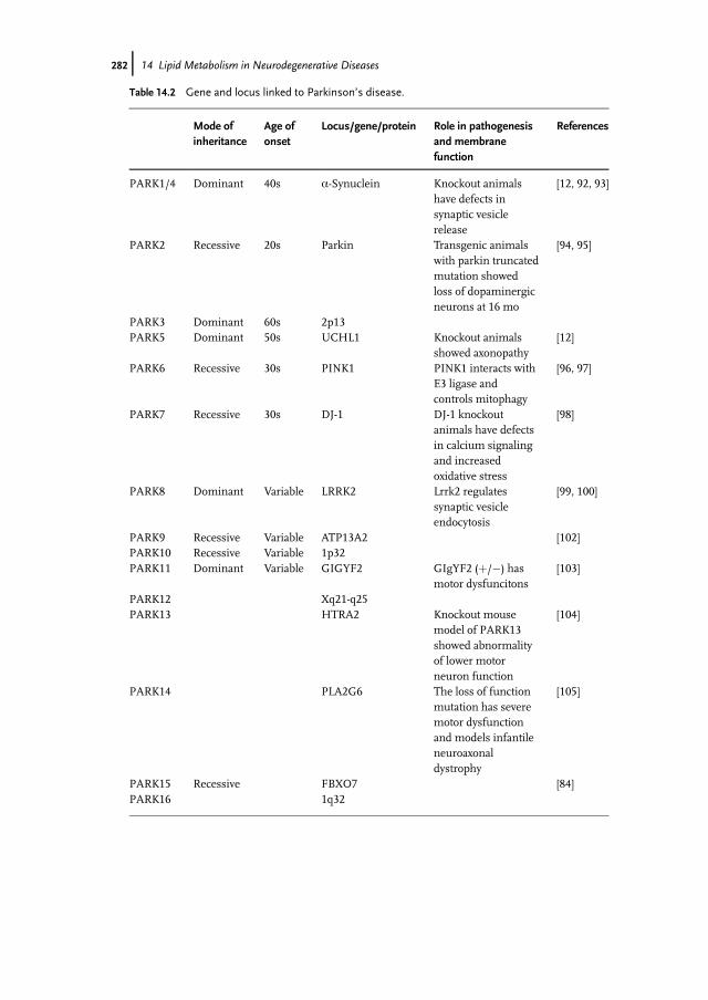
known genes (Table 14.2). Furthermore, the pathological mechanisms respon-sible for onset and progression of the disease are still unknown. The consen-sus is that PD is a multifactorial disease, whereby both extrinsic factors, suchas exposure to environmental toxins, and intrinsic factors, such as genetic
14.3 Parkinson’s Disease j281
Table 14.2 Gene and locus linked to Parkinson’s disease.
Mode ofinheritance
Age ofonset
Locus/gene/protein Role in pathogenesisand membranefunction
model of PARK13showed abnormalityof lower motorneuron function
[104]
PARK14 PLA2G6 The loss of functionmutation has severemotor dysfunctionand models infantileneuroaxonaldystrophy
[105]
PARK15 Recessive FBXO7 [84]PARK16 1q32
282j 14 Lipid Metabolism in Neurodegenerative Diseases
background, are important for pathogenesis. More importantly, there is agrowing list of components associated with PD etiology such as oxidativestress, ER protein misfolding, mitochondria dysfunction, transcription factorchanges, epigenetic changes, calcium toxicity, and cholesterol misregulation.It is not known if all these factors are related or are independent factors fordisease progression.
14.3.1Cerebrosides
Unlike AD, for which a dominant risk factor (APOE) was identified a few deca-des ago, there has been much less evidence for a direct and specific implicationof lipids in PD. This started to change around 2004 when the first clinical studyidentified an association of parkinsonism with mutation in the glucocerebrosi-dase gene (GBA) in Ashkenazi Jews [106]. Many other studies on differentpatients cohorts also found similar associations (see for review Ref. [107]). Todate, GBA variants are the most common risk factor associated with parkinson-ism, and PD patients are five times more likely to carry a GBA mutation thancontrol individuals [108].While the clinical link of GBA and PD has been well established, less is
known about the underlying mechanisms. Along with GBA–PD linkage studies,a number of groups have observed neuroprotective effects of gangliosides inmodels for PD [109, 110]. Short-term clinical trials have also demonstratedsome benefits to neurological functions in PD patients [109, 111, 112]. Studieson an animal cell line with inhibited glycosyl-ceramide synthase led to variousfeatures of lysosomal pathology, including compromised lysosomal activity,enhanced lysosomal membrane permeabilization, and increased cytotoxicity [113],suggesting a connection between ceramide metabolism and lysosomal functions.Glucosylceramide (GluCer), the substrate of glucocerebrosidase, stabilizes oligo-meric forms of a-syn and enhances amyloid formation. At the same time, a-synspecies lead to depletion of lysosomes. Loss of glucocerebrosidase activity causesaccumulation of a-synuclein but not that of tau, another aggregation-proneprotein [114].Studies in GBA models suggest that a-syn and lipids can coregulate each other.
However, lipidomic data to support these findings remain rather limiting. Forexample, untargeted lipidomics in transgenic mice with human a-syn or deletedendogenous a-syn reveal differences between ages and gender and smaller changesassociated with a-syn genotypes [115], thus providing little guidance on howchanges in lipids may affect progression of PD. Recently, a multicenter study pre-sented lipidomic analyses of human PD visual cortex (a region devoid of obviouspathology), amygdala and anterior cingulated cortex (regions with Lewy bodies butlacking degeneration). No significant differences were detected between the amyg-dala and the anterior cingulated cortex of controls and patients. In contrast, levelsof approximately 70 lipids were different in the visual cortex of controls versuspatients [29].
14.3 Parkinson’s Disease j283
14.3.2Coenzyme Q
In the list of rare genetic PD cases, many genes point toward mitochondrialdysfunction (Table 14.3). In addition, in patients with idiopathic PD, the cata-lytic activity of brain mitochondrial complex I is compromised [116]. A numberof environmental toxins that directly affect mitochondrial function induce par-kinsonism. Perhaps, the best example of toxin-induced PD is MPTP (or itsactive metabolite MPPþ). MPPþ selectively enters into dopaminergic neuronsvia the dopamine transporter (DAT), inhibits complex I of mitochondria, andthereby induces clinical symptoms that are reminiscent of those found inpatients with PD [117].Because MPTP/MPPþ toxicity emulates PD symptoms, it has been widely
used in animal and cellular models to study neuronal cell death and to screenfor neuroprotective agents. Many of the identified neuroprotective agents arefound to modulate mitochondrial function, including L-carnitine, creatine, andthe endogenous lipid electron carrier, coenzyme Q10 (CoQ) [127]. In MPTProdent and primate animal models and some cohorts of patients, the supple-mentation of CoQ is partially protective or beneficial [120–124]. Similar toCoQ, methylene blue, an FDA-approved drug and an antidote to cyanide poi-soning, is neuroprotective in PD models and acts by rerouting electrons simi-lar to known uncouplers [126].
Table 14.3 Summary of lipids that exacerbate or ameliorate degeneration in Parkinson’s disease.
Lipid name Abbreviation Role/mechanism in neuroprotection References
Anandamide Anandamide levels are increased in PDpatients’ CSF. Exogenous anadamideactivates the indirect pathway innigrostriatal synapse
[118, 119]
Coenzyme Q CoQ CoQ is protective in mouse/primatemodel of PD
[120–124]
Glucosylceramideand ganglioside
GluCer GluCer stabilizes oligomeric form ofa-syn. Increased GluCer is observed ina-syn PD mouse model. Gangliosidesare neuroprotective in PD models
[111, 112, 114]
Lanosterol Lanosterol levels are decreased inmouse model of PD. Exogenouslanosterol uncouples neuronalmitochondria
[125]
Methylene blue MB MB reroutes electrons and bypassescomplex I, II, III, and IV, similar touncouplers
[126]
284j 14 Lipid Metabolism in Neurodegenerative Diseases
Understanding the modulation of mitochondrial redox states has gained a lotof interest in the context of PD research in recent years. Uncoupling proteins(UCPs), which depend on fatty acids, are neuroprotective in the MPTP modelof PD [128–130], and their expression is downregulated in mice lacking DJ-1, agene linked to early onset of PD [98]. Using a targeted approach based on GC-MS for measurement of cholesterol (and some of its precursors and oxidizedderivatives), we discovered that lanosterol is substantially reduced in theaffected brain regions of mice treated with MPTP. We further uncovered anovel role of lanosterol in neuroprotection and showed that it induces uncou-pling, thus providing a link between sterol biosynthesis and mitochondrialfunction [125].
14.3.3Endocannabinoids
Another lipid class that can ameliorate movement deficits in PD is that ofendocannabinoids. Using a targeted approach (via GC-MS), Giuffrida et al.found that motor activity stimulated the release of anandamide but no otherspecies of endogenous cannabinoids in the brain [131]. This discovery becamean important starting point for subsequent studies that link PD to anandamidesignaling. In a rodent model of Parkinson’s disease (induced by unilaterallesion with 6-hydroxydopamine), the striatal levels of anandamide, but not thatof the other endocannabinoid, were affected [132]. Anandamides were alsodetected at higher levels in the MPTP mouse model than in control animals.Finally, anandamide is twice as abundant in the CSF of PD patients than it isin age-matched controls [119], though levels do not seem to correlate with dis-ease severity.One question arising from these observations is whether an increase in ananda-
mide is part of the pathology of PD or a protective mechanism upon loss of dopa-minergic neurons. The answer to this question came only after several studieselucidated the role of endocannabinoids in the basal ganglia circuitry. Fine move-ment in the mammalian system depends largely on basal ganglia, which integratesinputs from dopaminergic, cortical, and thalamic neurons via the striatum (Fig-ure 14.5). Striatal projection signals to the thalamic neurons via a direct or indirectpathway. Imbalances by hypoactivity in the direct and overactivity in the indirectpathways have been proposed to underlie the motor deficits in Parkinson’s disease[133]. In models of Parkinson’s disease, indeed, the indirect pathway endocannabi-noid-mediated depression is absent causing the overactivity, which can be rescuedby inhibitors of endocannabinoid degradation. Administration of these drugs invivo reduces parkinsonian motor deficits, suggesting that endocannabinoid-medi-ated activation of the indirect pathway synapses has a critical role in the control ofmovement [118].While it is quite clear that endocannabinoid signaling is linked to neuro-
transmission in the basal ganglia, it is important to note that each species of
14.3 Parkinson’s Disease j285
286j 14 Lipid Metabolism in Neurodegenerative Diseases
endocannabinoid may play different physiological roles in different neuronaltypes. For example, reuptake of GABA in striatal neurons is modulated by 2-arachidonoyl-glycerol (2-AG) but not anandamides [134]. In addition, exogenousanandamide produces either inhibitory or excitatory effects on subthalmicnucleus (STN) neurons depending on its localization in the STN [135]. Thus,the challenge for neuronal lipidomics is to understand cell type-specific lipidclasses. It is still unclear if there is a class of neurons that synthesize andsecrete endocannabinoid analogous to neurons that secrete and produce dopa-mine. Future advances in mass spectrometry with single-cell resolutions couldanswer some of these questions.
14.4Conclusions
Mass spectrometry of lipids is an outstanding new tool for lipid research. It is mostpromising when combined with complementary approaches. In this short chapter,we have provided examples on how such integration could be guided in the case oftwo major neurodegenerative diseases.
Acknowledgments
Work in our laboratory is supported by the National University of Singaporeand by grants from the Singapore National Research Foundation under CRPAward No. 2007-04 and from the Biomedical Research Council of Singapore(R-183-000-234-305).
3Figure 14.5 The nigrostriatal pathway inhealthy and disease states. Graphical drawing ofthe sagittal plane of a rodent brain (leftrepresents anterior and right representsposterior). (a) In a healthy state, dopaminergicneurons in the substantial nigra par compacta(SNpc) send excitatory and inhibitory signals totwo classes of GABAergic neurons of thecaudate putamen (striatum), consisting of D1and D2 receptors, respectively. In the directpathway, D1 GABAergic neurons synapse ontoGABAergic neurons of the globus pallidusinternal segment (GPi). GPi GABAergic neuronssynapse onto gluatmergic neurons of thethalamus (THAL), sending signal to the motorcortex. In the indirect pathway, D2 GABAergicneurons synapse onto GABAergic neurons ofthe globus pallidus external segment (GPe).
GPe GABAergic neurons synapse ontoglutamatergic neurons of the subthalmicnucleus (STN). From STN, glutamatergicneurons synapse onto GPi GABAerigc neurons.The balance between this direct and indirectpathway results in fine motor coordination suchas speech. (b) In PD, upon the loss ofdopaminergic neurons, D1 (direct) pathway isless active wheras D2 (indirect) is hyperactive.The final result is overinhibition of thalamicneurons, reducing glutaminergic synapses fromthe thalamus to motor cortex. Movementdeficits are thus in patients with defects in thenigrostriatal circuit. (c) In PD model, upon lossof dopaminergic neurons, exogenous additionof endocannbinoids causes partial defects inthe indirect pathway and improves motordeficits.
14.4 Conclusions j287
References
1 Nimmrich, V. and Ebert, U. (2009) IsAlzheimer’s disease a result ofpresynaptic failure? Synapticdysfunctions induced by oligomeric beta-amyloid. Rev. Neurosci., 20, 1–12.
2 Hirokawa, N., Niwa, S., and Tanaka, Y.(2011) Molecular motors in neurons:transport mechanisms and roles in brainfunction, development, and disease.Neuron, 68, 610–638.
3 Bezprozvanny, I. and Mattson, M.P.(2008) Neuronal calciummishandlingand the pathogenesis of Alzheimer’sdisease. Trends Neurosci., 31, 454–463.
4 Vives-Bauza, C. and Przedborski, S.(2011) Mitophagy: the latest problem forParkinson’s disease. Trends Mol. Med.,17, 158–165.
5 Goedert, M. and Spillantini, M.G. (2006)A century of Alzheimer’s disease.Science, 314, 777–781.
6 Piomelli, D., Astarita, G., and Rapaka, R.(2007) A neuroscientist’s guide tolipidomics. Nat. Rev. Neurosci., 8,743–754.
7 DiNitto, J.P., Cronin, T.C., andLambright, D.G. (2003) Membranerecognition and targeting by lipid-binding domains. Sci. STKE, 2003, re16.
8 Fukuda, M., Kojima, T., and Mikoshiba,K. (1996) Phospholipid compositiondependence of Ca2þ-dependentphospholipid binding to the C2A domainof synaptotagmin IV. J. Biol. Chem., 271,8430–8434.
9 Fukuda, M., Kojima, T., and Mikoshiba,K. (1997) Regulation by bivalent cationsof phospholipid binding to the C2Adomain of synaptotagmin III.Biochem. J., 323 (Pt 2), 421–425.
10 Wenk, M.R., Lucast, L., Di Paolo, G.,Romanelli, A.J., Suchy, S.F., Nussbaum,R.L., Cline, G.W., Shulman, G.I.,McMurray, W., and De Camilli, P. (2003)Phosphoinositide profiling in complexlipid mixtures using electrosprayionization mass spectrometry. Nat.Biotechnol., 21, 813–817.
11 Lim, L. and Wenk, M. (2009) Neuronalmembrane lipids: their role in thesynaptic vesicle cycle, inHandbook of
Neurochemistry and MolecularNeurobiology, 3rd edn, Springer Science,New York, pp. 224–238.
12 Dauer, W. and Przedborski, S. (2003)Parkinson’s disease: mechanisms andmodels. Neuron, 39, 889–909.
13 Huang, X., Abbott, R.D., Petrovitch, H.,Mailman, R.B., and Ross, G.W. (2008)Low LDL cholesterol and increased riskof Parkinson’s disease: prospectiveresults from Honolulu-Asia Aging Study.Mov. Disord., 23, 1013–1018.
14 Huang, X., Chen, H., Miller, W.C.,Mailman, R.B., Woodard, J.L., Chen,P.C., Xiang, D., Murrow, R.W., Wang, Y.Z., and Poole, C. (2007) Lower low-density lipoprotein cholesterol levels areassociated with Parkinson’s disease.Mov.Disord., 22, 377–381.
15 de Lau, L.M., Koudstaal, P.J., Hofman,A., and Breteler, M.M. (2006) Serumcholesterol levels and the risk ofParkinson’s disease. Am. J. Epidemiol.,164, 998–1002.
16 Sharon, R., Bar-Joseph, I., Mirick, G.E.,Serhan, C.N., and Selkoe, D.J. (2003)Altered fatty acid composition ofdopaminergic neurons expressing alpha-synuclein and human brains with alpha-synucleinopathies. J. Biol. Chem., 278,49874–49881.
18 Koob, A.O., Ubhi, K., Paulsson, J.F.,Kelly, J., Rockenstein, E., Mante, M.,Adame, A., and Masliah, E. (2010)Lovastatin ameliorates alpha-synucleinaccumulation and oxidation in transgenicmouse models of alpha-synucleinopathies. Exp. Neurol., 221,267–274.
19 Liu, J.P., Tang, Y., Zhou, S., Toh, B.H.,McLean, C., and Li, H. (2010) Cholesterolinvolvement in the pathogenesis ofneurodegenerative diseases.Mol. CellNeurosci., 43, 33–42.
20 Madine, J., Doig, A.J., and Middleton, D.A. (2006) A study of the regional effectsof alpha-synuclein on the organization
288j 14 Lipid Metabolism in Neurodegenerative Diseases
and stability of phospholipid bilayers.Biochemistry, 45, 5783–5792.
21 Naito, A. and Kawamura, I. (2007) Solid-state NMR as a method to revealstructure and membrane-interaction ofamyloidogenic proteins and peptides.Biochim. Biophys. Acta, 1768, 1900–1912.
22 Pfefferkorn, C.M. and Lee, J.C. (2011)Tryptophan probes at the alpha-synucleinand membrane interface. J. Phys. Chem.,114, 4615–4622.
23 Rantham Prabhakara, J.P., Feist, G.,Thomasson, S., Thompson, A.,Schommer, E., and Ghribi, O. (2008)Differential effects of 24-hydroxycholesterol and 27-hydroxycholesterol on tyrosinehydroxylase and alpha-synuclein inhuman neuroblastoma SH-SY5Ycells.J. Neurochem., 107, 1722–1729.
25 Sheikh, A.M. and Nagai, A. (2011)Lysophosphatidylcholine modulates fibrilformation of amyloid beta peptide. FEBSJ., 278, 634–642.
26 O’Brien, J.S. and Sampson, E.L. (1965)Fatty acid and fatty aldehyde compositionof the major brain lipids in normalhuman gray matter, white matter, andmyelin. J. Lipid Res., 6, 545–551.
27 Di Paolo, G. and De Camilli, P. (2006)Phosphoinositides in cell regulation andmembrane dynamics. Nature, 443,651–657.
28 Wenk, M.R. (2005) The emerging field oflipidomics. Nat. Rev. Drug Discov., 4,594–610.
29 Cheng, D., Jenner, A.M., Shui, G.,Cheong, W.F., Mitchell, T.W., Nealon,J.R., Kim, W.S., McCann, H., Wenk, M.R., Halliday, G.M., and Garner, B. (2011)Lipid pathway alterations in Parkinson’sdisease primary visual cortex. PLoS One,6, e17299.
and plasma lipids in human APOEepsilon2, epsilon3, and epsilon4 knock-inmice using electrospray ionization massspectrometry. J. Alzheimer’s Dis., 20,105–111.
31 Foley, P. (2010) Lipids in Alzheimer’sdisease: a century-old story. Biochim.Biophys. Acta, 1801, 750–753.
32 Graeber, M.B., Kosel, S., Egensperger, R.,Banati, R.B., Muller, U., Bise, K.,Hoff, P., Moller, H.J., Fujisawa, K., andMehraein, P. (1997) Rediscovery of thecase described by Alois Alzheimer in1911: historical, histological andmolecular genetic analysis. Neurogenetics,1, 73–80.
33 Graeber, M.B. and Mehraein, P. (1999)Reanalysis of the first case ofAlzheimer’s disease. Eur. Arch. Psy.Clin. N., 249 (Suppl. 3), 10–13.
34 Nishikawa, T., Tomori, Y., Yamashita, S.,and Shimizu, S. (1989) Inhibition ofNaþ,Kþ-ATPase activity by phospholipaseA2 and several lysophospholipids:possible role of phospholipase A2 innoradrenaline release from cerebralcortical synaptosomes. J. Pharm.Pharmacol., 41, 450–458.
35 Sanchez-Mejia, R.O., Newman, J.W.,Toh, S., Yu, G.Q., Zhou, Y., Halabisky, B.,Cisse, M., Scearce-Levie, K., Cheng, I.H.,Gan, L., Palop, J.J., Bonventre, J.V., andMucke, L. (2008) Phospholipase A2reduction ameliorates cognitive deficitsin a mouse model of Alzheimer’sdisease. Nat. Neurosci., 11, 1311–1318.
41 Bryleva, E.Y., Rogers, M.A., Chang, C.C.,Buen, F., Harris, B.T., Rousselet, E.,Seidah, N.G., Oddo, S., LaFerla, F.M.,Spencer, T.A., Hickey, W.F., and Chang,T.Y. (2010) ACAT1 gene ablationincreases 24(S)-hydroxycholesterolcontent in the brain and amelioratesamyloid pathology in mice with AD. Proc.Natl. Acad. Sci. USA, 107, 3081–3086.
42 Arsenault, D., Julien, C., Tremblay, C.,and Calon, F. (2011) DHA improvescognition and prevents dysfunction ofentorhinal cortex neurons in 3xTg-ADmice. PLoS One, 6, e17397.
43 Cole, G.M., Ma, Q.L., and Frautschy, S.A.(2009) Omega-3 fatty acids and dementia.Prostaglandins Leukot. Essent. Fatty Acids,81, 213–221.
44 Pomponi, M., Di Gioia, A., Bria, P., andPomponi, M.F. (2008) Fatty aspirin: anew perspective in the prevention ofdementia of Alzheimer’s type? Curr.Alzheimer Res., 5, 422–431.
46 Lee, J., Kang, J.H., Han, K.C., Kim, Y.,Kim, S.Y., Youn, H.S., Mook-Jung, I.,Kim, H., Lo Han, J.H., Ha, H.J., Kim, Y.H., Marquez, V.E., Lewin, N.E., Pearce, L.V., Lundberg, D.J., and Blumberg, P.M.(2006) Branched diacylglycerol-lactonesas potent protein kinase C ligands andalpha-secretase activators. J. Med. Chem.,49, 2028–2036.
47 Berman, D.E., Dall’armi, C., Voronov,S.V., McIntire, L.B., Zhang, H., Moore,A.Z., Staniszewski, A., Arancio, O., Kim,T.W., and Di Paolo, G. (2008) Oligomericamyloid-beta peptide disruptsphosphatidylinositol-4,5-bisphosphatemetabolism. Nat. Neurosci., 11, 547–554.
48 Di Paolo, G., Sankaranarayanan, S.,Wenk, M.R., Daniell, L., Perucco, E.,Caldarone, B.J., Flavell, R., Picciotto,M.R., Ryan, T.A., Cremona, O., and DeCamilli, P. (2002) Decreased synapticvesicle recycling efficiency and cognitivedeficits in amphiphysin 1 knockout mice.Neuron, 33, 789–804.
49 Goodenowe, D.B., Cook, L.L., Liu, J.,Lu, Y., Jayasinghe, D.A., Ahiahonu, P.W.,Heath, D., Yamazaki, Y., Flax, J.,Krenitsky, K.F., Sparks, D.L., Lerner, A.,Friedland, R.P., Kudo, T., Kamino, K.,Morihara, T., Takeda, M., and Wood, P.L.(2007) Peripheral ethanolamineplasmalogen deficiency: a logicalcausative factor in Alzheimer’sdisease and dementia. J. Lipid Res., 48,2485–2498.
50 Grimm, M.O., Kuchenbecker, J.,Rothhaar, T.L., Grosgen, S.,Hundsdorfer, B., Burg, V.K., Friess, P.,Muller, U., Grimm, H.S.,Riemenschneider, M., and Hartmann, T.(2011) Plasmalogen synthesis isregulated via alkyl-dihydroxyacetonephosphate-synthase byamyloid precursor protein processingand is affected in Alzheimer’s disease.J. Neurochem., 116, 916–925.
51 Han, X., Holtzman, D.M., and McKeel,D.W., Jr. (2001) Plasmalogen deficiencyin early Alzheimer’s disease subjects andin animal models: molecularcharacterization using electrosprayionization mass spectrometry.J. Neurochem., 77, 1168–1180.
52 Svennerholm, L. and Gottfries, C.G.(1994) Membrane lipids, selectivelydiminished in Alzheimer brains, suggestsynapse loss as a primary event in early-onset form (type I) and demyelination inlate-onset form (type II). J. Neurochem.,62, 1039–1047.
290j 14 Lipid Metabolism in Neurodegenerative Diseases
role for apolipoprotein E in the centralnervous system. Modulation of sulfatidecontent. J. Biol. Chem., 278, 8043–8051.
54 Han, X.D.M.H., McKeel, D.W., Jr., Kelley,J., and Morris, J.C. (2002) Substantialsulfatide deficiency and ceramideelevation in very early Alzheimer’sdisease: potential role in diseasepathogenesis. J. Neurochem., 82,809–818.
55 Han, X., Fagan, A.M., Cheng, H., Morris,J.C., Xiong, C., and Holtzman, D.M.(2003) Cerebrospinal fluid sulfatide isdecreased in subjects with incipientdementia. Ann. Neurol., 54, 115–119.
56 Chartier-Harlin, M.C., Crawford, F.,Houlden, H., Warren, A., Hughes, D.,Fidani, L., Goate, A., Rossor, M.,Roques, P., Hardy, J. et al. (1991) Early-onset Alzheimer’s disease caused bymutations at codon 717 of the beta-amyloid precursor protein gene. Nature,353, 844–846.
57 Laudon, H., Winblad, B., and Naslund, J.(2007) The Alzheimer’s disease-associated gamma-secretase complex:functional domains in the presenilin 1protein. Physiol. Behav., 92, 115–120.
58 Murrell, J., Farlow, M., Ghetti, B., andBenson, M.D. (1991) A mutation in theamyloid precursor protein associatedwith hereditary Alzheimer’s disease.Science, 254, 97–99.
59 Corder, E.H., Saunders, A.M.,Strittmatter, W.J., Schmechel, D.E.,Gaskell, P.C., Small, G.W., Roses, A.D.,Haines, J.L., and Pericak-Vance, M.A.(1993) Gene dose of apolipoprotein Etype 4 allele and the risk of Alzheimer’sdisease in late onset families. Science,261, 921–923.
60 Pericak-Vance, M.A. and Haines, J.L.(1995) Genetic susceptibility toAlzheimer disease. Trends Genet., 11,504–508.
61 Strittmatter, W.J. (2000) Apolipoprotein Eand Alzheimer’s disease. Ann. N. Y.Acad. Sci., 924, 91–92.
62 Anstey, K.J., Lipnicki, D.M., and Low, L.F. (2008) Cholesterol as a risk factor fordementia and cognitive decline: asystematic review of prospective studies
with meta-analysis. Am. J. Geriatr.Psychiatry, 16, 343–354.
63 Notkola, I.L., Sulkava, R., Pekkanen, J.,Erkinjuntti, T., Ehnholm, C., Kivinen, P.,Tuomilehto, J., and Nissinen, A. (1998)Serum total cholesterol, apolipoprotein Eepsilon 4 allele, and Alzheimer’s disease.Neuroepidemiology, 17, 14–20.
64 Bryleva, E.Y., Rogers, M.A., Chang, C.C.,Buen, F., Harris, B.T., Rousselet, E.,Seidah, N.G., Oddo, S., LaFerla, F.M.,Spencer, T.A., Hickey, W.F., and Chang,T.Y. (2011) ACAT1 gene ablationincreases 24(S)-hydroxycholesterolcontent in the brain and amelioratesamyloid pathology in mice with AD. Proc.Natl. Acad. Sci. USA, 107, 3081–3086.
65 Rodriguez, E., Mateo, I., Infante, J.,Llorca, J., Berciano, J., and Combarros,O. (2006) Cholesteryl ester transferprotein (CETP) polymorphism modifiesthe Alzheimer’s disease risk associatedwith APOE epsilon4 allele. J. Neurol.,253, 181–185.
66 Sanders, A.E., Wang, C., Katz, M., Derby,C.A., Barzilai, N., Ozelius, L., and Lipton,R.B. (2010) Association of a functionalpolymorphism in the cholesteryl estertransfer protein (CETP) gene withmemory decline and incidence ofdementia. JAMA, 303, 150–158.
67 Gomez-Ramos, P. and Asuncion Moran,M. (2007) Ultrastructural localization ofintraneuronal Abeta-peptide inAlzheimer disease brains. J. Alzheimer’sDis., 11, 53–59.
68 Hutter-Paier, B., Huttunen, H.J.,Puglielli, L., Eckman, C.B., Kim, D.Y.,Hofmeister, A., Moir, R.D., Domnitz,S.B., Frosch, M.P., Windisch, M., andKovacs, D.M. (2004) The ACAT inhibitorCP-113,818 markedly reduces amyloidpathology in a mouse model ofAlzheimer’s disease. Neuron, 44,227–238.
69 Huttunen, H.J., Havas, D., Peach, C.,Barren, C., Duller, S., Xia, W., Frosch,M.P., Hutter-Paier, B., Windisch, M., andKovacs, D.M. (2010) The acyl-coenzymeA: cholesterol acyltransferase inhibitorCI-1011 reverses diffuse brain amyloidpathology in aged amyloid precursor
References j291
protein transgenic mice. J. Neuropathol.Exp. Neurol., 69, 777–788.
70 Emory, C.R., Ala, T.A., and Frey, W.H.,2nd (1987) Ganglioside monoclonalantibody (A2B5) labels Alzheimer’sneurofibrillary tangles. Neurology, 37,768–772.
71 Majocha, R.E., Jungalwala, F.B.,Rodenrys, A., and Marotta, C.A. (1989)Monoclonal antibody to embryonic CNSantigen A2B5 provides evidence for theinvolvement of membrane componentsat sites of Alzheimer degeneration anddetects sulfatides as well as gangliosides.J. Neurochem., 53, 953–961.
72 Han, X. (2010) The pathogenicimplication of abnormal interactionbetween apolipoprotein E isoforms,amyloid-beta peptides, and sulfatides inAlzheimer’s disease.Mol. Neurobiol., 41,97–106.
73 Vance, J.E., Karten, B., and Hayashi, H.(2006) Lipid dynamics in neurons.Biochem. Soc. Trans., 34, 399–403.
74 Zoeller, R.A., Lake, A.C., Nagan, N.,Gaposchkin, D.P., Legner, M.A., andLieberthal, W. (1999) Plasmalogens asendogenous antioxidants: somatic cellmutants reveal the importance of thevinyl ether. Biochem. J., 338 (Pt 3),769–776.
75 Sun, G.Y., Xu, J., Jensen, M.D., andSimonyi, A. (2004) Phospholipase A2 inthe central nervous system: implicationsfor neurodegenerative diseases. J. LipidRes., 45, 205–213.
76 Green, J.T., Orr, S.K., and Bazinet, R.P.(2008) The emerging role of group VIcalcium-independent phospholipase A2in releasing docosahexaenoic acid frombrain phospholipids. J. Lipid Res., 49,939–944.
77 Bernard, J., Lahsaini, A., and Massicotte,G. (1994) Potassium-induced long-termpotentiation in area CA1 of thehippocampus involves phospholipaseactivation.Hippocampus, 4, 447–453.
78 Miller, B., Sarantis, M., Traynelis, S.F.,and Attwell, D. (1992) Potentiation ofNMDA receptor currents by arachidonicacid. Nature, 355, 722–725.
79 Schaeffer, E.L. and Gattaz, W.F. (2005)Inhibition of calcium-independent
phospholipase A2 activity in rathippocampus impairs acquisition ofshort- and long-term memory.Psychopharmacology, 181, 392–400.
80 Talbot, K., Young, R.A., Jolly-Tornetta, C.,Lee, V.M., Trojanowski, J.Q., and Wolf,B.A. (2000) A frontal variant ofAlzheimer’s disease exhibits decreasedcalcium-independent phospholipase A2activity in the prefrontal cortex.Neurochem. Int., 37, 17–31.
81 Gregory, A., Westaway, S.K., Holm, I.E.,Kotzbauer, P.T., Hogarth, P., Sonek, S.,Coryell, J.C., Nguyen, T.M., Nardocci, N.,Zorzi, G., Rodriguez, D., Desguerre, I.,Bertini, E., Simonati, A., Levinson, B.,Dias, C., Barbot, C., Carrilho, I., Santos,M., Malik, I., Gitschier, J., and Hayflick,S.J. (2008) Neurodegeneration associatedwith genetic defects in phospholipase A(2). Neurology, 71, 1402–1409.
82 Morgan, N.V., Westaway, S.K.,Morton, J.E., Gregory, A., Gissen, P.,Sonek, S., Cangul, H., Coryell, J.,Canham, N., Nardocci, N., Zorzi, G.,Pasha, S., Rodriguez, D., Desguerre,I., Mubaidin, A., Bertini, E.,Trembath, R.C., Simonati, A.,Schanen, C., Johnson, C.A., Levinson,B., Woods, C.G., Wilmot, B., Kramer,P., Gitschier, J., Maher, E.R., andHayflick, S.J. (2006) PLA2G6,encoding a phospholipase A2, ismutated in neurodegenerativedisorders with high brain iron. Nat.Genet., 38752–754.
84 Zhao, T., De Graaff, E., Breedveld, G.J.,Loda, A., Severijnen, L.A., Wouters, C.H., Verheijen, F.W., Dekker, M.C.,Montagna, P., Willemsen, R., Oostra, B.A., and Bonifati, V. (2011) Loss of nuclearactivity of the FBXO7 protein in patientswith parkinsonian-pyramidal syndrome(PARK15). PLoS One, 6, e16983.
85 Halapin, N.A. and Bazan, N.G. (2010)NPD1 induction of retinal pigment
292j 14 Lipid Metabolism in Neurodegenerative Diseases
87 Oliveira, T.G., Chan, R.B., Tian, H.,Laredo, M., Shui, G., Staniszewski, A.,Zhang, H., Wang, L., Kim, T.W., Duff, K.E., Wenk, M.R., Arancio, O., and DiPaolo, G. (2010) Phospholipase d2ablation ameliorates Alzheimer’sdisease-linked synaptic dysfunction andcognitive deficits. J. Neurosci., 30,16419–16428.
88 Kennedy, M.A., Kabbani, N., Lambert, J.P., Swayne, L.A., Ahmed, F., Figeys, D.,Bennett, S.A., Bryan, J., and Baetz, K.(2010) Srf1 is a novel regulator ofphospholipase D activity and is essentialto buffer the toxic effects of C16:0 plateletactivating factor. PLoS Genet., 7,e1001299.
89 Elbaz, A. and Moisan, F. (2008) Updatein the epidemiology of Parkinson’sdisease. Curr. Opin. Neurol., 21, 454–460.
90 de Lau, L.M. and Breteler, M.M. (2006)Epidemiology of Parkinson’s disease.Lancet Neurol., 5, 525–535.
91 Lim, A., Tsuang, D., Kukull, W., Nochlin,D., Leverenz, J., McCormick, W., Bowen,J., Teri, L., Thompson, J., Peskind, E.R.,Raskind, M., and Larson, E.B. (1999)Clinico-neuropathological correlation ofAlzheimer’s disease in a community-based case series. J. Am. Geriatr. Soc., 47,564–569.
92 Larsen, K., Hedegaard, C., Bertelsen, M.F., and Bendixen, C. (2009) Threonine 53in alpha-synuclein is conserved in long-living non-primate animals. Biochem.Biophys. Res. Commun., 387, 602–605.
93 Lo Bianco, C., Ridet, J.L., Schneider, B.L.,Deglon, N., and Aebischer, P. (2002)Alpha-synucleinopathy and selectivedopaminergic neuron loss in a ratlentiviral-based model of Parkinson’sdisease. Proc. Natl. Acad. Sci. USA, 99,10813–10818.
94 Frank-Cannon, T.C., Tran, T., Ruhn, K.A.,Martinez, T.N., Hong, J., Marvin, M.,Hartley, M., Trevino, I., O’Brien, D.E.,Casey, B., Goldberg, M.S., and Tansey,M.G. (2008) Parkin deficiency increasesvulnerability to inflammation-relatednigral degeneration. J. Neurosci., 28,10825–10834.
95 Lu, X.H., Fleming, S.M., Meurers, B.,Ackerson, L.C., Mortazavi, F., Lo, V.,Hernandez, D., Sulzer, D., Jackson, G.R.,Maidment, N.T., Chesselet, M.F., andYang, X.W. (2009) Bacterial artificialchromosome transgenic mice expressinga truncated mutant parkin exhibit age-dependent hypokinetic motor deficits,dopaminergic neuron degeneration, andaccumulation of proteinase K-resistantalpha-synuclein. J. Neurosci., 29,1962–1976.
96 Geisler, S., Holmstrom, K.M., Skujat, D.,Fiesel, F.C., Rothfuss, O.C., Kahle, P.J.,and Springer, W. (2010) PINK1/Parkin-mediated mitophagy is dependent onVDAC1 and p62/SQSTM1. Nat. CellBiol., 12, 119–131.
97 Narendra, D.P., Jin, S.M., Tanaka, A.,Suen, D.F., Gautier, C.A., Shen, J.,Cookson, M.R., and Youle, R.J. (2010)PINK1 is selectively stabilized onimpaired mitochondria to activateParkin. PLoS Biology, 8, e1000298.
98 Guzman, J.N., Sanchez-Padilla, J.,Wokosin, D., Kondapalli, J., Ilijic, E.,Schumacker, P.T., and Surmeier, D.J.(2010) Oxidant stress evoked bypacemaking in dopaminergic neurons isattenuated by DJ-1. Nature, 468, 696–700.
99 Li, Y., Liu, W., Oo, T.F., Wang, L., Tang,Y., Jackson-Lewis, V., Zhou, C.,Geghman, K., Bogdanov, M.,Przedborski, S., Beal, M.F., Burke, R.E.,and Li, C. (2009) Mutant LRRK2(R1441G) BAC transgenic micerecapitulate cardinal features ofParkinson’s disease. Nat. Neurosci., 12,826–828.
100 Ramonet, D., Daher, J.P., Lin, B.M.,Stafa, K., Kim, J., Banerjee, R.,Westerlund, M., Pletnikova, O., Glauser,L., Yang, L., Liu, Y., Swing, D.A., Beal, M.F., Troncoso, J.C., McCaffery, J.M.,Jenkins, N.A., Copeland, N.G., Galter, D.,
References j293
Thomas, B., Lee, M.K., Dawson, T.M.,Dawson, V.L., and Moore, D.J. (2011)Dopaminergic neuronal loss, reducedneurite complexity and autophagicabnormalities in transgenic miceexpressing G2019S mutant LRRK2. PLoSOne, 6, e18568.
104 Jones, J.M., Albin, R.L., Feldman, E.L.,Simin, K., Schuster, T.G., Dunnick, W.A.,Collins, J.T., Chrisp, C.E., Taylor, B.A.,and Meisler, M.H. (1993) mnd2: a newmouse model of inherited motor neurondisease. Genomics, 16, 669–677.
105 Wada, H., Yasuda, T., Miura, I., Watabe,K., Sawa, C., Kamijuku, H., Kojo, S.,Taniguchi, M., Nishino, I., Wakana, S.,Yoshida, H., and Seino, K. (2009)Establishment of an improved mousemodel for infantile neuroaxonaldystrophy that shows early disease onsetand bears a point mutation in Pla2g6.Am. J. Pathol., 175, 2257–2263.
106 Aharon-Peretz, J., Rosenbaum, H., andGershoni-Baruch, R. (2004) Mutations inthe glucocerebrosidase gene and
Parkinson’s disease in Ashkenazi Jews.N. Engl. J. Med., 351, 1972–1977.
107 DePaolo, J., Goker-Alpan, O., Samaddar,T., Lopez, G., and Sidransky, E. (2009)The association between mutations inthe lysosomal protein glucocerebrosidaseand parkinsonism.Mov. Disord., 24,1571–1578.
108 Velayati, A., Yu, W.H., and Sidransky, E.(2010) The role of glucocerebrosidasemutations in Parkinson disease and Lewybody disorders. Curr. Neurol. Neurosci.Rep., 10, 190–198.
109 Schneider, J.S., Roeltgen, D.P., Mancall,E.L., Chapas-Crilly, J., Rothblat, D.S., andTatarian, G.T. (1998) Parkinson’s disease:improved function with GM1 gangliosidetreatment in a randomized placebo-controlled study. Neurology, 50,1630–1636.
110 Wei, J., Fujita, M., Sekigawa, A.,Sekiyama, K., Waragai, M., andHashimoto, M. (2009) Gangliosides’protection against lysosomal pathologyof synucleinopathies. Autophagy, 5,860–861.
111 Schneider, J.S. (1998) GM1 gangliosidein the treatment of Parkinson’s disease.Ann. N. Y. Acad. Sci., 845, 363–373.
112 Schneider, J.S., Sendek, S., Daskalakis,C., and Cambi, F. (2010) GM1ganglioside in Parkinson’s disease:results of a five year open study. J. Neurol.Sci., 292, 45–51.
113 Wei, J., Fujita, M., Nakai, M., Waragai,M., Sekigawa, A., Sugama, S.,Takenouchi, T., Masliah, E., andHashimoto, M. (2009) Protective role ofendogenous gangliosides for lysosomalpathology in a cellular model ofsynucleinopathies. Am. J. Pathol., 174,1891–1909.
114 Mazzulli, J.R., Xu, Y.H., Sun, Y., Knight,A.L., McLean, P.J., Caldwell, G.A.,Sidransky, E., Grabowski, G.A., andKrainc, D. (2011) Gaucher diseaseglucocerebrosidase and alpha-synucleinform a bidirectional pathogenic loop insynucleinopathies. Cell, 146, 37–52.
115 Rappley, I., Myers, D.S., Milne, S.B.,Ivanova, P.T., Lavoie, M.J., Brown, H.A.,and Selkoe, D.J. (2009) Lipidomicprofiling in mouse brain reveals
294j 14 Lipid Metabolism in Neurodegenerative Diseases
differences between ages and genders,with smaller changes associated withalpha-synuclein genotype. J. Neurochem.,111, 15–25.
116 Keeney, P.M., Xie, J., Capaldi, R.A., andBennett, J.P., Jr. (2006) Parkinson’sdisease brain mitochondrial complex Ihas oxidatively damaged subunits and isfunctionally impaired andmisassembled. J. Neurosci., 26,5256–5264.
117 Watanabe, Y., Himeda, T., and Araki, T.(2005) Mechanisms of MPTP toxicity andtheir implications for therapy ofParkinson’s disease.Med. Sci. Monit., 11,RA17–RA23.
118 Kreitzer, A.C. and Malenka, R.C. (2007)Endocannabinoid-mediated rescue ofstriatal LTD and motor deficits inParkinson’s disease models. Nature, 445,643–647.
119 Pisani, A., Fezza, F., Galati, S., Battista,N., Napolitano, S., Finazzi-Agro, A.,Bernardi, G., Brusa, L., Pierantozzi, M.,Stanzione, P., and Maccarrone, M. (2005)High endogenous cannabinoid levels inthe cerebrospinal fluid of untreatedParkinson’s disease patients. Ann.Neurol., 57, 777–779.
120 Cleren, C., Yang, L., Lorenzo, B.,Calingasan, N.Y., Schomer, A., Sireci, A.,Wille, E.J., and Beal, M.F. (2008)Therapeutic effects of coenzyme Q10(CoQ10) and reduced CoQ10 in theMPTP model of parkinsonism.J. Neurochem., 104, 1613–1621.
121 Galpern, W.R. and Cudkowicz, M.E.(2007) Coenzyme Q treatment ofneurodegenerative diseases of aging.Mitochondrion, (7 Suppl.), S146–S153.
122 Horvath, T.L., Diano, S., Leranth, C.,Garcia-Segura, L.M., Cowley, M.A.,Shanabrough, M., Elsworth, J.D.,Sotonyi, P., Roth, R.H., Dietrich, E.H.,Matthews, R.T., Barnstable, C.J., andRedmond, D.E., Jr. (2003) Coenzyme Qinduces nigral mitochondrial uncouplingand prevents dopamine cell loss in aprimate model of Parkinson’s disease.Endocrinology, 144, 2757–2760.
symptomatic benefit in patients withParkinson’s disease. Neurosci. Lett., 341,201–204.
124 Yang, L., Calingasan, N.Y., Wille, E.J.,Cormier, K., Smith, K., Ferrante, R.J.,and Beal, M.F. (2009) Combinationtherapy with coenzyme Q10 and creatineproduces additive neuroprotective effectsin models of Parkinson’s andHuntington’s diseases. J. Neurochem.,109, 1427–1439.
125 Lim, L., Jackson-Lewis, V., Wong, L.C.,Shui, G.H., Goh, A.X., Kesavapany, S.,Jenner, A.M., Fivaz, M., Przedborski, S.,and Wenk, M.R. (2011) Lanosterolinduces mitochondrial uncoupling andprotects dopaminergic neurons from celldeath in a model for Parkinson’s disease.Cell Death Differ. DOI: 10.1038/cdd.2011.105.
126 Wen, Y., Li, W., Poteet, E.C., Xie, L., Tan,C., Yan, L.J., Ju, X., Liu, R., Qian, H.,Marvin, M.A., Goldberg, M.S., She, H.,Mao, Z., Simpkins, J.W., and Yang, S.H.(2011) Alternative mitochondrial electrontransfer as a novel strategy forneuroprotection. J. Biol. Chem., 286,16504–16515.
127 Virmani, A., Gaetani, F., and Binienda,Z. (2005) Effects of metabolic modifierssuch as carnitines, coenzyme Q10, andPUFAs against different forms ofneurotoxic insults: metabolic inhibitors,MPTP, and methamphetamine. Ann. N.Y. Acad. Sci., 1053, 183–191.
128 Andrews, Z.B., Diano, S., and Horvath,T.L. (2005) Mitochondrial uncouplingproteins in the CNS: in support offunction and survival. Nat. Rev. Neurosci.,6, 829–840.
129 Andrews, Z.B., Horvath, B., Barnstable,C.J., Elsworth, J., Yang, L., Beal, M.F.,Roth, R.H., Matthews, R.T., and Horvath,T.L. (2005) Uncoupling protein-2 iscritical for nigral dopamine cell survivalin a mouse model of Parkinson’s disease.J. Neurosci., 25, 184–191.
Uncoupling protein 2 protectsdopaminergic neurons from acute1,2,3,6-methyl-phenyl-tetrahydropyridinetoxicity. J. Neurochem., 93, 493–501.
131 Giuffrida, A., Parsons, L.H., Kerr, T.M.,Rodriguez de Fonseca, F., Navarro, M.,and Piomelli, D. (1999) Dopamineactivation of endogenous cannabinoidsignaling in dorsal striatum. Nat.Neurosci., 2, 358–363.
132 Gubellini, P., Picconi, B., Bari, M.,Battista, N., Calabresi, P., Centonze, D.,Bernardi, G., Finazzi-Agro, A., andMaccarrone, M. (2002) Experimentalparkinsonism alters endocannabinoiddegradation: implications for striatalglutamatergic transmission. J. Neurosci.,22, 6900–6907.
133 Kravitz, A.V., Freeze, B.S., Parker, P.R.,Kay, K., Thwin, M.T., Deisseroth, K., andKreitzer, A.C. (2010) Regulation ofparkinsonian motor behaviours byoptogenetic control of basal gangliacircuitry. Nature, 466, 622–626.
134 Venderova, K., Brown, T.M., andBrotchie, J.M. (2005) Differential effectsof endocannabinoids on [(3)H]-GABAuptake in the rat globus pallidus. Exp.Neurol., 194, 284–287.
135 Morera-Herreras, T., Ruiz-Ortega, J.A.,Linazasoro, G., and Ugedo, L. (2011)Nigrostriatal denervation changes theeffect of cannabinoids on subthalamicneuronal activity in rats.Psychopharmacology, 214,379–389.
296j 14 Lipid Metabolism in Neurodegenerative Diseases