1 Lipidomics Perspective: From Molecular Lipidomics to Validated Clinical Diagnostics Kim Ekroos 1.1 Introduction Lipids are recognized as extremely diversified molecules, with nearly 10 4 different structures of lipids currently being stored in the most comprehensive lipid struc- ture database (LIPID MAPS, http://www.lipidmaps.org). The complexity is con- founded by the fact that the absolute quantity of individual molecular lipids can differ among lipid species up to several million-fold depending on the matrix of origin. These features unprecedentedly complicate a precise assessment of the actual number of lipid entities and their identities and quantities making up a lip- idome of a biological system. The accomplishment of this task now relies on lipido- mics. Cutting edge lipidomics has already demonstrated its supremacy by revealing, for example, over 500 lipid species in human plasma [1, 2] and 250 lipid species in yeast [3]. With the currently ongoing meticulous developments in this field, it is highly anticipated that it will in the coming years facilitate delivery of a close to complete lipidomic content and precisely determined. If this increases the total number of species, for instance, in the human plasma lipidome to comprise thousand lipid species or more still remains to be seen. The lipidome of eukaryotic cells is believed to contain thousands of lipid entities that structurally and chemically regulate cell membranes, store energy, or become precursors to bioactive metabolites [4, 5]. Lipids primarily reside in cellular mem- branes. The individual membranes of a cell have unique lipid compositions, required for serving their vital biological functions. For example, the free choles- terol (FC) to total phospholipid (PL) ratio in the endoplasmic reticulum (ER) mem- brane in mammals has been shown to be 0.15, whereas in the plasma membrane the ratio is 1 [6]. Although ER is the main site of lipid synthesis, it is the local lipid metabolism that is the prime determinant of the unique compositions of organ- elles. Moreover, further membrane specialization is orchestrated by lateral organi- zation to form dynamic platforms, that is, lipid rafts, within the cellular membranes serving as functional assemblies for diverse processes such as signal transduction, membrane trafficking, and cell adhesion [7]. The physiological response and bioactive output of such lipid raft domain or cellular membrane will Lipidomics, First Edition. Edited by Kim Ekroos. # 2012 Wiley-VCH Verlag GmbH & Co. KGaA. Published 2012 by Wiley-VCH Verlag GmbH & Co. KGaA. j1
Transcript
1Lipidomics Perspective: From Molecular Lipidomicsto Validated Clinical DiagnosticsKim Ekroos
1.1Introduction
Lipids are recognized as extremely diversified molecules, with nearly 104 differentstructures of lipids currently being stored in the most comprehensive lipid struc-ture database (LIPID MAPS, http://www.lipidmaps.org). The complexity is con-founded by the fact that the absolute quantity of individual molecular lipids candiffer among lipid species up to several million-fold depending on the matrix oforigin. These features unprecedentedly complicate a precise assessment of theactual number of lipid entities and their identities and quantities making up a lip-idome of a biological system. The accomplishment of this task now relies on lipido-mics. Cutting edge lipidomics has already demonstrated its supremacy byrevealing, for example, over 500 lipid species in human plasma [1, 2] and 250 lipidspecies in yeast [3]. With the currently ongoing meticulous developments in thisfield, it is highly anticipated that it will in the coming years facilitate delivery of aclose to complete lipidomic content and precisely determined. If this increases thetotal number of species, for instance, in the human plasma lipidome to comprisethousand lipid species or more still remains to be seen.The lipidome of eukaryotic cells is believed to contain thousands of lipid entities
that structurally and chemically regulate cell membranes, store energy, or becomeprecursors to bioactive metabolites [4, 5]. Lipids primarily reside in cellular mem-branes. The individual membranes of a cell have unique lipid compositions,required for serving their vital biological functions. For example, the free choles-terol (FC) to total phospholipid (PL) ratio in the endoplasmic reticulum (ER) mem-brane in mammals has been shown to be 0.15, whereas in the plasma membranethe ratio is 1 [6]. Although ER is the main site of lipid synthesis, it is the local lipidmetabolism that is the prime determinant of the unique compositions of organ-elles. Moreover, further membrane specialization is orchestrated by lateral organi-zation to form dynamic platforms, that is, lipid rafts, within the cellularmembranes serving as functional assemblies for diverse processes such as signaltransduction, membrane trafficking, and cell adhesion [7]. The physiologicalresponse and bioactive output of such lipid raft domain or cellular membrane will
Lipidomics, First Edition. Edited by Kim Ekroos.# 2012 Wiley-VCH Verlag GmbH & Co. KGaA. Published 2012 by Wiley-VCH Verlag GmbH & Co. KGaA.
j1
collectively be defined by the present molecular lipid structures, their local concen-trations, and spatial distributions [8]. In addition, several studies have demon-strated and highlighted the importance and specificity of single-molecule lipidstructures in determining the biofunctionality. Thus, based on these facts, it ishighly anticipated that a defect in the underlying lipid regulation can lead to delete-rious effects on the cell or organism and assist in the pathophysiology of diseases.Precise determination of molecular lipid species becomes a prerequisite not only
to gain their biological functions that might vary depending on the localization butalso to gain their roles in a lipid collective. It is highly envisioned that this opens upnew avenues in cell biology, biochemistry, and biophysics as it will untie the organi-zation and function of the complex lipid metabolism machinery and its associationwith the construction and formation of unique cellular membranes. An essentialaspect is that this information will accelerate our understanding of human dis-eases. This applies not only to pharmaceutical drug discovery programs but also tonutrition programs. It is known that major diseases such as atherosclerosis, infec-tious diseases, Alzheimer’s disease, and cancer all have a lipid component in theirepidemiology. Through precisely defined maps of the lipid metabolism and its reg-ulation, more targeted delineations of the underlying dysfunctional metabolic path-ways and cellular events can be obtained that are likely to result in the discovery ofthe culprit(s) associated with a particular disease. As it will unravel the mechanismsof action, one can envision that this will advance the discovery of new drug targetsand efficacy and diagnostic biomarkers. In the field of nutrition research, thegained know-how will facilitate innovation of healthier food formulas. Finally, asthe valid drug efficacy and disease diagnostic lipid biomarkers are discovered, theyneed to be transferred into a regulatory environment. Not only will this demandstringent analytical quality fulfilling the regulatory guidelines, but will also requirethe assays to be cost-effective and high-throughput oriented.This chapter describes the types of lipid information the currently applied analyt-
ical platforms produce and how these differently assist in understanding biology.Future viewpoints of lipidomics in respect to its expected deliveries and upcomingchallenges will be given. Special focus has been put on molecular lipids as thesehold the answers in lipid biology.
1.2Hierarchical Categorization of the Analytical Lipid Outputs
The applied analytical approach determines the level of details of delivered lipidinformation. According to the currently available techniques, the following hierar-chical categorization can be made: (i) lipid class, (ii) sum compositions, (iii) molec-ular lipids and its related category, and (iv) structurally defined molecular lipids(Figure 1.1). Following this hierarchy, the number of entries belonging to phospha-tidylcholine (PC) in human red blood cells would, for instance, be 1 lipid class, 18sum compositions, more than 40 molecular lipids, and finally more than 100 struc-turally defined lipids [9].
2j 1 Lipidomics Perspective: From Molecular Lipidomics to Validated Clinical Diagnostics
1.2.1Lipid Class
The first lipid information level is lipid class. This output principally originatesfrom the use of traditional techniques such as thin-layer chromatography (TLC)and normal-phase liquid chromatography (NPLC). These techniques together withgas chromatography (GC) have been the principal tools for assessing lipid mea-surements over decades. Profound descriptions of their principles and applicationsexist in literature. The benefit of these techniques is their capability to separate lip-ids into respective classes. This ability has been enormously utilized for exploringthe lipid content of biological tissues and biofluids, results that strongly impactedthe evolution of the lipid biology framework. However, their major drawbacks arerecognized in the incapability of elucidating individual lipid entities, low detectionsensitivity, and time ineffectiveness.Lipid class measurements can also be obtained using the current lipidomic tech-
niques. This can, for example, be achieved using shotgun lipidomics by monitoringlipid class selected fragments. For instance, precursor ion scanning (PIS) analysesof m/z 184.1 in positive ion mode selectively detects phosphorylcholine containinglipids such as phosphatidylcholine (PC) and sphingomyelin (SM) [10]. The
Figure 1.1 Hierarchical categorization of lipidoutputs based on the analytical approach. Thenumber of entries per category is based on PCof human red blood cells [9]. The structure of
PC 16:1n7/18:1n9 is shown. aExpected numberof entries including all positional isomers.bLikelihood of number of entries, although itstill remains unknown.
1.2 Hierarchical Categorization of the Analytical Lipid Outputs j3
corresponding PC or SM lipid class levels would be obtained by adding up all sig-nals of the identified species, for example, 50. We have previously proven thisapproach to be valid. Here, we monitored the molecular composition of lipid spe-cies analyzed by shotgun lipidomic analysis of total HepG2 lipid extracts before andafter NPLC fractionation [11]. The molecular species composition was not affectedby the NPLC separation. Moreover and most importantly, direct analysis of totallipid extracts by PIS estimated the total amount of cholesteryl ester (CE) in HepG2cells to be 57 nmol/mg protein. In comparison, quantification using evaporativelight scattering (ELS) detection together with the NPLC fractionation determinedthe total amount of CE to be 53 nmol/mg protein. Thus, two independentapproaches and detector systems were shown to produce identical outputs. Thisstrongly indicates that the applied lipidomic method is both qualitatively and quan-titatively valid. Notably, factors such as methodological approach, selection of inter-nal standards, and isotopic correction will influence the quantification accuracy.Global or untargeted lipidomics, such as liquid chromatography-based full scan
mass spectrometry (MS) analyses (i.e., LC-MS), could also in theory be utilized todetermine total lipid class content. However, it has been recognized that ion sup-pression, which strongly influences quantification, might be more complex duringLC-MS analysis. Since ion suppression is likely to vary during the chromatographicrun due to the difference in the eluting mobile-phase composition (for gradient LC-MS methods) and sample matrix, it can lead to unequal signal responses of thedifferent lipid species of the same lipid class even though they are present at equi-molar concentrations. Data supporting this idea were recently published [1].Optionally, the collision energy could be optimized for each analyte to correct thesuppression effects. However, this becomes difficult or even unpractical as the set-tings are likely to be different, depending on, for example, sample matrix (differentbackground) and LC conditions. This issue is best solved by using stable isotope-labeled lipid standards that are structurally similar to the endogenous species.Under such circumstances, targeted LC-MS approaches have shown to be superiorfor absolute quantification. However, since synthetic standards for each endoge-nous lipid species are still unavailable, lipid class quantification in absoluteamounts by summing multiple various lipid species of the same class is not feasi-ble by this method. This has been described in greater detail in Chapter 5. Thus,the quantification accuracy related to absolute lipid class content from reverse-phase LC-MS-based lipidomic data still awaits to be proven. Until then, it is recom-mended that the available published results should only be considered asestimates.
1.2.2Sum Compositions
The level following lipid class is sum composition or brutto lipids. Common lipido-mic approaches are capable of elucidating lipids with different sum compositions,for example, phosphatidylethanolamine (PE) 36:4, where 36 represent the totalnumber of carbon and 4 the total number of double bonds in the attached fatty
4j 1 Lipidomics Perspective: From Molecular Lipidomics to Validated Clinical Diagnostics
acids [10]. In a full mass spectrometry analysis, such type of information canalready be obtained. Since no selective analysis modes are usually required, a pro-file of the sum lipid composition can be very rapidly acquired, either in conjunctionwith LC or with direct infusion approaches. For instance, in the latter, by takingadvantage of the high mass-resolving power of instruments, such as an orbitrap ora Fourier transform ion cyclotron resonance mass spectrometer, a broad profile ofbrutto lipids can be readily identified and quantified in only minutes fromunresolved samples [12]. Here, the high mass accuracy is used to separate theactual lipid peaks from the chemical noise. The simplicity, reliability, and the speedof such methods assisted by the lipid software advancements [13] have become notonly attractive for standard lipidomic analyses but also very appealing for high-throughput lipidomic screenings. However, the grave weakness of this approach isthat the results are still difficult to biologically interpret due to the missing detailsof the molecular lipids. This is more thoroughly discussed below.
1.2.3Molecular Lipids
After sum compositions follow molecular lipids in the hierarchy (Figure 1.1). Tar-geted or focused lipidomic approaches such as LC-MRM and shotgun-based PISand neutral loss scanning (NLS) are well-established techniques for the identifica-tion and quantification of molecular lipids. Their common lipid output could, forexample, be PC 16:0/18:1, where the information on the type of fatty acids andtheir positions attached to the glycerol backbone making up the particular lipidmolecule are revealed. Alternatively, this could be output as PC 16:0–18:1, where“� ” describes that the positions of the fatty acids are not determined. The basis ofthese approaches is to monitor the lipid characteristic fragmentation ions, forexample, head groups and acyl anions, to delineate the molecular species. MRM,PIS, and NLS techniques are described in greater detail in other chapters of thisbook and therefore only a brief overview is given here.In MRM, m/z of both precursor and fragment ions are specified. Precursors of
interests are isolated in quadrupole Q1 and subjected to fragmentation in quadru-pole Q2. Subsequently, selected fragment ions are set to pass in Q3, and the abun-dance of the specified fragment ions is monitored by the detector allowingquantification of targeted molecular lipids. In conjunction with LC, this approachbecomes highly selective as the latter system facilitates a vast and reproduciblesample cleanup prior to MRM. Reduction of sample complexity prior to MSanalysis improves not only the success rate of monitoring molecular lipids but alsothe sensitivity of the method. However, a drawback of this approach is the limitedtransitions, that is, number of molecular lipids, which can be covered during ananalysis run due to insufficient chromatographic peak collection caused by the lim-ited acquisition speed of the MS.In contrast, a shotgun lipidomics-based PIS and NLS analysis is typically not lim-
ited to acquisition time, as it has been shown that minute sample extracts can berobustly infused for an hour or even more [11]. Therefore, the lipid coverage can be
1.2 Hierarchical Categorization of the Analytical Lipid Outputs j5
significantly greater with this approach. On a quadrupole time-of-flight (QTOF)instrumentation, we previously demonstrated the possibility to simultaneouslyacquire 40–50 PIS using multiple precursor ion scanning (MPIS) [14]. Using therecent QTRAP technology, we can rapidly and sensitively acquire a total of 70–80PIS and 20–30 NLS that cover both fatty acids and lipid head group fragment ionswithin the quadrupole Q3, while the quadrupole Q1 is scanning lipid precursors [1].Typically such an analysis identifies and quantifies several hundred differentmolecular species in approximately 30min. These shotgun lipidomic methodshave proven suitable for high-throughput lipidomic screenings.Molecular lipid information could also be retrieved by, for instance, fragmenting
(i.e., MS/MS) all eluting peaks during a chromatographic run or all precursorsdetected in a direct infusion full scan (i.e., MS) analysis. An example of the latter isthe recently described technique sequential precursor ion fragmentation [15]. Here,precursors in a selected mass range are stepwise isolated (1 amu) in Q1 at unit-based resolution and subjected to collision-induced dissociation (CID) in Q2, whilecollecting more than a thousand MS/MS spectra covering every precursor in themass range of each cycle. The power of this methodology is that it collects full MSand MS/MS of every precursor, and therefore nothing is left behind. Utilizing thehigh acquisition speed and mass accuracy of the recent QTOF technology, a com-plete lipidomic analysis covering over 400 molecular lipids in human plasma couldbe accomplished in less than 12min including positive and negative polarities [15].This is the best performance of a molecular lipidomic methodology at present. Evi-dently, this type of emerging instrumental technologies in combination withmatching software tools will create new opportunities in molecular lipidomics as itamends the extensive acquisition times and maintains outstanding data quality andcomprehensiveness. Thus, the outlooks are most promising and positively willopen up new solutions for high-throughput screenings.
1.2.4Structurally Defined Molecular Lipids
The molecular lipid information will immensely facilitate the untying of theunknown knowledge in lipid biology. Further advancements will be achieved oncemore structural information of the particular molecular species can be determined,such as the double bond position determination in the attached fatty acids. Analyti-cal approaches for elucidating this type of information have recently emerged.Although the technologies are still rather immature, they deliver essential biologi-cal information. Therefore, the final level in the hierarchy is defined as structurallydefined molecular lipids (Figure 1.1).A most promising technique is OzID, which is described in greater detail in
Chapter 6. The basis of this technology is that ozone vapor is introduced to thecollision cell of the mass spectrometer, which will react with double bonds, forexample, of fatty acids, and selectively dissociate them. This process therefore gen-erates characteristic fragment ions that facilitate determination of the double bondposition [16, 17]. This technique applies in principle to all types of double bonds.
6j 1 Lipidomics Perspective: From Molecular Lipidomics to Validated Clinical Diagnostics
For example, Mitchell and colleagues have shown that OzID facilitates proper iden-tification of ether lipids, that is, containing alkyl and alkylen bonds, which are typi-cally difficult to assess by conventional MS approaches [18]. Preliminary resultsalso suggest that this technique could distinguish cis- and trans-bonds, howeverthis still needs to be proven. Thus, the emergence of completely structurallydefined molecular lipids is awaited in the near future. Clearly, this evolution willdepend on OzID and other similar nascent techniques.
1.3The Type of Lipid Information Delivers Different Biological Knowledge
It is critical to underscore that the various detail levels represent different implica-tions in biology. For example, lipid class information does not reveal the detailedcomposition of a plasma membrane, whereas molecular lipid species informationis required to fulfill this task. In contrast, triacylglycerol (TAG) level in humanplasma enables us to better understand the health condition. For instance, highTAG levels in human plasma (hypertriglyceridemia), has been identified as a riskfactor for coronary artery disease (CAD). Thus, the different types of lipid outputsguide us to understand a biological system from diverse angles.As mentioned above, our current know-how in lipid biology has been strongly
impacted by the extensive measurements of lipid classes performed over decades.Lipid class information produces an essential overview of a biological system. Forexample, it is well known that high level of cholesterol in low-density lipoprotein(LDL) is a hallmark for increased risk of atherosclerosis. Another example wouldbe monitoring of membrane fluidity by measuring the PC to PE ratio. Here, it hasbeen implicated that a decrease in the ratio might induce a loss of membrane integ-rity [19, 20]. The repertoire of biological examples is extensive and well docu-mented. Thus, substantial biological understanding has already been gainedthrough studying deviations in lipid classes from their normal levels. Undoubtedly,this will remain as an essential asset for upcoming lipid research.Information on brutto lipids has been rapidly emerging during the recent years.
Sum composition information has been, for example, utilized to estimate the lipidcontent of cellular membranes [21], isolated viruses [22], and cells [23, 24]. Theavailable information on total double bonds has further been used to determinethe degree of saturation, that is, saturation index, which is useful for studyingmembrane behavior. However, sum compositions to a great extent have beenassessed in studies related to diseases or dysfunctions. Here, the main objectivehas been to identify diagnostic or prognostic biomarkers based on observed devia-tions in brutto species between healthy controls and cases. Many studies of thiskind have been described over the recent years. However, less focus has been puton elucidating the underlying biological mechanisms causing the observedchanges. A prime reason for this is that the obtained results are normally difficultto interpret as such sum compositions do not exist in biological systems, rather itrepresents a collection of lipids. Moreover, there is a high risk that such a collection
1.3 The Type of Lipid Information Delivers Different Biological Knowledge j7
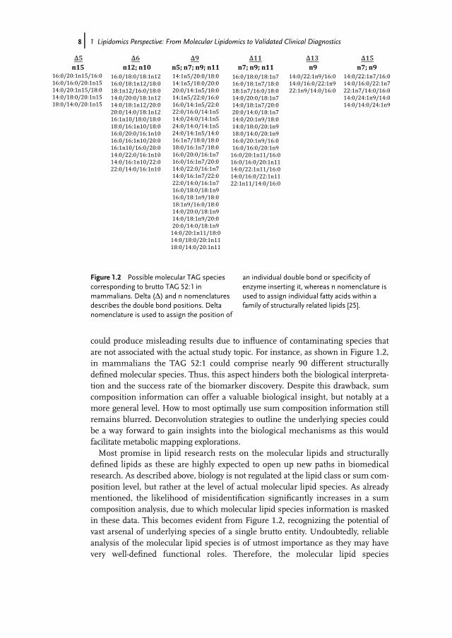
could produce misleading results due to influence of contaminating species thatare not associated with the actual study topic. For instance, as shown in Figure 1.2,in mammalians the TAG 52:1 could comprise nearly 90 different structurallydefined molecular species. Thus, this aspect hinders both the biological interpreta-tion and the success rate of the biomarker discovery. Despite this drawback, sumcomposition information can offer a valuable biological insight, but notably at amore general level. How to most optimally use sum composition information stillremains blurred. Deconvolution strategies to outline the underlying species couldbe a way forward to gain insights into the biological mechanisms as this wouldfacilitate metabolic mapping explorations.Most promise in lipid research rests on the molecular lipids and structurally
defined lipids as these are highly expected to open up new paths in biomedicalresearch. As described above, biology is not regulated at the lipid class or sum com-position level, but rather at the level of actual molecular lipid species. As alreadymentioned, the likelihood of misidentification significantly increases in a sumcomposition analysis, due to which molecular lipid species information is maskedin these data. This becomes evident from Figure 1.2, recognizing the potential ofvast arsenal of underlying species of a single brutto entity. Undoubtedly, reliableanalysis of the molecular lipid species is of utmost importance as they may havevery well-defined functional roles. Therefore, the molecular lipid species
n7; n9; n11∆11
16:0/18:0/18:1n7
16:0/18:1n7/18:0
18:1n7/16:0/18:0
14:0/20:0/18:1n7
14:0/18:1n7/20:0
20:0/14:0/18:1n7
14:0/20:1n9/18:0
14:0/18:0/20:1n9
18:0/14:0/20:1n9
16:0/20:1n9/16:0
16:0/16:0/20:1n9
16:0/20:1n11/16:0
16:0/16:0/20:1n11
14:0/22:1n11/16:0
14:0/16:0/22:1n11
22:1n11/14:0/16:0
n5; n7; n9; n1114:1n5/20:0/18:0
14:1n5/18:0/20:0
20:0/14:1n5/18:0
14:1n5/22:0/16:0
16:0/14:1n5/22:0
22:0/16:0/14:1n5
14:0/24:0/14:1n5
24:0/14:0/14:1n5
24:0/14:1n5/14:0
16:1n7/18:0/18:0
18:0/16:1n7/18:0
16:0/20:0/16:1n7
16:0/16:1n7/20:0
14:0/22:0/16:1n7
14:0/16:1n7/22:0
22:0/14:0/16:1n7
16:0/18:0/18:1n9
16:0/18:1n9/18:0
18:1n9/16:0/18:0
14:0/20:0/18:1n9
14:0/18:1n9/20:0
20:0/14:0/18:1n9
14:0/20:1n11/18:0
14:0/18:0/20:1n11
18:0/14:0/20:1n11
∆9n12; n10
∆6
16:0/18:0/18:1n12
16:0/18:1n12/18:0
18:1n12/16:0/18:0
14:0/20:0/18:1n12
14:0/18:1n12/20:0
20:0/14:0/18:1n12
16:1n10/18:0/18:0
18:0/16:1n10/18:0
16:0/20:0/16:1n10
16:0/16:1n10/20:0
16:1n10/16:0/20:0
14:0/22:0/16:1n10
14:0/16:1n10/22:0
22:0/14:0/16:1n10
n15∆5
16:0/20:1n15/16:0
16:0/16:0/20:1n15
14:0/20:1n15/18:0
14:0/18:0/20:1n15
18:0/14:0/20:1n15
∆15
14:0/22:1n7/16:0
14:0/16:0/22:1n7
22:1n7/14:0/16:0
14:0/24:1n9/14:0
14:0/14:0/24:1n9
n7; n9n9∆13
14:0/22:1n9/16:0
14:0/16:0/22:1n9
22:1n9/14:0/16:0
Figure 1.2 Possible molecular TAG speciescorresponding to brutto TAG 52:1 inmammalians. Delta (D) and n nomenclaturesdescribes the double bond positions. Deltanomenclature is used to assign the position of
an individual double bond or specificity ofenzyme inserting it, whereas n nomenclature isused to assign individual fatty acids within afamily of structurally related lipids [25].
8j 1 Lipidomics Perspective: From Molecular Lipidomics to Validated Clinical Diagnostics
information (together with structurally defined molecular species) should give usthe highest success rate in identifying the culprits in the causal lipid metabolic net-works leading to metabolic dysfunction states. This is discussed in more detail inthe following section.
1.4Untying New Biological Evidences through Molecular Lipidomic Applications
Chapter 2 intriguingly reviews the multifaceted lipid architecture in cells withemphasis on the capability of lipids to form morphologically different membranestructures for maintaining the cell function. Fascinatingly, a functional human redblood cell requires over 100 distinct PC molecules at individual concentrations anddistribution in its plasma membrane (Figure 1.1). Why such a high number of indi-vidual molecules is required still remains unknown. However, it demonstrates thebiological complexity of lipids. Moreover, the authors describe that platforms, thatis, lipid rafts, existing within membranes have highly specific functions. However,the knowledge of which lipids make up the lipid rafts is still limited. In addition,the platforms are likely to undergo reformations in their compositions, both in lip-ids and proteins, to alter their function. Obviously, the molecular lipid compositionof this platform plays a vital role and therefore needs to be delineated in great detailto understand its function. Only molecular lipidomics accompanied with structur-ally defined molecular species can address this type of questions. This fulfills pre-cise identification and quantification of each present lipid species, although issuessuch as insufficient sensitivity will remain as the sample amounts will be extremelysmall. Undoubtedly, this is an enormous challenge that lipidomics will be facing inthe coming years; hence, lipidomics should be orchestrated with biophysical andbiochemistry experiments to answer the cell biological questions.Several studies have demonstrated the importance and specificity of single-mole-
cule lipid structures rather than a lipid (and protein) collective in determining thebiofunctionality. An impressive work has been shown by Shinzawa-Itoh et al.. Theyshowed by sophisticated experiments that the oxygen transfer mechanism in cyto-chrome c oxidase requires a specific phosphatidylglycerol molecular lipid withpalmitate (C16:0) and vaccenate (C18:1n7) at the sn-1 and sn-2 positions, respec-tively, on the glycerol backbone [26]. Altering the double position from n7 to n9(from vaccenate to oleate) inactivated the function. Thus, a small conformationalchange in the attached fatty acid is sufficient to inactivate the oxygen transfer mech-anism. Moreover, Menuz et al. showed that C24–C26 carbon ceramides mediatedthe death of a Caenorhabditis elegans mutant that failed to resist asphyxia, whereasceramides with shorter chains had the opposite effect [27]. We have recently beenable to obtain similar findings in mammalians. Here, we showed that the C24 car-bon ceramide induced ER stress, whereas the shorter chain (C20–C22 and C16)ceramides had no effect on HL-1 cardiomyocyte cells [28]. Finally, Ewers et al.recently showed that the structure of the ganglioside GM1 determines the simianvirus 40 (SV40)-induced membrane invagination and infection [29]. They
1.4 Untying New Biological Evidences through Molecular Lipidomic Applications j9
demonstrated that GM1 molecules with long acyl chains facilitated entering ofSV40 through the host cell plasma membrane, while GM1 molecular species withshort hydrocarbon chains failed to support the invagination and endocytosis andinfection. Evidently, these examples already underscore the essence of molecularlipids and technologies for their discoveries.Exhaustive research lies behind our current knowledge of lipid metabolic path-
ways. Although the lipid metabolism is well characterized, information about themolecular lipid metabolism still remains unknown. The main reason for this isthat the applied technologies were capable of determining only those lipids that areat the lipid class level. Even though gas chromatography-based analyses havedirectly allowed tracking of the fatty acid and oxidized fatty acid metabolisms, therestill remain a significant number of unknowns in their metabolisms, especially ofthe latter. This is mainly due to lack of sensitivity and specificity of the appliedapproaches.Much promise now relies on lipidomics to untie the lipid metabolism at molecu-
lar level (and structural defined). Lipidomics exhibits the analytical preferences forthis quest. We have recently demonstrated the power of lipidomics in elucidatingthe lipid metabolism in yeast [3]. Although the pathways are outlined in the formof lipid classes, the experiments identified that the individual classes comprise ofunique lipid species, thus indicating that the metabolism is regulated at the molec-ular level. However, there is still no solution in respect to how to connect the molec-ular lipids in the metabolism. An utmost challenge lies in the measurements ofsingle metabolic events. It can be expected that a whole-cell measurement repre-sents the total sum of events that concurrently occur in the cell. Thus, how to sepa-rate, for instance, a local metabolism of PC in the plasma membrane from acoexisting PC synthesis in ER or metabolism in other organelles remains unclear.Fortunately, enlightening approaches tackling this are emerging. One approach isto perform metabolic flux experiments using stable isotopes that incorporate selec-tively and efficiently into lipids of interest. The approach will then be to specificallymeasure the labeled lipids during a time course to produce a kinetic readout of thelipids of interest. In this way, the synthesis or catabolism rates of lipids could beestablished. Haynes et al. recently described the use of a stable isotope-labeled pre-cursor ([U-13C]palmitate) to analyze de novo sphingolipid biosynthesis by tandemmass spectrometry [30]. Moreover, Pynn et al. incorporated a deuterated methyl-D9-labeled choline chloride to quantify biosynthesis fluxes through both the PC syn-thetic pathways in vivo in human volunteers and compared these fluxes with thosein mice [31]. In conjunction with sophisticated lipidomics, they were able for thefirst time to show that phosphatidylethanolamine-N-methyltransferase (PEMT)pathway in human liver is selective for polyunsaturated PC species, especially thosecontaining docosahexaenoic acid. Kuerschner et al. utilized a highly unique isotopiclabel facilitating detailed lipidomics-assisted tracking of labeled molecular lipidsand in concert with their cellular localization by fluorescent microscopy [32]. Thelabel does not necessary need to be detectable by advanced microcopy techniques.For instance, a general (nonfluorescent) labeled lipid could be precisely determinedby imaging lipidomic techniques (described in Chapter 7). Very recently, this
10j 1 Lipidomics Perspective: From Molecular Lipidomics to Validated Clinical Diagnostics
labeling approach in conjunction with lipidomics lent to the discovery of exclusivelyone sphingomyelin species, namely, d18:1/18:0, interacting directly and beinghighly specific with the transmembrane domain (TMD) of the COPI machineryprotein p24 [33]. The results demonstrate that the exclusive molecular sphingomye-lin acts as cofactor to regulate the function of a transmembrane protein and thusagain point out why biological membranes are assembled from such a large varietyof different lipids and the essence of single entities. Taken together, labeling experi-ments in concert with molecular and imaging lipidomics compose the right ingre-dients for tackling the delineation of the molecular lipid metabolism. Together withsubcellular dissection of the cell, enzymatic silencing or inhibitory experiments,and supported bioinformatics tools, these show the most promise for the discoveryof the biological roles of molecular lipids and mapping of the molecular lipidpathways.
The foreseeable biological specificity residing in molecular lipids make them primecandidates for drug and biomarker discovery. However, the success rate will subs-tantially depend on the selected experimental design and its accomplishment. Theuse of isotopic tracers, lipid imaging, and subcellular dissections are likely to be keyassets. These types of experiments are rather trivial to perform in vitro, however, forinstance, isotopic labeling in vivo is still a very challenging task. Another asset is theintegration of genomic and proteomic data with the molecular lipidomic data set.However, it still remains blurry in how to mine such large data sets. How biologicalrepresentative the currently existing results are remain very unclear, bearing inmind that the applied lipidomics results are mainly based on lipid sum composi-tions. No clear evidences exist demonstrating that the “omic” mining results corre-spond to the results from basic biochemistry and biology. Thus, the risk formisinterpretation can still be rather high in such hypothesis-driven experiments. Afinal strength is the combination of biochemistry, analytical chemistry, biophysics,biology, bioinformatics, and medicine know-how and expertise. Taken together, thereceipt for discovery of new drugs and lipid biomarkers rests on how to retrievemost out of the above-described assets (and others) alone and/or collectively, and inmost accurate way. Convincingly, an optimal component setup will take us beyondour current understanding in lipid biochemistry and biology. It is anticipated thatthe localization and function of the lipid metabolic machinery, including its activecomponents and interactive companions, to be inclusively illuminated. The result-ing novel lipid maps will foster the discovery of novel mechanisms of action (MoA),drug targets and drug efficacy and disease diagnostic biomarkers.As clearly illuminated in other chapters, lipids are highly awaited serving as drug
efficacy and disease diagnostic and prognostic biomarkers. A main reason for thisis that lipids are physiological readouts of the complex gene-driven system that isaffected by environmental factors. Molecular lipidomics in combination with the
appropriate clinical samples and biobank material can therefore be highly consid-ered for escalating the improvement of disease diagnostics and prediction. Thisapplies not only to a certain disease but also to many therapeutic areas, includingcardiovascular diseases, neurological states, cancers, metabolic diseases such asdiabetes, and inflammatory processes. It is anticipated that a single or up to a hand-ful of molecular lipids rather than 20–50 different biological molecules fulfill thediagnostic purposes. Moreover, as lipids are considered as intermediate phenotypesthat are actually much closer to the disease state in question than for instancegenetic information, they could also serve as candidates for companion diagnosticsin the pharmaceutical arena, which is moving increasingly toward the specializedtherapeutics model. Similarly, as lipids have been highly preserved throughout theevolution of life, that is, highly similar lipid contents throughout the mammalianspecies, they can be highly considered for the assessment of translational medicineand thereby help in identifying the optimal experimental animal model mostclosely mimicking the human disease. A promising example has recently beendescribed by Chan et al. [34]. They could identify a correlating behavior of GM3and CE in certain Alzheimer’s transgenic mouse models and in Alzheimer’s dis-ease patients. Although these results are most encouraging, they need to be furtherproved considering that the transgenic mice displayed highly dramatic lipidchanges that were not seen in humans.Once novel drug efficacy and prognostic and diagnostic molecular lipid bio-
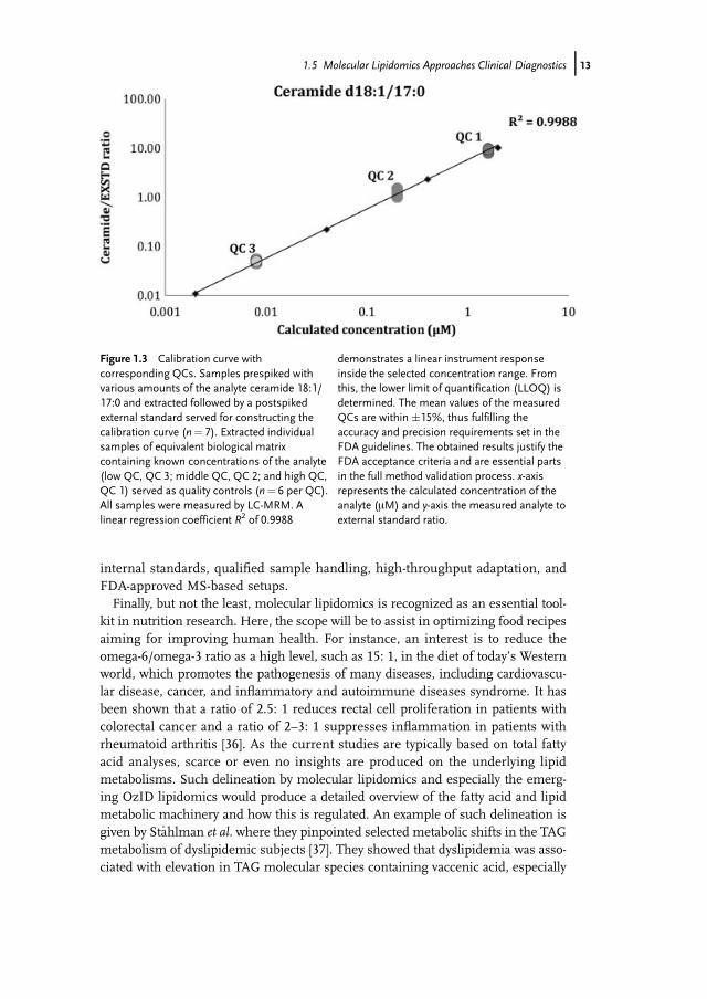
markers are discovered, the next step will be to move their monitoring into clin-ical laboratories. However, before this can take place it is required that thebiomarkers are thoroughly validated. For instance, the identity and quantity ofthe discovered lipid biomarkers should be verified simultaneously as the analy-sis of other independent cohorts should validate the findings. Optimally, thebiomarker validation should be performed in different clinical or diagnostic lab-oratories. In this cascade of the biomarker development, it will be required toadapt the lipidomic assay according to the regulatory requirements followingthe US Food and Drug Administration (FDA) guidelines. Consequently, the lip-idomic assay has to undergo a thorough validation procedure, which includes,for instance, the determination of the method accuracy, precision, lower limitof quantification (LLOQ), long- and short-term stability, freeze–thaw cycles, androbustness. An example of a calibration curve of ceramide d18:1/17:0 includingcorresponding quality controls (QC) is shown in Figure 1.3. Here, the perform-ance of the used LC-MRM method can be verified by the observed linear instru-ment response and performance of minimum three different QC samples inaccordance with the FDA guidelines. Thus, this type of results confidently indi-cates that validated methods for measuring molecular lipids such as ceramidescan be established. Indeed, Scherer et al. recently demonstrated a rapid and val-idated LC-MS assay for the measurement of plasma sphingosine 1-phosphate,sphinganine-1-phosphate, and lysophosphatidic acid [35]. Bearing in mind thelabiality and the analysis difficulty of these lipids, these results prosperouslydemonstrate the feasibility of using lipids as validated diagnostic endpoints.The upcoming challenges, however, include the availabilities of qualified
12j 1 Lipidomics Perspective: From Molecular Lipidomics to Validated Clinical Diagnostics
internal standards, qualified sample handling, high-throughput adaptation, andFDA-approved MS-based setups.Finally, but not the least, molecular lipidomics is recognized as an essential tool-
kit in nutrition research. Here, the scope will be to assist in optimizing food recipesaiming for improving human health. For instance, an interest is to reduce theomega-6/omega-3 ratio as a high level, such as 15: 1, in the diet of today’s Westernworld, which promotes the pathogenesis of many diseases, including cardiovascu-lar disease, cancer, and inflammatory and autoimmune diseases syndrome. It hasbeen shown that a ratio of 2.5: 1 reduces rectal cell proliferation in patients withcolorectal cancer and a ratio of 2–3: 1 suppresses inflammation in patients withrheumatoid arthritis [36]. As the current studies are typically based on total fattyacid analyses, scarce or even no insights are produced on the underlying lipidmetabolisms. Such delineation by molecular lipidomics and especially the emerg-ing OzID lipidomics would produce a detailed overview of the fatty acid and lipidmetabolic machinery and how this is regulated. An example of such delineation isgiven by Sta
�hlman et al. where they pinpointed selected metabolic shifts in the TAG
metabolism of dyslipidemic subjects [37]. They showed that dyslipidemia was asso-ciated with elevation in TAG molecular species containing vaccenic acid, especially
Figure 1.3 Calibration curve withcorresponding QCs. Samples prespiked withvarious amounts of the analyte ceramide 18:1/17:0 and extracted followed by a postspikedexternal standard served for constructing thecalibration curve (n¼ 7). Extracted individualsamples of equivalent biological matrixcontaining known concentrations of the analyte(low QC, QC 3; middle QC, QC 2; and high QC,QC 1) served as quality controls (n¼ 6 per QC).All samples were measured by LC-MRM. Alinear regression coefficient R2 of 0.9988
demonstrates a linear instrument responseinside the selected concentration range. Fromthis, the lower limit of quantification (LLOQ) isdetermined. The mean values of the measuredQCs are within �15%, thus fulfilling theaccuracy and precision requirements set in theFDA guidelines. The obtained results justify theFDA acceptance criteria and are essential partsin the full method validation process. x-axisrepresents the calculated concentration of theanalyte (mM) and y-axis the measured analyte toexternal standard ratio.
TAG 16:0/18:1n7/16:0, indicating an involvement of delta-9 desaturase and elovl-5.It needs to be noted that this is the first time complex endogenous lipid specieshave been identified and quantified at this detailed level. In the case of omega-6/omega-3 ratio, a target of a similar outlining could be to identify and utilize themetabolic switches that favor the omega-3 production or other beneficialmetabolites.
1.6Current Roadblocks in Lipidomics
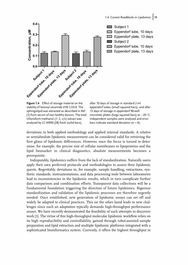
The success of a MoA, drug, or biomarker study decidedly depends on the bioana-lytical quality. Since the stability information of molecular lipids in various matricesor milieus remains scarce or unknown, the sample handling should be performedwith precaution. Here, not only storage of the samples but also the sample prepara-tion practices and processes throughout the workflow should be considered. Care-ful sample collection is required where samples are preferably quickly frozen andstored at the appropriate storage conditions if they cannot directly be subjected tolipidomic analysis. This has recently been reviewed by Jung et al. [1]. It has beenshown that certain biological matrices can be safely stored for years at �80 �C [38].However, the stability can dramatically vary depending on the time, type of lipid,type of sample, and type of storage material and solvent. For instance, Hammad etal. recently showed that ethylenediaminetetraacetic acid (EDTA) is the preferredanticoagulant for sphingolipids [39]. This indicates that the material should care-fully be selected to avoid unspecific reactions and interferences with the lipids ofinterest. Furthermore, the materials should also be resistant to the harsh solventtreatment, as typically a lipid extraction is performed in hazardous organic solventssuch as chloroform, which itself can unfavorably react with lipids [40]. An exampleof the stability of lactosyl ceramide d18:1/24:0 from human serum is shown in Fig-ure 1.4. This sphingolipid is shown to be rather stable when stored (10 days) inchloroform:methanol (1: 2, v/v) at �20 �C in standard eppendorf tubes (EppendorfAG). Unexpectedly, its concentration is more than threefold elevated when storedin 96-well plates from the same vendor. The reason for this remains unknown.However, this shows that a small change in the storage condition can have fatalconsequences and thus points out the essence in sample handling. Moreover, it isrecognized that the number of freeze and thaw cycles can influence the end result[41]; however, two freeze–thaw cycles did not affect the levels of this sphingolipid.Another challenge is that the end results are strongly influenced by the chosen
extraction methodology and lipidomic instrumentations, leading to deviations inthe final lipidomic outputs. Although much effort is currently put in making ofsynthetic standards, a substantial lack of proper nonendogenous standards is still afacto. Therefore, users have been stranded using the available synthetic standards,which regrettably in most cases have been insufficient for absolute quantification ofmonitored lipids. Thus, most of the currently available lipidomic reports are basedon relative or semiabsolute lipid outputs, which can be difficult to compare due to
14j 1 Lipidomics Perspective: From Molecular Lipidomics to Validated Clinical Diagnostics
deviations in both applied methodology and applied internal standards. A relativeor semiabsolute lipidomic measurement can be considered valid for retrieving thefirst glims of lipidomic differences. However, once the focus is turned to deter-mine, for example, the precise size of cellular membranes or lipoproteins and thelipid biomarker in clinical diagnostics, absolute measurements becomes aprerequisite.Indisputably, lipidomics suffers from the lack of standardization. Naturally users
apply their own preferred protocols and methodologies to assess their lipidomicquests. Regrettably, deviations in, for example, sample handling, extractions, syn-thetic standards, instrumentations, and data processing tools between laboratorieslead to inconsistencies in the lipidomic results, which in turn complicate furtherdata comparison and combination efforts. Transparent data collections will be afundamental foundation triggering the direction of future lipidoimcs. Rigorousstandardization and validation of the lipidomic processes are therefore urgentlyneeded. Once established, new generation of lipidomic assays can set off andwidely be adapted to clinical practices. This on the other hand leads to new chal-lenges since such an adaptation typically demands high-throughput performanceassays. We have recently demonstrated the feasibility of such attempts in discoverywork [1]. The virtue of this high-throughput molecular lipidomic workflow relies onits high reproducibility and controllability, gained through robot-assisted samplepreparation and lipid extraction and multiple lipidomic platforms integrated with asophisticated bioinformatics system. Currently, it offers the highest throughput in
Figure 1.4 Effect of storage material on thestability of lactosyl ceramide d18:1/24:0. Thesphingolipid was extracted as described in Ref.[1] from serum of two healthy donors. The totalchloroform:methanol (1: 2, v/v) extract wasanalyzed by LC-MRM [28] fresh (solid bars),
after 10 days of storage in standard 2mleppendorf tubes (small squared bars), and after13 days of storage in eppendorf 96-wellmicrotiter plates (large squared bars) at �20 �C.Independent samples were analyzed and errorbars indicate standard deviation (n¼ 6).
1.6 Current Roadblocks in Lipidomics j15
delivering simultaneously the most comprehensive and quantitative lipidomic out-puts at the molecular lipid level. For instance, approximately 5 days are required todetermine the concentration of over 500 molecular lipids in 20 different lipid clas-ses of 96 human plasma samples. Thus, this setup illuminates and demonstratesthe first attempts toward high-throughput quantitative molecular lipidomics and,although not confirmed, prosperously supports its suitability in a regulatorysetting.
1.7Conclusions
The lipidomics era is currently occurring. Applications in basic and appliedresearch have clearly pointed out and demonstrated its indispensable value. It willopen up new avenues in the biomedical research community, with high expecta-tions that this discovery toolkit will enhance biomarker discovery and provide novelinformation to target discovery programs as it will prospectively shed new lightonto the affected metabolic and signaling pathways. Undoubtedly, it is the delinea-tion of molecular lipids and their precise determination that will lead the way for-ward and accelerate our understanding of molecular lipids, the integratedlipidomic networks, and decoding the coordinately regulated pathways.New attempts will be taken to overcome the challenges lipidomics currently faces.
The standardization of sample preparation and analytical and bioinformatic proce-dures has to be properly addressed. Once solved, this launches the transition of lipi-domics into clinical laboratories. In parallel, the demand for high-throughputtechnologies that do not compromise on the data quality is required. Emerging newMS technologies and methodologies already show promises by offering quantitativeinformation on over 400 molecular lipid species obtained in less than 12min [15]. Ifthese platforms fulfill the regulatory requirements still needs to be seen.It is now the time for researchers in biochemistry, analytical chemistry, bio-
physics, biology, bioinformatics, and medicine to gather and utilize lipidomics inthe most productive way. It is the collective wisdom that will take us beyond ourcurrent know-how in lipid biology.
References
1 Jung, H.R., Sylvanne, T., Koistinen, K.M.,Tarasov, K., Kauhanen, D., and Ekroos, K.(2011) High throughput quantitativemolecular lipidomics. Biochim. Biophys.Acta, 1811, 925–934.
Sullards, C.M., Wang, E., Murphy,R.C., Barkley, R.M., Leiker, T.J., Raetz,C.R., Guan, Z., Laird, G.M., Six,D.A., Russell, D.W., McDonald, J.G.,Subramaniam, S., Fahy, E., andDennis, E.A. (2010) Lipidomicsreveals a remarkable diversity oflipids in human plasma. J. Lipid. Res.,51, 3299–3305.
16j 1 Lipidomics Perspective: From Molecular Lipidomics to Validated Clinical Diagnostics
3 Ejsing, C.S., Sampaio, J.L., Surendranath,V., Duchoslav, E., Ekroos, K., Klemm, R.W., Simons, K., and Shevchenko, A.(2009) Global analysis of the yeastlipidome by quantitative shotgun massspectrometry. Proc. Natl. Acad. Sci. USA,106, 2136–2141.
4 Shevchenko, A. and Simons, K. (2010)Lipidomics: coming to grips with lipiddiversity. Nat. Rev. Mol. Cell Biol., 11,593–598.
5 van Meer, G. (2005) Cellular lipidomics.EMBO J., 24, 3159–3165.
6 van Meer, G., Voelker, D.R., andFeigenson, G.W. (2008) Membrane lipids:where they are and how they behave. Nat.Rev. Mol. Cell Biol., 9, 112–124.
7 Balasubramanian, N., Scott, D.W., Castle,J.D., and Casanova, J.E., and Schwartz,M.A. (2007) Arf6 and microtubules inadhesion-dependent trafficking of lipidrafts. Nat. Cell Biol., 9, 1381–1391.
8 Lingwood, D., Binnington, B., Rog, T.,Vattulainen, I., Grzybek, M., Coskun, U.,Lingwood, C.A., and Simons, K. (2011)Cholesterol modulates glycolipidconformation and receptor activity. Nat.Chem. Biol., 7, 260–262.
9 Ekroos, K., Ejsing, C.S., Bahr, U., Karas,M., Simons, K., and Shevchenko, A.(2003) Charting molecular composition ofphosphatidylcholines by fatty acidscanning and ion trap MS3fragmentation. J. Lipid Res., 44,2181–2192.
10 Brugger, B., Erben, G., Sandhoff, R.,Wieland, F.T., and Lehmann, W.D. (1997)Quantitative analysis of biologicalmembrane lipids at the low picomole levelby nano-electrospray ionization tandemmass spectrometry. Proc. Natl. Acad. Sci.USA, 94, 2339–2344.
11 Stahlman, M., Ejsing, C.S., Tarasov, K.,Perman, J., Boren, J., and Ekroos, K.(2009) High-throughput shotgunlipidomics by quadrupole time-of-flightmass spectrometry. J. Chromatogr. B.Analyt. Technol. Biomed. Life Sci., 877,2664–2672.
12 Schuhmann, K., Almeida, R., Baumert,M., Herzog, R., Bornstein, S.R., andShevchenko, A. (2012) Shotgunlipidomics on a LTQ orbitrap mass
spectrometer by successive switchingbetween acquisition polarity modes.J. Mass Spectrom., 47,, 96–104.
13 Herzog, R., Schuhmann, K., Schwudke,D., Sampaio, J.L., Bornstein, S.R.,Schroeder, M., and Shevchenko, A. (2012)LipidXplorer: a software for consensualcross-platform lipidomics. PLoS One, 7,e29851.
14 Ekroos, K., Chernushevich, I.V.,Simons, K., and Shevchenko, A. (2002)Quantitative profiling of phospholipids bymultiple precursor ion scanning on ahybrid quadrupole time-of-flight massspectrometer. Anal. Chem., 74, 941–949.
15 Simons, B., Kauhanen, D., Sylv€anne, T.,Tarasov, K., Duchoslav, E., and Ekroos, K.(2012) Shotgun lipidomics by sequentialprecursor ion fragmentation on a hybridquadrupole time-of-flight massspectrometer.Metabolites, 2, 195–213.
17 Poad, B.L., Pham, H.T., Thomas, M.C.,Nealon, J.R., Campbell, J.L., Mitchell, T.W., and Blanksby, S.J. (2010) Ozone-induced dissociation on a modifiedtandem linear ion-trap: observations ofdifferent reactivity for isomeric lipids. J.Am. Soc. Mass Spectrom., 21,1989–1999.
18 Deeley, J.M., Thomas, M.C., Truscott, R.J.,Mitchell, T.W., and Blanksby, S.J. (2009)Identification of abundant alkyl etherglycerophospholipids in the human lensby tandemmass spectrometry techniques.Anal. Chem., 81, 1920–1930.
19 Li, Z., Agellon, L.B., Allen, T.M., Umeda,M., Jewell, L., Mason, A., and Vance, D.E.(2006) The ratio of phosphatidylcholine tophosphatidylethanolamine influencesmembrane integrity and steatohepatitis.Cell Metab., 3, 321–331.
20 Sergent, O., Ekroos, K., Lefeuvre-Orfila,L., Rissel, M., Forsberg, G.B., Oscarsson,J., Andersson, T.B., and Lagadic-Gossmann, D. (2009) Ximelagatranincreases membrane fluidity and changesmembrane lipid composition in primary
References j17
human hepatocytes. Toxicol. In Vitro, 23,1305–1310.
21 Zech, T., Ejsing, C.S., Gaus, K., de Wet, B.,Shevchenko, A., Simons, K., and Harder,T. (2009) Accumulation of raft lipids in T-cell plasma membrane domains engagedin TCR signalling. EMBO J., 28, 466–476.
22 Gerl, M.J., Sampaio, J.L., Urban, S.,Kalvodova, L., Verbavatz, J.M.,Binnington, B., Lindemann, D.,Lingwood, C.A., Shevchenko, A.,Schroeder, C., and Simons, K. (2012)Quantitative analysis of the lipidomes ofthe influenza virus envelope and MDCKcell apical membrane. J. Cell Biol., 196,213–221.
23 Dennis, E.A., Deems, R.A., Harkewicz, R.,Quehenberger, O., Brown, H.A., Milne, S.B., Myers, D.S., Glass, C.K., Hardiman,G., Reichart, D., Merrill, A.H., Jr.,Sullards, M.C., Wang, E., Murphy, R.C.,Raetz, C.R., Garrett, T.A., Guan, Z., Ryan,A.C., Russell, D.W., McDonald, J.G.,Thompson, B.M., Shaw, W.A., Sud, M.,Zhao, Y., Gupta, S., Maurya, M.R., Fahy,E., and Subramaniam, S. (2010) A mousemacrophage lipidome. J. Biol. Chem.,28539976–39985.
24 Sampaio, J.L., Gerl, M.J., Klose, C.,Ejsing, C.S., Beug, H., Simons, K., andShevchenko, A. (2011) Membranelipidome of an epithelial cell line. Proc.Natl. Acad. Sci. USA, 108,1903–1907.
25 Vance, D.E. and Vance, J.E. (1996)Biochemistry of Lipids, Lipoproteins, andMembranes, Elsevier, Amsterdam.
26 Shinzawa-Itoh, K., Aoyama, H.,Muramoto, K., Terada, H., Kurauchi, T.,Tadehara, Y., Yamasaki, A., Sugimura, T.,Kurono, S., Tsujimoto, K., Mizushima, T.,Yamashita, E., Tsukihara, T., andYoshikawa, S. (2007) Structures andphysiological roles of 13 integral lipids ofbovine heart cytochrome c oxidase. EMBOJ., 26, 1713–1725.
27 Menuz, V., Howell, K.S., Gentina, S.,Epstein, S., Riezman, I., Fornallaz-Mulhauser, M., Hengartner, M.O.,Gomez, M., Riezman, H., and Martinou,J.C. (2009) Protection of C. elegans fromanoxia by HYL-2 ceramide synthase.Science, 324, 381–384.
28 Perman, J.C., Bostrom, P., Lindbom, M.,Lidberg, U., StAhlman, M., Hagg, D.,Lindskog, H., Scharin Tang, M.,Omerovic, E., Mattsson Hulten, L.,Jeppsson, A., Petursson, P., Herlitz, J.,Olivecrona, G., Strickland, D.K., Ekroos,K., Olofsson, S.O., and Boren, J. (2011)The VLDL receptor promotes lipotoxicityand increases mortality in mice followingan acute myocardial infarction. J. Clin.Invest., 121, 2625–2640.
29 Ewers, H., Romer, W., Smith, A.E., Bacia,K., Dmitrieff, S., Chai, W., Mancini, R.,Kartenbeck, J., Chambon, V., Berland, L.,Oppenheim, A., Schwarzmann, G., Feizi,T., Schwille, P., Sens, P., Helenius, A., andJohannes, L. (2010) GM1 structuredetermines SV40-induced membraneinvagination and infection. Nat. Cell Biol.,12, 11–18.
30 Haynes, C.A., Allegood, J.C., Wang, E.W.,Kelly, S.L., Sullards, M.C., and Merrill, A.H., Jr. (2011) Factors to consider in using[U-C]palmitate for analysis of sphingolipidbiosynthesis by tandem massspectrometry. J. Lipid Res., 52, 1583–1594.
31 Pynn, C.J., Henderson, N.G., Clark, H.,Koster, G., Bernhard, W., and Postle, A.D.(2011) Specificity and rate of human andmouse liver and plasmaphosphatidylcholine synthesis analyzed invivo. J. Lipid Res., 52, 399–407.
32 Kuerschner, L., Ejsing, C.S., Ekroos, K.,Shevchenko, A., Anderson, K.I., andThiele, C. (2005) Polyene-lipids: a new toolto image lipids. Nat. Methods, 239–45.
33 Contreras, F.X., Ernst, A.M., Haberkant,P., Bjorkholm, P., Lindahl, E., Gonen, B.,Tischer, C., Elofsson, A., von Heijne, G.,Thiele, C., Pepperkok, R., Wieland, F., andBrugger, B. (2012) Molecular recognitionof a single sphingolipid species by aprotein’s transmembrane domain. Nature,481, 525–529.
34 Chan, R.B., Oliveira, T.G., Cortes, E.P.,Honig, L.S., Duff, K.E., Small, S.A., Wenk,M.R., Shui, G., and Di Paolo, G. (2012)Comparative lipidomic analysis of mouseand human brain with Alzheimer disease.J. Biol. Chem., 287, 2678–2688.
35 Scherer, M., Schmitz, G., and Liebisch,G. (2009) High-throughput analysis ofsphingosine 1-phosphate, sphinganine
18j 1 Lipidomics Perspective: From Molecular Lipidomics to Validated Clinical Diagnostics
1-phosphate, and lysophosphatidic acidin plasma samples by liquidchromatography-tandem massspectrometry. Clin. Chem., 55,1218–1222.
36 Simopoulos, A.P. (2002) The importance ofthe ratio of omega-6/omega-3 essential fattyacids. Biomed. Pharmacother., 56, 365–379.
37 Stahlman, M., Pham, H.T., Adiels, M.,Mitchell, T.W., Blanksby, S.J., Fagerberg,B., Ekroos, K., and Boren, J. (2012)Clinical dyslipidaemia is associated withchanges in the lipid composition andinflammatory properties ofapolipoprotein-B-containing lipoproteinsfrom women with type 2 diabetes.Diabetologia, 55 (4), 1156–1166.
triglyceride, and phospholipid fractions.J. Lipid Res., 51, 2826–2832.
39 Hammad, S.M., Pierce, J.S., Soodavar,F., Smith, K.J., Al Gadban, M.M.,Rembiesa, B., Klein, R.L., Hannun, Y.A., Bielawski, J., and Bielawska, A.(2010) Blood sphingolipidomics inhealthy humans: impact of samplecollection methodology. J. Lipid Res., 51,3074–3087.
40 Owen, J.S., Wykle, R.L., Samuel, M.P.,and Thomas, M.J. (2005) An improvedassay for platelet-activating factor usingHPLC-tandem mass spectrometry. J. LipidRes., 46, 373–382.
41 Zivkovic, A.M., Wiest, M.M., Nguyen,U.T., Davis, R., Watkins, S.M., andGerman, J.B. (2009) Effects of samplehandling and storage on quantitativelipid analysis in human serum.Metabolomics, 5, 507–516.