This study aims at improving the buccal delivery of vitamin B6 (VB6) as a model highly water-soluble, lowpermeable vitamin. Two main strategies were combined;first VB6 was entrapped in liposomes, whichwere then formulated as mucoadhesivefilm. Both plain and VB6-loaded liposomes (LPs) containingLipoid S100 and propylene glycol (200 nm) were then incorporated into mucoadhesivefilm composedof SCMC and HPMC. Results showed prolonged release of VB6 (72.65%, T50% diss 105 min) after 6 h fromLP-film compared to controlfilm containing free VB6 (96.37%, T50% diss 30 min). Mucoadhesion wasassessed both ex vivo on chicken pouch and in vivo in human. Mucoadhesive force of 0.2 N and residencetime of 4.4 h were recorded. Ex vivo permeation of VB6, across chicken pouch mucosa indicated increasedpermeation from LP-systems compared to corresponding controls. Interestingly, incorporation of thevesicles in mucoadhesivefilm reduced theflux by 36.89% relative to LP-dispersion. Meanwhile, bothfilmsprovided faster initial permeation than the liquid forms. Correlating the cumulative percent permeatedex vivo with the cumulative percent released in vitro indicated that LPs retarded VB6 release but improvedpermeation. These promising results represent a step forward in thefield of buccal delivery of water-soluble vitamins
Liposomal buccal mucoadhesive film for improved delivery and permeation of water-soluble vitamins Heba Abd El Azim a, b , Noha Nafee a, *, Alyaa Ramadan a , Nawal Khalafallah a a Department of Pharmaceutics, Faculty of Pharmacy, Alexandria University, Alexandria, Egypt b Department of Pharmaceutics, Faculty of Pharmacy, Damanhour University, Damanhour, Egypt A R T I C L E I N F O Article history: Received 6 February 2015 Received in revised form 11 April 2015 Accepted 16 April 2015 Available online 18 April 2015 Keywords: Liposomes Mucoadhesive film Water-soluble vitamins Vitamin B6 Buccal permeation A B S T R A C T This study aims at improving the buccal delivery of vitamin B6 (VB6) as a model highly water-soluble, low permeable vitamin. Two main strategies were combined; first VB6 was entrapped in liposomes, which were then formulated as mucoadhesive film. Both plain and VB6-loaded liposomes (LPs) containing Lipoid S100 and propylene glycol (200 nm) were then incorporated into mucoadhesive film composed of SCMC and HPMC. Results showed prolonged release of VB6 (72.65%, T50% diss 105 min) after 6 h from LP-film compared to control film containing free VB6 (96.37%, T50% diss 30 min). Mucoadhesion was assessed both ex vivo on chicken pouch and in vivo in human. Mucoadhesive force of 0.2 N and residence time of 4.4 h were recorded. Ex vivo permeation of VB6, across chicken pouch mucosa indicated increased permeation from LP-systems compared to corresponding controls. Interestingly, incorporation of the vesicles in mucoadhesive film reduced the flux by 36.89% relative to LP-dispersion. Meanwhile, both films provided faster initial permeation than the liquid forms. Correlating the cumulative percent permeated ex vivo with the cumulative percent released in vitro indicated that LPs retarded VB6 release but improved permeation. These promising results represent a step forward in the field of buccal delivery of water- soluble vitamins. ã 2015 Elsevier B.V. All rights reserved. 1. Introduction A great deal of interest has focused on the implementation of buccal mucosa for systemic delivery of various drugs and dietary supplements including vitamins (Puratchikody and Mathew, 2011). Drug delivery across buccal mucosa offers a variety of advantages such as providing direct entry into the systemic circulation through the jugular vein, thus bypassing the stomach environment and first-pass liver metabolism leading possibly to increased bioavailability of drugs suffering from gut or first pass liver metabolism. Ease of drug administration, accessibility and improved patient compliance all provide acceptable alternative for pediatrics, geriatric and nauseous patients. The buccal route allows for both prolonged and rapid drug release for local or systemic action, in addition to versatility in designing uni- or multi- directional release systems (Gilhotra et al., 2014). Among the buccal dosage forms, caffeine chewing gum, (Stay Alert 1 ) and nicotine chewing gums (e.g., Nicorette 1 and Nic- otinell 1 ) have been marketed for systemic drug delivery (Kami- mori et al., 2002; Patel et al., 2011). A novel liquid aerosol insulin formulation (Oralin 1 , Generex Biotechnology) was developed allowing pain-free, user-friendly administration as well as precise insulin dose delivery into the mouth (comparable to injection) (Modi et al., 2002). Attempts for prolonged and improved drug contact with the mucosa have lead to the development of mucoadhesive delivery systems (e.g., patches, flexible films and wafers) as extensively reported (Kianfar et al., 2012; Patel et al., 2011; Sharma et al., 2012). Intraoral polymeric films have gained a wide range of applications from breath fresheners to local anesthetics, vitamin supplements, vaccines, in allergic remedies, emetic condition and in chronic attacks of some diseases (Chaturvedi et al., 2011). Both the controlled release and fast dissolving films can be suitable for young children and for patients with difficulty in swallowing (dysphagic patients) (Boateng et al., 2013). * Corresponding author at: Department of Pharmaceutics, Faculty of Pharmacy, Alexandria University, 21521-El Khartoom Square, Alexandria, Egypt. Tel.: +20 34868482; fax: +20 34871668. E-mail addresses: [email protected], [email protected](N. Nafee). http://dx.doi.org/10.1016/j.ijpharm.2015.04.052 0378-5173/ ã 2015 Elsevier B.V. All rights reserved. International Journal of Pharmaceutics 488 (2015) 78–85 Contents lists available at ScienceDirect International Journal of Pharmaceutics journa l home page : www.e lsevier.com/loca te/ijpharm
Transcript
International Journal of Pharmaceutics 488 (2015) 78–85
Liposomal buccal mucoadhesive film for improved delivery andpermeation of water-soluble vitamins
Heba Abd El Azim a,b, Noha Nafee a,*, Alyaa Ramadan a, Nawal Khalafallah a
aDepartment of Pharmaceutics, Faculty of Pharmacy, Alexandria University, Alexandria, EgyptbDepartment of Pharmaceutics, Faculty of Pharmacy, Damanhour University, Damanhour, Egypt
A R T I C L E I N F O
Article history:Received 6 February 2015Received in revised form 11 April 2015Accepted 16 April 2015Available online 18 April 2015
This study aims at improving the buccal delivery of vitamin B6 (VB6) as a model highly water-soluble, lowpermeable vitamin. Two main strategies were combined; first VB6 was entrapped in liposomes, whichwere then formulated as mucoadhesive film. Both plain and VB6-loaded liposomes (LPs) containingLipoid S100 and propylene glycol (�200 nm) were then incorporated into mucoadhesive film composedof SCMC and HPMC. Results showed prolonged release of VB6 (72.65%, T50% diss 105 min) after 6 h fromLP-film compared to control film containing free VB6 (96.37%, T50% diss 30 min). Mucoadhesion wasassessed both ex vivo on chicken pouch and in vivo in human. Mucoadhesive force of 0.2 N and residencetime of 4.4 h were recorded. Ex vivo permeation of VB6, across chicken pouch mucosa indicated increasedpermeation from LP-systems compared to corresponding controls. Interestingly, incorporation of thevesicles in mucoadhesive film reduced the flux by 36.89% relative to LP-dispersion. Meanwhile, both filmsprovided faster initial permeation than the liquid forms. Correlating the cumulative percent permeatedex vivo with the cumulative percent released in vitro indicated that LPs retarded VB6 release but improvedpermeation. These promising results represent a step forward in the field of buccal delivery of water-soluble vitamins.
ã 2015 Elsevier B.V. All rights reserved.
Contents lists available at ScienceDirect
International Journal of Pharmaceutics
journa l home page : www.e l sev ier .com/ loca te / i jpharm
1. Introduction
A great deal of interest has focused on the implementation ofbuccal mucosa for systemic delivery of various drugs and dietarysupplements including vitamins (Puratchikody and Mathew,2011). Drug delivery across buccal mucosa offers a variety ofadvantages such as providing direct entry into the systemiccirculation through the jugular vein, thus bypassing the stomachenvironment and first-pass liver metabolism leading possibly toincreased bioavailability of drugs suffering from gut or first passliver metabolism. Ease of drug administration, accessibility andimproved patient compliance all provide acceptable alternative forpediatrics, geriatric and nauseous patients. The buccal route allowsfor both prolonged and rapid drug release for local or systemic
* Corresponding author at: Department of Pharmaceutics, Faculty of Pharmacy,Alexandria University, 21521-El Khartoom Square, Alexandria, Egypt.Tel.: +20 34868482; fax: +20 34871668.
http://dx.doi.org/10.1016/j.ijpharm.2015.04.0520378-5173/ã 2015 Elsevier B.V. All rights reserved.
action, in addition to versatility in designing uni- or multi-directional release systems (Gilhotra et al., 2014).
Among the buccal dosage forms, caffeine chewing gum, (StayAlert1) and nicotine chewing gums (e.g., Nicorette1 and Nic-otinell1) have been marketed for systemic drug delivery (Kami-mori et al., 2002; Patel et al., 2011). A novel liquid aerosol insulinformulation (Oralin1, Generex Biotechnology) was developedallowing pain-free, user-friendly administration as well as preciseinsulin dose delivery into the mouth (comparable to injection)(Modi et al., 2002).
Attempts for prolonged and improved drug contact with themucosa have lead to the development of mucoadhesive deliverysystems (e.g., patches, flexible films and wafers) as extensivelyreported (Kianfar et al., 2012; Patel et al., 2011; Sharma et al., 2012).Intraoral polymeric films have gained a wide range of applicationsfrom breath fresheners to local anesthetics, vitamin supplements,vaccines, in allergic remedies, emetic condition and in chronicattacks of some diseases (Chaturvedi et al., 2011). Both thecontrolled release and fast dissolving films can be suitable foryoung children and for patients with difficulty in swallowing(dysphagic patients) (Boateng et al., 2013).
H. Abd El Azim et al. / International Journal of Pharmaceutics 488 (2015) 78–85 79
Meanwhile, colloidal systems such as micelles, liposomes,nanoemulsions and polymeric nanoparticles showed great prom-ise in drug delivery by improving the therapeutic index of drugs,modifying their distribution, thus increasing their efficacy and/orreducing their toxicity (Tan and Liu, 2011). Among colloidalsystems, liposomes (LPs) have gained considerable attention due totheir ability to entrap both lipophilic (within the phospholipidsbilayer) and hydrophilic drugs (in the inner aqueous compartment)(Kumar et al., 2010; Xu et al., 2012). Various buccal liposomaldosage forms reported in the literature include silymarinliposomes for improved buccal permeation through chicken pouch(El-Samaligy et al., 2006), buccal micellar insulin spray and buccaldeformable liposomes to deliver insulin with significantly greaterbioavailability than conventional LPs and subcutaneous insulinsolution in rabbits (Yang et al., 2002). Nevertheless, drug leakageand colloidal instability upon storage are reported as the maindrawbacks of liposomal carriers.
Multiple-unit mucoadhesive carriers combine the abilities ofthe mucoadhesive dosage forms with the advantageous featuresof the multiparticulate delivery systems (e.g., uniform dispersionin the targeting site, more reproducible drug absorption andreduced local irritation) (Albertini et al., 2009; Nitin et al., 2010).Mucoadhesive liposomal ointment for the buccal delivery ofpeptides increased the intimate contact with mucosa andaccordingly local drug concentration (Veuillez et al., 2001).As well, liposome encapsulated corticosteroids paste was devel-oped for the treatment of oral lichen planus (Hearnden et al.,2012). In another study, mucoadhesive ointment containingliposomal triamcinolone acetonide was found to be welltolerated with no local irritation (Sveinsson and Peter Holbrook,1993).
Water-soluble vitamins represent a structurally and function-ally diverse set of organic molecules that play a crucial role inmaintaining the metabolic, energy, differentiation and prolifera-tion status of cells (Said and Seetharam, 2006). Vitamin B6 (VB6)as member of the vitamin B complex group is involved in themetabolism of carbohydrates, amino acids, lipids and neuro-transmitters (Ball, 2004). It also regulates the expression factorsresponsible for efficient absorption of microelements, e.g.,calcium or transport drugs or antigens (Zieli�nska-Dawidziaket al., 2008). VB6 deficiency in humans leads to a variety ofclinical abnormalities that include sideroblastic anemia, weakness,insomnia, and neurologic disorders (Kevadiya et al., 2010). One ofthe applications of interest of VB6 is as an antiemetic. Many oraland parenteral products are already on the market, yet a buccalprolonged release product if developed, could be less nauseatingthan oral product to be swallowed and less painful compared toparenteral VB6.
Therefore, the aim of this study was to develop a prolongedrelease buccal formulation of VB6 as a model highly water-soluble,low permeable vitamin. In this context, two main strategies werecombined; first VB6 was entrapped in liposomal carrier in anattempt to improve permeability, and second, this liposomaldispersion was formulated as mucoadhesive buccal film in order toprolong residence time and release profile. Loaded liposomes,control drug solution and control drug film were tested in parallelfor drug release and permeation for comparison.
2. Materials and methods
2.1. Materials
Lipoid S100 (LS100) also known as phosphatidylcholine (PC)from soybean lecithin, containing not less than 94% PC, was a kindgift from Lipoid GmbH (Ludwigshafen, Germany). Vitamin B6 was a
kind gift from European Egyptian Pharmaceuticals (Alexandria,Egypt). Propylene glycol (PG), anhydrous ethanol were purchasedfrom ADWIC, El-Nasr Pharmaceutical Chemicals Co. (Abu Zaabal,Egypt). Hydroxypropyl methyl cellulose (HPMC, molecular weight4 kDa) and carboxymethyl cellulose sodium (SCMC) were fromProlabo Pharmaceutical Chemicals Co. (Cairo, Egypt).
2.2. Methods
2.2.1. Preparation of plain and VB6-loaded liposomesThe formulation prepared contained 2, 5 and 0.6% w/v of LS100,
PG and VB6, respectively, (calculated with respect to final LP-dispersion). A minimum amount of ethanol was used to dissolveVB6. LS100 was dissolved in the ethanolic VB6 solution. Ethanolwas slowly removed at reduced pressure, using a rotary evaporator(Rotavapor, Büchi, Germany) above the lipid transition tempera-ture. The lipid film was hydrated with deionized water containingthe PG.
The resulting vesicle dispersion was subjected to sonication inice bath (30 min intermittent) and to extrusion cycles throughmembrane filters of descending pore size (once through nylonfilter 0.45 mm and twice through cellulose acetate filter 0.20 mm).The final LP-dispersion were stored in refrigerator at 4 �C. Plain(VB6-free) LPs were similarly prepared.
2.2.2. Colloidal characterization of liposomesVesicle size, polydispersity index (PdI) and zeta potential for
plain and loaded LPs were determined using Malvern ZetasizerNano ZS (Malvern Instruments, Malvern, UK). Deionized water wasused as dilution medium (20 fold dilution). For morphologicalcharacterization, LPs were examined by TEM (Jeol-100CX, Japan)after negative staining using uranyl acetate.
2.2.3. Determination of drug entrapment efficiency by dialysisA volume of LP-dispersion (0.2 ml equivalent to 1.2 mg VB6) was
filled in dialysis bags (Carolina1 dialysis tubing, 12–14 kDamolecular weight cut off, North Carolina, USA) and suspended indeionized water (pH 6.5 � 0.05) for 2 h at 4 �C, to ensure aninternal: external volume ratio of 1:300. VB6 in dialysate wasmeasured spectrophotometrically at 292 nm (Thermospectronic,Helios alpha, NC 9423 UVA 1002E, UK) a wavelength correspondingto maximum UV absorption of VB6, as pyridoxine HCl, in water(Moffat et al., 2004; Ristilä et al., 2006).
Entrapment efficiency (EE) was calculated using the followingequation:
EE ¼ Total VB6 added � VB6 in dialysateTotal VB6 added
� 100
2.2.4. Preparation of mucoadhesive buccal liposomal filmSolvent casting method was used to prepare buccal mucoad-
hesive films containing VB6-loaded LPs. HPMC and SCMC (1% w/veach, calculated with respect to LP-dispersion volume) were addedto the LP dispersion while stirring till complete dissolution of thepowders. The gels formed were left overnight at room temperaturein a dessicator to ensure clear, bubble-free gel. PG already presentin the LPs formula served as plasticizer. The gel was cast into glassPetri dishes and allowed to dry at 40 �C. Film discs (1 cm2,containing 1.4 mg VB6) were wrapped in aluminum foil, and storedin glass containers at 4 �C. A control film containing free VB6 wassimilarly prepared.
80 H. Abd El Azim et al. / International Journal of Pharmaceutics 488 (2015) 78–85
2.3. Characterization of liposomal mucoadhesive films (LP-film)
2.3.1. Physicochemical characterizationFilm thickness, weight uniformity, surface pH, swelling and
endurance were assessed (n = 3) as detailed in Supplementarymaterials as previously reported (Mohamed et al., 2011).
2.3.2. Integrity of the liposomes in mucoadhesive filmsFilms were dissolved in deionized water. Vesicle size, PdI and
surface charge of restored LPs in the film solution were measured.Morphology was examined by TEM.
2.3.3. In vitro release of VB6 from LP-filmsVB6 release from the buccal film was assessed by dialysis at
37 �C. A film disc (equivalent to 1.4 mg drug) was placed in thedialysis bag and immersed in 20 ml simulated saliva (pH6.75 � 0.05), prepared by dissolving 2.38 g Na2HPO4, 0.19 g KH2PO4
and 8.0 g NaCl in 1000 ml of distilled water (Wong et al., 1999). Atpredetermined time intervals, samples (0.5 ml) were withdrawnand replaced with fresh release medium. VB6 released wasanalyzed at 254 nm, a wavelength corresponding to maximumabsorption of VB6 at pH 6.75 (Moffat et al., 2004; Ristilä et al.,2006). A calibration curve in simulated saliva was used.
2.3.4. Stability on storageLP-film was stored for three months at 4 �C. Vesicle size, PdI and
VB6 release were measured. Morphology was examined by TEM.Films were dissolved in deionized water before size measurementsand morphological examination. Release was tested by dialysis ofthe films (Section 2.2.3). Data for LP-dispersion stability were alsogenerated under the same storage conditions for comparison.
2.4. Mucoadhesion
2.4.1. Ex vivo determination of mucoadhesive strengthThe mucoadhesion strength was determined using an in house
balance method based on the representative sketch illustrated inFig. S1 (Supplementary materials) (Nafee et al., 2003, 2004). Thechicken pouch membrane was used as model mucosa for thesestudies. The film (attached to one arm of the balance) was broughtinto contact with the chicken pouch fitted on a rubber cork andkept moistened with simulated saliva (pH 6.75, 37 �C). The weightof water, in grams (poured into a beaker hanging from the otherarm of the balance), required to detach the film from the mucosalsurface gave the measure of mucoadhesive strength. Averagevalues were recorded (n = 3). The force of adhesion was calculatedusing the equation (Hamzah et al., 2010):
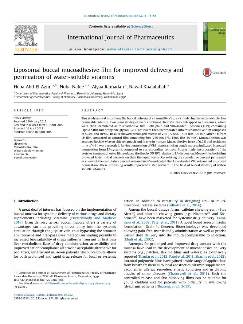
Fig. 1. TEM micrographs of VB6-loaded LPs, (A) fresh
Force of adhesionðNÞ ¼ Bioadhesive strength1000
� �� 9:81
2.4.2. In vivo testing of mucoadhesion in humanMucoadhesion was also assessed in vivo in three volunteers. The
study followed the rules approved by the Faculty of Pharmacy(Alexandria University, Alexandria, Egypt) ethics committee forresearch involving volunteers. Volunteers were asked to rinse theirmouth with distilled water before a unit of the film was placedbetween the cheek and gingiva in the region of the upper canineand gently pressed onto the mucosa for about 30 s. The time forcomplete erosion or detachment of the film from the buccalmucosa was recorded. Volunteers were allowed to eat and drinkduring the testing period. Possible irritation, bad taste, swelling,dry mouth or increase in salivary flow were monitored (Nafee et al.,2003).
2.5. Ex vivo permeation using chicken pouch mucosa
2.5.1. Tissue preparationExcised chicken pouch mucosa was carefully cleaned and kept
frozen at �20 �C in saline, thawed to room temperature directlybefore use and washed with simulated saliva (pH 6.75 � 0.05).Tissue integrity was confirmed histologically by H&E staining.
2.5.2. Ex vivo permeation testingLPs, LP-film, control VB6 solution and control VB6 film were
tested for VB6 permeation through chicken pouch mucosa usingthe QuixSep Micro Dializer (Roth, Karlsruhe, Germany). The unit isa two-piece capsule that securely locks with a push fit. The donorchamber was filled with 230 ml LP-dispersion or control solution.In case of mucoadhesive films, the donor chamber was filled with230 ml simulated saliva (pH 6.75 � 0.05) and the film was pressedon the mucosal side of chicken pouch for 30 s.
In all cases, the mucosal side of chicken pouch was stretchedover the capsule open end towards the donor compartment andcovered tightly with the lid collar. A beaker containing phosphatebuffer (15 ml, pH 7.4 � 0.05) was used as the recipient solution. Thewhole set was put in thermostatically controlled shaking waterbath at 50 rpm and 37 � 0.5 �C for 6 h. Samples were withdrawnover 6 h and compensated with fresh buffer. The concentrations ofpermeated drug were calculated from the absorbance measured at254 nm, a wave length corresponding to maximum absorption ofVB6 at pH 7.4. For blank readings, microdialyser units fitted withchicken pouch membranes, containing donor and recipientsolutions, were placed in the shaking water bath; samples from
ly prepared; (B) after six months storage at 4 �C.
Fig. 2. In vitro release profiles of VB6 from LP-film, control film and control solution.
H. Abd El Azim et al. / International Journal of Pharmaceutics 488 (2015) 78–85 81
the recipient compartment were used as blanks. Experiments wereperformed in triplicate and mean value was used to calculate theflux and permeability coefficient (Hamzah et al., 2010).
2.5.3. Calculation of permeation parametersCumulative amount of permeated drug per square centimeter
was plotted versus time, and steady-state flux was estimated fromthe slope of the linear portion of the plot using the followingequation:
Flux ¼ Jss ¼ dQ=dtA
where, Jss is the steady-state flux (mg cm�2 h�1); dQ/dt is thepermeation rate (mg cm�2); A is the active diffusion area (cm2).
The VB6 permeability coefficient (P) across chicken pouchmucosa was calculated using the relation derived from Fick’s firstlaw of diffusion as follows:
P ¼ JssCd
where, P is the permeability coefficient (cm s�1) and Cd is the donordrug concentration (mg ml�1).
2.6. Statistical analysis
Results are expressed as mean � standard deviation. Statisticalanalysis was performed using Student t-test or One-way ANOVA.Differences were considered significant at a level of p < 0.05.
3. Results and discussion
3.1. Colloidal characteristics of VB6-loaded LPs
VB6-loaded LPs were 207.4 �17.1 nm in diameter (PdI0.228 � 0.03). Zeta potential of loaded LPs was �1.98 mV comparedto an average value of �15.5 mV for corresponding plain LPsindicating possible surface adsorption of the positively-chargedvitamin on the phospholipid bilayer. The weak surface potential ofthe loaded vesicles suggests stabilization mechanisms other thanelectrostatic repulsion.
TEM micrographs of VB6-loaded LPs (freshly-prepared and6-months stored LPs) showed spherical, evenly distributed vesicleswith smooth surface (Fig. 1A and B). Estimated TEM size was in thenano-range in accordance with size measurement by photoncorrelation spectroscopy. However, LPs appear relatively smaller;this might be attributed to drying and staining processes prior toTEM measurement.
3.2. Entrapment efficiency
Percent entrapment efficiency value was 45.99 � 1.75. Deter-mination of EE by dialysis was favored over ultracentrifugation asthe strong energy transmitted to the samples disrupted thevesicles in spite of cooling (Lopez-Pinto et al., 2005). Preliminarytrials indicated that 2 h was enough period for complete dialysis ofthe free drug.
Generally speaking, the entrapment efficiency of drugs inliposomes is a function of various factors such as the preparationtechnique, the geometry of the prepared vesicles (size andlamellarity), the lipid concentration, drug physicochemical prop-erties, possible drug–lipid interactions, aqueous phase composi-tion among others (Adrian and Huang,1979; Dominak and Keating,2007; Nii and Ishii, 2005; Pidgeon et al., 1987; Sun and Chiu, 2005).Passive encapsulation of water-soluble drugs depends on theability of liposomes to trap aqueous medium containing thedissolved drug during vesicle formation. Trapping effectiveness is
limited by the trapped volume in the liposomes and drug solubility(Akbarzadeh et al., 2013).
3.3. Characteristics of LP-film
The mucoadhesive film was 0.1 mm thick, with an averageweight of 15.46 mg cm�2 and a surface pH of 5.93. The filmsshowed good flexibility; no cracks appeared when folded morethan 250 times (Table S1, Supplementary materials). Swellingassessment indicated that the film weight was 5 times the initialweight in 5 min and 25 times in 120 min (Fig. S2, Supplementarymaterials). The film remained intact after 2 h immersion indeionized water in spite of the increase in weight.
3.4. In vitro VB6 release from LP-film
The in vitro release profiles of VB6 from control solution, LP-filmand control film (Fig. 2) indicated that the films provided slowerVB6 release from the dialysis bag compared to VB6 solution.Hydrophilic polymers in water produce a water-swollen gel-likestate that reduces the penetration of the dissolution medium intothe films and lengthens the diffusion path length resulting inretarded drug release (Sekhar et al., 2008; Vishnu et al., 2007).
Apart from the impact of hydrophilic polymers, entrapment ofVB6 in the vesicles contributed to the controlled release. After 6 h,LP-film achieved slower release (72.65 %, T50% diss 105 min) thanthe control film (96.37 %, T50% diss 30 min). Initial amount of VB6released from LP-film (after 30 min) was significantly lower (t-test,p < 0.05) than from control film. The slower release profile fromthe LP-film compared to the control film (Fig. 2) also suggested thatgelling and film casting process did not compromise the integrityof the incorporated vesicles.
Release kinetics followed the Higuchi model; Korsmeyer–Peppas n value (0.321) indicated diffusion release mechanism.
3.5. Stability attributes
3.5.1. Integrity of liposomes in the film before and after storageAn important prerequisite for the effective use of liposomes as a
drug carrier is demonstration of physical and performance stabilityin the final dosage form (Kirby et al., 1980). The data generated inthis study suggested maintenance of vesicle integrity afterincorporation in the film and also after 3 months-storage of thefilm (in both cases, the film was dissolved in deionized water topermit measuring vesicle size (Fig. 3A) and obtaining TEMmicrographs) (Fig. 3B and C, respectively).
Fig. 3. Stability on storage at 4 �C: (A) particle size and PdI of LPs in fresh and stored films, TEM micrograph of: (B) VB6-loaded LP-film, (C) same film after storage, (D) in vitrorelease of VB6 from fresh and stored mucoadhesive films.
82 H. Abd El Azim et al. / International Journal of Pharmaceutics 488 (2015) 78–85
Concerning size measurements, insignificant fluctuation in themean size of LPs and polydispersity was evident before and after LPincorporation in the film (Fig. 3A). TEM micrographs indicated thatLP retained their morphology (spherical shape and smooth, intactsurface) before and after incorporation in the film (Figs. 1A and 3B),respectively.
3.5.2. Effect of storage on the in vitro release of VB6 from LP-filmNo change in VB6 release profiles was observed after three
months of storage of the LP-film at 4 �C (Fig. 3D). Korsmeyer–Peppas kinetics (n = 0.321) revealed the persistence of diffusionrelease mechanism with ageing of the film.
Release profiles also lent further evidence; superimposedrelease profiles (dialysis of films before and after storage,Fig. 3D) is less likely to occur from destroyed and reformed LPscompared to stable LPs. Based on these data, the suggestion thatLPs remained stable and preserved their integrity during filmformation appears better evidenced.
The stability attributes of LP-film were in contrast to LPdispersion; the latter showed gradual drug leakage on storage at4 �C (EE data, Table 1), while no change in vesicle size and PdI wasrecorded (Table 1).
In the present study, the film provided a possible patient-friendly buccal dosage form and improved stability against leakageof the LPs by immobilizing them in the film, as evident in the lack of
H. Abd El Azim et al. / International Journal of Pharmaceutics 488 (2015) 78–85 83
change in VB6 release profiles from the film before and afterstorage.
3.6. Ex vivo and in vivo mucoadhesion
The mucoadhesive strength determined ex vivo was20.55 � 0.2 g corresponding to a force of adhesion of 0.2 N. Thesemucoadhesive properties resulted in an in vivo duration ofattachment of LP-film to the buccal mucosa of 4.43 � 0.07 h inthree volunteers (Table S2, Supplementary materials). Incorpo-ration of LPs in the film had no remarkable apparent impact onmucoadhesion. None of the volunteers complained of irritation,
Fig. 4. (A) Chicken pouch membrane (H & E staining), (B) permeation of VB6 controlsolution and VB6-loaded LPs across chicken pouch membrane, (C) permeation ofVB6 from control film and VB6-loaded LP-film across chicken pouch membrane.
bad taste, dry mouth or increase in salivary flow during the in vivostudy.
3.7. Ex vivo permeation through chicken pouch membrane
Histological examination of the isolated chicken pouchconfirmed membrane integrity (Fig. 4A).
3.7.1. LP systems versus controlsEx vivo permeation of VB6, across chicken pouch mucosa, from
control solution, LP-dispersion, control film and LP-film (Fig. 4Band C), indicated significant increase in permeation from LPsystems compared to corresponding controls (One-way ANOVA:p = 0.0005 for LP-film versus control film, p = 0.014 for LP-dispersionversus control solution of free VB6).
Flux values (Table 2), confirmed the increased VB6 flux from LPsystems (dispersion and film) by 13.06 and 35.21%, respectively,compared to their controls.
The data indicate the permeation enhancing potential of LPs. Inaccordance with our findings, a novel post-expansile hydrogelfoam containing PG-liposomes ensured drug permeation throughporcine vaginal mucosa higher than hydrogel foam aerosol, PG-liposome aerosol and hydrogel (Li et al., 2012). The penetrationenhancing effect of phospholipids and propylene glycol waspreviously reported (Elsayed et al., 2007; Manconi et al., 2009).However, the data generated in the present study, did not allowconcluding whether the vesicles themselves permeate or whetherthey release the drug at the permeation surface, while thephospholipid solely acts as permeation enhancer.
3.7.2. Mucoadhesive films versus liquid systemsComparing LP-film with LP-dispersion as well as control film
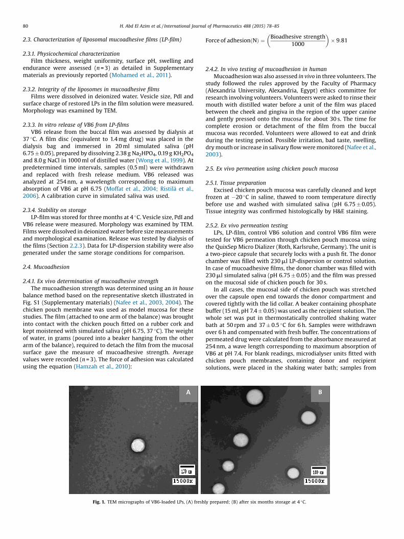
with control solution, mucoadhesive films reduced the cumulativepermeated amount of VB6 relative to the corresponding disper-sion, Fig. 4B and C. Fig. 5A shows that, at 6 h, the amount of VB6permeated from control VB6 solution was 1.5 times that permeatedfrom control film. Similarly, VB6 permeated from LP-dispersionwas 1.2 times that permeated from LP-film. In terms of flux values,incorporation of LPs in mucoadhesive film reduced the flux by36.89% relative to LP-dispersion (113.10 and 179.20 mg cm�2 h�1,respectively, Table 2). Similar results were previously recorded formucoadhesive-coated curcumin-loaded liposomes for vaginaldelivery (Berginc et al., 2014).
However, looking at the rate of permeation and judging by theamounts of VB6 permeated across the chicken pouch membrane in30 min (Fig. 5A), both films provided faster initial permeation thanthe liquid forms. The films, being mucoadhesive, presumablyallowed better drug contact with the permeating membrane. Bytime, swelling of the polymeric film increased the diffusion pathlength of VB6 leading to reduced permeation in case of LP-filmcompared to the dispersion.
The permeability coefficient calculated from flux value of thecontrol VB6 solution (7.3 � 10�6 cm s�1,Table 2) was of the samemagnitude reported for other hydrophilic drugs such as caffeine
Table 2Ex vivo permeation parameters of VB6 across chicken pouch mucosa determined at37 �C.
a Steady state flux obtained from the linear portion of the cumulative amountpermeated (mg) plotted against time (h).
b Permeability coefficient.
Fig. 5. (A) Amount of VB6 permeated in 30 min and in 6 h from LPs, control film, LP-film and control solution across chicken pouch membrane, (B) correlation betweenpercent VB6 dialysed and permeated from LP-film, (C) correlation between percentVB6 dialysed and permeated from control film.
84 H. Abd El Azim et al. / International Journal of Pharmaceutics 488 (2015) 78–85
(8.14 �10�6 cm s�1) determined across porcine mucosa (Kulkarniet al., 2011) but lower than that recorded for diltiazemhydrochloride from HPC/SCMC mucoadhesive film across chickenpouch mucosa (25.7 � 10�6 cm s�1) (Mohamed et al., 2011). Incomparison, the permeability coefficient of PEG hydrophilicmolecules was in the range 1–3 �10�6 cm s�1according tomolecular weight (Goswami et al., 2009).
Relevant permeability studies necessitate careful choice of themucosal membrane; the oral epithelia of a number of experimentalanimals such as rats and rabbits are entirely keratinised (Harris andRobinson, 1992), with a very thick keratinised buccal mucosa(Shojaei, 1998). In contrast, chicken pouch represents a betteralternative as it resembles the human thin and non-keratinisedoral lining mucosa (Erjavec et al., 2006; Hamzah et al., 2010).
Concerning the mechanism of VB6 transport across mucosalmembranes, Zieli�nska-Dawidziak et al. (2008) suggested a dualmechanism of VB6 transport; at low concentrations, the uptake
process was concentration dependant via simple diffusion, whileat high concentrations, a concentration independent, carrier-mediated mechanism was recognized. Heard and Annison (1986)reported that VB6 absorption through chicken intestinal epitheli-um occurred by simple diffusion.
3.8. Diffusion-permeation relation
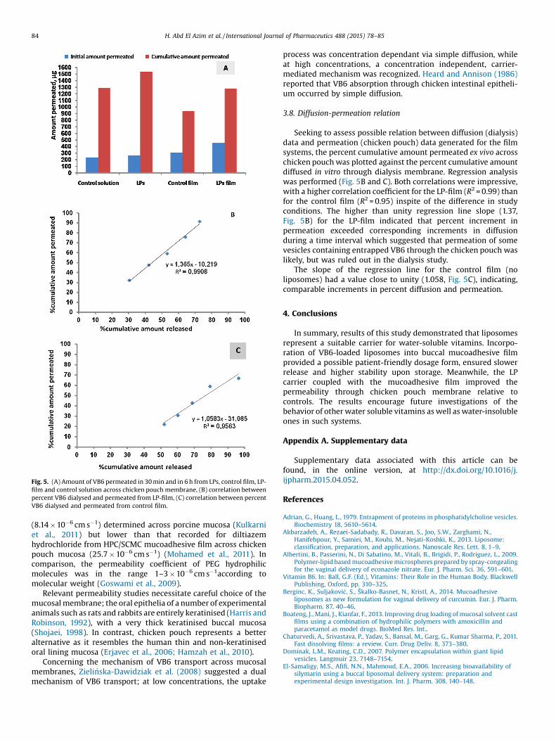
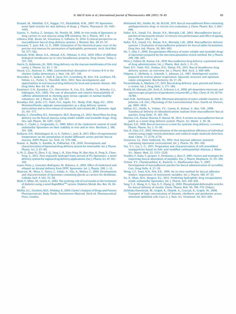
Seeking to assess possible relation between diffusion (dialysis)data and permeation (chicken pouch) data generated for the filmsystems, the percent cumulative amount permeated ex vivo acrosschicken pouch was plotted against the percent cumulative amountdiffused in vitro through dialysis membrane. Regression analysiswas performed (Fig. 5B and C). Both correlations were impressive,with a higher correlation coefficient for the LP-film (R2 = 0.99) thanfor the control film (R2 = 0.95) inspite of the difference in studyconditions. The higher than unity regression line slope (1.37,Fig. 5B) for the LP-film indicated that percent increment inpermeation exceeded corresponding increments in diffusionduring a time interval which suggested that permeation of somevesicles containing entrapped VB6 through the chicken pouch waslikely, but was ruled out in the dialysis study.
The slope of the regression line for the control film (noliposomes) had a value close to unity (1.058, Fig. 5C), indicating,comparable increments in percent diffusion and permeation.
4. Conclusions
In summary, results of this study demonstrated that liposomesrepresent a suitable carrier for water-soluble vitamins. Incorpo-ration of VB6-loaded liposomes into buccal mucoadhesive filmprovided a possible patient-friendly dosage form, ensured slowerrelease and higher stability upon storage. Meanwhile, the LPcarrier coupled with the mucoadhesive film improved thepermeability through chicken pouch membrane relative tocontrols. The results encourage future investigations of thebehavior of other water soluble vitamins as well as water-insolubleones in such systems.
Appendix A. Supplementary data
Supplementary data associated with this article can befound, in the online version, at http://dx.doi.org/10.1016/j.ijpharm.2015.04.052.
References
Adrian, G., Huang, L., 1979. Entrapment of proteins in phosphatidylcholine vesicles.Biochemistry 18, 5610–5614.
Akbarzadeh, A., Rezaei-Sadabady, R., Davaran, S., Joo, S.W., Zarghami, N.,Hanifehpour, Y., Samiei, M., Kouhi, M., Nejati-Koshki, K., 2013. Liposome:classification, preparation, and applications. Nanoscale Res. Lett. 8, 1–9.
Albertini, B., Passerini, N., Di Sabatino, M., Vitali, B., Brigidi, P., Rodriguez, L., 2009.Polymer-lipid based mucoadhesive microspheres prepared by spray-congealingfor the vaginal delivery of econazole nitrate. Eur. J. Pharm. Sci. 36, 591–601.
Vitamin B6. In: Ball, G.F. (Ed.), Vitamins: Their Role in the Human Body. BlackwellPublishing, Oxford, pp. 310–325.
Berginc, K., Suljakovi�c, S., Škalko-Basnet, N., Kristl, A., 2014. Mucoadhesiveliposomes as new formulation for vaginal delivery of curcumin. Eur. J. Pharm.Biopharm. 87, 40–46.
Boateng, J., Mani, J., Kianfar, F., 2013. Improving drug loading of mucosal solvent castfilms using a combination of hydrophilic polymers with amoxicillin andparacetamol as model drugs. BioMed Res. Int..
Chaturvedi, A., Srivastava, P., Yadav, S., Bansal, M., Garg, G., Kumar Sharma, P., 2011.Fast dissolving films: a review. Curr. Drug Deliv. 8, 373–380.
H. Abd El Azim et al. / International Journal of Pharmaceutics 488 (2015) 78–85 85
Elsayed, M., Abdallah, O.Y., Naggar, V.F., Khalafallah, N.M., 2007. PG-liposomes:novel lipid vesicles for skin delivery of drugs. J. Pharm. Pharmacol. 59, 1447–1450.
Erjavec, V., Pavlica, Z., Sentjurc, M., Petelin, M., 2006. In vivo study of liposomes asdrug carriers to oral mucosa using EPR oximetry. Int. J. Pharm. 307, 1–8.
Gilhotra, R.M., Ikram, M., Srivastava, S., Gilhotra, N., 2014. A clinical perspective onmucoadhesive buccal drug delivery systems. J. Biomed. Res. 28, 81–97.
Goswami, T., Jasti, B.R., Li, X., 2009. Estimation of the theoretical pore sizes of theporcine oral mucosa for permeation of hydrophilic permeants. Arch. Oral Biol.54, 577–582.
Hamzah, M.M., Reem, K.A., Ahmad, A.N., Othman, A.-H.A., 2010. Effect of differentbiological membranes on in vitro bioadhesion property. Drug Invent. Today 2,155–159.
Harris, D., Robinson, J.R., 1992. Drug delivery via the mucous membranes of the oralcavity. J. Pharm. Sci. 81, 1–10.
Heard, G.S., Annison, E., 1986. Gastrointestinal absorption of vitamin B-6 in thechicken (Gallus domesticus). J. Nutr. 116, 107–120.
Hearnden, V., Sankar, V., Hull, K., Juras, D.V., Greenberg, M., Kerr, A.R., Lockhart, P.B.,Patton, L.L., Porter, S., Thornhill, M.H., 2012. New developments andopportunities in oral mucosal drug delivery for local and systemic disease. Adv.Drug Deliv. Rev. 64, 16–28.
Kamimori, G.H., Karyekar, C.S., Otterstetter, R., Cox, D.S., Balkin, T.J., Belenky, G.L.,Eddington, N.D., 2002. The rate of absorption and relative bioavailability ofcaffeine administered in chewing gum versus capsules to normal healthyvolunteers. Int. J. Pharm. 234, 159–167.
Kevadiya, B.D., Joshi, G.V., Patel, H.A., Ingole, P.G., Mody, H.M., Bajaj, H.C., 2010.Montmorillonite–alginate nanocomposites as a drug delivery system:intercalation and in vitro release of vitamin B1 and vitamin B6. J. Biomater. Appl.25, 161–177.
Kianfar, F., Chowdhry, B.Z., Antonijevic, M.D., Boateng, J.S., 2012. Novel films for drugdelivery via the buccal mucosa using model soluble and insoluble drugs. DrugDev. Ind. Pharm. 38, 1207–1220.
Kirby, C., Clarke, J., Gregoriadis, G., 1980. Effect of the cholesterol content of smallunilamellar liposomes on their stability in vivo and in vitro. Biochem. J. 186,591–598.
Kulkarni, U.D., Mahalingam, R., Li, X., Pather, I., Jasti, B., 2011. Effect of experimentaltemperature on the permeation of model diffusants across porcine buccalmucosa. AAPS Pharm. Sci. Tech. 12, 579–586.
Kumar, A., Badde, S., Kamble, R., Pokharkar, V.B., 2010. Development andcharacterization of liposomal drug delivery system for nimesulide. Int. J. Pharm.Pharm. Sci. 2, 87–89.
Li, W.-Z., Zhao, N., Zhou, Y.-Q., Yang, L.-B., Xiao-Ning, W., Bao-Hua, H., Peng, K., Chun-Feng, Z., 2012. Post-expansile hydrogel foam aerosol of PG-liposomes: a noveldelivery system for vaginal drug delivery applications. Eur. J. Pharm. Sci. 47,162–169.
Lopez-Pinto, J., Gonzalez-Rodriguez, M., Rabasco, A., 2005. Effect of cholesterol andethanol on dermal delivery from DPPC liposomes. Int. J. Pharm. 298, 1–12.
Manconi, M., Mura, S., Sinico, C., Fadda, A., Vila, A., Molina, F., 2009. Developmentand characterization of liposomes containing glycols as carriers for diclofenac.Colloids Surf. A 342, 53–58.
Modi, P., Mihic, M., Lewin, A., 2002. The evolving role of oral insulin in the treatmentof diabetes using a novel RapidMistTM system. Diabetes Metab. Res. Rev. 18, 38–42.
Moffat, A.C., Osselton, M.D., Widdop, B., 2004. Clarke’s Analysis of Drugs and PoisonsPharmaceuticals, Body Fluids, Postmortem Material, third ed. PharmaceuticalPress, London.
Mohamed, M.I., Haider, M., Ali, M.A.M., 2011. Buccal mucoadhesive films containingantihypertensive drug: in vitro/in vivo evaluation. J. Chem. Pharm. Res. 3, 665–686.
Nafee, N.A., Ismail, F.A., Boraie, N.A., Mortada, L.M., 2003. Mucoadhesive buccalpatches of miconazole nitrate: in vitro/in vivo performance and effect of ageing.Int. J. Pharm. 264, 1–14.
Nafee, N.A., Ismail, F.A., Boraie, N.A., Mortada, L.M., 2004. Mucoadhesive deliverysystems. I. Evaluation of mucoadhesive polymers for buccal tablet formulation.Drug Dev. Ind. Pharm. 30, 985–993.
Nii, T., Ishii, F., 2005. Encapsulation efficiency of water-soluble and insoluble drugsin liposomes prepared by the microencapsulation vesicle method. Int. J. Pharm.298, 198–205.
Nitin, J., Pallavi, M., Kumar, S.K., 2010. Buccoadhesive drug delivery-a potential routeof drug administration. Int. J. Pharm. Biol. Arch. 1, 11–23.
Puratchikody, A., Mathew, S.T., 2011. Buccal drug delivery: past, present and future –
a review. Int. J. Drug Deliv. 3, 171–184.Ristilä, M., Matxain, J.M., Strid, Å., Eriksson, L.A., 2006. pH-dependent electronic and
spectroscopic properties of pyridoxine (vitamin B6). J. Phys. Chem. B 110,16774–16780.
Said, H.M., Seetharam, B., 2006. INtestinal absorption of water-soluble vitamins, In:Johnson, L.R. (Ed.), Physiology of the Gastrointestinal Tract. fourth ed. Elsevier,pp. 1809–1810.
Sekhar, K.C., Naidu, K., Vishnu, Y.V., Gannu, R., Kishan, V., Rao, Y.M., 2008.Transbuccal delivery of chlorpheniramine maleate from mucoadhesive buccalpatches. Drug Deliv. 15, 185–191.
Sharma, G.K., Kumar Sharma, P., Bansal, M., 2012. A review on mucoadhesive buccalpatch as a novel drug delivery system. Pharm. Sci. Monit. 3, 30–38.
Shojaei, A.H., 1998. Buccal mucosa as a route for systemic drug delivery: a review. J.Pharm. Pharm. Sci. 1, 15–30.
Sun, B., Chiu, D.T., 2005. Determination of the encapsulation efficiency of individualvesicles using single-vesicle photolysis and confocal single-molecule detection.Anal. Chem. 77, 2770–2776.
Sveinsson, S.J., Peter Holbrook, W., 1993. Oral mucosal adhesive ointmentcontaining liposomal corticosteroid. Int. J. Pharm. 95, 105–109.
Tan, Y.-l., Liu, C.-G., 2011. Preparation and characterization of self-assembliednanoparticles based on folic acid modified carboxymethyl chitosan. J. Mater.Sci.: Mater. Med. 22, 1213–1220.
Veuillez, F., Kalia, Y., Jacques, Y., Deshusses, J., Buri, P., 2001. Factors and strategies forimproving buccal absorption of peptides. Eur. J. Pharm. Biopharm. 51, 93–109.
Vishnu, Y.V., Chandrasekhar, K., Ramesh, G., Madhusudan Rao, Y., 2007.Development of mucoadhesive patches for buccal administration of carvedilol.Curr. Drug Deliv. 4, 27–39.
Wong, C.F., Yuen, K.H., Peh, K.K., 1999. An in-vitro method for buccal adhesionstudies: importance of instrument variables. Int. J. Pharm. 180, 47–57.
Xu, X., Khan, M.A., Burgess, D.J., 2012. Predicting hydrophilic drug encapsulationinside unilamellar liposomes. Int. J. Pharm. 423, 410–418.
Zieli�nska-Dawidziak, M., Grajek, K., Olejnik, A., Czaczyk, K., Grajek, W., 2008.Transport of high concentration of thiamin, riboflavin and pyridoxine acrossintestinal epithelial cells Caco-2. J. Nutr. Sci. Vitaminol. 54, 423–429.