Lipoxygenases Mediate the Effect of Essential Fatty Acid inSkin Barrier FormationA PROPOSED ROLE IN RELEASING OMEGA-HYDROXYCERAMIDE FOR CONSTRUCTION OFTHE CORNEOCYTE LIPID ENVELOPE*□S �
Received for publication, April 15, 2011, and in revised form, May 9, 2011 Published, JBC Papers in Press, May 10, 2011, DOI 10.1074/jbc.M111.251496
Yuxiang Zheng‡, Huiyong Yin‡, William E. Boeglin‡, Peter M. Elias§, Debra Crumrine§, David R. Beier¶,and Alan R. Brash‡1
From the ‡Department of Pharmacology and the Vanderbilt Institute of Chemical Biology, Vanderbilt University School ofMedicine, Nashville, Tennessee 37232, the §Department of Dermatology, University of California, San Francisco, San Francisco,California 94122, and the ¶Division of Genetics, Department of Medicine, Brigham and Women’s Hospital, Harvard Medical School,Boston, Massachusetts 02115
A barrier to water loss is vital to maintaining life on dryland. Formation of the mammalian skin barrier requires boththe essential fatty acid linoleate and the two lipoxygenases12R-lipoxygenase (12R-LOX) and epidermal lipoxygenase-3(eLOX3), although their roles are poorly understood. Linole-ate occurs in O-linoleoyl-�-hydroxyceramide, which, afterhydrolysis of the linoleate moiety, is covalently attached toprotein via the free �-hydroxyl of the ceramide, forming thecorneocyte lipid envelope, a scaffold between lipid and pro-tein that helps seal the barrier. Here we show using HPLC-UV, LC-MS, GC-MS, and 1H NMR that O-linoleoyl-�-hy-droxyceramide is oxygenated in a regio- and stereospecificfashion by the consecutive actions of 12R-LOX and eLOX3and that these products occur naturally in pig and mouseepidermis. 12R-LOX forms 9R-hydroperoxy-linoleoyl-�-hy-droxyceramide, further converted by eLOX3 to specificepoxyalcohol (9R,10R-trans-epoxy-11E-13R-hydroxy) and9-keto-10E,12Z esters of the ceramide; an epoxy-ketonederivative (9R,10R-trans-epoxy-11E-13-keto) is the mostprominent oxidized ceramide in mouse skin. These productsare absent in 12R-LOX-deficient mice, which crucially dis-play a near total absence of protein-bound �-hydroxycera-mides and of the corneocyte lipid envelope and die shortlyafter birth from transepidermal water loss. We conclude thatoxygenation of O-linoleoyl-�-hydroxyceramide is requiredto facilitate the ester hydrolysis and allow bonding of the�-hydroxyceramide to protein, providing a coherent expla-nation for the roles of multiple components in epidermal bar-rier function. Our study uncovers a hitherto unknown bio-chemical pathway in which the enzymic oxygenation ofceramides is involved in building a crucial structure of theepidermal barrier.
In 1929, Burr and Burr (1) famously reported the existence ofessential fatty acids (EFA)2 and noted the most obvious symp-tom of EFA deficiency as “an abnormal, scaly condition of theskin.” This phenotype is associated with accelerated transepi-dermal water loss that reflects defects in water barrier function(2) and thus resembles the human genetic disorders of ichthy-osis, diseases with a dry, thickened, scaly skin (3, 4). Linoleate(C18:2) is by far themost abundant EFA in the epidermis, beingmainly esterified to the �-hydroxyl of the amide-linked verylong chain fatty acid (VLFA) in a unique class of ceramides,namely esterified omega-hydroxyacyl-sphingosine (EOS) (5). Aportion of EOS is further converted to omega-hydroxyacyl-sphingosine (OS) and �-hydroxy-VLFA that are covalentlyattached to the external face of the cornified cell envelope (CE)composed of cross-linked proteins (Fig. 1) (6–9). These cova-lently bound lipids are the main component of the corneocytelipid envelope (CLE) (10), which, together with the underlyingCE, constitutes a structure indispensable to the integrity of theepidermal water barrier (5, 11).EFA contain at least two CH2-interrupted cis double bonds,
and as such they are potential substrates for oxygenation bylipoxygenase (LOX) enzymes. In fact, there are two epidermis-specific lipoxygenases, ALOX12B and ALOXE3, that, if mu-tated to inactive forms, cause a barrier-related disease, auto-somal recessive congenital ichthyosis (4, 12). Mouse knock-outmodels confirm a severely defective barrier phenotype (13–15).Because the same symptoms are caused by mutation of eithergene, the encoded proteins 12R-lipoxygenase (12R-LOX) andepidermal lipoxygenase-3 (eLOX3) are proposed to function inthe same pathway of skin barrier formation (12, 16). Biochem-ical studies also suggest that the two enzymes act in tandem:12R-LOX first makes a specific hydroperoxide from a fatty acidsubstrate, and eLOX3 in turn converts the 12R-LOX product
* This work was supported, in whole or in part, by National Institutes ofHealth Grants AR051968 (to A. R. B.), AR019098 (to P. M. E.), andHD36404 (to D. R. B.).
� This article was selected as a Paper of the Week.□S The on-line version of this article (available at http://www.jbc.org) contains
supplemental Table S1 and Figs. S1–S6.1 To whom correspondence should be addressed: Dept. of Pharmacology,
into a specific epoxyalcohol (16). However, the physiologicalsubstrate of 12R-LOX and eLOX3 in the epidermis and thespecific function of the LOX products in skin barrier formationremain to be identified.Two further observations lead us to propose amodel that can
explain simultaneously the roles of 12R-LOX, eLOX3, and EFAin the mammalian epidermis. In essential fatty acid deficiency,in which linoleate in the EOS ceramides is replaced by oleate(not a lipoxygenase substrate) (17), the protein-bound �-hy-droxyceramides are significantly decreased (18). Similarly,there is evidence that in the 12R-LOX�/�mouse epidermis, theprotein-bound �-hydroxyceramides are also decreased (14).Although LOX enzymes typically act on free fatty acids (andindeed 12R-LOX gets its name from its reaction with arachi-
donic acid), we hypothesized that the two LOX enzymes oxy-genate the linoleate esterified in EOS and that this LOX-cata-lyzed oxygenation is required to facilitate hydrolysis of the(oxidized) linoleate moiety, exposing the �-hydroxyl of theVLFAmoiety for coupling to the CE proteins and thus formingthe CLE. To test this hypothesis, we examined the proposedreactions using recombinant human LOX enzymes and ana-lyzed lipid extracts from pig and mouse epidermis for the pres-ence of naturally occurring oxidized ceramides.With the aid ofa 12R-LOX�/� mouse model, we evaluated the impact of theputative 12R-LOX-eLOX3 pathway on the profiles of both freeand covalently bound ceramides and on the CLE structure inthe stratum corneum of mouse epidermis.
EXPERIMENTAL PROCEDURES
Materials
HODEmethyl ester standards were synthesized by autoxida-tion of methyl linoleate (NuChek Prep Inc.) followed by triph-enylphosphine reduction and HPLC purification. HODE freeacid standards were generated by alkaline hydrolysis of thecorresponding methyl esters or by reacting soybean LOX-1(Sigma-Aldrich) or Anabaena LOX (19) with linoleic acid(NuChek Prep Inc.). The generation of 12R-LOX-deficientmice via N-ethyl-N-nitrosourea mutagenesis and methods fortheir identification by genotype analysis have been describedpreviously (13). Human 12R-LOX and human eLOX3 wereexpressed and purified as described previously (20).
Preparation of Epidermis
Sections of pig skin (�10 cm � 10 cm) were immersed in65 °C water for 2 min, and then the epidermis was separatedfrom the dermis using a razor blade. Mouse epidermis wasisolated after treating the skin with 1.5 mg/ml Dispase II(Roche Applied Science) in phosphate-buffered saline at 4 °Covernight.
Lipid Extraction
The epidermis was homogenized in chloroform-methanolmixtures (1:1 and 2:1, v/v). The organic layer was separatedfrom the protein pellet by centrifugation. This was repeatedfour times. The lipids were extracted by the Bligh and Dyermethod (21). The lipid extract was dried using a rotary evapo-rator or under a stream of N2 and then redissolved in chloro-form-hexane (1:1, v/v). The lipid extract was then loaded onto apre-equilibrated solid phase silica column (Bond Elut, Varian,Inc.), washed with chloroform-hexane (1:1, v/v), and elutedwith chloroform and chloroform-methanol (2:1, v/v).
Isolation of Glc-EOS and EOS from Pig Epidermis
Glc-EOS and EOS were isolated from the lipid extract bynormal phase HPLC with APCI-MS detection (22). NormalphaseHPLCused aWaters 2695 pump, a Beckman SilicaUltra-sphere column (5 �m, 1 � 25 cm), a gradient program fromchloroform to chloroform-isopropanol (1:1, v/v) in 15min, anda flow rate of 5 ml/min. An Upchurch Scientific TEE was usedto split the flow between the fraction collector and the massspectrometer, a Finnigan LCQ deca XP, or a Finnigan LTQ
FIGURE 1. Esterified ceramides of the mammalian epidermal barrier.A, EOS in the outer epidermis is esterified mainly with linoleic acid (C18:2) andis glucosylated at C-1 of the sphingosine (Glc-EOS) early in differentiation.After hydrolysis of the glycosidic and ester bonds, the resulting OS is esteri-fied by transglutaminase (TGase) to glutamines in the cross-linked proteins ofthe CE (9). The ester linkage in the resulting glutamates bonds the monomo-lecular lipid coating, the CLE, to the outer face of the CE. �-Hydroxy-VLFA(OAcid, shown on the right side) are also ester-linked components of the CLE.B, the CE is a highly cross-linked protein coat at the cell periphery, bondedwith the extracellular lipid milieu via the CLE, with a detailed view illustratedin the segment below.
Lipoxygenases, Ceramides, and the Epidermal Barrier
JULY 8, 2011 • VOLUME 286 • NUMBER 27 JOURNAL OF BIOLOGICAL CHEMISTRY 24047
instrument (Thermo Electron). The APCI vaporizer tempera-ture was set to 500 °C, and the capillary temperature was set to150 °C. The spectrawere obtained in full scanmodemonitoringpositive ions betweenm/z 400 andm/z 1500.
Reactions of 12R-LOX and eLOX3 with Glc-EOS or EOS in Vitro
Isolated Glc-EOS (final concentration, 60 �M) or EOS(final concentration, 40 �M), together with the antioxidant4-hydroxy-TEMPO (4-hydroxy-2,2,6,6-tetramethylpiperi-din-1-oxyl; final concentration, 1 mM; Sigma-Aldrich) (23),was taken to dryness under a stream of N2. The lipids weredispersed in 2 ml of sodium phosphate, pH 6.0, with 10 mM
CHAPS (Sigma-Aldrich) by sonication on ice and then incu-bated with 12R-LOX (final concentration, 5 �M) at room tem-perature for 2 h. To study the reaction of eLOX3, HPLC-puri-fied 12R-LOX products (final concentration, 10–15 �M) weredispersed in 1 ml of sodium phosphate, pH 7.5, with 10 mM
CHAPS by sonication on ice and then incubated with eLOX3(final concentration, 5 �M) at room temperature for 2 h. Both12R-LOX and eLOX3 products were extracted by the Bligh andDyer method (21).
HPLC-UV and LC-MS Analysis of the 12R-LOX and eLOX3Reactions with Glc-EOS
All of the HPLC-UV analyses used an Agilent 1100 or 1200systemwith diode array detection. The reactions with Glc-EOSwere analyzed by RP-HPLC-UV analysis using a Zorbax EclipseXDB-C8 column (5 �m, 4.6 �m � 15 cm), an isocratic solventsystem ofmethanol/hexane (10:1, v/v) for 12R-LOX reaction ormethanol/water (100:2, v/v) for eLOX3 reaction, and a flow rateof 1 ml/min. The reactions with EOS were analyzed by normalphase HPLC-UV analysis using a Beckman Silica Ultraspherecolumn (5 �m, 4.6 mm � 25 cm), a solvent system of hexane/isopropanol/acetic acid (90:10:0.1, v/v/v), and a flow rate of 1ml/min. Prior to LC-MS analysis, 12R-LOX products werereduced with excess triphenylphosphine, because hydroperox-ides were not stable under the used APCI conditions. LC-MSanalysis used the same chromatography conditions as the cor-responding HPLC-UV analysis and the same APCI-MS condi-tions as described under “Isolation of Glc-EOS and EOS fromPig Epidermis.”
Recovery of Ester-linked Protein-bound Lipids
After removal of free and reversibly bound lipids, proteinpellets were incubated in 1 M KOH in 95% methanol at roomtemperature overnight. The methanolic layer was removedafter centrifugation, neutralized with 1 N HCl, and thenextracted by the Bligh andDyermethod (21). The protein pelletwas washed by chloroform-methanol (1:1, v/v), which was thenextracted by the Bligh and Dyer method. The organic phaseswere combined, dried, and redissolved in the LC solvent.
HPLC-UV and LC-MS Analysis of Naturally OccurringEpidermal Ceramides
All HPLC-UV analysis was performed on an Agilent 1100 or1200 systemwith diode array detection. NP-HPLC-UV analysisused a Beckman Silica Ultrasphere column (5�m, 4.6mm� 25cm), a solvent system of hexane/isopropanol/acetic acid (90:10:
0.1, v/v/v), and a flow rate of 1 ml/min. After the analysis, polarlipids retained on the column were eluted with hexane/isopro-panol/acetic acid (50:50:0.1, v/v/v) at a flow rate of 1 ml/min.The same chromatography conditions were used for LC-MSanalysis. For analysis of free and reversibly protein-bound cera-mides, the APCI vaporizer temperature was set to 500 °C, andthe capillary temperature was set to 150 °C. For analysis ofester-linked protein-bound �-hydroxyceramides, the APCIvaporizer temperature was set to 275 °C, and the capillary tem-perature was set to 250 °C. The spectra were obtained in fullscan mode monitoring positive ions.
Transesterification of Oxygenated Ceramides and HPLC-UVand GC-MS Analysis of the Methyl Esters
HPLC-purified samples were incubated with 0.16 M sodiummethoxide in chloroform-methanol (2:1, v/v) at room temper-ature for 1 h. After the reaction, the mixture was acidified by 1M KH2PO4, and the organic phase was washed twice with waterbefore being dried and redissolved in the HPLC solvent. Nor-mal phase HPLC-UV analysis of HODE methyl esters used aBeckman Silica Ultrasphere column (5�m, 4.6mm� 25 cm), asolvent system of hexane/isopropanol (100:1.5 or 100:2, v/v),and a flow rate of 1ml/min. Chiral HPLC-UV analysis ofHODEmethyl esters used a solvent system of hexane/methanol/etha-nol (100:5:5, v/v/v), with either a Chiralpak AD column (5 �m,4.6 mm � 25 cm) eluted at 1 ml/min, or a Chiralpak AD-Hcolumn (5 �m, 2.1 mm � 15 cm) eluted at 0.2 ml/min. Normalphase HPLC-UV analysis of epoxyalcohol methyl esters used aBeckman Silica Ultrasphere column (5�m, 4.6mm� 25 cm), asolvent system of hexane/isopropanol (100:2 or 100:1.5, v/v),and a flow rate of 1 ml/min. GC-MS analysis of epoxyalcoholmethyl esters was performed as described previously (24).
Preparation, Identification, Assignment of Chirality, andAbsolute Configuration of the 9,10-Epoxy-11E-13-hydroxy-octadecenoate Epoxyalcohols
Preparation of 9S-HPODE and Racemic 9-HPODE—9S-HPODE was prepared from linoleic acid using an extract ofpotato tuber (containing 9S-LOX) (25). Racemic 9-HPODEmethyl ester was prepared by vitamin E-controlled autoxida-tion of methyl linoleate as described (26). The hydroperoxideswere purified by normal phase HPLC.Preparation of Epoxyalcohols—Epoxyalcohols were gener-
ated by reacting 9S-HPODE or racemic 9-HPODE with hema-tin as described previously (27). A group of four isomeric 9,10-epoxy-10E-13-hydroxy epoxyalcohols was resolved by normalphase HPLC using an Ultrasphere silica column, 5�, 25 � 0.46cm, with a solvent hexane/isopropanol/glacial acetic acid (100:2:0.02, v/v/v) at 1 ml/min. These products were converted tothemethyl ester derivatives using diazomethane and repurifiedby normal phase HPLC using the solvent of hexane/isopropa-nol 100:2 (v/v) at a flow rate of 1 min/min with UV detection at205 nm.
1H NMR—The proton NMR spectrum and COSY of each ofthe four main epoxyalcohol methyl esters purified from thehematin reaction was recorded in C6D6, allowing their char-acterization as four isomers of 9,10-epoxy-11E-13-hydroxy-octadecenoates, a more abundant pair of trans-epoxides
Lipoxygenases, Ceramides, and the Epidermal Barrier
24048 JOURNAL OF BIOLOGICAL CHEMISTRY VOLUME 286 • NUMBER 27 • JULY 8, 2011
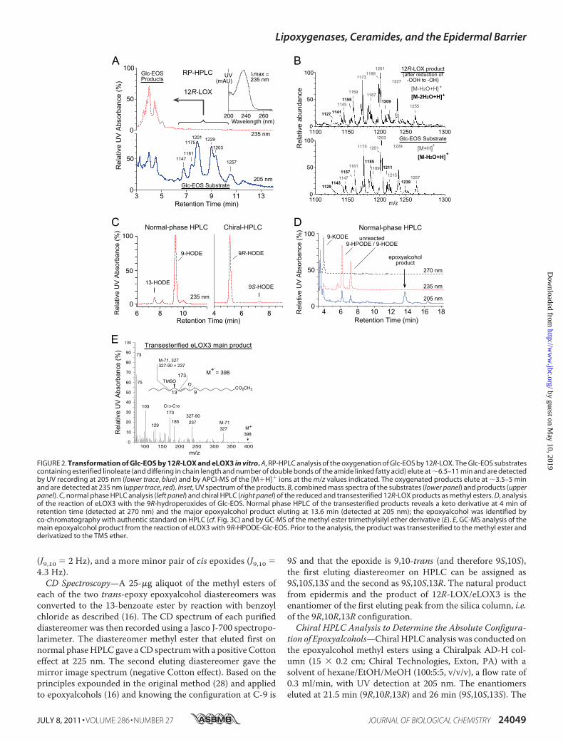
(J9,10 � 2 Hz), and a more minor pair of cis epoxides (J9,10 �4.3 Hz).CD Spectroscopy—A 25-�g aliquot of the methyl esters of
each of the two trans-epoxy epoxyalcohol diastereomers wasconverted to the 13-benzoate ester by reaction with benzoylchloride as described (16). The CD spectrum of each purifieddiastereomer was then recorded using a Jasco J-700 spectropo-larimeter. The diastereomer methyl ester that eluted first onnormal phaseHPLC gave aCD spectrumwith a positive Cottoneffect at 225 nm. The second eluting diastereomer gave themirror image spectrum (negative Cotton effect). Based on theprinciples expounded in the original method (28) and appliedto epoxyalcohols (16) and knowing the configuration at C-9 is
9S and that the epoxide is 9,10-trans (and therefore 9S,10S),the first eluting diastereomer on HPLC can be assigned as9S,10S,13S and the second as 9S,10S,13R. The natural productfrom epidermis and the product of 12R-LOX/eLOX3 is theenantiomer of the first eluting peak from the silica column, i.e.of the 9R,10R,13R configuration.Chiral HPLC Analysis to Determine the Absolute Configura-
tion of Epoxyalcohols—Chiral HPLC analysis was conducted onthe epoxyalcohol methyl esters using a Chiralpak AD-H col-umn (15 � 0.2 cm; Chiral Technologies, Exton, PA) with asolvent of hexane/EtOH/MeOH (100:5:5, v/v/v), a flow rate of0.3 ml/min, with UV detection at 205 nm. The enantiomerseluted at 21.5 min (9R,10R,13R) and 26 min (9S,10S,13S). The
FIGURE 2. Transformation of Glc-EOS by 12R-LOX and eLOX3 in vitro. A, RP-HPLC analysis of the oxygenation of Glc-EOS by 12R-LOX. The Glc-EOS substratescontaining esterified linoleate (and differing in chain length and number of double bonds of the amide linked fatty acid) elute at �6.5–11 min and are detectedby UV recording at 205 nm (lower trace, blue) and by APCI-MS of the [M�H]� ions at the m/z values indicated. The oxygenated products elute at �3.5–5 minand are detected at 235 nm (upper trace, red). Inset, UV spectrum of the products. B, combined mass spectra of the substrates (lower panel) and products (upperpanel). C, normal phase HPLC analysis (left panel) and chiral HPLC (right panel) of the reduced and transesterified 12R-LOX products as methyl esters. D, analysisof the reaction of eLOX3 with the 9R-hydroperoxides of Glc-EOS. Normal phase HPLC of the transesterified products reveals a keto derivative at 4 min ofretention time (detected at 270 nm) and the major epoxyalcohol product eluting at 13.6 min (detected at 205 nm); the epoxyalcohol was identified byco-chromatography with authentic standard on HPLC (cf. Fig. 3C) and by GC-MS of the methyl ester trimethylsilyl ether derivative (E). E, GC-MS analysis of themain epoxyalcohol product from the reaction of eLOX3 with 9R-HPODE-Glc-EOS. Prior to the analysis, the product was transesterified to the methyl ester andderivatized to the TMS ether.
Lipoxygenases, Ceramides, and the Epidermal Barrier
JULY 8, 2011 • VOLUME 286 • NUMBER 27 JOURNAL OF BIOLOGICAL CHEMISTRY 24049
absolute configuration was established using the 9S,10S,13Sstandard prepared from 9S-HPODE; it eluted as the secondpeak from the chiral column, indicating that the first elutingenantiomer has the 9R,10R,13R configuration.
LC-MS Analysis of �-Hydroxy VLFA and Epoxy-ketone AcylAcid
RP-HPLC used a Waters Symmetry C18 column (5 mm, 2.1mm � 15 cm), an isocratic solvent system of methanol/aceticacid (100:0.01 v/v) for �-hydroxy VLFA or methanol/hexane/acetic acid (100:10:0.01, v/v/v) for epoxy-ketone acyl acid, and aflow rate of 0.2 ml/min. Electrospray ionization-MS useda Thermo Finnigan LTQ instrument. Mass spectra wereacquired over the mass rangem/z 200–1000 at 2 s/scan underthe negative ion mode.
Electron Microscopy
Mouse skin samples, �1 mm2, were cut from frozen tissueand prefixed with Karnovsky’s fixative, either before or afterimmersion in absolute pyridine for 2 h, a lipid extraction tech-nique that enhances visualization of the CLE (29). The sampleswere postfixed in either 1%OsO4 in 0.1 M cacodylate buffer, pH7.3, or in RuO4. Ultrathin sections were examined in a Zeiss10A electron microscope.
RESULTS
Lipoxygenase Metabolism of EOS in Vitro—To test whether12R-LOX and eLOX3 react with the acyl(glucosyl)ceramides invitro, first we isolated and characterized the acylglucosylcer-amides, Glc-EOS, from pig epidermis. In agreement with pre-vious studies of others (30, 31), the isolated Glc-EOS were agroup of structural analogues varying in chain length and num-ber of double bounds of the amide-linked VLFA and the sphin-gosine base, the major species of which were detected by RP-HPLC-APCI-MS as m/z 1175, m/z 1201, m/z 1203, and m/z1229 (Fig. 2, A and B). The identity of these compounds asGlc-EOS was further confirmed by in-source fragmentationduring the APCI-MS analysis, giving rise to acylceramides(EOS) with a loss of glucose.
SCHEME 1
FIGURE 3. Identification of oxygenated EOS ceramides in pig epidermis.A, normal phase HPLC with UV detection reveals KODE-EOS (270 nm, blacktrace), containing a keto derivative of linoleate, HODE-EOS (235 nm, red trace)containing hydroxy-linoleate, and EpOH-EOS (205 nm, blue trace) containingan epoxyalcohol derivative of linoleate. Unmodified EOS and other knownceramides are also detected at 205 nm (5–7 min). B, following transesterifica-tion, the main hydroxy product is identified as 9-HODE, and chiral HPLCreveals that it is predominantly 9R-HODE. C, transesterified EpOH-EOS showsa single peak (top panels), corresponding to a standard of 9,10-trans-epoxy-13-hydroxy-octadeca-11E-enoate methyl ester (details in SI Methods). ChiralHPLC analysis of the natural diastereomer (top panels) shows that it corre-sponds exclusively to the 9R,10R,13R enantiomer of a racemic standard (bot-tom panels).
Lipoxygenases, Ceramides, and the Epidermal Barrier
24050 JOURNAL OF BIOLOGICAL CHEMISTRY VOLUME 286 • NUMBER 27 • JULY 8, 2011
Recombinant 12R-LOX reacted with either Glc-EOS (orEOS, shown in supplemental Fig. S1) to give a group of morepolar Glc-EOS derivatives with absorbance at 235 nm, eachdisplaying the characteristic UV spectrum of a conjugateddiene, as expected fromLOX-catalyzed oxygenation of the lino-leate to a hydroperoxide, with conjugation of the double bonds(Fig. 2A). The product peaks were pooled, reduced to thehydroxy derivative, transesterified to themethyl ester, and sub-sequently analyzed by HPLC including chiral HPLC in com-parison to authentic standards. 9-Hydroxy-octadecadienoate(9-HODE) was identified as the predominant product, and itwas almost exclusively of the 9R chirality (Fig. 2C).Next, we incubated 9R-HPODE-Glc-EOS (the hydroperox-
ide produced by 12R-LOX) with eLOX3. RP-HPLC-APCI-MSanalysis identified a group of products with retention timesshorter than those of the 9R-HPODE-Glc-EOS substrate andmolecular masses 32 atomic mass units (i.e. the mass of twooxygen atoms) higher thanGlc-EOS.When transesterified intothe methyl esters, these compounds gave rise to a single epoxy-alcohol (EpOH) methyl ester as the major product, which wasidentified by normal phase HPLC and GC-MS analysis in com-parison with authentic standards (and assuming the 9R config-uration is retained) as methyl 9R,10R-trans-epoxy-13R-hy-droxy-octadeca-11E-enoate (Fig. 2, D and E; and supplementalTable S1 gives the 1H-NMRdata of this epoxyalcohol formedbyeLOX3 from 9R-HPODE methyl ester). A minor keto product(Fig. 1D) was identified as 9-keto-octadecadienoate (9-KODE;Fig. 2D).Nonglycosylated 9R-HPODE-EOS reacted similarly with
eLOX3 (supplemental Fig. S1). Together, these results identifya pathway in which 12R-LOX and eLOX3 can act in tandem onthe linoleate moiety of Glc-EOS or EOS to generate specific
derivatives of 9R-hydroperoxy-linoleoyl-Glc-EOS with re-markable regio- and stereospecificity (Scheme 1).Transformation of Glc-EOS or EOS by 12R-LOX and eLOX3
in vitro was exceptionally slow compared with typical LOXreactions (e.g. 5 �M 12R-LOX catalyzed only �20% conversionof 40�MEOS to product in 2 h).We attribute this slow reactionto the very marked insolubility of the EOS esters in aqueoussolution. In our best attempts, we found that micromolar con-centrations of Glc-EOS or EOS could be partially dissolvedupon sonication in aqueous buffers, pH 5–7, in the presence of10 mM CHAPS. Nonetheless, regiospecific and chiral productsare formed, indicating that the transformation proceeds undertight enzymatic control.LOXMetabolites of EOS in Pig Epidermis—Having observed
the reaction of 12R-LOX and eLOX3 with the acylceramides invitro, we searched for their metabolites in vivo, initially in pigepidermis. Because the LOX metabolites could be present inrelatively small amounts, we developed a sensitive method forceramide analysis using isocratic normal phase HPLC elutionfollowed by either diode array or APCI-MS detection. Withnormal phase HPLC, we detected three groups of novel cera-mides in pig epidermis with HPLCmobility and UV absorptionconsistentwith LOXmetabolites of EOS (Fig. 3A). LC-MS anal-ysis indicated that these three groups of EOS derivatives were,in order of elution, KODE-EOS, HODE-EOS, and EpOH-EOS.For example, themolecular masses of EpOH-EOSwere heavierthan those of EOS by 32 atomic mass units, corresponding tothe two incorporated oxygen atoms in EpOH-EOS (Table 1 andsupplemental Fig. S2A). The ceramide structures are difficult tointerpret from mass spectral data alone, because the presenceof different chain length amide-linked VLFA components inEOS gives rise to a complex array of ions. Accordingly, our
TABLE 1APCI-mass spectrometry of EOS and EpOH-EOS
Lipoxygenases, Ceramides, and the Epidermal Barrier
JULY 8, 2011 • VOLUME 286 • NUMBER 27 JOURNAL OF BIOLOGICAL CHEMISTRY 24051
structural analysis, although supported by the LC-MS data onthe intact, oxidized ceramides, is also founded on the UV spec-tral characteristics of the oxidation products (conjugated diene,enone, or dieone) and on HPLC-UV and GC-MS analysis bycomparison with authentic standards of the oxidized linoleatemoiety following its transesterification from the parental cera-mide. The major hydroxy derivative in EOS was identified as9-HODE, almost exclusively of the 9R chirality (Fig. 3B). Themain keto derivative was 9-KODE (supplemental Fig. S2B).Transesterification of EpOH-EOS released one main epoxyal-cohol, identified by normal phase HPLC, chiral HPLC, andGC-MS as 9R,10R-trans-epoxy-11E-13R-hydroxy-octadeceno-ate (Fig. 3C and supplemental Fig. S2C).LOXMetabolites of Ceramides in Mouse Epidermis; Absence
in 12R-LOX�/�Mice—The almost perfectmatch of in vitro andin vivo products made a compelling case for involvement of12R-LOX and eLOX3 in oxygenation of the EOS ceramides inmammalian epidermis. This connection was further examinedby analysis of endogenous LOX metabolites in mouse epider-
mis, in which the overall hypothesis could be tested using 12R-LOX�/� mice. Indeed, LOXmetabolites of EOS were detectedin wild-type mouse epidermis: 9R-HODE-EOS, and EpOH-EOSwere present, although inminor abundance, and absent in12R-LOX�/� epidermis (supplemental Figs. S3 and S4, A andG). The main LOX product in the mouse epidermal ceramidesis a distinctive derivative of EpOH-EOS identified as epoxy-keto-EOS (see below), detected both as the free lipid (Fig. 4A,indicated on the red 235-nm trace, and supplemental Fig. S4, Aand F) and as reversibly bound lipid in extracts of the proteins(Fig. 4B and supplemental Figs. S4, C andH, and S5). The ratioof the free to the reversibly protein-bound epoxy-keto-EOSwas0.89 � 0.06 (S.E., n � 4).The UV spectrum of the epoxy-ketone-EOS (Fig. 4B, inset) is
characteristic of a conjugated enone with allylic epoxide (32).Its structural identification entailed reduction with sodiumborohydride, transesterification to the methyl ester, andHPLC-UV and GC-MS analysis of the resulting epoxyalcoholmethyl esters in comparison with authentic standards (Fig. 4,C
FIGURE 4. Oxygenated EOS ceramides of mouse epidermis; absence in 12R-LOX�/� mice. A, normal phase HPLC of the ceramides extracted from wild-type(top panel) and 12R-LOX-deficient mouse epidermis (bottom panel). Ceramides identified by MS include EOS, non-hydroxy-fatty acid sphingosine (NS), �-hy-droxy-fatty acid sphingosine (AS), and non-hydroxy-fatty acid hydroxysphingosine (NH). B, normal phase HPLC of mouse epidermal lipids recovered from a finalsoak for 24 h at room temperature after exhaustive chloroform-methanol extractions at 0 °C, from wild-type epidermis (top panel) and 12R-LOX�/� (bottompanel). Top panel, inset, typical UV spectrum of products I and II in normal phase HPLC solvent. C, because of its instability during transesterification, theepoxy-ketone derivative in the ceramides was first reduced using NaBH4 and then analyzed as the resulting epoxyalcohol diastereomers. D, normal phase HPLCseparation of the NaBH4 reduction products. Chiral HPLC analysis shows that the first eluting diastereomer is 9R,10R-epoxy-11E-13R-hydroxy-octadecenoate,identical to the epoxyalcohol product of 12R-LOX and eLOX3 observed in pig epidermis and in vitro (cf. Fig. 2D and supplemental Fig. S2).
Lipoxygenases, Ceramides, and the Epidermal Barrier
24052 JOURNAL OF BIOLOGICAL CHEMISTRY VOLUME 286 • NUMBER 27 • JULY 8, 2011
and D). This main oxidized ceramide in mouse epidermis wasthus identified as the purely chiral 9R,10R-trans-epoxy-11E-13-keto-octadecenoate-EOS (Fig. 4D). We have observed suchepoxy-ketone derivatives as minor products in reactions ofeLOX3 with fatty acid hydroperoxides (33), although its prom-inence in mouse epidermis suggests that a dehydrogenase maybe involved in biosynthesis from its epoxyalcohol precursor.The surprising finding of reversible binding of the epoxy-
ketone to proteins was made during our procedures to exhaus-tively extract the free lipids (supplemental Fig. S5). Followingfour or five cycles of sonication and extraction with chloro-form/methanol (1:1 or 2:1) for 1–2 h at 0 °C, HPLC analysisindicated no further detectable free lipid in the extracts. How-ever, upon a further soak of the protein pellet in chloroform/methanol (1:1) at room temperature overnight, two classes oflipids were recovered: an early eluting epoxy-keto-�-hydroxy-VLFA (Fig. 4B and supplemental Fig. S4, D and I) and epoxy-keto-EOS identical to that in the free ceramides (except that itsVLFA component was mainly the longer C34:1 species, Fig. 4Band supplemental Fig. S4, C andH). To completely extract thispool of lipids, six or seven cycles of such prolonged incubationsat room temperaturewere required. Like the other LOXmetab-olites, these products were completely absent in 12R-LOX�/�
epidermis (Fig. 4B, lower panel).We note that in the 12R-LOX�/� mouse epidermis, unme-
tabolized EOS is present at over twice the level of wild type
(2.5 � 0.2-fold, mean � S.E., n � 5; Fig. 4A), a selective accu-mulation pointing to EOS as the LOX substrate. In essentialfatty acid deficiency, a selective accumulation of oleate-substi-tuted EOS is seen (17), which in light of our results is under-standable because oleate is not a LOX substrate, and thereforeits further metabolism is precluded. We also examined Glc-EOS, the precursor of EOS (34), and found no oxygenatedmetabolites in either pig or mouse epidermis, further suggest-ing that the LOX metabolism occurs during or after the trans-formation of Glc-EOS to EOS.Identification of Free 9R-HODE in Mouse Epidermis—These
results confirm the role of 12R-LOX and eLOX3 in productionof the oxidized ceramides in the epidermis. Furthermore, anadditional LOX product in wild-type epidermis was identifiedas free 9R-HODE, and this was also absent in the 12R-LOX�/�
mice (Fig. 5). Because mouse 12R-LOX is inactive toward freelinoleic acid (35, 36), the observed occurrence of free 9R-HODEpoints to its origin via hydrolysis of an esterified precursor,probably 9R-HODE-EOS. As proposed in our hypothesis, sucha hydrolytic event following LOXmetabolism is a crucial step inepidermal barrier formation.Ester-linked Protein-bound Lipids in Mouse Epidermis—
Central to our hypothesis is that the ester-linked protein-bound OS ceramides and �-hydroxy-VLFA, the main compo-nents of the CLE, should be reduced or absent in 12R-LOX�/�
mice. Although this has been suggested by prior TLC analysis
FIGURE 5. Analysis of free oxygenated fatty acid metabolites from wild-type mouse epidermis (A) and from 12R-LOX�/� mouse epidermis (B). Leftpanels, RP-HPLC analysis of free HODE and HETE. Middle panels, HODE was collected and further analyzed by normal phase HPLC. Right panels, 9-HODE wascollected and further analyzed by chiral HPLC Free HODE and HETE eluted between 2 and 4 min in the normal phase HPLC analysis of total lipid extract as in Fig.4A. Therefore, the fraction from 2 to 4 min was collected and then subjected to the RP-HPLC analysis shown here. Chiral HPLC analysis indicated that 12-HETEfrom both wild-type and 12R-LOX�/� epidermis was in S configuration (data not shown). Identical aliquots were injected in this wild type versus 12R-LOX�/�
comparison. Shown are representative chromatograms from three independent experiments on three sets of wild-type and 12R-LOX�/� littermates. Note the4- or 10-fold more sensitive y axis absorbance scale on the 12R-LOX�/� chromatograms.
Lipoxygenases, Ceramides, and the Epidermal Barrier
JULY 8, 2011 • VOLUME 286 • NUMBER 27 JOURNAL OF BIOLOGICAL CHEMISTRY 24053
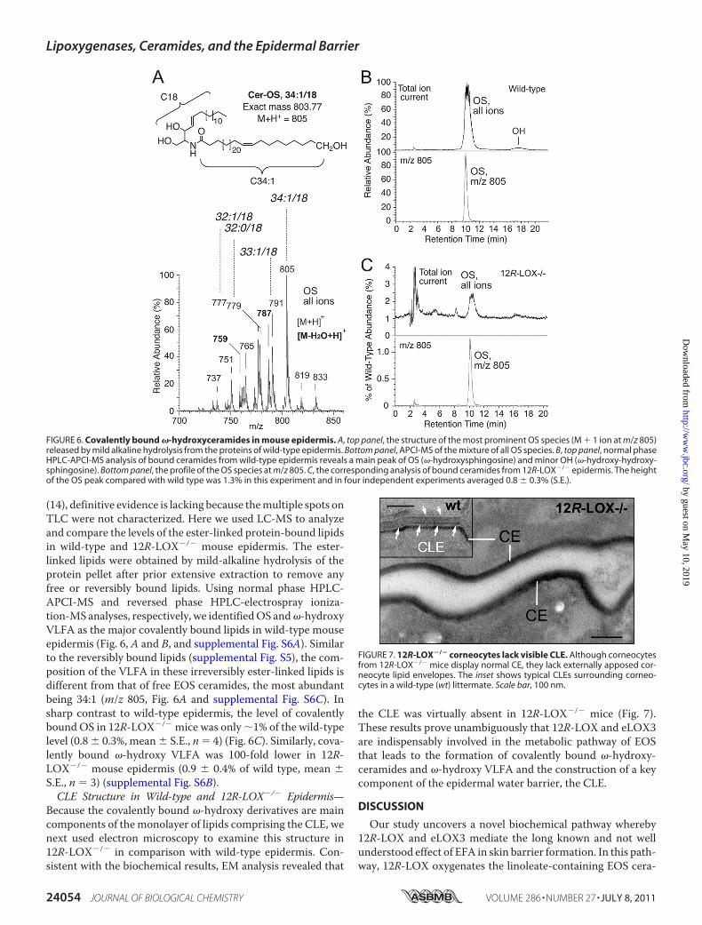
(14), definitive evidence is lacking because themultiple spots onTLC were not characterized. Here we used LC-MS to analyzeand compare the levels of the ester-linked protein-bound lipidsin wild-type and 12R-LOX�/� mouse epidermis. The ester-linked lipids were obtained by mild-alkaline hydrolysis of theprotein pellet after prior extensive extraction to remove anyfree or reversibly bound lipids. Using normal phase HPLC-APCI-MS and reversed phase HPLC-electrospray ioniza-tion-MS analyses, respectively, we identifiedOS and�-hydroxyVLFA as the major covalently bound lipids in wild-type mouseepidermis (Fig. 6, A and B, and supplemental Fig. S6A). Similarto the reversibly bound lipids (supplemental Fig. S5), the com-position of the VLFA in these irreversibly ester-linked lipids isdifferent from that of free EOS ceramides, the most abundantbeing 34:1 (m/z 805, Fig. 6A and supplemental Fig. S6C). Insharp contrast to wild-type epidermis, the level of covalentlyboundOS in 12R-LOX�/� mice was only �1% of the wild-typelevel (0.8 � 0.3%, mean � S.E., n � 4) (Fig. 6C). Similarly, cova-lently bound �-hydroxy VLFA was 100-fold lower in 12R-LOX�/� mouse epidermis (0.9 � 0.4% of wild type, mean �S.E., n � 3) (supplemental Fig. S6B).CLE Structure in Wild-type and 12R-LOX�/� Epidermis—
Because the covalently bound �-hydroxy derivatives are maincomponents of themonolayer of lipids comprising the CLE, wenext used electron microscopy to examine this structure in12R-LOX�/� in comparison with wild-type epidermis. Con-sistent with the biochemical results, EM analysis revealed that
the CLE was virtually absent in 12R-LOX�/� mice (Fig. 7).These results prove unambiguously that 12R-LOX and eLOX3are indispensably involved in the metabolic pathway of EOSthat leads to the formation of covalently bound �-hydroxy-ceramides and �-hydroxy VLFA and the construction of a keycomponent of the epidermal water barrier, the CLE.
DISCUSSION
Our study uncovers a novel biochemical pathway whereby12R-LOX and eLOX3 mediate the long known and not wellunderstood effect of EFA in skin barrier formation. In this path-way, 12R-LOX oxygenates the linoleate-containing EOS cera-
FIGURE 6. Covalently bound �-hydroxyceramides in mouse epidermis. A, top panel, the structure of the most prominent OS species (M � 1 ion at m/z 805)released by mild alkaline hydrolysis from the proteins of wild-type epidermis. Bottom panel, APCI-MS of the mixture of all OS species. B, top panel, normal phaseHPLC-APCI-MS analysis of bound ceramides from wild-type epidermis reveals a main peak of OS (�-hydroxysphingosine) and minor OH (�-hydroxy-hydroxy-sphingosine). Bottom panel, the profile of the OS species at m/z 805. C, the corresponding analysis of bound ceramides from 12R-LOX�/� epidermis. The heightof the OS peak compared with wild type was 1.3% in this experiment and in four independent experiments averaged 0.8 � 0.3% (S.E.).
FIGURE 7. 12R-LOX�/� corneocytes lack visible CLE. Although corneocytesfrom 12R-LOX�/� mice display normal CE, they lack externally apposed cor-neocyte lipid envelopes. The inset shows typical CLEs surrounding corneo-cytes in a wild-type (wt) littermate. Scale bar, 100 nm.
Lipoxygenases, Ceramides, and the Epidermal Barrier
24054 JOURNAL OF BIOLOGICAL CHEMISTRY VOLUME 286 • NUMBER 27 • JULY 8, 2011
mides to give the 9R-hydroperoxide derivative, and eLOX3 inturn converts the 12R-LOX product to the specific 9R,10R-epoxy-13R-hydroxy-epoxyalcohol derivative. Furthermore,based on our biochemical and EM evidence that the CLE ismissing in 12R-LOX�/� mice in correlation with a completeabsence of LOX metabolites, we deduce that this LOX-cata-lyzed oxygenation is required to facilitate hydrolysis of the (oxi-dized) linoleate moiety and leave free the �-hydroxyl on theceramides for coupling to the cross-linked proteins of the CE(Fig. 8). This covalent coupling is an important step in sealingthe water barrier (5, 37). If EFA are deficient, there is no LOXsubstrate available, and thus the fatty acid cannot be cleaved.Absence of one or the other of the LOX enzymes has a similareffect. Thus in both cases, theCLE is eithermissing or defective,leading to a skin barrier defect.Our model linking EFA with the action of 12R-LOX and
eLOX3 also provides a simple explanation for the findings ofHoutsmuller and van der Beek (2) on the structural character-istics of EFA: for a fatty acid (natural or synthetic) to be able tocure the effects of EFA deficiency in the skin, it has to have atleast two cis double bonds in the arrangement of bond-CH2-bond and placed near the �6 position from the end of the car-bon chain (2), which fits perfectly with the structural require-ment of LOX substrates. Thus, although other fatty acids suchas 9-trans,12-cis-18:2�6 or 9-trans,12-cis-18:2�6 (which differfrom linoleic acid only in the configuration of one double bond)will be incorporated into the EOS ceramides if topically appliedto essential fatty acid-deficient skin (34), they invariably fail tocure the skin defects, according to our model because they arenot LOX substrates. This line of thinking is not mutually exclu-sive with evidence that the special physical properties broughtby linoleate itself in acylceramides contribute to another facetof epidermal barrier function, the proper phase behavior andstructural organization of the intercellular lipid lamellae(38, 39).
In further support of our hypothesis, many papers over theyears indicate that oxidized polyunsaturated fatty acid estersare more susceptible to hydrolysis from membrane lipids, andindeednowadays it is generally accepted that oxidation disruptsmembranes and provokes clearance of oxidized lipids (40–43).In our model, there is also the possibility that the specific oxy-genation pattern brought by 12R-LOX and eLOX3 is strin-gently required by the downstream esterase for hydrolysis tooccur.It is worth mentioning at this point that EOS-related cera-
mides were proposed to be LOX substrates in the mid-1980s(34), at a time when this class of ceramides was newly discov-ered (44) and over 10 years before the discovery of 12R-LOXand eLOX3 (45, 46). When Nugteren et al. (34) applied[1-14C]linoleic acid to the skin of EFA-deficient rats, 2 or 3 dayslater some of the radioactivity was recovered in a group of oxi-dized ceramides that were suggested to be LOX metabolites.Because of technical limitations at the time, these putative LOXmetabolites were not structurally defined. Our present studydemonstrates that the oxygenated metabolites of EOS found invivo exhibit the same remarkably high regio- and stereospeci-ficity as the LOX products in vitro, thus definitively resolvingthe arguments over whether the oxygenated metabolites weretruly of enzymatic origin (47). Moreover, we have advanced afurther step in rationalizing the functions these LOX metabo-lites may serve in the skin, i.e. to help build the CLE.
It was once assumed that arachidonic acid is the natural sub-strate of the putative 12R-LOX-eLOX3 pathway in the epider-mis, being converted via 12R-HPETE to 8R-hydroxy-11R,12R-epoxyeicosa-5Z,9E,14Z-trienoic acid (16). A major problemwith this assumption was that arachidonic acid is not a sub-strate of mouse 12R-LOX (35). Instead, the enzyme prefers touse fatty acid esters such as methyl arachidonate as substrate.This no longer presents as a problem, because we now suggestin this study that arachidonic acid oxygenation by 12R-LOX
FIGURE 8. Proposed model linking ceramides, EFA, and LOX in skin barrier formation. Early events (not illustrated) involve fusion of lipid-containinglamellar granules with the corneocyte plasma membrane, extruding the lipid lamellar discs extracellularly and combining the granule limiting membrane(comprised of Glc-EOS) with the cell plasma membrane, initiating formation of the CLE (shown greatly expanded). Progression toward the mature barrierentails LOX-catalyzed oxygenation of the linoleate. The resultant “oxygen signal” permits esterase-catalyzed hydrolysis of the oxidized linoleate, freeing theceramide �-hydroxyl for transglutaminase-catalyzed covalent coupling of the lipids to the cross-linked proteins of the CE, thus forming the CLE and helping toseal the barrier. Lipoxygenase-catalyzed oxygenation of Glc-EOS may be initiated earlier in the process than illustrated here.
Lipoxygenases, Ceramides, and the Epidermal Barrier
JULY 8, 2011 • VOLUME 286 • NUMBER 27 JOURNAL OF BIOLOGICAL CHEMISTRY 24055
and eLOX3 is of little relevance to the physiology of skin barrierformation. Nonetheless, the 12R-LOX products of arachidonicacid do occur in the inflammatory skin disease of psoriasis (48,49) and may have a separate role in that condition.In mouse epidermis, the EpOH-EOS product of eLOX3 was
detected only as a minor metabolite, and the major metabolitewas identified as 9R,10R-epoxy-13-keto-EOS. Such epoxy-ke-tone compounds are often formed as byproducts in the reac-tions of eLOX3with fatty acid hydroperoxides (33). Because thedetected LOX metabolites in vivo are likely to be “leftovers”from the downstream hydrolysis, one interpretation is that theepoxyalcohol derivative is preferentially hydrolyzed, whereasthe byproducts are hydrolyzed to a lesser extent and thus accu-mulate. An alternative is an enzymatic step catalyzing the alco-hol-to-ketone oxidation. Intriguingly approximately half of theepoxy-ketone-EOS in mouse epidermis, together with epoxy-ketone-acyl acids, is directly coupled to proteins through areversible linkage. Most likely, the chemistry involves Michaeladdition of the �,�-unsaturated ketone to His on proteins (32).Notably, the CE protein involucrin is enriched in His (50).These potential non-ester-bound species cannot be distin-guished from the ester-bound �-hydroxyceramides using mildalkaline hydrolysis, because free �-hydroxyceramides will alsobe recovered, whereas the oxidized linoleate will remainattached to proteins. Binding via the �,�-unsaturated ketonemoiety could help juxtapose the EOS derivatives on the CEproteins, prior to the more permanent covalent linkage.Our findings delineate the events in construction of the CLE
and link EFA with LOX metabolism, an understanding withpotential therapeutic implications related to treatments of theichthyoses. Patients with inactivating mutations in ALOX12Bor ALOXE3 lack the ability to oxidize the ceramides and cleavethe linoleate moiety, and potentially this defect could be by-passed by topical application of LOXmetabolites or free �-hy-droxyceramides. Understanding this pathway could also bebeneficial in the treatment of patients with other forms ofichthyosis.
Acknowledgments—We thank Drs. David Hachey and Wade Calcuttfor helpwith LC-MS, Jonas Perez andDr. DavidWright for helpwith theCD spectroscopy, Melanie Hupe for preparing the EM figure, and Drs.RonaldB. Emeson andXiangli Yang for providing normalmouse tissues.
REFERENCES1. Burr, G. O., and Burr, M. M. (1929) J. Biol. Chem. 82, 345–3672. Houtsmuller, U. M., and van der Beek, A. (1981) Prog. Lipid Res. 20,
219–2243. Akiyama, M., and Shimizu, H. (2008) Exp. Dermatol. 17, 373–3824. Fischer, J. (2009) J. Invest. Dermatol. 129, 1319–13215. Uchida, Y., and Holleran, W. M. (2008) J. Dermatol. Sci. 51, 77–876. Wertz, P. W., and Downing, D. T. (1987) Biochim. Biophys. Acta 917,
108–1117. Hedberg, C. L., Wertz, P. W., and Downing, D. T. (1988) J. Invest. Derma-
tol. 91, 169–1748. Doering, T., Brade, H., and Sandhoff, K. (2002) J. Lipid Res. 43, 1727–17339. Nemes, Z., Marekov, L. N., Fesus, L., and Steinert, P. M. (1999) Proc. Natl.
Acad. Sci. U.S.A. 96, 8402–840710. Swartzendruber, D. C., Wertz, P. W., Madison, K. C., and Downing, D. T.
(1987) J. Invest. Dermatol. 88, 709–71311. Elias, P. M. (2005) J. Invest. Dermatol. 125, 183–200
12. Jobard, F., Lefevre, C., Karaduman, A., Blanchet-Bardon, C., Emre, S.,Weissenbach, J., Ozquc, M., Lathrop, M., Prud’homme, J. F., and Fischer,J. (2002) Hum. Mol. Genet. 11, 107–113
13. Moran, J. L., Qiu, H., Turbe-Doan, A., Yun, Y., Boeglin,W. E., Brash, A. R.,and Beier, D. R. (2007) J. Invest. Dermatol. 127, 1893–1897
14. Epp, N., Furstenberger, G., Muller, K., de Juanes, S., Leitges, M., Hausser,I., Thieme, F., Liebisch, G., Schmitz, G., and Krieg, P. (2007) J. Cell Biol.177, 173–182
15. Krieg, P. (2010) Exp. Dermatol. 19, 941–94316. Yu, Z., Schneider, C., Boeglin,W. E., Marnett, L. J., and Brash, A. R. (2003)
Proc. Natl. Acad. Sci. U.S.A. 100, 9162–916717. Wertz, P.W., Cho, E. S., andDowning, D. T. (1983)Biochim. Biophys. Acta
753, 350–35518. Meguro, S., Arai, Y., Masukawa, Y., Uie, K., and Tokimitsu, I. (2000)Arch.
Dermatol. Res. 292, 463–46819. Zheng, Y., Boeglin, W. E., Schneider, C., and Brash, A. R. (2008) J. Biol.
Chem. 283, 5138–514720. Coffa, G., and Brash, A. R. (2004) Proc. Natl. Acad. Sci. U.S.A. 101,
15579–1558421. Bligh, E. G., and Dyer, W. J. (1959) Can. J. Biochem. Physiol. 37, 911–91722. Farwanah, H., Wohlrab, J., Neubert, R. H., and Raith, K. (2005) Anal.
Bioanal. Chem. 383, 632–63723. Butovich, I. A., and Reddy, C. C. (2001) Biochim. Biophys. Acta 1546,
379–39824. Zheng, Y., and Brash, A. R. (2010) J. Biol. Chem. 285, 13427–1343625. Galliard, T., and Phillips, D. R. (1971) Biochem. J. 124, 431–43826. Peers, K. E., and Coxon, D. T. (1983) Chem. Phys. Lipids 32, 49–5627. Chang, M. S., Boeglin, W. E., Guengerich, F. P., and Brash, A. R. (1996)
Biochemistry 35, 464–47128. Schneider, C., Schreier, P., andHumpf,H. YU. (1997)Chirality 9, 563–56729. Behne, M., Uchida, Y., Seki, T., de Montellano, P. O., Elias, P. M., and
Holleran, W. M. (2000) J. Invest. Dermatol. 114, 185–19230. Wertz, P. W., and Downing, D. T. (1983) J. Lipid Res. 24, 753–75831. Hamanaka, S., Asagami, C., Suzuki, M., Inagaki, F., and Suzuki, A. (1989)
J. Biochem. 105, 684–69032. Lin, D., Zhang, J., and Sayre, L. M. (2007) J. Org. Chem. 72, 9471–948033. Zheng, Y., and Brash, A. R. (2010) J. Biol. Chem. 285, 39876–3988734. Nugteren, D. H., Christ-Hazelhof, E., van der Beek, A., and Houtsmuller,
U. M. (1985) Biochim. Biophys. Acta 834, 429–43635. Siebert, M., Krieg, P., Lehmann, W. D., Marks, F., and Furstenberger, G.
(2001) Biochem. J. 355, 97–10436. Meruvu, S., Walther, M., Ivanov, I., Hammarstrom, S., Furstenberger, G.,
Krieg, P., Reddanna, P., and Kuhn, H. (2005) J. Biol. Chem. 280,36633–36641
37. Madison, K. C. (2003) J. Invest. Dermatol. 121, 231–24138. Bouwstra, J. A., Gooris, G. S., Dubbelaar, F. E., and Ponec, M. (2002)
J. Invest. Dermatol. 118, 606–61739. Groen, D., Gooris, G. S., and Bouwstra, J. A. (2010) Langmuir 26,
4168–417540. Sevanian, A., and Kim, E. (1985) J. Free Radic. Biol. Med. 1, 263–27141. Feussner, I., Wasternack, C., Kindl, H., and Kuhn, H. (1995) Proc. Natl.
Acad. Sci. U.S.A. 92, 11849–1185342. Belkner, J., Stender, H., Holzhutter, H. G., Holm, C., and Kuhn, H. (2000)
Biochem. J. 352, 125–13343. Stafforini, D. M., Sheller, J. R., Blackwell, T. S., Sapirstein, A., Yull, F. E.,
McIntyre, T. M., Bonventre, J. V., Prescott, S. M., and Roberts, L. J., 2nd.(2006) J. Biol. Chem. 281, 4616–4623
44. Wertz, P. W., and Downing, D. T. (1982) Science 217, 1261–126245. Boeglin, W. E., Kim, R. B., and Brash, A. R. (1998) Proc. Natl. Acad. Sci.
U.S.A. 95, 6744–674946. Kinzig, A., Heidt, M., Furstenberger, G., Marks, F., and Krieg, P. (1999)
Genomics 58, 158–16447. Wertz, P. W., and Downing, D. T. (1990) J. Lipid Res. 31, 1839–184448. Brash, A. R., Yu, Z., Boeglin, W. E., and Schneider, C. (2007) FEBS J. 274,
3494–350249. Woollard, P. M. (1986) Biochem. Biophys. Res. Commun. 136, 169–17650. Downing, D. T. (1992) J. Lipid Res. 33, 301–313
Lipoxygenases, Ceramides, and the Epidermal Barrier
24056 JOURNAL OF BIOLOGICAL CHEMISTRY VOLUME 286 • NUMBER 27 • JULY 8, 2011


![Billing Code 3410-9R DEPARTMENT OF AGRICULTURE · Billing Code 3410-9R DEPARTMENT OF AGRICULTURE 7 CFR Part 15 [OES-2014-0002] RIN: 051-AA70 Guidance to Federal Financial Assistance](https://static.documents.pub/doc/80x56/5f09452f7e708231d426052d/billing-code-3410-9r-department-of-agriculture-billing-code-3410-9r-department-of.jpg)


