1Department of Psychology, ‘‘Sapienza’’ University of Rome, Rome, Italy2IRCCS Fondazione Santa Lucia, Rome, Italy
r r
Abstract: Behavioral studies indicate that directional gaze and hand pointing are fundamental social sig-nals that may capture spatial attention more powerfully than directional arrows. By using fMRI, weexplored whether reflexive shifts of attention triggered by different distracters were influenced by themotor effector used for performing an overt response. In separate blocks, healthy participants performeda directional saccadic or a hand pointing movement. Color changes of a central black fixation point con-stituted the imperative instruction signal to make a leftward (red color) or a rightward (blue color) move-ment while ignoring distracting leftward or rightward oriented gaze, hand pointing, or arrow. Distractersthat were directionally incongruent with the instruction cue impaired the saccadic and pointing-releaseRTs. The comparison of incongruent versus congruent conditions showed an increase of BOLD signal inthe frontal eye field (FEF), the intraparietal sulcus (IPS), and the posterior parietal cortex (PPC) bilaterally.Importantly, a specific relationship between distracter and effector used for the response was found inthese frontal and parietal regions. In particular, higher activity in the FEF, for distracting gaze was foundmainly during the saccadic response task. In the same vein, higher activity in the left and right IPSregions was found for the distracting hand mainly in the hand pointing task. The results suggest thatreflexive shifts of attention triggered by social signals are coded in the fronto-parietal cortex according toeffector-specific mapping rules. Hum Brain Mapp 00:000–000, 2011. VC 2011 Wiley-Liss, Inc.
Allocation of attention to a specific point in space maybe automatically triggered by biological (e.g., averted
gaze or pointing hands) as well as non-biological direc-tional signals (e.g., regulatory or warning road arrows)[Frischen et al., 2007; Itier and Batty, 2009; Langton et al.,2000]. Whether biological cues are pre-eminent in deter-mining attentional shifts with respect to non-biologicalcues is hotly debated [Bonato et al., 2009; Eimer, 1997;Friesen and Kingstone, 1998; Friesen et al., 2004, 2005;Hietanen, 1999; Jonides, 1981; Ristic et al., 2002, 2007;Stevens et al., 2008; Tipples, 2002], mainly because labo-ratory based paradigms use impoverished tasks thathardly reproduce the situational complexity of real lifehuman interactions [Birmingham and Kingstone, 2009;Kingstone, 2009]. Indeed, fundamental socio-cognitiveoperations, for example, intention and mind reading, areinherently linked to the power of gaze in capturing the
*Correspondence to: Aglioti Salvatore Maria, ‘‘Sapienza’’ Universityof Rome, Via dei Marsi 78, 00185 Roma, Italy. E-mail: [email protected] or Cazzato Valentina, Department of Psychol-ogy, ‘‘Sapienza’’ University of Rome, Via dei Marsi 78, 00185 Roma,Italy. E-mail: [email protected]
Received for publication 26 May 2010; Revised 24 September 2010;Accepted 20 October 2010
DOI: 10.1002/hbm.21202Published online 00 Month 2010 in Wiley Online Library(wileyonlinelibrary.com)
ID: kumarpr I Black Lining: [ON] I Time: 00:32 I Path: N:/3b2/HBM#/Vol00000/100235/APPFile/JW-HBM#100235
VC 2011 Wiley-Liss, Inc.
attention of an observer and in triggering reflexive jointattention under daily life conditions [Kuhn and Land,2006; Smilek et al., 2006].
Possibly, because eye contact is a hallmark of inter-personal interactions, a considerable number of behavioralstudies focused on the role of gaze perception in modulat-ing social attention [Kingstone, 2009; Klein et al., 2009;Nummenmaa and Calder, 2009]. Importantly, unlike non-social orienting cues such as arrows, gaze cues not onlysignal a seen agent’s direction of attention but are alsoused to infer current goals and intentions of other individ-uals. This difference raises the important issue, exploredby recent functional neuroanatomy and electrophysiologi-cal studies, of whether orienting attention to biological,socially relevant cues, such as gaze, may engage neuralmechanisms distinct from those engaged by orienting tonon-social cues. Hietanen et al. [2006], for example,explored at behavioral and neural levels the effect ofresponding to left or right visual targets preceded by cen-tral non-predictive gaze or arrow cues pointing to same oropposite direction. Although the interference effect (IE) ofcue-target directional incongruence was found for bothgaze and arrows, changes of BOLD signal revealed thatwhile gaze-cued orienting recruits occipital regions, arrow-cued orienting also recruits parietal and frontal regions.That arrow-cues related orienting activates a larger net-work with respect to gaze-cue related orienting is also sug-gested by an event-related study showing that changes ofparietal and frontal attention-directed neuroelectric signa-tures are found for arrow—but not for gaze-cues [Hieta-nen et al., 2008]. However, using an ingenious event-related fMRI design in which the central cue was an am-biguous stimulus that could appear as an eye in profile oran arrow, Tipper and colleagues [2008] demonstrated thatattention to social and non-social cues activates a largelyoverlapping neural network centered upon ventral anddorsal fronto-parietal and lateral occipital regions. Sinceactivation in two regions of this network, namely the ven-tral frontal cortex and the lateral occipital, was higher forgaze- than arrow-cues, the authors suggested that quanti-tative more than qualitative differences underlie the socialversus non-social mapping of attentional shifts [Tipperet al., 2008].
Although most of the original studies focused on theimportance of gaze in social attention, body parts otherthan the eyes play a fundamental role in triggering jointattention. Studies demonstrate, for example, that fullbody/head orientation as well as hand orientation of amodel modulates attentional shifts of an observer [Langtonand Bruce, 2000; Langton, 2000; Pierno et al., 2008]. Muchless is known on whether shifts of attention are similarlytriggered by different person-related cues. Information onwhether reflexive social attention triggered by differentperson-related cues is mapped according to the socialvalence of the cue or in body-centered coordinates is veryscanty. Studies indicate that social attention may recruit amore extensive neural network with respect to non-social
spatial attention. Indeed, areas involved in face, gaze,hand, and even full body perception may be called intoplay specifically in social attention tasks [Nummenmaaand Calder, 2009]. This raises the question of whethersocial spatial attention may be coded according to body-centered coordinate systems.
In a recent behavioral study, we explored whether theIE of person-related cues (averted gaze and pointinghands) and of non-social stimuli (arrow) was specificallyinfluenced by the type of effector used for respondingnamely, saccadic movements and hand pointing [Crostellaet al., 2009]. We expected that a non-specific spatial inter-ference of social stimuli would produce higher interferenceof gaze and pointing hands than arrows, regardless of thebody part performing the action. By contrast, we hypothe-sized that finding a relation between the type of distract-ing stimulus and the type of response would suggest thatadditional reference frames are called into action in thetask. The results showed that distracting gaze stimuliinterfere specifically with saccadic performance and dis-tracting hand stimuli with pointing performance. Relevantto this issue is the fMRI study showing that mere observa-tion of directional and non-directional eyes, hands, andarrows in the absence of any motor response, activatedoverlapping neural regions that included the posteriorsuperior temporal sulcus (STS), the inferior parietal lobule(IPL), the inferior frontal gyrus (IFG), and the occipitalcortices in the right hemisphere [Sato et al., 2009].
Capitalizing on such behavioral and neuroimaging evi-dence we sought to determine whether the neural activityin the network underpinning the observation of person-and non-person related signals was modulated by the rela-tionship between type of distracter and type of effectorused for the response. We recorded changes of BOLDfMRI signal associated to conditions where three differentdistracters (gaze, hand, or arrows) influenced overt direc-tional saccadic or hand responses triggered by centralinstruction signals. This design allowed us to highlight:(i) the neural network activated during reflexive shifts ofattention triggered by social and non-social distracters; (ii)the possible modulatory role of gaze and hand distracterson saccadic and hand pointing responses, respectively.
We predicted a specific involvement of dorsal fronto-parietal structures in modulating attentional shifts trig-gered by directional, socially relevant stimuli (i.e., eyesand hand vs. arrow). The fronto-parietal attention system,which includes portions of the intraparietal cortex (e.g.,the intraparietal sulcus, IPS) and of the superior frontalcortex (e.g., frontal eye field, FEF) [Corbetta et al., 2002], isinvolved in the selection of stimuli and goal-directedresponses for goal-directed actions. Importantly, specificsections of this system (FEF and some parts of IPS) maybe differentially active when subjects plan and performvisually guided hand movements, instead than eye move-ments [Astafiev et al., 2003; Corbetta and Shulman, 2002;Corbetta et al., 2008]. It is also relevant that clinical and
ID: kumarpr I Black Lining: [ON] I Time: 00:32 I Path: N:/3b2/HBM#/Vol00000/100235/APPFile/JW-HBM#100235
r Cazzato et al. r
r 2 r
brain imaging studies suggest the presence in humans of asegregated pattern of effector representations in the parie-tal lobe [De Renzi, 1982; Jeannerod, 1986; Seitz et al., 1991].
On the basis of this evidence, we investigated whetherthe tendency of an onlooker to imitate the actions of theobserved model reflects the activity of a resonant systemthat works according to body-part specific reference frames.
MATERIALS AND METHODS
Participants
Eighteen right-handed volunteers (10 males and 8females, mean age ¼ 28 years, range: 23–36 years) tookpart in the study. All subjects had normal or contact-cor-rected-to-normal visual acuity. All were in good health,free of psychotropic or vasoactive medication, with no his-tory of psychiatric or neurological disease. After havingreceived an explanation of the procedures, participantsgave their written consent. The study was approved bythe independent Ethics Committee of the Santa LuciaFoundation (Scientific Institute for Research Hospitaliza-tion and Health Care). Behavioral and imaging data wereanalyzed for subjects who showed reliable IE (slowerresponses for incongruent vs. congruent condition both forsaccade and pointing task). Five subjects did not meet thiscriterion and therefore were not included in the analysesthat were performed on 13 subjects (8 males, mean age:27.5 years; 5 female, mean age: 27 years; range: 23–32years).
Stimuli and Procedure
Participants were positioned in the scanner, in a dimlylit environment. The experimental visual stimuli were pre-sented via a mirror mounted on the MRI headcoil (totaldisplay size 19.5� � 14.6� degrees of visual angle, 1.024 �
768 screen resolution, 60 Hz refresh rate). The visual stim-uli were back-projected on a screen behind the magnet.Stimulus presentation was controlled with Cogent2000(www.vislab.ucl.ac.uk/Cogent/).
Each trial started with the appearance of a black centralfixation mark (0.5� � 0.5� in size), presented centrallyagainst a gray background, and of two black squares (1.4�
� 1.4� in size), presented for 500 ms at 7.5� of eccentricityin the left and the right visual field. The distracting stimuliconsisted of digital Photoshop 8.0.1 (Adobe, CA) modifiedphotographs of gaze, hand, or arrow. The three distracterswere created by using colored photographs of: (i) an emo-tional neutral-expression, full-face of a young womanlooking to the right; (ii) a man hand pointing to the right;(iii) an arrow pointing to the right obtained by digitallyscrambling the hand distracter. The mirror images of thesepictures were created to produce leftward directed stimuli.To make the attention-capture effect conspicuous and thescenario reminiscent of what can be encountered under
daily life conditions, the stimuli were animated by pre-senting two frames in rapid sequence. The first framedepicted a straight gaze, an upward pointing fist or a T-like shape. The second frame, which depicted a leftwardor rightward oriented gaze, extended finger or arrow,replaced the first frame. The direction of the distracter andthe one indicated by the instruction cue could be 50% ofthe time congruent or incongruent. Before starting thefMRI acquisition, each participant was asked to performoutside the scanner a training task in which they had tolearn with 100% accuracy on 30 consecutive trials pertask, the association between instruction signal (red orblue) with leftward or rightward saccadic or pointingmovements.
In the scanner, each trial started with the presentationbehind the black fixation mark of a straight gaze, anupward pointing fist or a T-like shape which lasted 500ms. At 500 ms, a second frame, that depicted leftward orrightward oriented gaze, extended finger or an arrow,replaced the first frame and created a strong animationeffect. The directional distracters remained on until theend of the trial and 75 ms after the oriented distracter pre-sentation, the black central fixation mark (imperative cue)changed to either blue or red color. This was the instruc-tion signal for the subjects to make, in separate runs, a sac-cade or a right index pointing movement towards the left(change into red) or the right (change into blue) targetsquare (for saccades) and the left or right button of ahome-made keypad (for pointing). Thus, the direction ofthe distracter and that indicated by the instruction cuecould be congruent (left-red or right-blue) or incongruent(left-blue or right-red). The colored cue remained visibleuntil the end of the trial (See Fig. F11).
To engage automatic processes and minimize expecta-tions, the directional cues were equiprobable (50% congru-ent) and non-predictive. It is worth noting that thesubjects were instructed to ignore the distracters and tofocus on the central mark color change. Moreover, theywere explicitly informed that the instruction cue was notinformative about the direction of the distracters. In thehand pointing task, subjects were also instructed to fixatethe central cross for the entire trial. This allowed us tomeasure attentional shifts independent of eye movements.To avoid subjects anticipating stimuli, a random inter-trialinterval ranging from 3.5 to 4.5 s was used.
Twelve event types were organized in a 3 � 2 � 2 facto-rial design. One factor was the Distracter: gaze, hand (bothbiological distracters with social valence), and arrow (nonbiological and non social distracter). The second factorwas the type of Effector: saccadic versus pointing move-ments. To minimize any task-switching requirements, eachparticipant performed three fMRI runs of saccadic move-ment and three fMRI runs of pointing movement. Theorder of the effectors was counterbalanced across partici-pants. On each run, participants were verbally instructedabout the motor response to be performed (saccadic orpointing task). The third factor was the Condition:
ID: kumarpr I Black Lining: [ON] I Time: 00:32 I Path: N:/3b2/HBM#/Vol00000/100235/APPFile/JW-HBM#100235
r Reflexive Shifts of Attention r
r 3 r
congruent vs. incongruent direction between instructionsignal and distracter. Congruent and incongruent direc-tional combinations of instruction cues and distracterswere presented in unpredictable and randomized order.Thus, fMRI data were acquired via a mixed, blocked(Distracter, Effector)/event-related (Condition) protocol.All participants underwent six fMRI runs. Each participantcompleted a total of 720 trials (360 for each effector), there-fore each imaging session consisted of 40 repetitions foreach of the three distracters (Gaze/Hand/Arrow), respec-tively, 20 for congruent and 20 for incongruent conditions(balanced for left/right direction and red-blue imperative-cues). Each scanning session lasted �8 min for a totalexperiment duration of about 50 min.
Eye Movements Recording
In the training session outside the scanner, subjects satin front of a computer screen. In all subjects, eye positionand saccadic movements were monocularly monitoredusing an infrared video camera (Sony EVI D31, color videocamera, Sony JP). Participants were instructed to look atthe location indicated by the instruction cue and then toquickly look back at the fixation point. During the scan-ning session, again the participants’ saccadic movementswere monocularly monitored in real-time by means of anASL eye-tracking system that was adapted for use in thescanner (Applied Science Laboratories, Bedford, MA;Model 504, sampling rate: 60 Hz). For each subject, the
ID: kumarpr I Black Lining: [ON] I Time: 00:32 I Path: N:/3b2/HBM#/Vol00000/100235/APPFile/JW-HBM#100235
r Cazzato et al. r
r 4 r
eye-tracking system was calibrated before fMRI scanning.The calibration was repeated during the experiment when-ever necessary. Eye-position traces were examined in a1,175 ms time window, beginning with the imperative cueonset until the end of the trial. In the sessions requiringpointing movements, the maintenance of central fixationwas monitored throughout the trial. We defined losses offixation as changes in horizontal eye-position >�2� of vis-ual angle with durations at least 100 ms. For trials requir-ing saccadic responses (Saccadic Task), the saccadic RTswere calculated from the target onset time to when anhorizontal eye position exceeded 2�. Moreover, we did notcompute RTs for the trials in which subject made asaccade to the wrong side (e.g., saccade to the left targetafter the central cue turned into blue) or did not performany saccade at all.
Hand Movement Recording
In the training session outside the scanner, participantssat in front of a computer screen by keeping their rightindex finger on a central response key until the occurrenceof the instruction signal. Then, based on the directionalinstruction cue, subjects pointed towards a left or a rightkey located 2 cm laterally with respect to the centralposition. In the scanner, the right hand was positioned incorrespondence of the low abdomen in a relaxed posturewith the right index finger extended and all other fingersflexed. This position allowed participants to perform cen-tral-cue instructed index finger movements toward theright or the left button key. The right shoulder and armwere supported and immobilized with cushioning wedgedbetween the scanner bed and the coil surface. When thecentral mark changed color, subjects pointed as quickly aspossible in the direction of the target location (lateral keypresses) and then returned to the resting position. Pointinginvolved a minimal rotation of the wrist with extendingindex without movements of the shoulder or the arm [seeAstafiev et al., 2003]. For pointing data, we computed aRelease RTs measure and we only analyzed trials in whichsubjects maintained fixation on the central fixation mark.
Magnetic Resonance Imaging
A Siemens Allegra (Siemens Medical Systems, Erlangen,Germany) operating at 3T and equipped for echo-planarimaging (EPI) acquired functional magnetic resonance(MR) images. A quadrature volume head coil was used forradio frequency transmission and reception. Head move-ments were minimized by mild restraint and cushioning.Thirty-six slices of functional MR images were acquiredusing blood oxygenation level-dependent imaging (3.0 �
3.0 � 2.5 mm thick, 50% distance factor, TR ¼ 2.34 s, TE ¼
30 ms), covering the entire cortex.
Data Analysis
We used the statistical parametric mapping packageSPM5 (www.fil.ion.ucl.ac.uk) implemented in MATLAB(v 7.1, The MathWorks, Natick, MA) for data pre-process-ing and statistical analyses. For all participants, weacquired 1.290 fMRI volumes, 215 for each run. The firstfour image volumes of each run were used for stabilizinglongitudinal magnetization and were discarded from theanalysis. Pre-processing included rigid-body transforma-tion (realignment) and slice timing to correct for headmovement and slice acquisition delay. Residual effects ofhead motion were corrected including the six estimatedmotion parameters for each subject as regressors of no in-terest. Slice-acquisition delays were corrected using themiddle slice as a reference. All images were normalizedto the standard SPM5 EPI template, resampled to 2 mmisotropic voxel size, and spatially smoothed using an iso-tropic Gaussian kernel of 8 mm FWHM. Statistical infer-ence was based on a random effects approach [Pennyand Holmes, 2004]. First, for each participant, the datawere best-fitted at every voxel using a combination ofeffects of interest. These were delta functions representingthe onsets of the 12 conditions given by the crossing ofour 3 � 2 � 2 factorial design: Distracter (gaze/hand/arrow) � Condition (congruent/incongruent) � Effector(saccadic movement/pointing movement) convolved withthe SPM5 hemodynamic response function. The onset ofthe hemodynamic response function was aligned with theonset of the imperative cue with duration ¼ 0. Onsets oftrials in which an erroneous response or an eye move-ment toward the wrong side occurred were included inthe design matrix as covariates of no interest, butexcluded from any further analysis. Linear contrasts wereused to determine differential activation for incongruenceminus congruence conditions separately for 3 � 2 (Dis-tracter � Effector) (e.g., [Gaze (Incong) > Gaze (Cong)]for saccadic movement) factors, averaging the three fMRIruns (three for the saccade and three for the pointingtask). These six contrasts images were entered in a 3 � 2factorial ANOVA with Distracter (gaze, hand, and arrow)and Effector (saccadic movement, pointing movement).Finally, linear compounds (contrasts) were used to com-pare the Incongruency effect using between-participantsvariance (rather than between scans). Correction fornonsphericity [Friston et al., 2002] was used to accountfor possible differences in error variance across conditionsand non-independent error terms for the repeatedmeasures.
The analyses aimed at determining: (i) the brain regionscalled into action when directional cue and distractersprovided conflicting directional information (incongruentcondition); (ii) whether any modulation exerted by thebiological distracters (gaze and hand) was specificallylinked to the effector the onlookers used for responding;(iii) whether reflexive joint attention was differentiallymodulated by the biological (gaze and hand) vs.
ID: kumarpr I Black Lining: [ON] I Time: 00:32 I Path: N:/3b2/HBM#/Vol00000/100235/APPFile/JW-HBM#100235
r Reflexive Shifts of Attention r
r 5 r
non-biological distracters (arrow), irrespective of motoreffector.
We first sought to determine any specific cortical atten-tional network associated with the directional incongru-ence conditions (comparing incongruent vs. congruentcondition, irrespective of distracter and effector). Thus, themain effect of Incongruence allowed us to identify thenetwork activated by the directional conflict between task-irrelevant distracters and instruction signals. For this com-parison, the SPM threshold was set to P corr. ¼ <0.05 atcluster level (cluster extent estimated a P uncorr. ¼ 0.001),considering the whole brain as the volume of interest. Totest for the interaction between the IE with Motor-Effectorand Distracter, we created regions of interest (ROIs)extracting average BOLD signals (MarsBar 0.41, ‘‘MAR-Seille Boıte A Region d’Interet’’ SPM toolbox) from thepeak activity of the voxels that showed a main effect ofIncongruence. Each ROI was defined as a 10 mm radiussphere centered on the corresponding maxima of thewhole-brain analysis (see Table II), and P values were Bon-ferroni-corrected. We expected that our manipulations ofIE would affect activity within the dorsal fronto-parietalattentional systems depending on specific relationshipswith Distracter and Effector [Corbetta and Shulman, 2002;Crostella et al., 2009; Ricciardelli et al., 2002]. Accordingly,we used a combination of anatomical and functional crite-ria to identify six ROIs in the dorsal attentional system:the FEF, the posterior parietal cortex (PPC), the IPSbilaterally.
The bilateral frontal ROIs included a portion of middlefrontal gyrus (FEF) located laterally within the superiorfrontal sulcus [Paus, 1996]. Because of the large extensionof parietal cortex clusters, we decided to distinguishbetween posterior and anterior anatomical regions, bilat-eral PPC and IPS, respectively. Bilateral PPC included aportion of superior parietal lobule close to superior parie-tal gyrus and precuneus. Bilateral IPS ROIs were insteadlocated anteriorly and close to the inferior parietal lobule,the angular gyrus and the IPS.
For each ROI, we tested the three-way interaction IE �
Effector � Distracter to provide information on whetherthe cost of directional incongruence was mapped ondifferent brain regions depending on specific relation-ships with distracter and motor effector. For example,this interaction allowed exploring whether observation ofincongruent saccades performed by the distracting gazeinduced differential brain responses in the onlookers’when performing the saccadic with respect to the hand-pointing movement. It should be noted that main effectand interactions are orthogonal and, therefore, our ROIselection procedure was unbiased. Moreover, based onthe prediction that IEs are stronger when elicited bysocial (gaze and hand) than by non-social (arrow) dis-tracters, we tested the interaction between the IE and thebiological vs. non-biological distracters, irrespective ofmotor effector.
RESULTS
Behavioral Performance
Both saccadic and release mean RTs were calculated col-lapsing left and right target trials. Incorrect responses(movements performed following distracters instead thaninstruction cues), misses (no response), anticipations (RTs< 100 ms), and retards (RTs > 1.500 ms) were notincluded in the analysis. Overall, we discarded 12.7% oftrials for saccadic sessions and 7.5% of trials for pointingsessions. Following previous studies [Kitagawa andSpence, 2005; Murphy and Klein, 1998; Spence et al.,2001a,b], we computed an inverse efficiency score bydividing, for each condition and in each subject, the meancorrect RTs by the percentage of directionally correctresponses. The inverse efficiency score provides a way tocombine RT and accuracy measures of performance into asingle measure [Townsend and Ashby, 1983] and allowscontrolling for any speed-accuracy trade-off effects. As forRT and error measures, higher inverse efficiency scoresindicate worse performance. Table T1I reports inverse effi-ciency scores in the saccadic and hand pointing tasks, foreach distracter type and incongruent and congruent condi-tions, acquired during fMRI scanning.
The inverse efficiency scores were entered in two sepa-rate 3 � 2 repeated-measures ANOVAs (one for saccadicand one for pointing task) with Distracter (gaze, hand, andarrow), and Condition (congruent and incongruent) aswithin-subjects effects. In the saccadic task, the main effectof Condition [F (1, 12) ¼ 22.297, P ¼ 0.001] was explainedby the worse performance in the incongruent than congru-ent trials (629 vs. 505 ms/percentage of correct trials). Noother effects or interactions were significant. Also in thepointing task, the main effect of Condition was significant[F (1, 12) ¼ 8.521, P ¼ 0.01] because of the worse perform-ance in the incongruent than congruent trials (535 vs. 510ms/percentage of correct responses). Again, no othereffects or interactions were significant.
To sum up, saccadic and release RTs/percentage ofcorrect trials scores during MR scanning show that theincongruent cues worsened both saccadic and pointingperformances. However, this effect was independent from
Inverse efficiency scores (mean RT/percentage of correctresponses, S.E.M. in brackets) are represented separately for sacca-dic and release RTs as a function of Distracter (G ¼ Gaze/H ¼
Hand/A ¼ Arrow) and Condition (Congruent/Incongruent).
ID: kumarpr I Black Lining: [ON] I Time: 00:32 I Path: N:/3b2/HBM#/Vol00000/100235/APPFile/JW-HBM#100235
r Cazzato et al. r
r 6 r
the type of Distracter (as indicated by the non-significantDistracter � Effector interaction).
fMRI Data
Main effect of incongruence
To highlight the neural underpinnings of the IE trig-gered by incongruent distracters, we tested for the maineffect of incongruence irrespective of Distracter and Effec-tor (See TableT2 II).
This contrast revealed the expected activation of theventral and dorsal frontal and parietal regions. The parie-tal region consisted of a large cluster including the rightsuperior and inferior parietal cortex bilaterally. The frontalregion included the left precentral gyrus, the right middlefrontal cortex bilaterally, the right supplementary motorarea, the most posterior portion of the inferior frontalgyrus, the operculum, bilaterally, and the pars triangularisextending into the insula and the middle portion of theright cingulate cortex (See Fig.F2 2).
The main effect of Incongruence considering the threedistracters (G ¼ Gaze/H ¼ Hand/A ¼ Arrow, averagingacross saccadic/hand-pointing motor effector) was used todefine the center of each ROI in the two hemispheres.Within each ROI, we tested for: (i) the critical interaction:IE of Distracter (Gaze and Hand) on the paired saccadicand hand pointing task; (ii) the IE of biological (Gazeand Hand) vs. non-biological distracters (Arrow), irrespec-tive to effector.
IE of distracters in the saccadic and hand pointing tasks
We investigated the possible influence on BOLD signalof the pairing between body-part related (gaze or pointing
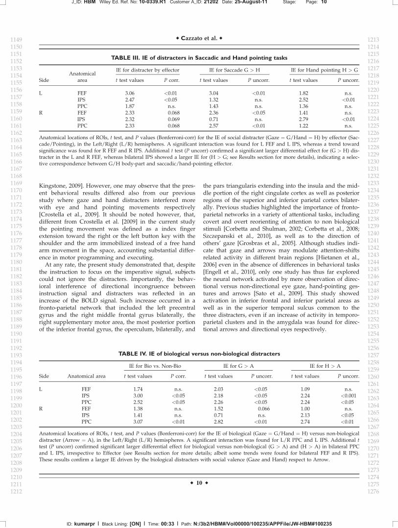
hand) distracter of motor-effector used for the response(eyes or hand) within each frontal and parietal ROI. Themean BOLD activation for each Distracter and Effector inthe frontal and parietal ROIs is shown in Figure F33. Statis-tics, for the interaction effect and additional t test in eachROI are reported in Table T3III.
Left FEF was specifically modulated by the interactionIE � Distracter � Effector, while right FEF showed a trendtoward significance. To further confirm the specificity ofthese effects, we compared the IE of gaze vs. hand dis-tracter for saccadic motor effector. This revealed that leftand right FEF were modulated by the selective correspon-dence between ‘‘Gaze’’ body-part and ‘‘Saccadic’’ effector(See bars Fig. 3, left panel: G > H). The opposite patternwas found in left IPS region; as for left FEF, this regionresulted specifically influenced by the interaction IE �
Distracter � Effector, whereas right IPS showed a trend to-ward significance. Additional t test confirmed that thiseffect was due to a larger IE for ‘‘Hand pointing’’than gaze-distracter during ‘‘Hand pointing’’ movements(See bars Fig. 3, right panel: H > G). This demonstratesthat activity in these regions is specially influenced by themotor effectors used to perform the task. This effect wasstronger in the left than in the right hemisphere. Finally,left PPC was not sensitive to this interaction given thatresults were not replicated (albeit a significant IE for Gazemore than Arrow was found for right PPC).
fMRI Activations Associated to the IE of
Biological Versus Non-Biological Distracters
To explore whether reflexive joint attention was differ-entially modulated by the different categories of distracters(e.g., biological and social vs. non-biological non-social
Anatomical locations, peak coordinates in MNI space (Montreal Neurological Institute), and statistical values for the main effect ofincongruence (incongruent > congruent trials, irrespective of distracter and effector). P values are corrected for multiple comparisons atthe cluster level, considering the whole brain as the volume of interest. R/L PPC ¼ Right/Left Posterior Parietal Cortex; R/L IPS ¼
Right/Left Intraparietal Sulcus; R/L FEF ¼ Right/Left Frontal Eye Field; R/L Insula ¼ Right/Left Insula; R Cingulum Mid ¼ RightMiddle Cingulum; L Precentral G ¼ left Precentral Gyrus. With the asterisk (*) we indicated the ROIs within the dorsal fronto-parietalattentional network. ROIs were extracted averaging BOLD signals (see Methods) from a 10 mm sphere centered on the cluster peak.
ID: kumarpr I Black Lining: [ON] I Time: 00:32 I Path: N:/3b2/HBM#/Vol00000/100235/APPFile/JW-HBM#100235
r Reflexive Shifts of Attention r
r 7 r
cues) independently from motor-effector, we compared theIE for biological (gaze and hand) vs. non-biologicaldistracters (arrow).
Statistics for the interaction effect and additional t test ineach ROI are reported in TableT4 IV.
Significant interactions, mainly in the left hemispherewere found within the parietal ROIs. In particular, in theleft and the right PPC, the activation for the IE triggeredby biological distracters (gaze and hand) was larger thanthe activation for the IE triggered by non-biological distracter (arrow; See Bars Fig.F4 4: G > A). Inother words, the BOLD signal in these regions was higherwhen the directional conflict between distracter-instructionsignals involved biological (gaze and hand) distractersthan when the conflict involved the non-biological (arrow)distracter. Confirmatory t tests demonstrated that thiseffect was due to both a significant IE for gaze versusarrow distracters and to a significant IE for hand versusarrow distracters in left IPS and bilateral PPC. Finally,these analyses did not reveal any significant interaction for
right and left FEF or right IPS, with the exception of alarger IE for G > A in bilateral FEF and a larger IE forH > A in right IPS.
DISCUSSION
The present study had two main aims: (i) to ascertainwhether the possible differential attention orienting-powerof directional gaze, hand, and arrow distracters reliesupon commons neural substrates; (ii) to explore whetherthe relationship between gaze and hand distracters andthe motor effector used in the experimental task (Saccadicor Hand pointing response) was reflected in a specificmodulation of the activity in the dorsal fronto-parietalnodes of the reflexive attention network. Finally, the studyexplored the architecture of the reflexive orienting trig-gered by biological (Gaze and Hand) and non-biological(Arrow) distracters, irrespective of motor effector.
ID: kumarpr I Black Lining: [ON] I Time: 00:32 I Path: N:/3b2/HBM#/Vol00000/100235/APPFile/JW-HBM#100235
r Cazzato et al. r
r 8 r
Behavioral and Neural Correlates of
Reflexive Attention
A cost of directional incongruence between distractersand instructions signals was found. All distracters in thebehavioral performance showed a congruency effect bothfor saccadic and pointing task. This is in keeping withstudies showing that attention is captured by gaze andarrows to a similar extent [Kuhn and Benson, 2007; Kuhnand Kingstone, 2009; Sato et al., 2009] and at variance
from studies showing that social distracters like avertedgaze or pointing hands induce stronger attentional capturemore than symbolic arrow [Langton and Bruce, 2000;Ricciardelli et al., 2002]. It is worth noting that in manycomplex daily life interactions, the tendency to followothers seems to be very strong. Thus, the lack of predomi-nance of gaze- over arrow-distracters in triggering reflex-ive attention of arrows in some studies may be due to afloor effect induced by the extremely simplified reality oflaboratory conditions [Birmingham and Kingstone, 2009;
ID: kumarpr I Black Lining: [ON] I Time: 00:32 I Path: N:/3b2/HBM#/Vol00000/100235/APPFile/JW-HBM#100235
r Reflexive Shifts of Attention r
r 9 r
Kingstone, 2009]. However, one may observe that the pres-ent behavioral results differed also from our previousstudy where gaze and hand distracters interfered morewith eye and hand pointing movements respectively[Crostella et al., 2009]. It should be noted however, that,different from Crostella et al. [2009] in the current studythe pointing movement was defined as a index fingerextension toward the right or the left button key with theshoulder and the arm immobilized instead of a free handarm movement in the space, accounting substantial differ-ence in motor programming and executing.
At any rate, the present study demonstrated that, despitethe instruction to focus on the imperative signal, subjectscould not ignore the distracters. Importantly, the behav-ioral interference of directional incongruence betweeninstruction signal and distracters was reflected in anincrease of the BOLD signal. Such increase occurred in afronto-parietal network that included the left precentralgyrus and the right middle frontal gyrus bilaterally, theright supplementary motor area, the most posterior portionof the inferior frontal gyrus, the operculum, bilaterally, and
the pars triangularis extending into the insula and the mid-dle portion of the right cingulate cortex as well as posteriorregions of the superior and inferior parietal cortex bilater-ally. Previous studies highlighted the importance of fronto-parietal networks in a variety of attentional tasks, includingcovert and overt reorienting of attention to non biologicalstimuli [Corbetta and Shulman, 2002; Corbetta et al., 2008;Szczepanski et al., 2010], as well as to the direction ofothers’ gaze [Grosbras et al., 2005]. Although studies indi-cate that gaze and arrows may modulate attention-shiftsrelated activity in different brain regions [Hietanen et al.,2006] even in the absence of differences in behavioral tasks[Engell et al., 2010], only one study has thus far exploredthe neural network activated by mere observation of direc-tional versus non-directional eye gaze, hand-pointing ges-tures and arrows [Sato et al., 2009]. This study showedactivation in inferior frontal and inferior parietal areas aswell as in the superior temporal sulcus common to thethree distracters, even if an increase of activity in temporo-parietal clusters and in the amygdala was found for direc-tional arrows and directional eyes respectively.
Anatomical locations of ROIs, t test, and P values (Bonferroni-corr) for the IE of social distracter (Gaze ¼ G/Hand ¼ H) by effector (Sac-cade/Pointing), in the Left/Right (L/R) hemispheres. A significant interaction was found for L FEF and L IPS, whereas a trend towardsignificance was found for R FEF and R IPS. Additional t test (P uncorr) confirmed a significant larger differential effect for (G > H) dis-tracter in the L and R FEF, whereas bilateral IPS showed a larger IE for (H > G; see Results section for more details), indicating a selec-tive correspondence between G/H body-part and saccadic/hand-pointing effector.
TABLE IV. IE of biological versus non-biological distracters
Side Anatomical area
IE for Bio vs. Non-Bio IE for G > A IE for H > A
t test values P corr. t test values P uncorr. t test values P uncorr.
Anatomical locations of ROIs, t test, and P values (Bonferroni-corr) for the IE of biological (Gaze ¼ G/Hand ¼ H) versus non-biologicaldistracter (Arrow ¼ A), in the Left/Right (L/R) hemispheres. A significant interaction was found for L/R PPC and L IPS. Additional ttest (P uncorr) confirmed significant larger differential effect for biological versus non-biological (G > A) and (H > A) in bilateral PPCand L IPS, irrespective to Effector (see Results section for more details; albeit some trends were found for bilateral FEF and R IPS).These results confirm a larger IE driven by the biological distracters with social valence (Gaze and Hand) respect to Arrow.
ID: kumarpr I Black Lining: [ON] I Time: 00:33 I Path: N:/3b2/HBM#/Vol00000/100235/APPFile/JW-HBM#100235
r Cazzato et al. r
r 10 r
Body-Part Specific Reference Frames for
Mapping Reflexive Social Attention in the
Fronto-Parietal Cortex
In keeping with previous neuroimaging studies[Grosbras et al., 2005; Hietanen et al., 2006; Sato et al.,2009; Tipper et al., 2008], our results highlight the funda-mental role of fronto-parietal structures in mediating gazeand hand related shifts of attention. However, our studyexpands significantly previous knowledge by combining,for the first time, two main issues, namely the possiblespecificity of the neural representation of different effec-tors used for response and the influence of social and non
social distracters in modulating reflexive attention. It iswidely held that movements performed with differenteffectors are coded in different cortical regions. Distinctposterior parietal modules, for example, may preferentiallycode for saccades and reaches, respectively [Colby andGoldberg, 1999; Glimcher, 2003]. More recent studies indi-cate that far from being a strict principle, effector-selectiv-ity implies a gradual transition of preference from oneeffector to another, with areas of balanced activation tosaccades and reaches and areas with significant preferencefor reaches [Levy et al., 2007]. Similarly, effector preferencewas found in parieto-frontal areas during eye or handmovement planning but no region responded exclusively
ID: kumarpr I Black Lining: [ON] I Time: 00:33 I Path: N:/3b2/HBM#/Vol00000/100235/APPFile/JW-HBM#100235
r Reflexive Shifts of Attention r
r 11 r
to either effector [Beurze et al., 2009]. A predominance ofleft lateralized maps for coding the preparation of pointingmovements in the presence of equivalent coding of sacca-dic and reaches preparation in frontal areas has also beenreported [Astafiev et al., 2003]. Testing the hypothesis of adifference in the visuospatial maps recruited by pointingand saccades, Hagler and colleagues [2007] identified mul-tiple maps in both PPC and superior frontal cortexrecruited for eye and hand movements, including mapsnot observed in previous studies. Although their analysisrevealed subtle differences between pointing and saccades,including hemispheric asymmetries, no evidence of point-ing-specific maps of visual space was found.
In the present study, we explored whether biologicaldirectional distracters such as directional gaze and pointinggestures, influenced the neural underpinnings of reflexiveshifts of attention in relation to the motor effectors used forthe response, namely eyes or hands. To this aim, we com-pared the BOLD signal in the fronto-parietal ROIs thatturned out to be involved in reflexive attention. We found afunctional dissociation in the frontal and parietal nodes ofthe reflexive joint attention network, hinting at a specificinfluence of gaze and hand distracters in the saccadic andhand pointing tasks, respectively. Overall, the fMRI dataindicated that the observed interference with voluntary ori-enting varied as a function of central distracter-type andmotor-effector. In particular, we observed greater IE-relatedactivation in the frontal ROIs for shifts of spatial attentiontriggered by gaze in the saccadic task and in the parietalROIs, specifically bilateral IPS, for shifts of attention trig-gered by hand in the pointing task. This result is in keepingwith previous studies indicating the importance of parietalregions in mediating interference of hand movements in-congruous with planning of a different hand movement[Grefkes et al., 2004] or of hand-related attention switchingtasks [Rushworth et al., 2001].
Tellingly, a main point of novelty of the present study isthat the fronto-parietal network subserving reflexive shiftsof social attention is specially sensitive to the relationshipbetween specific body-related distracters and the respond-ing body parts. Importantly, an effector-specific activationof fronto-parietal networks in humans has also been foundin a recent study on cortical temporal dynamics of visuallyguided behavior [Hinkley et al., 2010]. In this study, high-gamma activity was observed in SEF and subsequently invisual cortex and FEF bilaterally, followed by a low-betapower decrease over caudal PPC during saccade execution.Thus, hand or saccadic movements implied a differentfunctional connectivity between frontal and parietal areas.
Mirroring of Attention in the
Fronto-Parietal System
In our experimental paradigm, participants were specifi-cally instructed to ignore the visual distracting stimuli(gaze, hand, and arrow), to focus on the central imperative
go signal and to maintain the fixation on the central point.Given that the distracter was presented before the unpre-dictable central cue, the cost of re-orienting to fully irrele-vant-task distracters is likely due to interference withongoing action programs. This may be in keeping withpre-motor theories of attention [Rizzolatti et al., 1987] andwith the notion of mirroring others’ actions [Rizzolatti andSinigaglia, 2010]. Behavioral studies indicate that priminga given motor response is more effective if the visualprime shares specific properties with the requestedresponse suggesting that perceptual codes and actionplans may share a common representational medium[Craighero et al., 2002]. Neuroimaging studies indicatethat viewing hand, mouth and foot actions may induce aspecific increase of the BOLD signal in the frontal and pa-rietal representations of the acting body parts [Buccinoet al., 2001]. A clear link between action mirroring andsharing of attention between individuals has been estab-lished in a single cell recording study from the monkeyparietal lobe [Shepherd et al., 2009]. This study demon-strates an increase of activity of parietal neurons not onlywhen the monkey oriented his attention towards theirreceptive field, but also during observation of anothermonkey orienting in the same direction. It is also relevantthat overlapping fronto-parietal cortical representations arecalled into play during executed, observed, and imaginedreaching in humans [Filimon et al., 2007]. That reflexiveshifts of social attention may be coded in body-partspecific coordinates and may reflect a specific tendency toimitate other movements, is indirectly suggested by abehavioral study showing that distracting gaze and handpointing distracters impaired saccadic and pointing per-formance, respectively [Crostella et al., 2009]. The patternof activation found in the present study likely representsneural evidence that mirroring of attention may be codedaccording to body-part specific reference frames.
Influence of Social Versus Non-Social
Distracters on Changes of BOLD Signal
in the Fronto-Parietal Network Underlying
Reflexive Attention
As reported in the results section, the performance toincongruent trials was impaired with respect to congruenttrials irrespectively of the distracter (gaze, pointing hand,or arrow). Importantly, however, despite the equivalent IEof the three distracters at the behavioral level, higherchanges of BOLD signal for biological (gaze and hand-pointing) than non biological distracters were found in thebilateral PPC and left IPS regions. This suggests thathemodynamic brain responses may be more sensitive thanbehavioral responses in signaling selective influences onattentional shifts and thus in highlighting the special con-tribution of the parietal-frontal network to reflexive socialattention [Deaner and Platt, 2003]. Thus biological stimuli,possibly because of their social relevance, may have an
ID: kumarpr I Black Lining: [ON] I Time: 00:33 I Path: N:/3b2/HBM#/Vol00000/100235/APPFile/JW-HBM#100235
r Cazzato et al. r
r 12 r
inherently higher power in catching attention than non-biological stimuli even when this is not elected in thebehavioural performance. This result is in keeping with arecent fMRI study showing that even though the interfer-ence of gaze and arrows was comparable at the behaviorallevel, only the latter distracter modulated neural activityin the temporo-parietal attention network, thus indicatingthat different neural substrates underpin reflexive atten-tion mediated by biological and non-biological cues[Engell et al., 2010].
CONCLUSION
Our study indicates that frontal and parietal corticalregions map the conflict between a central cue instructingleftward or rightward saccadic or hand pointing move-ments directions and to-be-ignored distracters (gaze, hand,and arrow) pointing in opposite direction. Crucially, how-ever, the detrimental effect of the directional conflictinduced by gaze and hand distracters brought about dif-ferential activation in parietal and frontal structuresdepending on whether subjects performed a saccadic orhand-pointing task. In particular, the distracting effect ofpointing gestures is associated with higher parietal activitywhen the motor task is performed with the hand. By con-trast, the distracting effect of averted gaze is associatedwith high frontal activity when the motor task is per-formed with the eyes. It is worth noting that the distract-ing effect of arrows induced increased responses in thefronto-parietal network independently from the effectorused for the response but overall to lesser degree than bio-logical distracter. This pattern of results indicates, for thefirst time, that reflexive social attention is coded in thefronto-parietal cortex according to body-part centeredcoordinate systems.
ACKNOWLEDGMENTS
The authors thank Dr. Valerio Santangelo for providinghelpful comments and Dr. Paolo Alessandrini for histechnical assistance. The Neuroimaging Laboratory of theFondazione Santa Lucia is supported by The ItalianMinistry of Health. The financial contribution from MIUR(Ministero Italiano Universita e Ricerca) and IIT (ItalianInstitute of Technology, SEED, Prot. Num. 21538) toS.M.A. is gratefully acknowledged.
REFERENCES
Astafiev SV, Shulman GL, Stanley CM, Snyder AZ, Van EssenDC, Corbetta M (2003): Functional organization of humanintraparietal and frontal cortex for attending, looking, andpointing. J Neurosci 23:4689–4699.
Beurze SM, de Lange FP, Toni I, Medendorp WP (2009): Spatialand effector processing in the human parietofrontal networkfor reaches and saccades. J Neurophysiol 101:3053–3062.
Birmingham E, Kingstone A (2009): Human social attention. ProgBrain Res 176:309–320.
Bonato M, Priftis K, Marenzi R, Zorzi M (2009): Normal andimpaired reflexive orienting of attention after central nonpre-dictive cues. J Cogn Neurosci 21:745–759.
Buccino G, Binkofski F, Fink GR, Fadiga L, Fogassi L, Gallese V,Seitz RJ, Zilles K, Rizzolatti G, Freund H-J (2001): Action ob-servation activates premotor and parietal areas in a somato-topic manner: An fMRI study. Eur J Neurosci 13:400–404.
Colby CL, Goldberg ME (1999): Space and attention in parietalcortex. Annu Rev Neurosci 22:319–349.
Corbetta M, Patel G, Shulman GL (2008): The reorienting systemof the human brain: From environment to theory of mind.Neuron 58:306–324.
Corbetta M, Shulman GL (2002): Control of goal-directed and stim-ulus-driven attention in the brain. Nat Rev Neurosci 3:201–215.
Craighero L, Bello A, Fadiga L, Rizzolatti G (2002): Hand actionpreparation influences the response to hand pictures. Neuro-psychol 40:492–502.
Crostella F, Carducci F, Aglioti SM (2009): Reflexive social atten-tion is mapped according to effector-specific reference systems.Exp Brain Res 197:143–151.
Deaner RO, Platt ML (2003): Reflexive social attention in monkeysand humans. Curr Biol 13:1609–1613.
De Renzi E (1982): Disorders of Space Exploration and Cognition.New York: John Wiley & Sons, Inc.
Eimer M (1997): Uninformative symbolic cues may bias visual-spatial attention: Behavioral and electrophysiological evidence.Biol Psychol 46:67–71.
Engell AD, Nummenmaa L, Oosterhof NN, Henson RN, HaxbyJV, Calder AJ: Differential activation of fronto-parietal atten-tion networks by social and symbolic spatial cues. Soc CognAffect Neurosci (in press).
Filimon F, Nelson JD, Hagler DJ, Sereno MI (2007): Human corti-cal representations for reaching: Mirror neurons for execution,observation, and imagery. Neuroimage 37:1315–1328.
Friesen CK, Kingstone A (1998): The eyes have it: Reflexive orient-ing is triggered by nonpredictive gaze. Psychon Bull Rev5:490–495.
Friesen CK, Ristic J, Kingstone A (2004): Attentional effects ofcounterpredictive gaze and arrow cues. J Exp Psychol HumPercept Perform 30:319–329.
Friesen CK, Moore C, Kingstone A (2005): Does gaze directionreally trigger a reflexive shift of spatial attention? Brain Cogn57:66–69.
Frischen A, Bayliss AP, Tipper SP (2007): Gaze cueing of attention:Visual attention, social cognition, and individual differences.Psychol Bull 133:694–724.
Friston KJ, Glaser DE, Henson RN, Kiebel S, Phillips C, AshburnerJ (2002): Classical and Bayesian inference in neuroimaging:Applications. NeuroImage 16:484–512.
Glimcher PW (2003): The neurobiology of visual-saccadic decisionmaking. Annu Rev Neurosci 26:133–179.
Grefkes C, Ritzl A, Zilles K, Fink GR (2004): Human medial intra-parietal cortex subserves visuomotor coordinate transforma-tion. Neuroimage 23:1494–1506.
Grosbras MH, Laird AR, Paus T (2005): Cortical regions involvedin eye movements, shifts of attention, and gaze perception.Hum Brain Map 25:140–154.
Hagler DJ Jr, Riecke L, Sereno MI (2007): Parietal and superiorfrontal visuospatial maps activated by pointing and saccades.Neuroimage 35:1562–1577.
ID: kumarpr I Black Lining: [ON] I Time: 00:33 I Path: N:/3b2/HBM#/Vol00000/100235/APPFile/JW-HBM#100235
r Reflexive Shifts of Attention r
r 13 r
Hietanen JK (1999): Does your gaze direction and head orientationshift my visual attention? Neuroreport 10:3443–3447.
Hietanen JK, Nummenmaa L, Nyman MJ, Parkkola R, Hamalai-nen H (2006): Automatic attention orienting by social and sym-bolic cues activates different neural networks: An fMRI study.NeuroImage 33:406–413.
Hietanen JK, Leppanen JM, Nummenmaa L, Astikainen P (2008):Visuospatial attention shifts by gaze and arrow cues: An ERPstudy. Brain Res 1215:123–136.
Itier RJ, Batty M (2009): Neural bases of eye and gaze processing:The core of social cognition. Neurosci Biobehav Rev 33:843–863.
Jeannerod M (1986): The Neural and Behavioural Organisation ofGoal-Directed Movements. Oxford: Oxford University Press.
Jonides J (1981): Voluntary versus automatic control over themind’s eye’s movement. In: Long JB, Baddeley AD,editors. Attention and Performance IX. Hillsdale, NJ: Erlbaum.pp 187–203.
Kingstone A (2009): Taking a real look at social attention. CurrOpin Neurobiol 19:52–56.
Kitagawa N, Spence C (2005): Investigating the effect of a trans-parent barrier on the crossmodal congruency effect. Exp BrainRes 161:62–71.
Klein JT, Shepherd SV, Platt ML (2009): Social attention and thebrain. Curr Biol 19:958–962.
Kuhn G, Benson V (2007): The influence of eye-gaze and arrowpointing distractor cues on voluntary eye movements. PerceptPsychophys 69:966–971.
Kuhn G, Kingstone A (2009): Look away! Eyes and arrows engageoculomotor responses automatically. Atten Percept Psychophys71:314–327.
Kuhn G, Land MF (2006): There’s more to magic than meets theeye. Curr Biol 16:950–951.
Langton SR, Watt RJ, Bruce II (2000): Do the eyes have it? Cues tothe direction of social attention. Trends Cogn Sci 4:50–59.
Langton SR (2000): The mutual influence of gaze and head orien-tation in the analysis of social attention direction. Q J ExpPsychol A 53:825–845.
Langton SR, Bruce V (2000): You must see the point: Automaticprocessing of cues to the direction of social attention. J ExpPsychol Hum Percept Perform 26:747–757.
Levy I, Schluppeck D, Heeger DJ, Glimcher PW (2007): Specificityof human cortical areas for reaches and saccades. J Neurosci27:4687–4696.
Murphy FC, Klein RM (1998): The effects of nicotine on spatialand non-spatial expectancies in a covert orienting task. Neuro-psychologia 36:1103–1114.
Nummenmaa L, Calder AJ (2009): Neural mechanism of socialattention. Trends Cogn Sci 13:135–143.
Paus T (1996): Location and function of the human frontal eye-field: A selective review. Neuropsychologia 34:475–483.
Penny W, Holmes AP (2004): Random-effects analysis. In:Frackowiak RSJ, Ashburner JT, Penny WD, Zeki S, Friston KJ,Frith CD, Dolan RJ, Price CJ, editors. Human Brain Function.San Diego: Elsevier. pp 843–850.
Pierno AC, Becchio C, Tubaldi F, Turella L, Castiello U (2008):Motor ontology in representing gaze-object relations. NeurosciLett 430:246–251.
Ricciardelli P, Bricolo E, Aglioti SM, Chelazzi L (2002): My eyeswant to look where your eyes are looking: Exploring the tend-ency to imitate another individual’s gaze. Neuroreport13:2259–2264.
Ristic J, Friesen CK, Kingstone A (2002): Are eyes special? Itdepends on how you look at it. Psychon Bull Rev 9:507–513.
Ristic J, Wright A, Kingstone A (2007): Attentional control andreflexive orienting to gaze and arrow cues. Psychon Bull Rev14:964–969.
Rizzolatti G, Riggio L, Dascola I, Umilta C (1987): Reorientingattention across the horizontal and vertical meridians: Evi-dence in favor of a premotor theory of attention. Neuropsycho-logia 25:31–40.
Rizzolatti G, Sinigaglia C (2010): The functional role of the pari-eto-frontal mirror circuit: Interpretations and misinterpreta-tions. Nat Rev Neurosci 11:264–274.
Rushworth MF, Paus T, Sipila PK (2001): Attention systems andthe organization of the human parietal cortex. J Neurosci21:5262–5271.
Sato W, Kochiyama T, Uono S, Yoshikawa S (2009): Commonal-ities in the neural mechanisms underlying automatic atten-tional shifts by gaze, gestures, and symbols. NeuroImage45:984–992.
Seitz RJ, Roland PE, Bohm C, Greitz T, Stone-Elander S (1991):Somatosensory discrimination of shape: Tactile explorationand cerebral activation. Eur J Neurosci 3:481–492.
Shepherd SV, Klein JT, Deaner RO, Platt ML (2009): Mirroring ofattention by neurons in macaque parietal cortex. Proc NatlAcad Sci USA 106:9489–9494.
Smilek D, Birmingham E, Cameron D, Bischof W, Kingstone A(2006): Cognitive ethology and exploring attention in real-world scenes. Brain Res 1080:101–119.
Spence C, Kingstone A, Shore DI, Gazzaniga MS (2001a) Repre-sentation of visuotactile space in the split brain. Psychol Sci12:90–93.
Spence C, Nicholls ME, Driver J (2001b) The cost of expectingevents in the wrong sensory modality. Percept Psychophys63:330–336.
Stevens SA, West GL, Al-Aidroos N, Weger UW, Pratt J (2008):Testing whether gaze cues and arrow cues producereflexive or volitional shifts of attention. Psychon Bull Rev15:1148–1153.
Szczepanski SM, Konen CS, Kastner S (2010): Mechanisms of spa-tial attention control in frontal and parietal cortex. J Neurosci30:148–160.
Tipper CM, Handy TC, Giesbrecht B, Kingstone AF (2008):Brain responses to biological relevance. J Cogn Neurosci20:879–891.
Tipples J (2002): Eye gaze is not unique: Automatic orienting inresponse to uninformative arrows. Psychon Bull Rev 9:314–318.
Townsend JT, Ashby FG (1983): The Stochastic Modelling of Ele-mentary Psychological Processes. Cambridge: Cambridge Uni-versity Press.