35
Localization – a quick look

Localization – a quick look

Covering the basics
• Differential amplifiers
• Polarity convention
• 10-20 electrode system
• Basic montages: bipolar and referential
• Other aspects of displaying the EEG
• Localization

Montage in examples to follow
10-20 International electrode
system
The widely used longitudinal
bipolar montage

Bipolar method
• Chains of electrodes: front to back or transverse (right to left or left to right)
• Displays local potential gradients between adjacent electrodes
• Good for:
localization of discrete foci
general topography of background activity
identification and suppression of artefacts
• Not so good for:
local reduction in amplitude
asymmetries
waveform of diffuse events

Phase reversals in a bipolar montage: The electrode connected to
input 2 of the 1st amplifier is also connected to the 1st input of the
2nd amplifier
EEG
Amp
1
-- ----- - --
EEG
Amp
2

Epileptiform focus – standard bipolar montage

Epileptiform focus – common reference montage

Patient’s history
• Referring doctor
“Absent” (sic) seizures for 2 years
• Patient’s mother
Pt’s eyes turn upwards, mouth pulls to the
right, drools, arms and legs become stiff and
body shakes slightly. Pt doesn’t react properly,
is very tired afterwards.
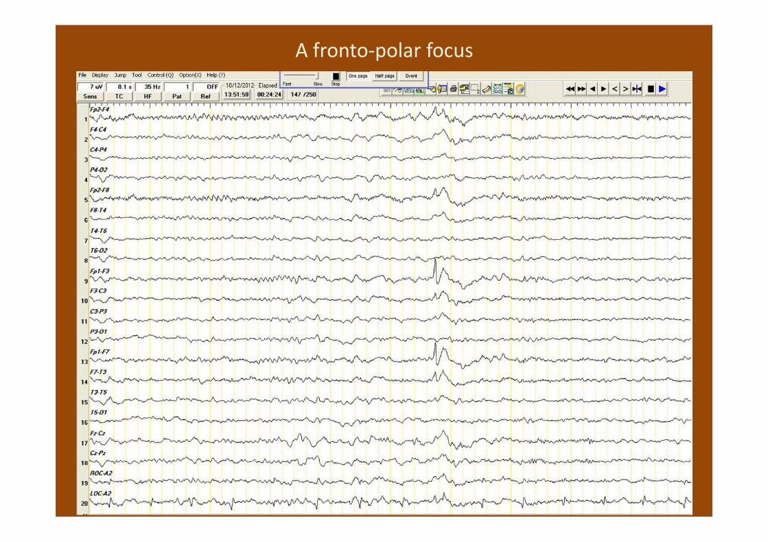
A fronto-polar focus
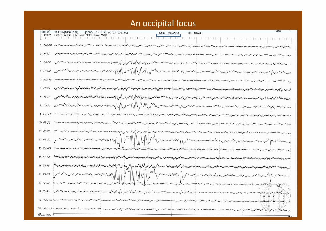
An occipital focus

Artefacts and the EEG

The MAULSBY Guidelines for Assessing Spikes (1971)
Be cautious and conservative:
1. Every spike-like wave is a normal variant or
artefact unless there are good reasons to suspect
otherwise.
2. Spikes of cerebral origin always occupy a
definable electrical field - appear in two or more
nearby electrode sites.

Artefacts• Perspective
Southern Florida surveys of incorrectly analyzed EEGs – artefacts - relatively innocent
• Problem
Do sometimes get classified as an epileptiform discharge
When excessive – slows down analysis – causes doubts - becomes a pain
• Procedure of dealing with artefacts:
Technologist/EEG technician is trained to identify, then to eliminate/reduce where possible and even to record with non-cerebral electrodes when appropriate
The analyzer has to learn to recognize them

Annotations by the technologist

Essential artefacts
1) Eye opening and closure
2) Eye movements – blinks
3) Eye movements – flutter
4) Muscle/body movements
5) Tremor
6) ECG
7) Environmental electrical noise
8) Electrode artefacts

Eye opening and closure (and saccadic eye movements)

Eye movements – blinks. Large downward deflections (V-shaped) in
fronto-polar derivations.
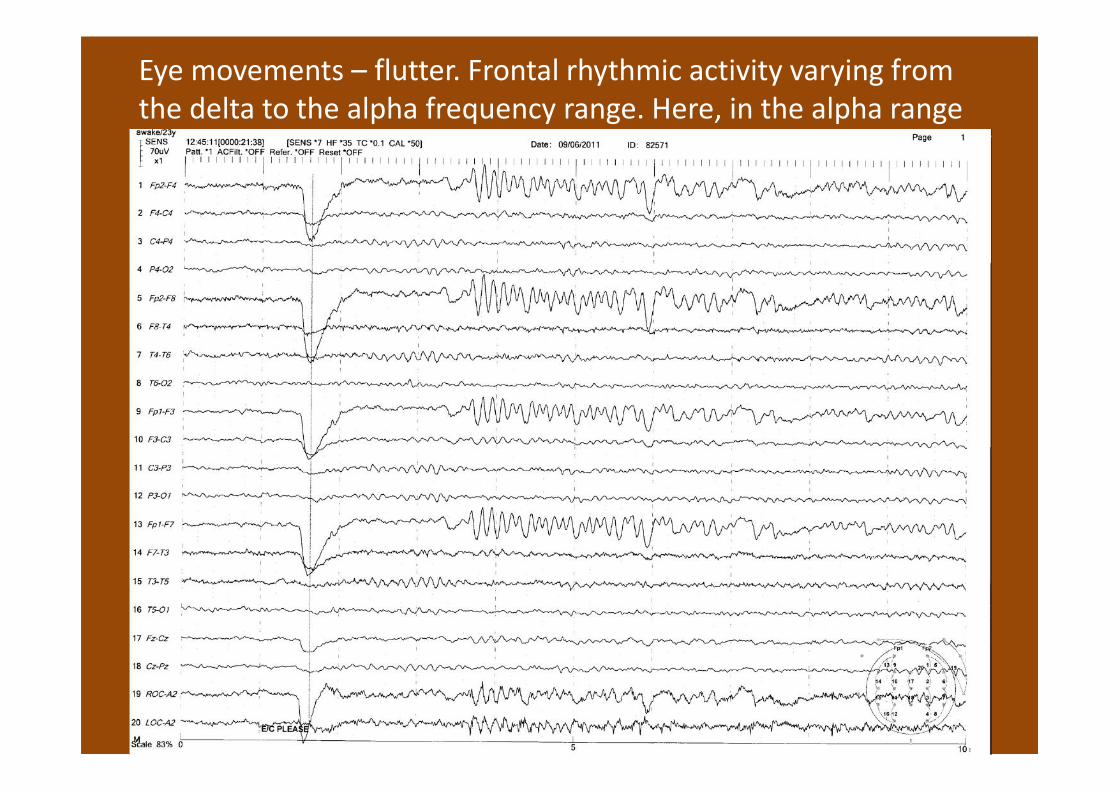
Eye movements – flutter. Frontal rhythmic activity varying from
the delta to the alpha frequency range. Here, in the alpha range

Eye movements – flutter in the delta frequency range.

Eye movements – lateral rectus spikes. Muscle spikes in fronto-
polar derivations. Can appear as spike and slow wave complexes.

Eye movements – several types.

EMG spikes.

Muscle/body movements on a major scale

Slight, subtle movements
sob sob sob

Monitoring muscle/movement

Displaying the EEGDisplaying the EEG

High frequency filter settings
The threat of diminishing epileptiform spikes

Muscle
artefacts
misinterpreted
as epileptiform
events.
(Hernandez-
Frau and
Benbadis 2011)

Tremor. Rhythmic activity usually in the delta or
theta frequency ranges.
A tremor artefact inducing head movements and causing a
rhythmical artefact maximal in posterior temporal derivations.

ECG. Commonly involves recording of the QRS
complex in the form of spikes.
These periodic spikes in the EEG can be attributed to contamination by the ECG on the
grounds of being time-locked to the R wave.

Pulse artefact – channel 12

Environmental electrical noise. Appears in EEG
recordings as 50 Hz rhythmical activity.
Reduction of 50 Hz mains interference by use of a 50 Hz AC filter in an EEG
recorded in ICU. Residual high frequency contamination is probably mostly
muscle artefact.

Electrode artefacts. Appear in various forms including
focal spikes or sharp waves, with or without slow
components.

Electrode artefact. Which electrode is poorly applied?

High impedance and electrode artefact.


