The Plant Cell, Vol. 8, 293-303, February 1996 O 1996 American Society of Plant Physiologists Localization of a Rho GTPase lmplies a Role in Tip Growth and Movement of the Generative Cell in Pollen Tubes Yakang Lin,a Yalai Wang,a Jian-kang Zhu,b and Zhenbiao Yangay’ a Department of Plant Biology, and Plant Molecular and Biotechnology Program, Ohio State University, Columbus, Ohio 43210 Department of Plant Sciences, University of Arizona, Tucson, Arizona 85721 The Rho family GTPases function as key molecular switches, controlling a variety of actin-dependent cellular processes, such as the establishment of cell polarity, cell morphogenesis, and movement in diverse eukaryotic organisms. A nove1 subfamily of Rho GTPases, Rop, has been identified in plants. Protein gel blot and RNA gel blot hybridization analyses indicated that one of these plant Rho GTPases, Ropl, is expressed predominantly in the male gametophyte (pollen and pollen tubes). Cell fractionation analysis of pollen tubes showed that Rop is partitioned into soluble and particulate frac- tions. The particulate Rop could be solubilized with detergents but not with salts, indicating that it is tightly bound to membranes. The membrane association appears to result from membrane anchoring via a geranylgeranyl group because an in vitro isoprenylation assay demonstrated that RoplPs is geranylgeranylated. Subcellular localization, using indirect immunofluorescence and confocal microscopy, showed that Rop is highly concentrated in the cortical region of the tube apex and in the periphery of the generative cell. The cortical Rop protein at the apex forms a gradient with decreasing concentration from tip to base and appears to be associated with the plasma membrane. These results suggest that the apical Rop GTPase may be involved in the signaling mechanism that controls the actin-dependent tip growth of pollen tubes. Localization of the Rop GTPase to the periphery of the generative cell is analogous to that of myosin, suggesting that the Rop GTPase plays an important role in the modulation of an actomyosin motor system involved in the movement of the generative cell. INTRODUCTION The Rho family of small GTPases (GTP binding proteins) plays a central role in the modulation of many cellular processes related to the actin cytoskeleton (reviewed in Chant, 1994; Hall, 1994; Chant and Stowers, 1995). Many Rho and related proteins have been identified in various eukaryotes and categorized into three major subfamilies: CDC42, Rac, and Rho. Their categorization is based on their cellular functions and se- quence homology (Chant, 1994; Hall, 1994; Chant and Stowers, 1995). In motile mammalian cells, each subfamily has a dis- tinct function, yet they constitute a hierarchical cascade of molecular switches for the control of actin-mediated cell mor- phogenesis, movement, and adhesion (Ridley and Hall, 1992; Ridley et al., 1992; Hall, 1994; Nobes and Hall, 1995). Within this cascade, CDC42 controls Rac, which in turn controls Rho. Similar Rho GTPase cascades also appear to be involved in cell morphogenesis in other diverse systems. In the yeast Saccharomyces cerevisiae, CDC42 controls the establishment of cell polarity associated with the asymmetrical distribution of cortical actin filaments at the site of bud emergence (Adams et al., 1990; Johnson and Pringle, 1990). CDC42 also appears ’ To whom correspondence should be addressed to regulate the activity of a series of Rho-related proteins (Rhol, Rho2, Rho3, and Rho4) that are required for subsequent bud growth (Matsui and Toh-e, 1992; Yamochi et al., 1994). Consistent with their functions, both CDC42 and Rhol are localized to the site of bud emergence and growth (Ziman et al., 1993; Yamochi et al., 1994). In Schizosaccharomyces pombe, CDC42 controls actin-mediatedasymmetricalgrowth (Miller and Johnson, 1994). CDC42 is also involved in the modulation of neuronal cell differentiation and cell coupling in the immune system (Lu0 et al., 1994; Stowers et al., 1995). Animal CDC42 is able to complement yeast cdc42 temperature- sensitive mutations, indicating remarkable functional conser- vation (Shinjo et al., 1990; Chen et al., 1993). The establishmentand maintenance of cell polarity are fun- damental cellular attributes to differentiation and development in plants. Polarized growth is exhibited in many plant cell types, such as zygotes, specialized epidermal cells (e.g., root hairs, trichomes, and guard cells), and pollen tubes. Although the actin cytoskeleton has been implicated in the polarized growth of plant cells, molecular mechanisms controlling the organi- zation of F-actin and growth polarity remain largely obscure. As a first step toward understanding the possible role of Rho in controlling these cellular behaviors in plants, we have cloned
Transcript
The Plant Cell, Vol. 8, 293-303, February 1996 O 1996 American Society of Plant Physiologists
Localization of a Rho GTPase lmplies a Role in Tip Growth and Movement of the Generative Cell in Pollen Tubes
Yakang Lin,a Yalai Wang,a Jian-kang Zhu,b and Zhenbiao Yangay’
a Department of Plant Biology, and Plant Molecular and Biotechnology Program, Ohio State University, Columbus, Ohio 43210
Department of Plant Sciences, University of Arizona, Tucson, Arizona 85721
The Rho family GTPases function as key molecular switches, controlling a variety of actin-dependent cellular processes, such as the establishment of cell polarity, cell morphogenesis, and movement in diverse eukaryotic organisms. A nove1 subfamily of Rho GTPases, Rop, has been identified in plants. Protein gel blot and RNA gel blot hybridization analyses indicated that one of these plant Rho GTPases, Ropl, is expressed predominantly in the male gametophyte (pollen and pollen tubes). Cell fractionation analysis of pollen tubes showed that Rop is partitioned into soluble and particulate frac- tions. The particulate Rop could be solubilized with detergents but not with salts, indicating that it is tightly bound to membranes. The membrane association appears to result from membrane anchoring via a geranylgeranyl group because an in vitro isoprenylation assay demonstrated that RoplPs is geranylgeranylated. Subcellular localization, using indirect immunofluorescence and confocal microscopy, showed that Rop is highly concentrated in the cortical region of the tube apex and in the periphery of the generative cell. The cortical Rop protein at the apex forms a gradient with decreasing concentration from tip to base and appears to be associated with the plasma membrane. These results suggest that the apical Rop GTPase may be involved in the signaling mechanism that controls the actin-dependent tip growth of pollen tubes. Localization of the Rop GTPase to the periphery of the generative cell is analogous to that of myosin, suggesting that the Rop GTPase plays an important role in the modulation of an actomyosin motor system involved in the movement of the generative cell.
INTRODUCTION
The Rho family of small GTPases (GTP binding proteins) plays a central role in the modulation of many cellular processes related to the actin cytoskeleton (reviewed in Chant, 1994; Hall, 1994; Chant and Stowers, 1995). Many Rho and related proteins have been identified in various eukaryotes and categorized into three major subfamilies: CDC42, Rac, and Rho. Their categorization is based on their cellular functions and se- quence homology (Chant, 1994; Hall, 1994; Chant and Stowers, 1995). In motile mammalian cells, each subfamily has a dis- tinct function, yet they constitute a hierarchical cascade of molecular switches for the control of actin-mediated cell mor- phogenesis, movement, and adhesion (Ridley and Hall, 1992; Ridley et al., 1992; Hall, 1994; Nobes and Hall, 1995). Within this cascade, CDC42 controls Rac, which in turn controls Rho.
Similar Rho GTPase cascades also appear to be involved in cell morphogenesis in other diverse systems. In the yeast Saccharomyces cerevisiae, CDC42 controls the establishment of cell polarity associated with the asymmetrical distribution of cortical actin filaments at the site of bud emergence (Adams et al., 1990; Johnson and Pringle, 1990). CDC42 also appears
’ To whom correspondence should be addressed
to regulate the activity of a series of Rho-related proteins (Rhol, Rho2, Rho3, and Rho4) that are required for subsequent bud growth (Matsui and Toh-e, 1992; Yamochi et al., 1994). Consistent with their functions, both CDC42 and Rhol are localized to the site of bud emergence and growth (Ziman et al., 1993; Yamochi et al., 1994). In Schizosaccharomyces pombe, CDC42 controls actin-mediated asymmetrical growth (Miller and Johnson, 1994). CDC42 is also involved in the modulation of neuronal cell differentiation and cell coupling in the immune system (Lu0 et al., 1994; Stowers et al., 1995). Animal CDC42 is able to complement yeast cdc42 temperature- sensitive mutations, indicating remarkable functional conser- vation (Shinjo et al., 1990; Chen et al., 1993).
The establishment and maintenance of cell polarity are fun- damental cellular attributes to differentiation and development in plants. Polarized growth is exhibited in many plant cell types, such as zygotes, specialized epidermal cells (e.g., root hairs, trichomes, and guard cells), and pollen tubes. Although the actin cytoskeleton has been implicated in the polarized growth of plant cells, molecular mechanisms controlling the organi- zation of F-actin and growth polarity remain largely obscure. As a first step toward understanding the possible role of Rho in controlling these cellular behaviors in plants, we have cloned
294 The Plant Cell
and characterized genes encoding several Rho-type GTPasesfrom pea and Arabidopsis (Yang and Watson, 1993; H. Li, D.Ware, D. Zhou, K.R. Davis, C.L. Cramer, and Z. Yang, un-published data). These plant Rho proteins belong to a novelsubfamily of Rho GTPases, Rop, which is distinct from thosefound in fungi and animals. In Arabidopsis, the expressionof Rop1 At, the Arabidopsis counterpart of pea Rop1 Ps (Yangand Watson, 1993), is correlated with pollination and pollentube growth (H. Li, D. Ware, D. Zhou, K.R. Davis, C.L. Cramer,and Z. Yang, unpublished data). This provides the first cluethat Rop1 may be involved in the control of pollen and pollentube function.
Pollen and pollen tubes are an attractive model systemfor investigating the mechanism controlling cellular polarity.First, pollen grains must establish a growth polarity before theemergence of the tube because only one tube emerges fromone of the three or four apertures on the grain during germi-nation. Second, growth occurs only at the apex by fusionof vesicles to the apical plasma membrane. This tip growthrequires targeted delivery of vesicles containing membranesand wall-matrix materials to the site of growth. Third, thedirection of pollen tube extension is highly regulated bothin vitro and in vivo. In vitro tube growth can be guided byelectrical potential and chemical gradients (Mascarenhas,1993). In vivo pollen tube growth is guided through the trans-mitting tract of styles and toward the embryo sac (Hulskampet al., 1995). Because the extension of pollen tubes is totallydependent on tip growth, pollen tube guidance must involvedirected fusion of vesicles to the site of growth. In additionto polarized growth, cellular polarity in pollen tubes is alsoreflected by the distinct functional zonation along the lengthof the pollen tube (Cresti and Tiezzi, 1992). Behind an apicalgrowing zone and an organelle-rich subapical zone, there isa wide nuclear zone that contains vegetative nuclei and agenerative cell. As the pollen tube extends, the generativecell, accompanied by the vegetative nucleus, moves down thetube as a male germ unit (Palevitz and Tiezzi, 1992).
There is evidence that an actomyosin system is involved inthe movement of vesicles to the tip and the generative cell(Heslop-Harrison and Heslop-Harrison, 1989; Palevitz andTiezzi, 1992; Pierson and Cresti, 1992; Pierson and Li, 1992).Actin filaments form extensive axial bundles that appear toguide the delivery of vesicles to the apical zone. Actin filamentsalso are associated with both the vegetative nucleus and thegenerative cell. Actin also has been proposed to determinethe polarity of pollen germinal aperture and tip growth (Tiwariand Polito, 1988). A fine actin network has been observed bothclose to the pore from which the tube will emerge and at thetip of the pollen tube (Tiwari and Polito, 1988; Pierson andCresti, 1992; Pierson and Li, 1992). Cytochalasin treatmentleads to inhibition of germination, random vesicle distribution,and inhibition of tip growth (reviewed in Pierson and Cresti,1992). Together, these studies suggest that the actin cytoskele-ton is dynamic during pollen activation and tube growth andplays an important role in the function of the male gameto-phyte. However, the mechanism controlling the dynamic
organization of the actin cytoskeleton in pollen tubes isunknown.
In this study, we report that a unique member of the con-served Rho GTPase family is expressed predominantly in peapollen and pollen tubes. Furthermore, our results indicate thatthe pea Rho GTPase is localized preferentially to what appearto be specific regions of the plasma membrane, that is, theapex of pollen tubes and the periphery of the generative cell.These results support the hypothesis that one or more RhoGTPases modulate both the tip growth of pollen tubes and themovement of the generative cell.
RESULTS
Preferential Accumulation of the Rop Protein andmRNA in the Male Gametophyte
We have shown that an Arabidopsis homolog of RoplPs,FtoplAt, is expressed specifically in fully developed flowersand that its mRNA accumulates to a maximum level duringpollination and fertilization, suggesting that Rop1 plays afundamental role in pollen and pollen tube function (H. Li, D.Ware, D. Zhou, K.R. Davis, C.L. Cramer, and Z. Yang, unpub-lished results). As a first step in testing this possibility, theaccumulation of Rop1 Ps protein in different parts of pea flowers
I
I
— sepal
— petal
— filament
— anther
— stigma/style, unpollinated
— stigma/style, pollinated
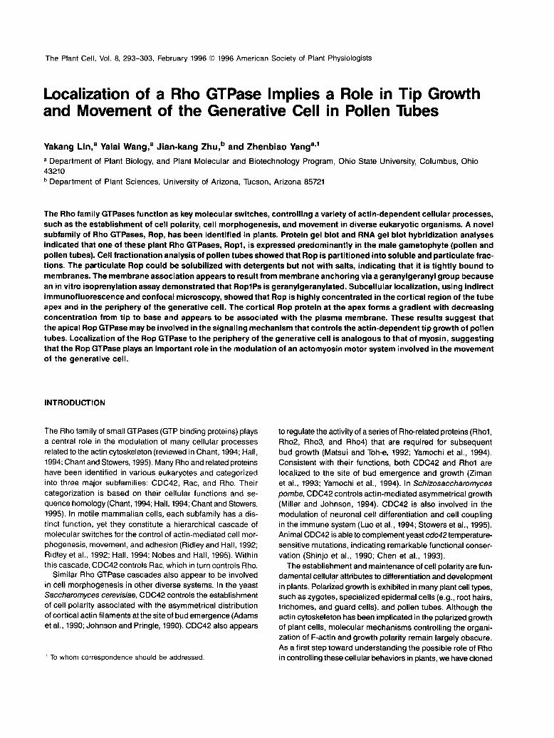
Figure 1. Protein Gel Blot Analysis of Rop Proteins in Different Partsof Pea Flowers.
Total proteins were extracted from different parts of flowers as indi-cated. Equivalent amounts of proteins were separated on an SDS-polyacrylamide gel, transferred to a nitrocellulose membrane, reactedwith the affinity-purified anti-Rop1Ps antibody, and detected with a che-miluminescence kit as described in Methods.
Localization of a Rho GTPase in Pollen Tubes 295
was estimated by protein gel blot analysis, using a polyclonalantibody raised against RoplPs. As shown in Figure 1, a spe-cific band of ~21 kD, corresponding to the estimated molecularmass of RoplPs, was detected. The intensity of this band wasstrongest in anthers and pollinated stigma and styles. A weaksignal was detected in filaments and petals only after extendedexposure. However, no Pop protein was found in unpollinatedstigma and styles. It should be noted that the anti-Rop1Ps poly-clonal antibody is likely to react with other members of the Ropsubfamily in pea, because all plant Rop members that havebeen characterized to date share >80°/o sequence identity (H.Li, D. Ware, D. Zhou, K.R. Davis, C.L. Cramer, and Z. Yang,unpublished data). Hence, these experiments did not addressspecifically whether any of the proteins detected by the anti-body raised against RoplPs are specifically encoded byRoplPs.
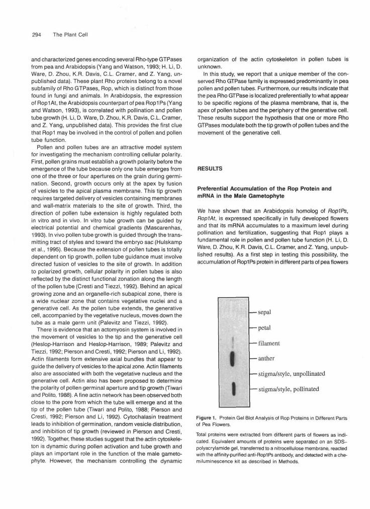
To study further the relationship of the Rop protein foundin different parts of the flower to RoplPs gene expression,RoplPs transcript levels from different vegetative and reproduc-tive pea organs were determined by RNA gel blot hybridizationanalysis. As shown in Figure 2, low levels of RoplPs mRNAwere also detected in floral buds, vegetative buds, and roottips, but the RoplPs transcript was most abundant in pollenamong various parts of pea plants, indicating that Rop pro-teins detected in the male gametophyte are indeed encodedprimarily if not specifically by RoplPs. This is consistent withthe expression pattern of RoplAt during floral developmentin Arabidopsis (H. Li, D. Ware, D. Zhou, K.R. Davis, C.L. Cramer,and Z. Yang, unpublished data).
Subcellular Localization of Rop Proteins inPollen Tubes
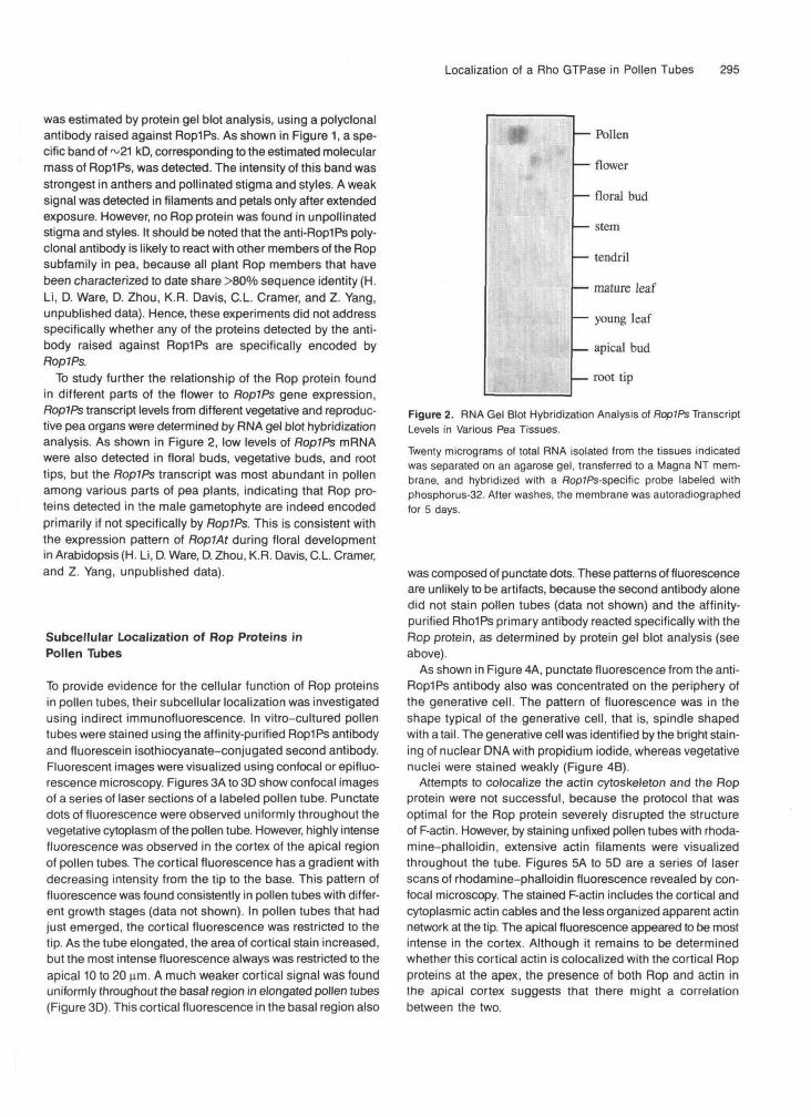
To provide evidence for the cellular function of Rop proteinsin pollen tubes, their subcellular localization was investigatedusing indirect immunofluorescence. In vitro-cultured pollentubes were stained using the affinity-purified RoplPs antibodyand fluorescein isothiocyanate-conjugated second antibody.Fluorescent images were visualized using confocal or epifluo-rescence microscopy. Figures 3A to 3D show confocal imagesof a series of laser sections of a labeled pollen tube. Punctatedots of fluorescence were observed uniformly throughout thevegetative cytoplasm of the pollen tube. However, highly intensefluorescence was observed in the cortex of the apical regionof pollen tubes. The cortical fluorescence has a gradient withdecreasing intensity from the tip to the base. This pattern offluorescence was found consistently in pollen tubes with differ-ent growth stages (data not shown). In pollen tubes that hadjust emerged, the cortical fluorescence was restricted to thetip. As the tube elongated, the area of cortical stain increased,but the most intense fluorescence always was restricted to theapical 10 to 20 urn. A much weaker cortical signal was founduniformly throughout the basal region in elongated pollen tubes(Figure 3D). This cortical fluorescence in the basal region also
*f — Pollen
— flower
— floral bud
— stem
— tendril
— mature leaf
— young leaf
— apical bud
— root tip
Figure 2. RNA Gel Blot Hybridization Analysis of RoplPs TranscriptLevels in Various Pea Tissues.
Twenty micrograms of total RNA isolated from the tissues indicatedwas separated on an agarose gel, transferred to a Magna NT mem-brane, and hybridized with a RopJPs-specific probe labeled withphosphorus-32. After washes, the membrane was autoradiographedfor 5 days.
was composed of punctate dots. These patterns of fluorescenceare unlikely to be artifacts, because the second antibody alonedid not stain pollen tubes (data not shown) and the affinity-purified RholPs primary antibody reacted specifically with theRop protein, as determined by protein gel blot analysis (seeabove).
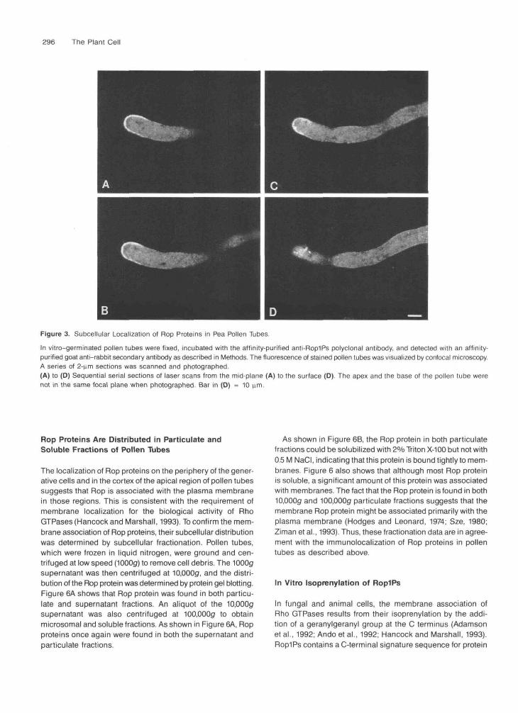
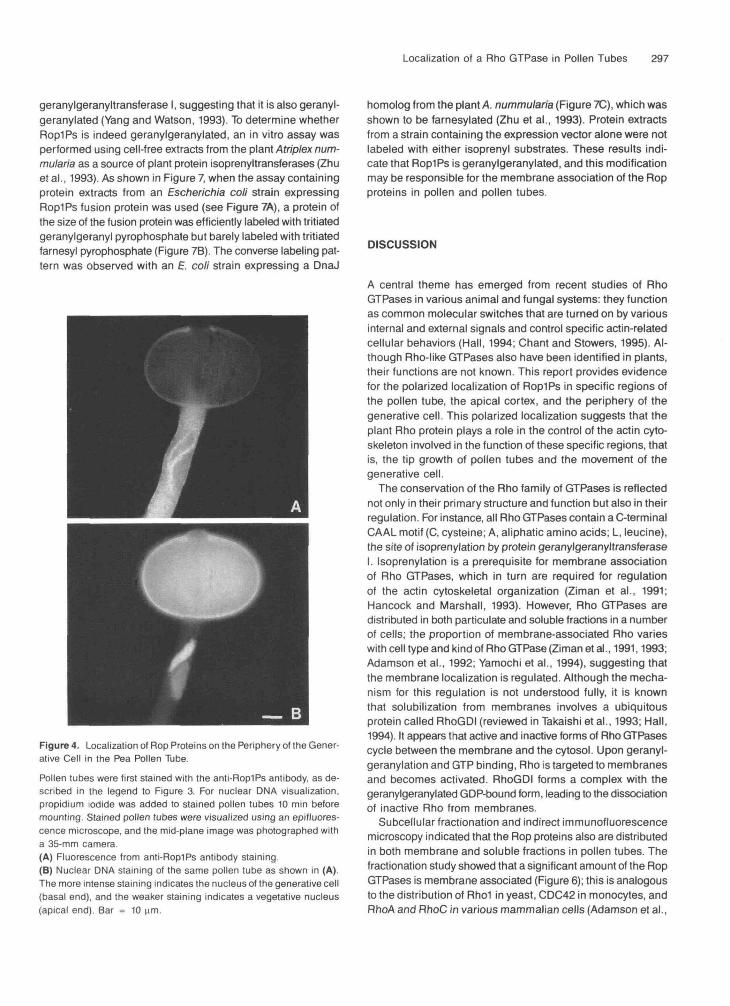
As shown in Figure 4A, punctate fluorescence from the anti-RoplPs antibody also was concentrated on the periphery ofthe generative cell. The pattern of fluorescence was in theshape typical of the generative cell, that is, spindle shapedwith a tail. The generative cell was identified by the bright stain-ing of nuclear DMA with propidium iodide, whereas vegetativenuclei were stained weakly (Figure 4B).
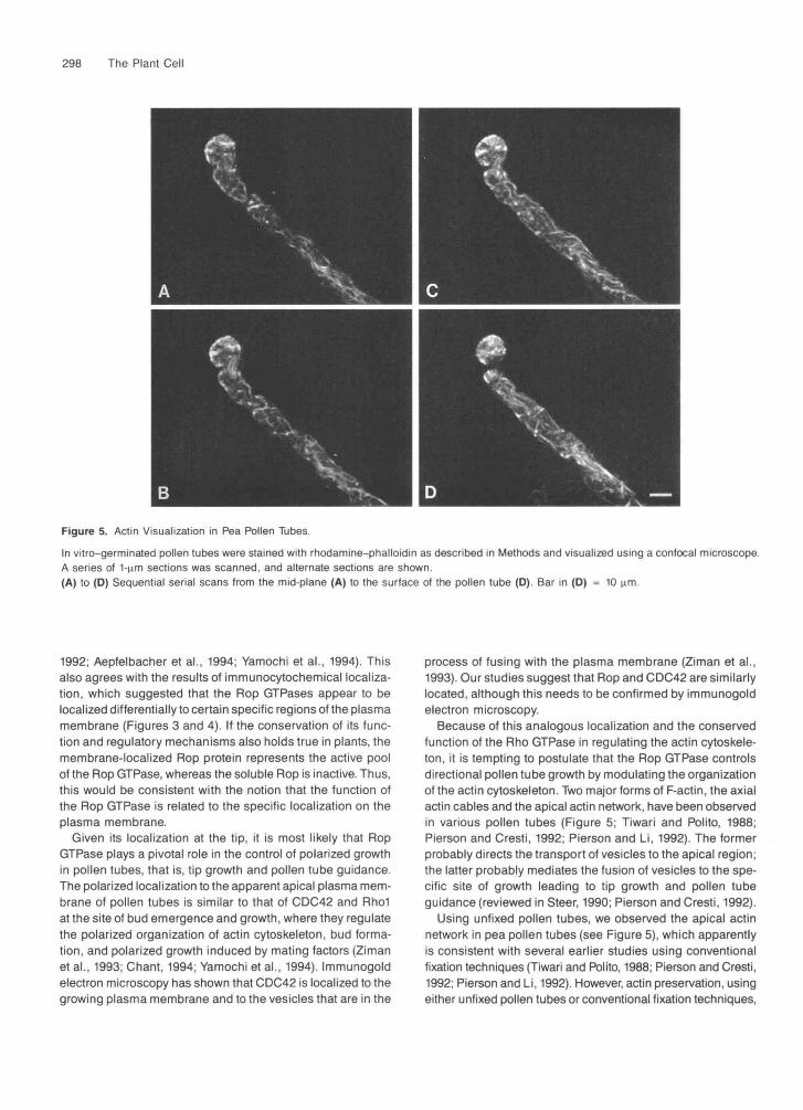
Attempts to colocalize the actin cytoskeleton and the Ropprotein were not successful, because the protocol that wasoptimal for the Rop protein severely disrupted the structureof F-actin. However, by staining unfixed pollen tubes with rhoda-mine-phalloidin, extensive actin filaments were visualizedthroughout the tube. Figures 5A to 5D are a series of laserscans of rhodamine-phalloidin fluorescence revealed by con-focal microscopy. The stained F-actin includes the cortical andcytoplasmic actin cables and the less organized apparent actinnetwork at the tip. The apical fluorescence appeared to be mostintense in the cortex. Although it remains to be determinedwhether this cortical actin is colocalized with the cortical Ropproteins at the apex, the presence of both Rop and actin inthe apical cortex suggests that there might a correlationbetween the two.
296 The Plant Cell
Figure 3. Subcellular Localization of Rop Proteins in Pea Pollen Tubes.
In vitro-germinated pollen tubes were fixed, incubated with the affinity-purified anti-Rop1Ps polyclonal antibody, and detected with an affinity-purified goat anti-rabbit secondary antibody as described in Methods. The fluorescence of stained pollen tubes was visualized by confocal microscopy.A series of 2-nm sections was scanned and photographed.(A) to (D) Sequential serial sections of laser scans from the mid-plane (A) to the surface (D) The apex and the base of the pollen tube werenot in the same focal plane when photographed. Bar in (D) = 10 urn.
Rop Proteins Are Distributed in Particulate andSoluble Fractions of Pollen Tubes
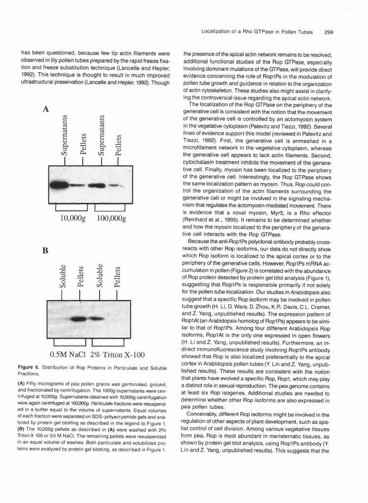
The localization of Rop proteins on the periphery of the gener-ative cells and in the cortex of the apical region of pollen tubessuggests that Rop is associated with the plasma membranein those regions. This is consistent with the requirement ofmembrane localization for the biological activity of RhoGTPases (Hancock and Marshall, 1993). To confirm the mem-brane association of Rop proteins, their subcellular distributionwas determined by subcellular fractionation. Pollen tubes,which were frozen in liquid nitrogen, were ground and cen-trifuged at low speed (1000g) to remove cell debris. The 1000gsupernatant was then centrifuged at 10,000g, and the distri-bution of the Rop protein was determined by protein gel blotting.Figure 6A shows that Rop protein was found in both particu-late and supernatant fractions. An aliquot of the 10,000gsupernatant was also centrifuged at 100,000g to obtainmicrosomal and soluble fractions. As shown in Figure 6A, Ropproteins once again were found in both the supernatant andparticulate fractions.
As shown in Figure 6B, the Rop protein in both particulatefractions could be solubilized with 2% Triton X-100 but not with0.5 M NaCI, indicating that this protein is bound tightly to mem-branes. Figure 6 also shows that although most Rop proteinis soluble, a significant amount of this protein was associatedwith membranes. The fact that the Rop protein is found in both10,000g and 100,000g particulate fractions suggests that themembrane Rop protein might be associated primarily with theplasma membrane (Hodges and Leonard, 1974; Sze, 1980;Ziman et al., 1993). Thus, these fractionation data are in agree-ment with the immunolocalization of Rop proteins in pollentubes as described above.
In Vitro Isoprenylation of RoplPs
In fungal and animal cells, the membrane association ofRho GTPases results from their isoprenylation by the addi-tion of a geranylgeranyl group at the C terminus (Adamsonet al., 1992; Ando et al., 1992; Hancock and Marshall, 1993).RoplPs contains a C-terminal signature sequence for protein
Localization of a Rho GTPase in Pollen Tubes 297
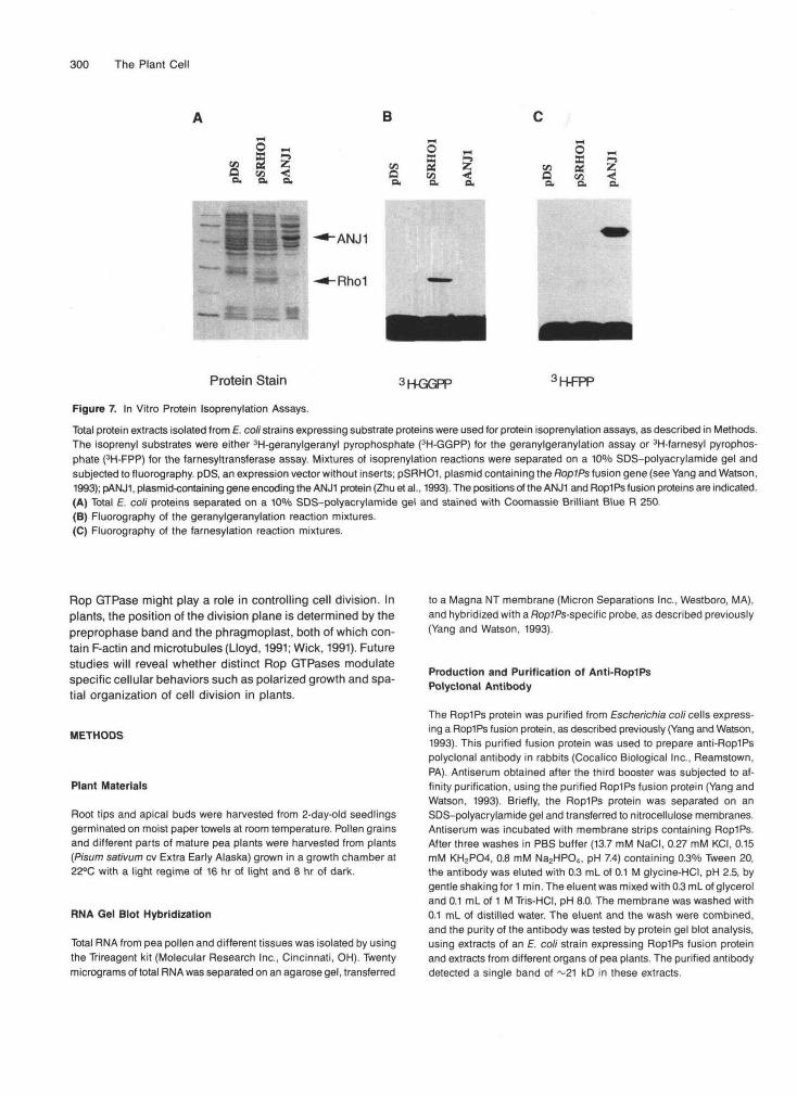
geranylgeranyltransferase I, suggesting that it is also geranyl-geranylated (Yang and Watson, 1993). To determine whetherRoplPs is indeed geranylgeranylated, an in vitro assay wasperformed using cell-free extracts from the plant Atriplex num-mularia as a source of plant protein isoprenyltransferases (Zhuet al., 1993). As shown in Figure 7, when the assay containingprotein extracts from an Escherichia coli strain expressingRoplPs fusion protein was used (see Figure 7A), a protein ofthe size of the fusion protein was efficiently labeled with tritiatedgeranylgeranyl pyrophosphate but barely labeled with tritiatedfarnesyl pyrophosphate (Figure 7B). The converse labeling pat-tern was observed with an £ coli strain expressing a DnaJ
Figure 4. Localization of Rop Proteins on the Periphery of the Gener-ative Cell in the Pea Pollen Tube.Pollen tubes were first stained with the anti-Rop1Ps antibody, as de-scribed in the legend to Figure 3. For nuclear DMA visualization,propidium iodide was added to stained pollen tubes 10 min beforemounting. Stained pollen tubes were visualized using an epifluores-cence microscope, and the mid-plane image was photographed witha 35-mm camera(A) Fluorescence from anti-Rop1Ps antibody staining.(B) Nuclear DNA staining of the same pollen tube as shown in (A).The more intense staining indicates the nucleus of the generative cell(basal end), and the weaker staining indicates a vegetative nucleus(apical end). Bar = 10 |im.
homolog from the plant A. nummularia (Figure 7C), which wasshown to be farnesylated (Zhu et al., 1993). Protein extractsfrom a strain containing the expression vector alone were notlabeled with either isoprenyl substrates. These results indi-cate that RoplPs is geranylgeranylated, and this modificationmay be responsible for the membrane association of the Ropproteins in pollen and pollen tubes.
DISCUSSION
A central theme has emerged from recent studies of RhoGTPases in various animal and fungal systems: they functionas common molecular switches that are turned on by variousinternal and external signals and control specific actin-relatedcellular behaviors (Hall, 1994; Chant and Stowers, 1995). Al-though Rho-like GTPases also have been identified in plants,their functions are not known. This report provides evidencefor the polarized localization of RoplPs in specific regions ofthe pollen tube, the apical cortex, and the periphery of thegenerative cell. This polarized localization suggests that theplant Rho protein plays a role in the control of the actin cyto-skeleton involved in the function of these specific regions, thatis, the tip growth of pollen tubes and the movement of thegenerative cell.
The conservation of the Rho family of GTPases is reflectednot only in their primary structure and function but also in theirregulation. For instance, all Rho GTPases contain a C-terminalCAAL motif (C, cysteine; A, aliphatic amino acids; L, leucine),the site of isoprenylation by protein geranylgeranyltransferaseI. Isoprenylation is a prerequisite for membrane associationof Rho GTPases, which in turn are required for regulationof the actin cytoskeletal organization (Ziman et al., 1991;Hancock and Marshall, 1993). However, Rho GTPases aredistributed in both paniculate and soluble fractions in a numberof cells; the proportion of membrane-associated Rho varieswith cell type and kind of Rho GTPase (Ziman et al., 1991,1993;Adamson et al., 1992; Yamochi et al., 1994), suggesting thatthe membrane localization is regulated. Although the mecha-nism for this regulation is not understood fully, it is knownthat solubilization from membranes involves a ubiquitousprotein called RhoGDI (reviewed in Takaishi et al., 1993; Hall,1994). It appears that active and inactive forms of Rho GTPasescycle between the membrane and the cytosol. Upon geranyl-geranylation and GTP binding, Rho is targeted to membranesand becomes activated. RhoGDI forms a complex with thegeranylgeranylated GDP-bound form, leading to the dissociationof inactive Rho from membranes.
Subcellular fractionation and indirect immunofluorescencemicroscopy indicated that the Rop proteins also are distributedin both membrane and soluble fractions in pollen tubes. Thefractionation study showed that a significant amount of the RopGTPases is membrane associated (Figure 6); this is analogousto the distribution of Rho1 in yeast, CDC42 in monocytes, andRhoA and RhoC in various mammalian cells (Adamson et al.,
298 The Plant Cell
Figure 5. Actin Visualization in Pea Pollen Tubes.
In vitro-germinated pollen tubes were stained with rhodamine-phalloidin as described in Methods and visualized using a confocal microscope.A series of 1-nm sections was scanned, and alternate sections are shown(A) to (D) Sequential serial scans from the mid-plane (A) to the surface of the pollen tube (D). Bar in (D) = 10 urn.
1992; Aepfelbacher et al., 1994; Yamochi et al., 1994). Thisalso agrees with the results of immunocytochemical localiza-tion, which suggested that the Pop GTPases appear to belocalized differentially to certain specific regions of the plasmamembrane (Figures 3 and 4). If the conservation of its func-tion and regulatory mechanisms also holds true in plants, themembrane-localized Rop protein represents the active poolof the Rop GTPase, whereas the soluble Rop is inactive. Thus,this would be consistent with the notion that the function ofthe Rop GTPase is related to the specific localization on theplasma membrane.
Given its localization at the tip, it is most likely that RopGTPase plays a pivotal role in the control of polarized growthin pollen tubes, that is, tip growth and pollen tube guidance.The polarized localization to the apparent apical plasma mem-brane of pollen tubes is similar to that of CDC42 and Rho1at the site of bud emergence and growth, where they regulatethe polarized organization of actin cytoskeleton, bud forma-tion, and polarized growth induced by mating factors (Zimanet al., 1993; Chant, 1994; Yamochi et al., 1994). Immunogoldelectron microscopy has shown that CDC42 is localized to thegrowing plasma membrane and to the vesicles that are in the
process of fusing with the plasma membrane (Ziman et al.,1993). Our studies suggest that Rop and CDC42 are similarlylocated, although this needs to be confirmed by immunogoldelectron microscopy.
Because of this analogous localization and the conservedfunction of the Rho GTPase in regulating the actin cytoskele-ton, it is tempting to postulate that the Rop GTPase controlsdirectional pollen tube growth by modulating the organizationof the actin cytoskeleton. Two major forms of F-actin, the axialactin cables and the apical actin network, have been observedin various pollen tubes (Figure 5; Tiwari and Polito, 1988;Pierson and Cresti, 1992; Pierson and Li, 1992). The formerprobably directs the transport of vesicles to the apical region;the latter probably mediates the fusion of vesicles to the spe-cific site of growth leading to tip growth and pollen tubeguidance (reviewed in Steer, 1990; Pierson and Cresti, 1992).
Using unfixed pollen tubes, we observed the apical actinnetwork in pea pollen tubes (see Figure 5), which apparentlyis consistent with several earlier studies using conventionalfixation techniques (Tiwari and Polito, 1988; Pierson and Cresti,1992; Pierson and Li, 1992). However, actin preservation, usingeither unfixed pollen tubes or conventional fixation techniques,
Localization of a Rho GTPase in Pollen Tubes 299
has been questioned, because few tip actin filaments wereobserved in lily pollen tubes prepared by the rapid freeze fixa-tion and freeze substitution technique (Lancelle and Hepler,1992). This technique is thought to result in much improvedultrastructural preservation (Lancelle and Hepler, 1992). Though
*2 S «£ a JaT! o. rr)JJ 3 OJC1- C/3 (X
JT10,000g 100,000g
B<u Cfl«_*
<D
o "o00 O-i
IO.SMNaCl 2% Triton X-100
Figure 6. Distribution of Rop Proteins in Paniculate and SolubleFractions.
(A) Fifty micrograms of pea pollen grains was germinated, ground,and fractionated by centrifugation. The 1000g supernatants were cen-trifuged at 10,000g. Supernatants obtained with 10,000g centrifugationwere again centrifuged at 100,000g. Paniculate fractions were resuspend-ed in a buffer equal to the volume of supernatants. Equal volumesof each fraction were separated on SDS-polyacryamide gels and ana-lyzed by protein gel blotting as described in the legend to Figure 1.(B) The 10,000g pellets as described in (A) were washed with 2%Triton-X 100 or 0.5 M NaCI. The remaining pellets were resuspendedin an equal volume of washes. Both paniculate and solubilized pro-teins were analyzed by protein gel blotting, as described in Figure 1.
the presence of the apical actin network remains to be resolved,additional functional studies of the Rop GTPase, especiallyinvolving dominant mutations of the GTPase, will provide directevidence concerning the role of RoplPs in the modulation ofpollen tube growth and guidance in relation to the organizationof actin cytoskeleton. These studies also might assist in clarify-ing the controversial issue regarding the apical actin network.
The localization of the Rop GTPase on the periphery of thegenerative cell is consistent with the notion that the movementof the generative cell is controlled by an actomyosin systemin the vegetative cytoplasm (Palevitz and Tiezzi, 1992). Severallines of evidence support this model (reviewed in Palevitz andTiezzi, 1992). First, the generative cell is enmeshed in amicrofilament network in the vegetative cytoplasm, whereasthe generative cell appears to lack actin filaments. Second,cytochalasin treatment inhibits the movement of the genera-tive cell. Finally, myosin has been localized to the peripheryof the generative cell. Interestingly, the Rop GTPase showsthe same localization pattern as myosin. Thus, Rop could con-trol the organization of the actin filaments surrounding thegenerative cell or might be involved in the signaling mecha-nism that regulates the actomyosin-mediated movement. Thereis evidence that a novel myosin, Myr5, is a Rho effector(Reinhard et al., 1995). It remains to be determined whetherand how the myosin localized to the periphery of the genera-tive cell interacts with the Rop GTPase.
Because the anti-Rop1Ps polyclonal antibody probably cross-reacts with other Rop isoforms, our data do not directly showwhich Rop isoform is localized to the apical cortex or to theperiphery of the generative cells. However, RoplPs mRNA ac-cumulation in pollen (Figure 2) is correlated with the abundanceof Rop protein detected by protein gel blot analysis (Figure 1),suggesting that RoplPs is responsible primarily if not solelyfor the pollen tube localization. Our studies in Arabidopsis alsosuggest that a specific Rop isoform may be involved in pollentube growth (H. Li, D. Ware, D. Zhou, K.R. Davis, C.L. Cramer,and 2. Yang, unpublished results). The expression pattern ofRoplAt (an Arabidopsis homolog of RoplPs) appears to be simi-lar to that of RoplPs. Among four different Arabidopsis Ropisoforms, RoplAt is the only one expressed in open flowers(H. Li and Z. Yang, unpublished results). Furthermore, an in-direct immunofluorescence study involving RoplPs antibodyshowed that Rop is also localized preferentially to the apicalcortex in Arabidopsis pollen tubes (Y. Lin and Z. Yang, unpub-lished results). These results are consistent with the notionthat plants have evolved a specific Rop, Rop1, which may playa distinct role in sexual reproduction. The pea genome containsat least six flop isogenes. Additional studies are needed todetermine whether other Rop isoforms are also expressed inpea pollen tubes.
Conceivably, different Rop isoforms might be involved in theregulation of other aspects of plant development, such as spa-tial control of cell division. Among various vegetative tissuesfrom pea, Rop is most abundant in meristematic tissues, asshown by protein gel blot analysis, using RoplPs antibody (YLin and Z. Yang, unpublished results). This suggests that the
300 The Plant Cell
B
fi en •<a, o. o.
O
•a 1 05•a
-ANJ1
-Rho1
Protein Stain 3H-GGPP 3H-FPP
Figure 7. In Vitro Protein Isoprenylation Assays.
Total protein extracts isolated from E coli strains expressing substrate proteins were used for protein isoprenylation assays, as described in Methods.The isoprenyl substrates were either 3H-geranylgeranyl pyrophosphate (3H-GGPP) for the geranylgeranylation assay or 3H-farnesyl pyrophos-phate (3H-FPP) for the farnesyltransferase assay. Mixtures of isoprenylation reactions were separated on a 10% SDS-polyacrylamide gel andsubjected to fluorography. pDS, an expression vector without inserts; pSRHOI, plasmid containing the RoplPs fusion gene (see Yang and Watson,1993); pANJ1, plasmid-containing gene encoding the ANJ1 protein (Zhu et al., 1993). The positions of the ANJ1 and RoplPs fusion proteins are indicated.(A) Total E coli proteins separated on a 10% SDS-polyacrylamide gel and stained with Coomassie Brilliant Blue R 250.(B) Fluorography of the geranylgeranylation reaction mixtures.(C) Fluorography of the farnesylation reaction mixtures.
Rop GTPase might play a role in controlling cell division. Inplants, the position of the division plane is determined by thepreprophase band and the phragmoplast, both of which con-tain F-actin and microtubules (Lloyd, 1991; Wick, 1991). Futurestudies will reveal whether distinct Rop GTPases modulatespecific cellular behaviors such as polarized growth and spa-tial organization of cell division in plants.
METHODS
Plant Materials
Root tips and apical buds were harvested from 2-day-old seedlingsgerminated on moist paper towels at room temperature. Pollen grainsand different parts of mature pea plants were harvested from plants(Pisum sativum cv Extra Early Alaska) grown in a growth chamber at22°C with a light regime of 16 hr of light and 8 hr of dark.
RNA Gel Blot Hybridization
Total RNA from pea pollen and different tissues was isolated by usingthe Trireagent kit (Molecular Research Inc., Cincinnati, OH). Twentymicrograms of total RNA was separated on an agarose gel, transferred
to a Magna NT membrane (Micron Separations Inc., Westboro, MA),and hybridized with a flop7Ps-specific probe, as described previously(Yang and Watson, 1993).
Production and Purification of Anti-Rop1PsPolyclonal Antibody
The RoplPs protein was purified from Escherichia coli cells express-ing a RoplPs fusion protein, as described previously (Yang and Watson,1993). This purified fusion protein was used to prepare anti-Rop1Pspolyclonal antibody in rabbits (Cocalico Biological Inc., Reamstown,PA). Antiserum obtained after the third booster was subjected to af-finity purification, using the purified RoplPs fusion protein (Yang andWatson, 1993). Briefly, the RoplPs protein was separated on anSDS-polyacrylamide gel and transferred to nitrocellulose membranes.Antiserum was incubated with membrane strips containing RoplPs.After three washes in PBS buffer (13.7 mM NaCI, 0.27 mM KCI, 0.15mM KH2PO4, 0.8 mM Na2HPO4, pH 7.4) containing 0.3% Tween 20,the antibody was eluted with 0.3 mL of 0.1 M glycine-HCI, pH 2.5, bygentle shaking for 1 min. The eluent was mixed with 0.3 mL of glyceroland 0.1 mL of 1 M Tris-HCI, pH 8.0. The membrane was washed with0.1 mL of distilled water. The eluent and the wash were combined,and the purity of the antibody was tested by protein gel blot analysis,using extracts of an E. coli strain expressing RoplPs fusion proteinand extracts from different organs of pea plants. The purified antibodydetected a single band of ~21 kD in these extracts.
Localization of a Rho GTPase in Pollen Tubes 301
Plant Protein Extraction, Electrophoresis, and Protein Gel Blot Analysis
For the isolation of total proteins from different pea tissues, 100 mg of tissues was frozen in liquid nitrogen, ground to fine powder with mortar and pestle, and resuspended in 1 mL of protein sample buffer (50 mM Tris-HCI, pH 6.8,2% SDS, 10% glycerol, 5% P-mercaptoethanol, 0.025% bromophenol blue, 1 pg/mL proteinase inhibitors (aprotinin, pepstatin A, chymostatin, and leupeptin]). The sample was boiled for 5 min and then centrifuged at 8000g for 10 min. Approximately 20 pL of supernatant containing equivalent amounts of protein for each sample was loaded on a 10% SDS-polyacrylamide gel and separated by elec- trophoresis. The protein was transferred to a nitrocellulose membrane (Schleicher i3 Schuell, Keene, NH) and reacted with the affinity-purified anti-RoplPs polyclonal antibody (x500 dilution). The antibody was de- tected, using a chemiluminescence kit (Chemiluminescence Western Blotting Kit; Boehringer Mannheim) and an anti-rabbit secondary an- tibody (IgG) conjugated with horseradish peroxidase (Boehringer Mannheim). Membranes were exposed to x-ray film for 1 min to sev- era1 minutes. To confirm equivalent protein loading, proteins were stained with Ponceau S concentrate (Sigma) after protein gel blot analysis.
lndirect lmmunofluorescence Microscopy
Pea pollen grains were germinated in a liquid medium (25% sucrose, 0.01% boric acid, i mM CaCI2) for 1 hr at room temperature. Pollen tubes were incubated in fixative (4% paraformaldehyde, 50 mM Pipes buffer, pH 6.9, 2 mM MgS04) for 1 hr. After washing with PBS, pollen tubes were treated with 2% cellulase R-10 and 1% macerozyme R-10 (Crescent Chemical Co., Hauppauge, NY) in a buffer containing 15 mM Mes, pH 5.5, 400 mM mannitol, 5 mM CaCI,, and 1 pg/mL pro- teinase inhibitors (see above) at room temperature for 5 min. Digested pollen tubes were washed with PBS twice for 5 min each and once with PBS containing 0.1% Triton X-100 for 5 min. Pollen tubes were transferred to polylysine-coated microscope slides, allowed to settle for 5 min, and blocked with 3% nonfat dry milk in PBS at room tem- perature for 1 hr. Slides were incubated with the purified anti-RoplPs polyclonal antibody (1:40 dilution with 1% nonfat milk in PBS) at 3OoC for 1 hr. After three 10-min washes in PBS containing 0.05% Triton X-100, slides were incubated with a secondary antibody (fluorescein isothiocyanate-conjugated, affinity-purified goat anti-rabbit IgG; Cappel Organon Teknika, Durham, NC) at 3OoC for 1 hr. After washes as de- scribed above, slides were mounted with 0.1% p-phenylenediamine and 50% glycerol in PBS. As controls, pollen tubes were stained as above except for the omission of the primary antibody. Observations and photography were conducted with an Axiophot photomicroscope equipped with epifluorescence optics and specific filters (Zeiss, Thorn- wood, NY) or a confocal microscope (model MRC-600; BioRad). For confocal microscopy, 2-pm seria1 sections were examined.
Visualization of Actin Microfilaments
Staining of F-actin in pollen tubes with rhodamine-phalloidin was per- formed according to the method of Pierson (1988). In vitro-cultured pea pollen tubes were incubated in a staining solution containing 130 nM rhodamine-phalloidin (Molecular Probes Inc., Eugene, OR), 5% DMSO, 25% sucrose, and 100 mM potassium phosphate buffer, pH 7.2, in the dark at room temperature for 2 hr. After a brief wash in 50
mM Pipes, pH 6.9, stained pollen tubes were mounted on microscope slides, examined, and photographed using confocal microscopy as described above.
Nuclear Staining
To visualize vegetative and generative nuclei in pea pollen tubes, pol- len tubes stained with the anti-RoplPs antibody as described above were incubated with 1 pg/mL propidium iodide in PBS for 10 min just before mounting on slides for observations.
In Vitro Geranylgeranylation of RoplPs
The E. coli-expressed RoplPs fusion protein (Yang and Watson, 1993) was used for an in vitro isoprenylation assay by employing cell-free extracts of the plant Atriplex nummu/aria as a source of plant protein isoprenyltransferases, as described previously (Zhu et al., 1993).
lsolation of Microsomal Membrane and Soluble Proteins
Pollen tubes germinated from 50 mg of pollen grains were frozen in liquid nitrogen, ground in a 1.5" Eppendorf tube, and resuspended in 500 pL of extraction buffer (10 mM Mops, pH 7.0, 0.8 M sorbitol, 1 mM EDTA, 1 pglmL proteinase inhibitors). Extracts were centrifuged at lOOOg at 4OC for 10 min. The pellet containing celi debris was dis- carded. Supernatants were centrifuged at 10,OOOg at 4OC for 10 min. The pellet was washed once with the extraction buffer and resuspended in the same buffer (volume equal to supernatant, -500 pL). The super- natant was again centrifuged at lO0,OOOg at 4OC for 1 hr. Pellets were resuspended and washed once with the extraction buffer and resuspended in the same buffer (volume equal to supernatants). Twenty microliters of each fraction was loaded on an SDS-polyacrylamide gel and analyzed by protein gel blotting as described above. For solubili- zation, NaCl or Triton X-100 was added to 150 pL of the 10,OOOg particulate fraction to a final concentration of 0.5 M or 2V0, respec- tively. This suspension was incubated at 4OC for 30 min and centrifuged at 10,OOOg for 10 min. Pellets were resuspended in an equal volume of extraction buffer. Twenty-five microliters of soluble or particulate frac- tions was used for protein blot analysis, as described above.
ACKNOWLEDGMENTS
We thank Drs. Fred Sack, Carole L. Cramer, Junko Katsuta, and Peter K. Hepler for helpful discussion and comments. Z.Y. is grateful to Dr. John C. Watson for his support; this work was initiated in his labora- tory. The work is supported by grants from the United States Department of Agriculture (No. 94-37304-1111) and the National Science Founda- tion (No. MCB 9496224).
Received September 1, 1995; accepted December 6, 1995.
REFERENCES
Adams, A.E.M., Johnson, D.I., Longnecker, R.M., Sloat, B.F., and Pringle, J.R. (1990). CDC42 and CDC43, two additional genes in-
302 The Plant Cell
volved in budding and the establishment of cell polarity in the yeast Saccharomyces cerevisiae. J. Cell Biol. 111, 131-142.
Adamson, P., Paterson, H.F., and Hall, A. (1992). lntracellular local- ization of the p21rh0 proteins. J. Cell Biol. 119, 617-627.
Aepfelbacher, M., Vauti, F., Weber, P.C., and Glomset, J.A. (1994). Spreading of differentiating human monocytes is associated with a major increase in membrane-bound CDC42. Proc. Natl. Acad. Sci.
Ando, S., Kaibuchi, K., Sasaki, T., Hiraoka, K., Nishiyama, T., Mizuno, T., Asada, M., Nunoi, H., Matsuda, I., Matsuura, Y., Polakis, I?, McCormick, F., and Takai, Y. (1992). Post-translational processing of rac p21 is important both for their interaction with the GDP/GTP exchange proteins and for their activation of NADPH oxi- dase. J. Biol. Chem. 267, 25709-25713.
Chant, J. (1994). Cell polarity in yeast. Trends Genet. 10, 328-333.
Chant, J., and Stowers, L. (1995). GTPase cascades choreograph- ing cellular behavior: Movement, morphogenesis, and more. Cell
Chen, W., Lim, H.H., and Lim, L. (1993). The CDC42 homologue from Caenorhabditis elegans. J. Biol. Chem. 268, 13280-13285.
Cresti, M., and Tieui, A. (1992). Pollen tube emission, organization and tip growth. In Sexual Plant Reproduction, M. Cresti and A. Tiezzi, eds (Berlin: Springer-Verlag), pp. .89-98.
Hall, A. (1994). Small GTP-binding proteins and the regulation of the actin cytoskeleton. Annu. Rev. Cell Biol. 10, 31-54.
Hancock, J.F., and Marshall, C.J. (1993). Posttranslational process- ing of ras and ras-related proteins. In The Ras Superfamily of GTPases, J.C. Laca1 and F. McCormick, eds (Boca Raton, FL: CRC Press), pp. 65-84.
Heslop-Harrison, J., and Heslop-Harrison, Y. (1989). Actomyosin and movement in the angiosperm pollen tube: An interpretation of some recent results. Sex. Plant Reprod. 2, 199-207.
Hodges, T.K., and Leonard, R.T. (1974). Purification of a plasma membrane-bound adenosine triphosphatase from plant roots. Methods Enzymol. 32, 392-406.
Hulskamp, M., Schneitz, K., and Pruitt, R.E. (1995). Genetic evi- dente for a long-range activity that directs pollen tube guidance in Arabidopsis. Plant Cell 7, 57-64.
Johnson, D.I., and Pringle, J.R. (1990). Molecular characterization of CDC42, a Saccharomyces cerevisiae gene involved in the devel- opment of cell polarity. J. Cell Biol. 111, 143-152.
Lancelle, S.A., and Hepler, P.K. (1992). Ultrastructure of freeze- substituted pollen tubes of Lilium longiflorum. Protoplasma 167, 215-230.
Lloyd, C.W. (1991). Cytoskeletal elements of the phragmosome estab- lish the division plane. in vacuolated higher plant cells. In The Cytoskeletal Basis of Plant Growth and Form, C.W. Lloyd, ed (Lon- don: Academic Press), pp. 245-257.
LUO, L., Liao, Y.J., Jan, L.Y., and Jan, Y.N. (1994). Distinct mor- phogenetic functions of similar small GTPases: Drosophila Dracl is involved in axonal outgrowth and myoblast fusion. Genes Dev.
Mascarenhas, J.P. (1993). Molecular mechanisms of pollen tube growth and differentiation. Plant Cell 5, 1303-1314.
Matsui, Y., and Toh-e, A. (1992). Yeast RH03 and RH04 ras super- family genes are necessary for bud growth, and their defect is
USA 91, 4263-4267.
81, 1-4.
8, 1787-1802.
suppressed by a high dose of bud formation genes CDC42 and BEMI. MOI. Cell. Biol. 12, 5690-5699.
Miller, P.J., and Johnson, D.I. (1994). Cdc42p GTPase is involved in controlling polarized cell growth in Schizosaccharomyces pombe. MOI. Cell. Biol. 14, 1075-1083.
Nobes, C.D., and Hall, A. (1995). Rho, Rac, and Cdc42 GTPases regu- late the assembly of multimolecular focal complexes associated with actin stress fiber, lamellipodia, and filopodia. Cell 81, 53-62.
Palevitz, B.A., and Tieui, A. (1992). Organization, composition, and function of the generative cell and sperm cytoskeleton. Int. Rev. Cytol.
Pierson, E.S. (1988). Rhodamine-phalloidin staining of F-actin in pol- len after dimethylsulfoxide permeabilization: A comparison with the conventional formaldehyde preparation. Sex. Plant Reprod. 1,8347.
Pierson, E.S., and Cresti, M. (1992). Cytoskeleton and cytoplasmic organization of pollen and pollen tubes. Int. Rev. Cytol. 140,73-125.
Pierson, E.S., and Li, Y.Q. (1992). The cytoskeleton of pollen grains and pollen tubes. In Sexual Plant Reproduction, M. Cresti and A. Tiezzi, eds (Berlin: Springer-Verlag), pp. 99-113.
Reinhard, J., Scheel, A.A., Diekmann, D., Hall, A., Ruppert, C., and Bahher, M. (1995). A nove1 type of myosin implicated in signal- ing by rho family GTPases. EMBO J. 14, 697-704.
Ridley, A.J., and Hall, A. (1992). The small GTP-binding protein rho regulates the assembly of focal adhesions and actin stress fibers in response to growth factors. Cell 70, 389-399.
Ridley, A.J., Paterson, H.F., Johnston, CL., Diekmann, D., and Hall, A. (1992). The small GTP-binding protein rac regulates growth factor- induced membrane ruffling. Cell 70, 401-410.
Shinjo, K., Koland, J.G., Hart, M.J., Narashimhan, V., Johnson, D.I., Evans, T., and Cerione, R.A. (1990). Molecular cloning of the gene for the human placenta1 GTP-binding protein Gp (G25K): Iden- tification of the GTP-binding protein as the human homolog of the yeast cell-divisioncycle protein CDC42. Proc. Natl. Acad. Sci. USA
Steer, M.W. (1990). Role of actin in tip growth. In Tip Growth in Plant and Funga1 Cells, 1.6. Heath, ed (San Diego, CA: Academic Press), pp. 119-145.
Stowers, L., Yelon, D., Berg, L., and Chant, J. (1995). Regulation of the polarization of T cells toward antigen-presenting cells by ras- related GTPase CDC42. Proc. Natl. Acad. Sci. USA92,5027-5031.
Sze, H. (1980). Nigerincin-stimulated ATPase activity in microsomal vesicles of tobacco callus. Proc. Natl. Acad. Sci. USA 77,5904-5908.
Takaishi, K., Kikuchi, A., Kuroda, S., Kotani, K., Sasaki, T., and Takai, Y. (1993). lnvolvement of rho p21 and its inhibitory GTP/GDP exchange protein (rho GDI) in cell motility. MOI. Cell. Biol. 13,72-79.
Tiwari, S.C., and Polito, V.S. (1988). Spatial and temporal organiza- tion of actin during hydration, activation, and germination of pollen in fyrus communis L.: A population study. Protoplasma 147, 5-15.
Wick, S.M. (1991). The preprophase band. In The Cytoskeletal Basis of Plant Growth and Form, C.W. Lloyd, eds (London: Academic Press),
Yamochi, W., Tanaka, K., Nonaka, H., Maeda,A., Takashi, M., and Takai, Y. (1994). Growth site localization of Rhol small GTP-binding protein and its involvement in bud formation in Saccharomyces cerevisiae. J. Cell Biol. 125, 1077-1093.
140, 149-185.
87, 9853-9857.
pp. 231-244.
Localization of a Rho GTPase in Pollen Tubes 303
Yang, Z., and Watson, J.C. (1993). Molecular cloning and character- ization of rho, a ras-related small GTP-binding protein from the garden pea. Proc. Natl. Acad. Sci. USA 90, 873243736,
Ziman, M., WBrien, J.M., Ouellette, L.A., Church, W.R., and Johnson, D.I. (1991). Mutational analysis of CDC42Sc, a Saccharomyces ceevisiae gene that encodes a putative GTP-binding protein involved in the control of cell polarity. MOI. Cell. Biol. 11, 353-3544,
Ziman, M., Preuss, D., Mulholland, J., OBrien, J.M., Botstein, D., and Johnson, D.I. (1993). Subcellular localization of Cdc42p, a Sac- chafomyces cerevisiae GTP-binding protein involved in the control of cell polarity. MOI. Biol. Cell 4, 1307-1316.
Zhu, J.-K., Bressan, R.A., and Hasegawa, P.M. (1993). Isoprenyla- tion of the plant molecular chaperone ANJl facilitates membrane association and function at high temperature. Proc. Natl. Acad. Sci. USA 90, 8557-8561.
This information is current as of January 13, 2009
DOI: 10.1105/tpc.8.2.293
1996;8;293-303 PLANT CELLY. Lin, Y. Wang, J. K. Zhu and Z. Yang
Cell in Pollen TubesLocalization of a Rho GTPase Implies a Role in Tip Growth and Movement of the Generative