Shinji Kurimoto" and Frank W. Newell Localization of phosphorylase in the alloxan-diabetic rat retina Phosphorylase was demonstrated histochemically in the retina of normal and alloxan-diahetic rats. In the normal rat, phosphorylase was present in the inner segments of visited cells, nuclear layers, plexiform layers, ganglion cell layer, and the neroe fiber layer, and was pre- dominant in the inner layers of the retina. The localization of phosphorylase in the alloxan- diabetic rat retina was generally similar to that in the normal rat retina, but the intensity was markedly increased, particularly in the inner layers of the retina. In general, the dis- tribution of phosphorylase corresponded to that of glycogen; however, phosphorylase was present in the absence of detectable glycogen. -L hosphorylase is an enzyme which, in conjunction with the branching and de- branching enzymes, catalyzes the reversible reaction: glycogen to glucose-1-phosphate. A method for the histochemical identifica- tion of phosphorylase was first described by Yin and Sun, 1 who demonstrated the enzyme in plant tissues. Cobb 2 demon- strated animal phosphorylase histochem- ically in sections of frozen dried cartilage. The phosphorylase method was extended by Takeuchi and Kuriaki, 3 who used activators (insulin and adenosine-5-phos- phate) and a primer (glycogen). Eranko and Palkaina" described improved results by adding polyvinyl pyrrolidine and an excess of glucose-1-phosphate to the in- cubating medium developed by Takeuchi and Kuriaki. From the Eye Research Laboratories, The Uni- versity of Chicago. This work was supported in part by the Douglas Smith Foundation for Medical Research of the University of Chicago. "Postdoctoral Research Fellow, financed in part by the Bob Hope Fight-for-Sight fund of the National Council to Combat Blindness, Inc., New York, N. Y. A number of investigators have identified the enzyme in various tissues, but there are few recorded histochemical studies of the enzyme in the ocular tissues. 5S The present investigation was undertaken to study distribution of phosphorylase and its relation to glycogen localization in the retina of alloxan-diabetic rats. Method Male albino Sprague-Dawley rats weighing between 312 and 355 grams received 20 mg. of alloxan per 100 gram of the body weight sub- cutaneously 0 and were fed a standard Purina rat chow. Seven of the rats were sacrificed 7 months later for this investigation. An equal number of untreated animals were kept for the same period as controls. The alloxan-diabetic rat urine was repeatedly positive for reducing substance. Blood glucose levels 10 ranged from 460 to 680 mg. per 100 ml. in the fourth month after the alloxan injection. On the day of sacrifice the diabetic rats weighed between 156 and 180 grams and blood glucose levels were 518 to 690 mg. per 100 ml. Cataract was present in all. The control rats weighed 560 to 620 grams, the blood glucose levels were 72 to 98 mg. per 100 ml., and no cataracts were present. The animals were anesthetized with ether and their eyes enucleated. The eyes were cut open at the equator and the vitreous was removed im- •f> A A A A A 24 Downloaded From: https://iovs.arvojournals.org/pdfaccess.ashx?url=/data/journals/iovs/932890/ on 09/16/2018
Transcript
Shinji Kurimoto" and Frank W. Newell
Localization of phosphorylase in thealloxan-diabetic rat retina
Phosphorylase was demonstrated histochemically in the retina of normal and alloxan-diaheticrats. In the normal rat, phosphorylase was present in the inner segments of visited cells,nuclear layers, plexiform layers, ganglion cell layer, and the neroe fiber layer, and was pre-dominant in the inner layers of the retina. The localization of phosphorylase in the alloxan-diabetic rat retina was generally similar to that in the normal rat retina, but the intensitywas markedly increased, particularly in the inner layers of the retina. In general, the dis-tribution of phosphorylase corresponded to that of glycogen; however, phosphorylase waspresent in the absence of detectable glycogen.
-L hosphorylase is an enzyme which, inconjunction with the branching and de-branching enzymes, catalyzes the reversiblereaction: glycogen to glucose-1-phosphate.A method for the histochemical identifica-tion of phosphorylase was first describedby Yin and Sun,1 who demonstrated theenzyme in plant tissues. Cobb2 demon-strated animal phosphorylase histochem-ically in sections of frozen dried cartilage.The phosphorylase method was extendedby Takeuchi and Kuriaki,3 who usedactivators (insulin and adenosine-5-phos-phate) and a primer (glycogen). Erankoand Palkaina" described improved resultsby adding polyvinyl pyrrolidine and anexcess of glucose-1-phosphate to the in-cubating medium developed by Takeuchiand Kuriaki.
From the Eye Research Laboratories, The Uni-versity of Chicago.
This work was supported in part by the DouglasSmith Foundation for Medical Research of theUniversity of Chicago.
"Postdoctoral Research Fellow, financed in partby the Bob Hope Fight-for-Sight fund of theNational Council to Combat Blindness, Inc.,New York, N. Y.
A number of investigators have identifiedthe enzyme in various tissues, but thereare few recorded histochemical studies ofthe enzyme in the ocular tissues.5S Thepresent investigation was undertaken tostudy distribution of phosphorylase andits relation to glycogen localization in theretina of alloxan-diabetic rats.
Method
Male albino Sprague-Dawley rats weighingbetween 312 and 355 grams received 20 mg. ofalloxan per 100 gram of the body weight sub-cutaneously0 and were fed a standard Purina ratchow. Seven of the rats were sacrificed 7 monthslater for this investigation. An equal number ofuntreated animals were kept for the same periodas controls.
The alloxan-diabetic rat urine was repeatedlypositive for reducing substance. Blood glucoselevels10 ranged from 460 to 680 mg. per 100 ml.in the fourth month after the alloxan injection.On the day of sacrifice the diabetic rats weighedbetween 156 and 180 grams and blood glucoselevels were 518 to 690 mg. per 100 ml. Cataractwas present in all. The control rats weighed 560to 620 grams, the blood glucose levels were 72to 98 mg. per 100 ml., and no cataracts werepresent.
The animals were anesthetized with ether andtheir eyes enucleated. The eyes were cut openat the equator and the vitreous was removed im-
•f>
A
A
A
AA
24
Downloaded From: https://iovs.arvojournals.org/pdfaccess.ashx?url=/data/journals/iovs/932890/ on 09/16/2018
Phosphonjlase in alloxan-diabetic rat retina 25
mediately. In half the eyes the remaining portionwas fixed in 100 per cent ethanol. The entire pro-cedure was carried out in daylight and at room
y temperature. After being dehydrated and em-bedded in paraffin, sections 8 M in thickness were
^ treated with periodic acid, and then stained with^ leukofuchsin.11 Counterstaining was done with• Harris' hematoxylin solution. Other sections were
treated with salivary amylase for one hour toremove glycogen.
r The posterior half of the fellow eye was im-mersed in a solution of isopentane, kept at -70°
f C. with a mixture of dry ice and acetone. Thefreshly frozen tissue was mounted on a chuck by
' ^ being frozen in place with water. Sections 15 f-in thickness were cut with a cold microtome
r (cryostat) at a temperature of -20° C. and. mounted on cover slips. The cover slips with
mounted alloxan-diabetic and normal sections»- were placed in the incubating medium of
Eriinko and Palkama" at 37° C. The sections&" were removed from the incubating fluid after 3
hours and allowed to dry. They were then im-^ mersed in 0.32 M sucrose for 5 minutes, stained. with Gram's iodine solution containing 0.32 M
sucrose for 5 minutes and mounted with glycerin4 jelly. The site of phosphorylase activity indicating
enzymatic synthesis of amylose-type polysaccharide4- appeared blue-black. The control sections were
placed in a similar incubating solution which didnot contain glucose-1-phosphate.
>- Results
^ In the normal rat no phosphorylase re-^ action was observed in the pigment epithe-
lium or in the outer segments of visualA cells. A few granules with the blue-black
color indicating phosphorylase activity ap-peared in the inner segments of some
•*• visual cells. However, this region was^ usually free of the enzyme. The outer
nuclear layer and external limiting mem-^ brane showed no reaction, but in some
areas fine blue-black granules were ar-ranged perpendicularly to the plane of the
*• retinal surface among the visual cell nuclei.^ The outer plexiform layer was stained dif-
fusely deep blue-black and showed the*s highest phosphorylase activity. In the inner
nuclear layer, many fine, deep blue-blackgranules arranged dendritically among the
*> nuclei were observed. The inner plexiform^ layer showed a slightly positive reaction,
and, in the middle of the layer, a strongly4 -staining zone which paralleled the surface
of the retina was present. A moderate tostrong reaction for phosphorylase was ob-served in the ganglion cell layer, but not inthe ganglion cell nuclei. The fine blue-black granules were present in the cyto-plasm of some ganglion cells. The nervefiber layer showed a weak reaction forphosphorylase and in some areas wasstained deep blue-black. In the region nearthe disc phosphorylase activity appealedon the fibers passing through the nervefiber layer in vertical direction to theretinal surface. Some parts of the internallimiting membrane took a blue-black stain(Fig. 1, left).
The localization of phosphorylase in theretinas of the alloxan-diabetic rats wasgenerally similar to that in the normal rats.However, the intensity of the reaction wasmarkedly increased, primarily in the innerlayers of the retina. The pigment epithe-lium and inner and outer segments of thevisual cells exhibited no increase of phos-phorylase. The outer limiting membraneshowed a weak reaction, staining faintblue. The outer nuclear layer was alsostained faintly, but not the nuclei. In manyplaces in the outer nuclear layer blue-blackgranules appeared arranged verticallyamong the visual cell nuclei. The outerplexiform layer stained strongly and had agreater number of the granules than thatof the normal rat retina.' These granuleswere located diffusely throughout thelayer. In the inner nuclear layer, the ac-cumulation of the granules was evidentlyincreased, and some nuclei in the layerwere covered by the granules. The innerplexiform, ganglion cell, and nerve fiberlayers were stained deeper than the samelayers of the normal rat retinas. Thin fibers,stained blue-black and running perpendic-ularly to the retinal surface from inner tolimiting membrane, were outlined throughthe retina in some sections. In the nervefiber layer near the disc, phosphorylaseactivity was also intensified along the fibersin a vertical direction. Some parts of theinternal limiting membrane had the blue-black granules (Fig. 1, right; Figs. 2 and
Downloaded From: https://iovs.arvojournals.org/pdfaccess.ashx?url=/data/journals/iovs/932890/ on 09/16/2018
26 Kurimoto and Newell atiue OphthalmologyFebruary 1963
3). No phosphorylase was found in theretinal vessels in the diabetic or normal ratretinas.
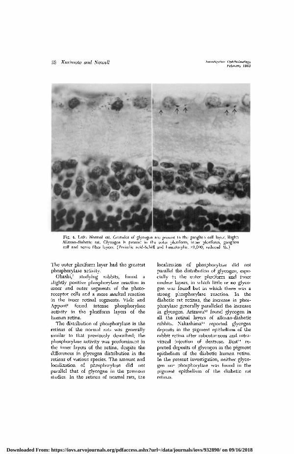
A few glycogen granules in the innersegments of some visual cells were found.Little or no glycogen was observed in theouter nuclear, outer plexiform, innernuclear layers, and in the inner plexiformlayer. Slight amounts of glycogen waspresent in the ganglion cell layer. Thenerve fiber layer had glycogen granulesarranged perpendicularly to the nervefiber bundles. Glycogen granules weresometimes seen on the internal limitingmembrane (Fig. 4, left). In the diabeticretina, deposits of glycogen were increased
in the inner plexiform, ganglion cell, andnerve fiber layers, and also in the internallimiting membrane. In the inner nuclearlayer and outer plexiform layer, the glyco-gen reaction was slightly pronounced. Someof the glycogen granules were arrangedalong fibers extending through the innerplexiform layer in a vertical direction. Thefibers were outlined through the nervefiber, ganglion cell, and inner plexiformlayers. No increase was observed in theouter layer of the retina (Fig. 4, right).No evidence of microaneurysms, capillaryhemorrhage, sclerosis of the vein, andproliferative retinitis could be found inthe diabetic rat retinas.
-GC
ON
V
P
Figs. 1-4. Key to abbreviations. N, Nerve fiber layer; GC, ganglion cell layer; IP, innerplexiform layer; IN, inner nuclear layer; OP, outer plexiform layer; ON, outer nuclear layer;V, rods and cones; P, pigment epithelium; O, optic nerve.Fig. 1. Left: Normal rat. Phosphorylase activity is indicated by the black areas in theplexiform, ganglion cell, and nerve fiber layers, as well as the inner nuclear layer. There is noactivity in the rods and cones. Right: Alloxan-diabetic rat. Phosphorylase activity is pro-nounced in the inner retinal layers (x425; reduced %.)
Downloaded From: https://iovs.arvojournals.org/pdfaccess.ashx?url=/data/journals/iovs/932890/ on 09/16/2018
Volume 2Number 1
Phosphorylase in alloxan-diabetic rat retina 27
Fig. 2. Alloxan-diabetic rat near the papilla. Phosphorylase activity is observed in the nervefiber layer in some areas. Intense phosphorylase activity is seen in the ganglion cell, plexiformand inner nuclear layers. (xl95; reduced approximately %.)
Fig. 3. Alloxan-diabetic rat. The phosphorylase activity is noted along fibers running per-pendicular to the retinal surface. (x425; reduced approximately Vi.)
Discussion
Nagaya" found phosphorylase activity inchicks in those parts of the inner segmentsof cones in which glycogen was present,in some of the cone feet, in the inner plexi-form layer, especially at its outer half along
supporting fibers, in the nerve fiber layer,and at the internal limiting membrane.Phosphorylase in the frog retinas was dis-tributed in the inner segments of cones,plexiform layers, nerve fiber layer, internallimiting membrane, and supporting fibers.
Downloaded From: https://iovs.arvojournals.org/pdfaccess.ashx?url=/data/journals/iovs/932890/ on 09/16/2018
28 Kurimoto and Newell Imiesligdtiva OphthalmologyFebruary 1963
"\?Fig. 4. Left: Normal rat. Granules of glycogen are present in the ganglion cell layer. Right:Alloxan-diabetic rat. Glycogen is present in the outer plexiform, inner plexiform, ganglioncell and nerve fiber layers. (Periodic acid-Schiff and hematoxylin. x2,000; reduced Vs.)
The outer plexiform layer had the greatestphospliorylase activity.
Ohashi,7 studying rabbits, found aslightly positive phospliorylase reaction ininner and outer segments of the photo-receptor cells and a more marked reactionin the inner retinal segments. Viale andApponi8 found intense phospliorylaseactivity in the plexiform layers of thehuman retina.
The distribution of phosphorylase in theretinas of the normal rats was generallysimilar to that previously described; thephosphorylase activity was predominant inthe inner layers of the retina, despite thecliffereTices in glycogen distribution in theretinas of various species. The amount andlocalization of phosphorylase did notparallel that of glycogen in the previousstudies. In the retinas of normal rats, the
localization of phosphorylase did notparallel the distribution of glycogen, espe-cially in the outer plexiform and innernuclear layers, in which little or no glyco-gen was found but in which there was astrong phosphorylase reaction. In thediabetic rat retinas, the increase in phos-phorylase generally paralleled the increasein glycogen. Arizawa12 found glycogen inall the retinal layers of alloxan-diabeticrabbits. Nakashima13 reported glycogendeposits in the pigment epithelium of therabbit retina after subcutaneous and intra-vitreal injection of dextrose. Best1' re-ported deposits of glycogen in the pigmentepithelium of the diabetic human retina.In the present investigation, neither glyco-gen nor phosphorylase was found in thepigment epithelium of the diabetic ratretinas.
Downloaded From: https://iovs.arvojournals.org/pdfaccess.ashx?url=/data/journals/iovs/932890/ on 09/16/2018
Phosphorylase in alloxan-diabetic rat retina 29
Phosphorylase activity in Miiller's fiberwas noted by Nagaya and Ohashi. Kuwa-bara and Cogan15 found that glycogen wassynthesized and stored in Miiller's cell ofthe retina of various species, but little ornone was present in the rat retina. How-ever, glycogen increased, especially inMiiller's fibers after perforating woundsof the rat eye. In the present study, phos-phorylase and glycogen deposits werefound arranged perpendicularly to theplane of the retinal surface. However, itwas not possible to determine whether thedeposits were in Miiller's fibers or in theaxons or dendrites of nerve cells.
Opinions concerning the role of theglycogen in retinal metabolism have beenvaried; e.g., Schmitz-Moormann10 con-sidered glycogen in the retina an energysource for contraction of cones, whileShimizu and Maeda37 regarded the glyco-gen as the energy source for restoration ofrhodopsin, phosphocreatine, and adenosinetriphosphate. Conversely, with the excep-tion of glycogen storage disease, which, ingeneral, shows no hyperglycemia, and sometypes of enzyme deficiency (glucose 6-phosphate, branching enzyme, debranchingenzyme and phosphorylase), abnormalaccumulations of glycogen in the cells ofthe body are a reflection of hyperglycemiaof which diabetes mellitus is the most im-portant cause. The glycogen depositsrepresent an individual cell accumulationof glucose in response to hyperglycemiaand are most important as anatomic indi-cators of deranged carbohydrate metab-olism.
Although phosphorylase synthesizesglycogen in vitro, the enzyme is concernedin vivo only with glycogen degradation.18"-0
The pathway of glycogen synthesis in vivois from uridine diphosphate glucosethrough the uridine diphosphate glucose-glycogen transference system. Nagaya0
found that glycogen appeared in the chickembryo retina on the eighteenth ortwentieth day of development, and phos-phorylase on the twenty-first day. The lateappearance of phosphorylase indicates that
the chick embryo retina can synthesizeglycogen without phosphorylase.
In our study, the accumulation of glyco-gen in the diabetic rat retina was accom-panied by increased phosphorylase activity.The increased amounts of phosphorylase inthe diabetic rat retina may be required todegrade the excess glycogen. The glycogenaccumulation in the diabetic rat retina maybe due to a greater increase in the rate ofglycogen synthesis compared to the rateof glycogen degradation which may beless markedly increased. However, theuridine diphosphate glucose-glycogentransferase system, through which glyco-gen synthesis is performed in vivo* couldnot be demonstrated histochemically inthe normal or in diabetic rat retinas.*
Another possibility is that the increaseof phosphorylase in the alloxan-diabeticrat retina may result from an increase ofphosphorylase B, the inactive form of theenzyme. Possibly, as suggested by Barany,-1
native glycogen acts as a primer for thephosphorylase. Thus Nagaya0 found nophosphorylase activity in the retina treatedwith amylase prior to incubation in hismedium, which contained no glycogen.However, phosphorylase activity was foundin the retina in the absence of demon-strable glycogen in the current study.Further investigation is required to clarifythis point.
The detection of phosphorylase activitymay be a more sensitive indicator of carbo-hydrate metabolism than periodic acid-Schiff glycogen staining. The abnormal in-crease of phosphorylase activity and ab-normal accumulation of glycogen in thediabetic rat retina are manifestations ofdisturbances of carbohydrate metabolismin the diabetic rat retina. Thus, it is in-teresting to recall the hypothesis that dis-turbances of metabolism in the retina pre-
;s, of 13"Fresh frozen sections, 50 /i in irat retinas and 10 severe alloxan-diabetic rat retinas weretreated by Takeuchi's method-" for uridine diphosphateglucose-glycogen transferase system. With this method,we were able to demonstrate glycogen synthesis in theocular recti muscles but net in the retinn.
Downloaded From: https://iovs.arvojournals.org/pdfaccess.ashx?url=/data/journals/iovs/932890/ on 09/16/2018
30 Kurimoto and Newell Investleatioe OphthalmologyFebruary 1963
cede diabetic retinopathy, which has beenpresented by Ashton,23 and de Roetth andYen.2"
Addendum
Hutchinson and Kuwabara25 have recently re-ported that phosphorylase activity has been demon-strated histochemically in the inner segments ofthe retinas of various species, and that minimalundine diphosphoglucose glycogen synthetase ac-tivity in the diabetic human retina and the calfretina has been demonstrated by histochemicalmethods, and the presence of the enzyme hasbeen shown biochemically in the calf retina.
Grateful acknowledgement is made to Dr.Bertha A. Klein, Professor of Ophthalmology, TheUniversity of Chicago, and Prof. Koku Kojima,Nagoya University School of Medicine, for theirvaluable advice.
REFERENCES1. Yin, H. C, and Sun, C. N.: Histochemical
method for the detection of phosphorylasein plant tissues, Science 105: 650, 1947.
2. Cobb, J.: Calcification of rat cartilage andbone, Anat. Rec. 103: 531, 1949.
3. Takeuchi, T., and Kuriaki, H.: Histochemicaldetection of phosphorylase in animal tissues,J. Histochem. 3: 153, 1955.
4. Eranko, O., and Palkama, A.: Improvedlocalization of phosphorylase by the use ofpolyvinyl pyrrolidine and high substrate con-centration, J. Histochem. 9: 585, 1961.
6. Nagaya, Y.: Histochemical study of phos-phorylase in the retina, Acta Soc. Ophth.Jap. 59: 1043, 1955.
7. Ohashi, K.: Histochemical studies of phos-phorylase in the ocular tissue, Acta Soc.Ophth. Jap. 64: 290, 435, 605, 1960.
8. Viale, G., and Apponi, G.: HistochemischeUntersuchung iiber Amylo-Phosphorylase undAmylo-1,4 -» 1,6-Transglukosylase in dermenschlichen Netzhaut, Ztschr. Zellforsch. 56:709, 1962.
9. Bailey, C. C, Bailey, O. T., and Leech, R.S.: Alloxan diabetes with diabetic complica-tions, New England J. Med. 230: 533, 1944.
10. Hoffman, W. S.: A rapid photoelectricmethod for the determination of glucose inblood and urine, J. Biol. Chem. 120: 51,1937.
11. McManus, J. F. A.: The periodic acid routineapplied to the kidney, Am. J. Path. 24: 643,1948.
12. Arizawa, T.: Ophthalmological studies of ex-
perimental diabetes due to alloxan, oxine, anddithizone, Acta Soc. Ophth. Jap. 55: 1239,1951.
13. Nakashima, C : Experimentelle Studien iiberdie Wirkung der Injektion von Proteinkorpernund Traubenzucker in den Glaskorper Arch,f. Ophth. 116: 403, 1926.
14. Best, F.: Demonstration mikroskopischerPriiparate vom diabetischen auge, Ophth. ges.Heidelberg 32: 315, 1905. Cited by Schmitz-Moormann.10
15. Kuwabara, T., and Cogan, D. G.: Retinalglycogen, A. M. A. Arch. Ophth. 66: 680,1961.
16. Schmitz-Moormann, P.: Uber den Glykogen-gehalt der Retina und seine Beziehungen zurZapfenkontraktion, Arch. f. Ophth. 118: 506,1927.
17. Shimizu, N., and Maeda, S.: Histochemicalstudies on glycogen of the retina, Anat. Rec.116: 427, 1953.
18. Leloir, L. F., and Cardini, C. E.: Bio-synthesis of glycogen from uridine diphos-phate glucose, J. Am. Chem. Soc. 79: 6340,1957.
19. Leloir, L. F., Olavorria, J. M., Goldenberg,S. H., and Corminatti, H.: Biosynthesis ofglycogen from uridine diphosphate glucose,Arch. Biochem. 81: 508, 1959.
20. Schmid, R., Robbins, P. W., and Traut, R.R.: Glycogen synthesis in muscle lackingphosphorylase, Proc. Nat. Acad. Sc. 45: 1236,1959.
21. Barany, E.: Personal communication.22. Takeuchi, T., and Glenner, G. G.: Histo-
chemical demonstration of uridine diphos-phate glucose-glycogen transferase in animaltissues, J. Histochem. 9: 304, 1961.
24. de Roetth, A., J., and Yen, F. P.: Experi-mental diabetic retinopathy; Retinal metab-olism in the alloxan diabetic rat, A. M. A.Arch. Ophth. 63: 226, 1960.
25. Hutchinson, B. T., and Kuwabara, T.: Phos-phorylase and uridine diphosphoglucose glyco-gen synthetase in the retina, A. M. A, Arch.Ophth. 68: 538, 1962.
DiscussionDr. B. Thomas Hutchinson, Boston, Mass. Drs.
Kurimoto and Newell have nicely demonstratedthe presence of phosphorylase activity in theretinas of the normal and the alloxan-diabetic ratand have presented a lucid, concise discussion ofthe metabolism of retinal glycogen. Previously,Kuwabara and Cogan reported the distribution ofretinal glycogen in a variety of animals and de-
Downloaded From: https://iovs.arvojournals.org/pdfaccess.ashx?url=/data/journals/iovs/932890/ on 09/16/2018
Phosphorylase in alloxan-diabetic rat retina 31
' scribed a species variation in native glycogencontent which showed glycogen, as demonstratedhistochemically, to be roughly inversely propor-
v- tional to the vascularity of the retina.Using essentially the same histochemical tech-
Y nique as employed in this presentation, Dr. Kuwa-g, bara and I have studied phosphorylase in the7 retinas of a variety of species; we, too, have
found abundant phosphorylase activity to be dis-tributed throughout the inner segments of the
r retina in all species, regardless of the relativenative glycogen content as demonstrated histo-
V chemically. Thus the mouse, having no histo-chemically demonstrable glycogen, the rat, hav-
^ ing glycogen only in the nerve fiber and ganglioncell layers, and the guinea pig, having glycogen
y throughout the inner portions of the retina, haveidentical distributions of phosphorylase.
We have been unable to demonstrate consistentlythe presence of UDPG glycogen synthetase in theretina by histochemical techniques but have beenable, by enzymatic assay, to demonstrate thisenzyme to be present in the calf, rat, and mouseretinas. It seems that synthetase, perhaps normallyattached to particulate glycogen, is solubilized byfreezing and is thus not histochemically evidentwhen "cryostat" techniques are employed.
We share the authors' view that the localiza-tion of phosphorylase activity might give a betterindication of sites of significant glycogen turn-over than is given by the histochemically demon-strable glycogen itself. Certainly the normal speciesvariation of retinal glycogen offers to the histo-chemist and the biochemist an unusual oppor-tunity, not found in other tissues, to further ex-plore differences in this important compound.
Downloaded From: https://iovs.arvojournals.org/pdfaccess.ashx?url=/data/journals/iovs/932890/ on 09/16/2018


![Untitled-13 [] · Title: Untitled-13 Author: PURIN Created Date: 1/21/2010 10:28:05 AM](https://static.documents.pub/doc/80x56/5f7a5cd244c75b6c3c68aa38/untitled-13-title-untitled-13-author-purin-created-date-1212010-102805.jpg)


