38
| Date post: | 19-Dec-2015 |
| Category: |
Documents |
| View: | 222 times |
| Download: | 2 times |
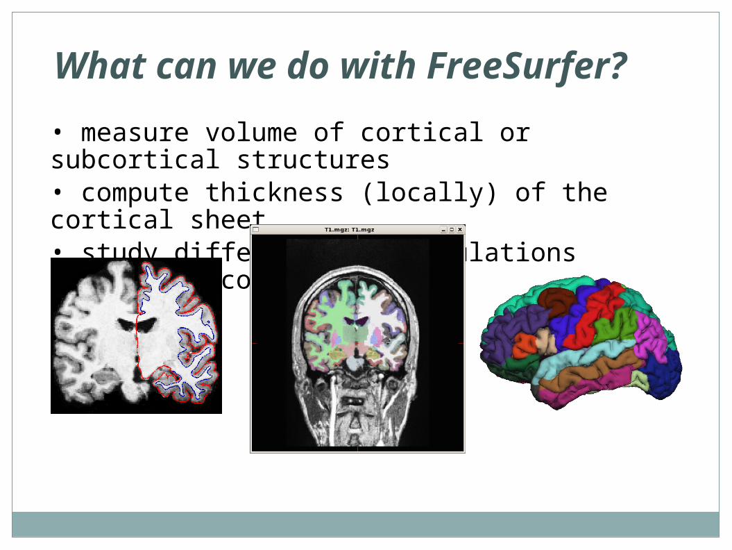
What can we do with FreeSurfer?
• measure volume of cortical or subcortical structures• compute thickness (locally) of the cortical sheet• study differences of populations (diseased, control)
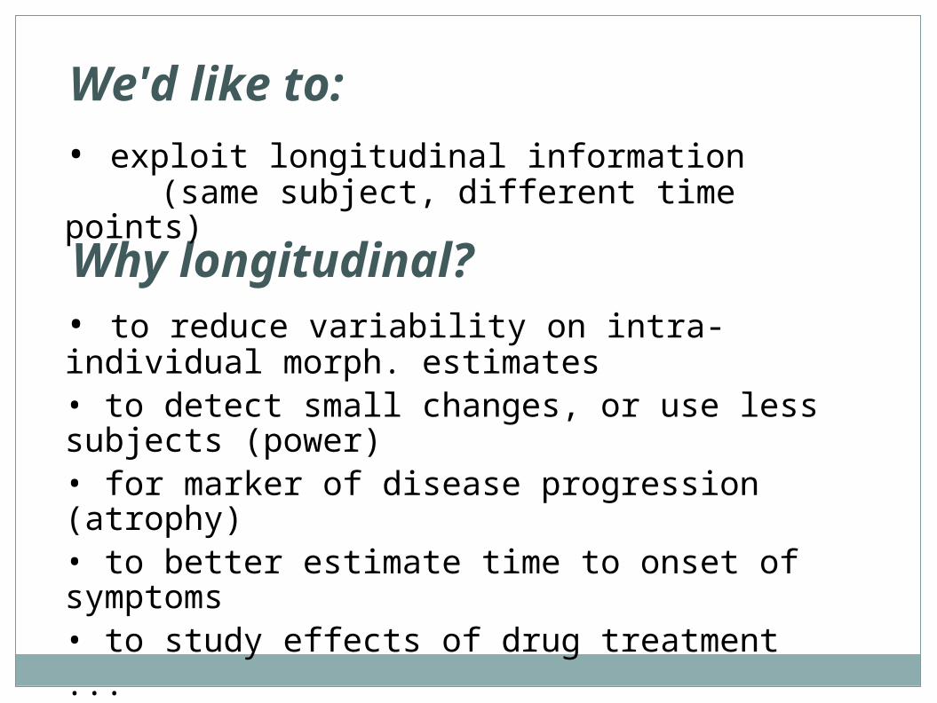
Why longitudinal?• to reduce variability on intra-individual morph. estimates• to detect small changes, or use less subjects (power)• for marker of disease progression (atrophy)• to better estimate time to onset of symptoms• to study effects of drug treatment...[Reuter et al, NeuroImage 2012]
We'd like to:• exploit longitudinal information
(same subject, different time points))
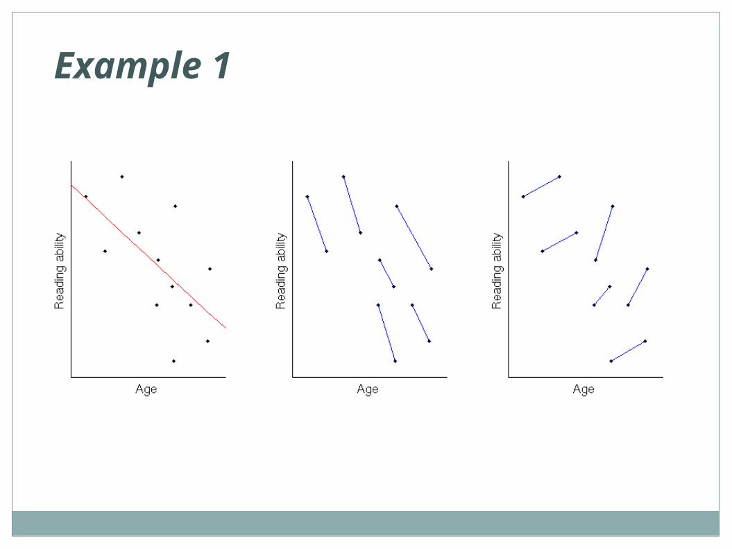
Example 1
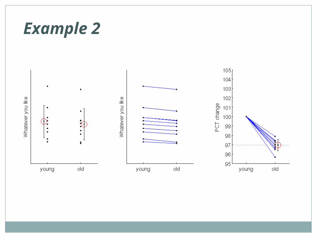
Example 2
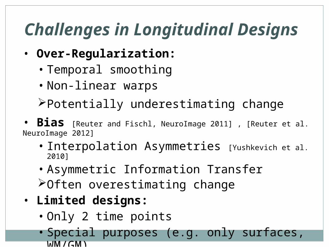
Challenges in Longitudinal Designs
• Over-Regularization:• Temporal smoothing• Non-linear warps
Potentially underestimating change
• Bias [Reuter and Fischl, NeuroImage 2011] , [Reuter et al. NeuroImage 2012]
• Interpolation Asymmetries [Yushkevich et al. 2010]
• Asymmetric Information TransferOften overestimating change
• Limited designs:• Only 2 time points• Special purposes (e.g. only surfaces, WM/GM)
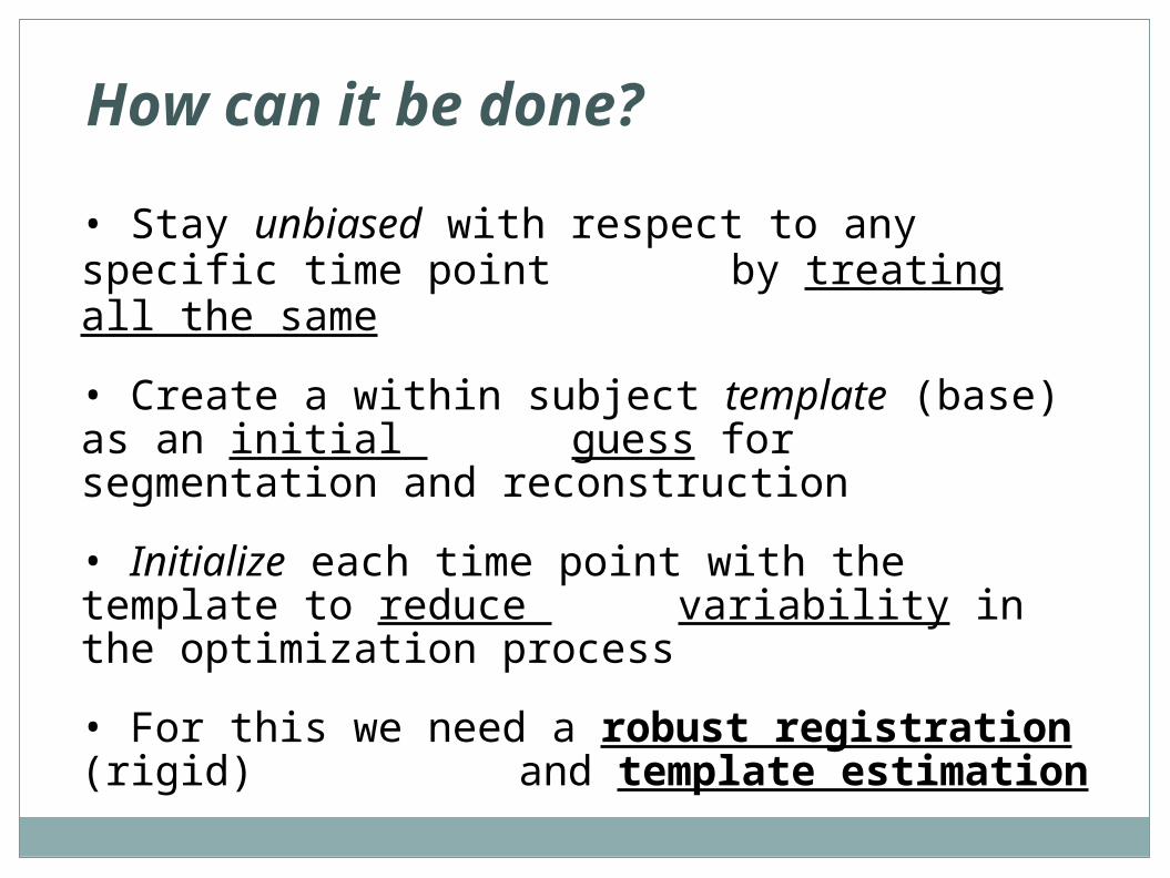
How can it be done?
• Stay unbiased with respect to any specific time point by treating all the same
• Create a within subject template (base) as an initial guess for segmentation and reconstruction
• Initialize each time point with the template to reduce variability in the optimization process
• For this we need a robust registration (rigid) and template estimation

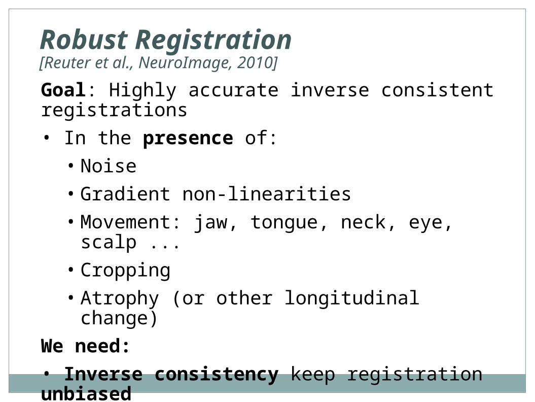
Robust Registration[Reuter et al., NeuroImage, 2010]
Robust Registration[Reuter et al., NeuroImage, 2010]
Goal: Highly accurate inverse consistent registrations
• In the presence of:
• Noise
• Gradient non-linearities
• Movement: jaw, tongue, neck, eye, scalp ...
• Cropping
• Atrophy (or other longitudinal change)
We need:
• Inverse consistency keep registration unbiased
• Robust statistics to reduce influence of outliers
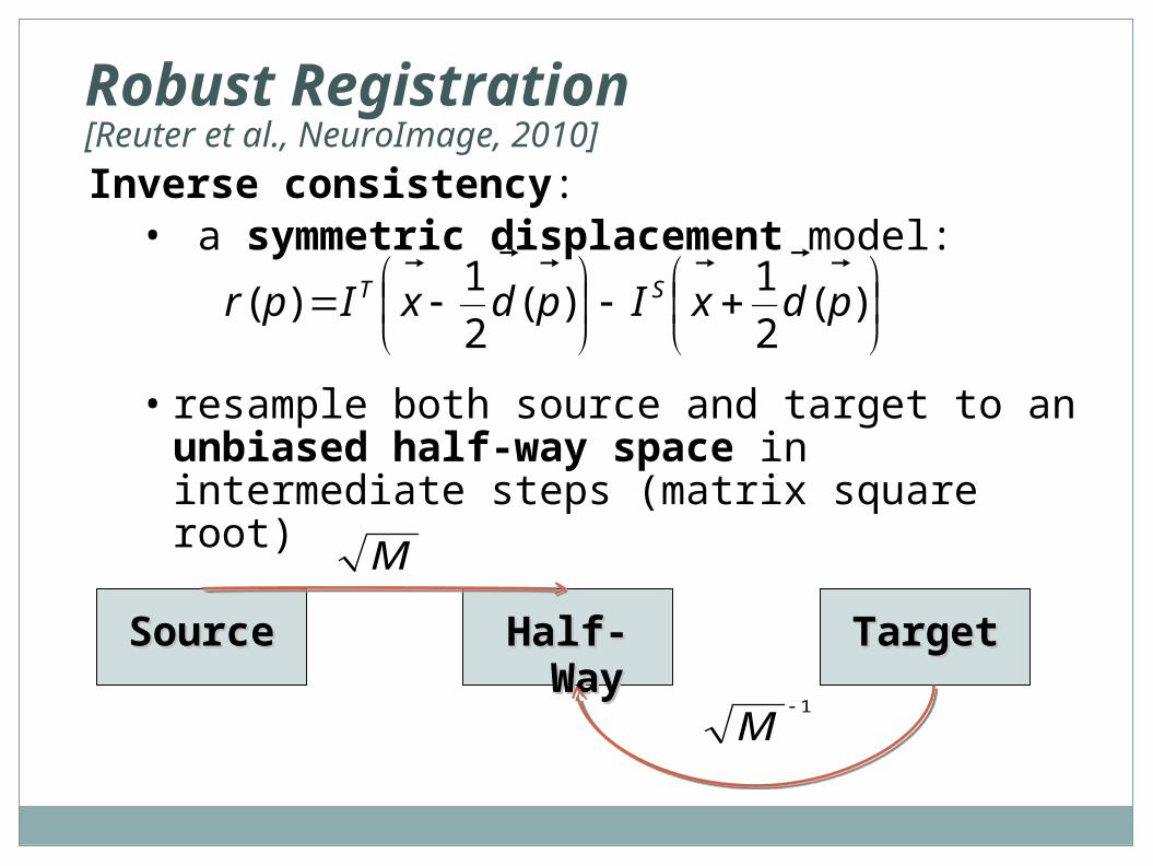
Inverse consistency:• a symmetric displacement model:
• resample both source and target to an unbiased half-way space in intermediate steps (matrix square root)
)(
2
1)(
2
1)( pdxIpdxIpr ST
SourceSource TargetTargetHalf-Half-WayWay
M
1
M
Robust Registration[Reuter et al., NeuroImage, 2010]
Robust Registration[Reuter et al., NeuroImage, 2010]
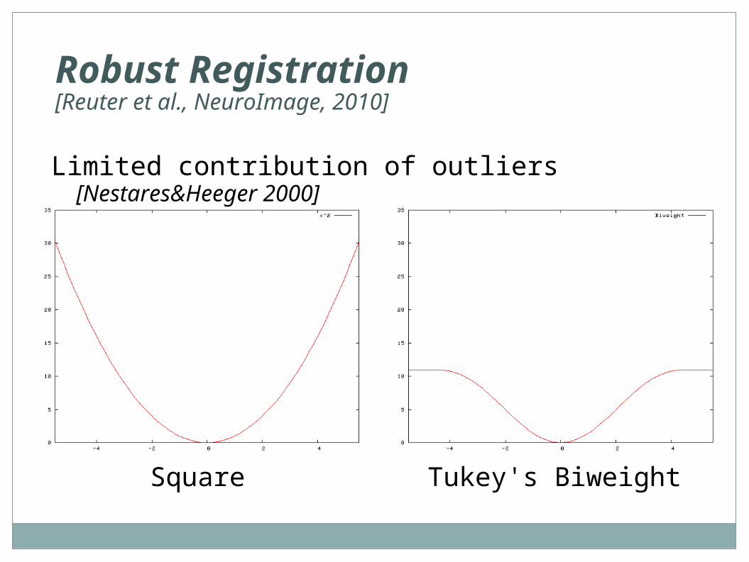
Square Tukey's Biweight
Limited contribution of outliers [Nestares&Heeger 2000]
Robust Registration[Reuter et al., NeuroImage, 2010]
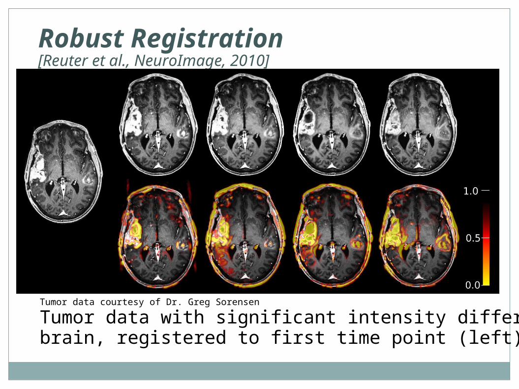
Tumor data courtesy of Dr. Greg Sorensen
Tumor data with significant intensity differences in thebrain, registered to first time point (left).
Robust Registration [Reuter et al 2010]
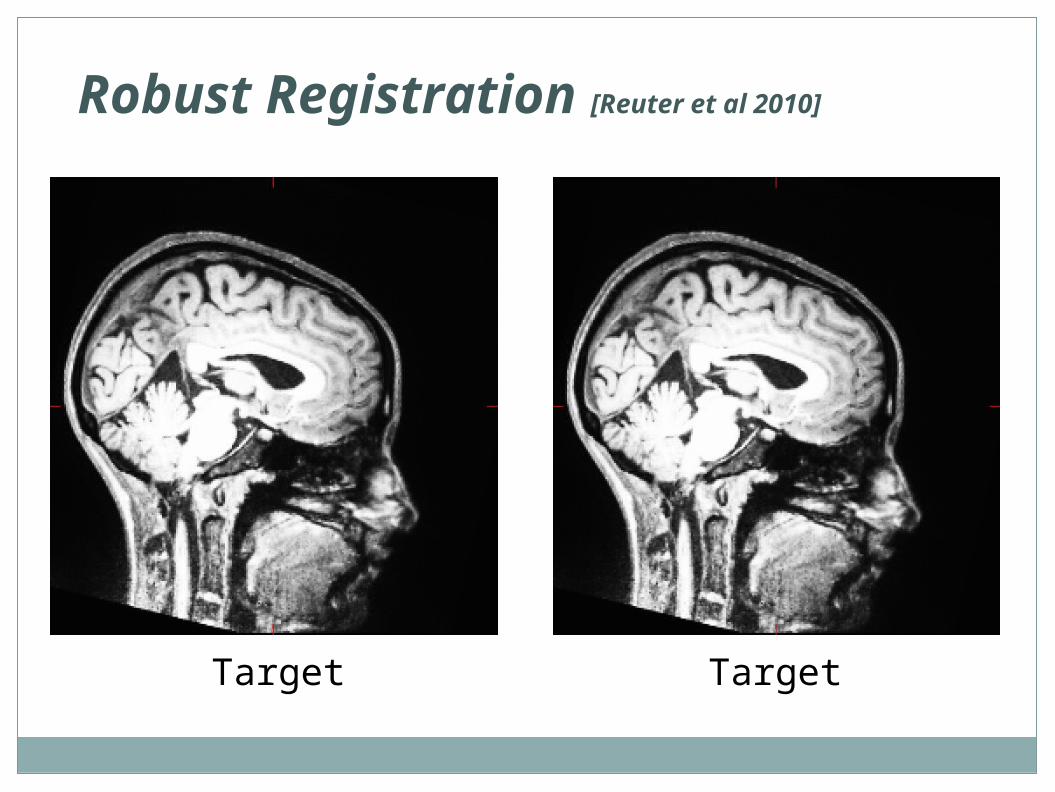
Target Target
Robust Registration [Reuter et al 2010]
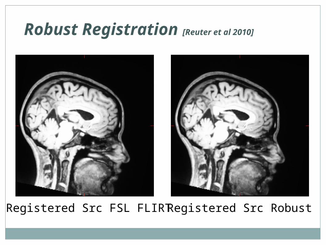
Registered Src FSL FLIRT Registered Src Robust
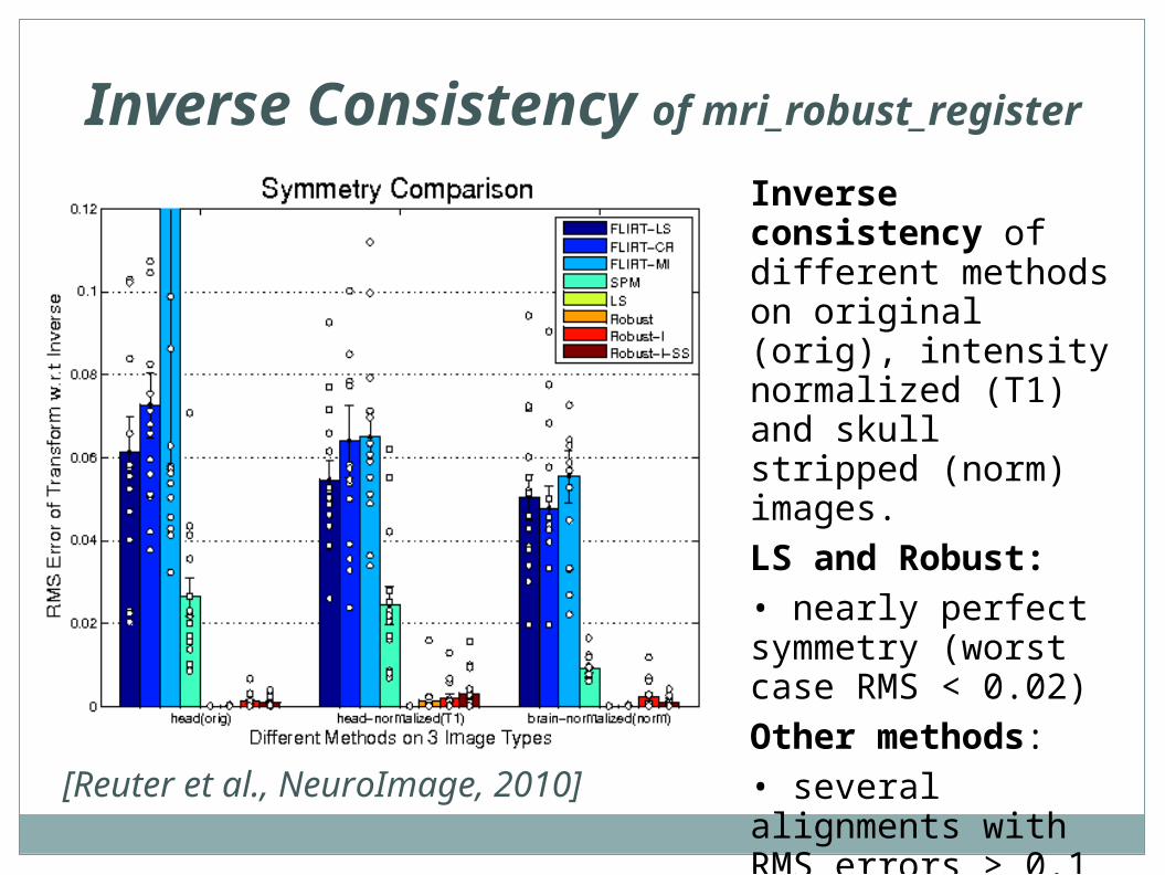
Inverse Consistency of mri_robust_register
Inverse consistency of different methods on original (orig), intensity normalized (T1) and skull stripped (norm) images.
LS and Robust:• nearly perfect symmetry (worst case RMS < 0.02)
Other methods:• several alignments with RMS errors > 0.1
[Reuter et al., NeuroImage, 2010]
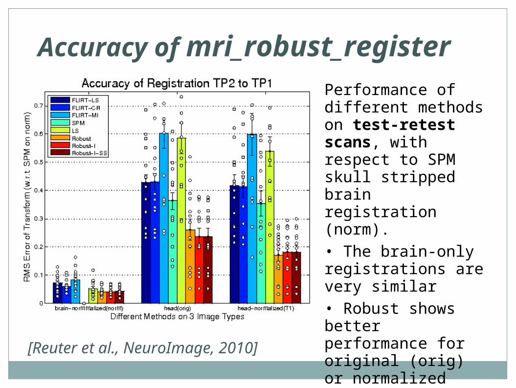
Accuracy of mri_robust_registerPerformance of different methods on test-retest scans, with respect to SPM skull stripped brain registration (norm). • The brain-only registrations are very similar• Robust shows better performance for original (orig) or normalized (T1) full head images
[Reuter et al., NeuroImage, 2010]
mri_robust_register• mri_robust_register is part of FreeSurfer
• can be used for pair-wise registration (optimally within subject, within modality)
• can output results in half-way space
• can output ‘outlier-weights’
• see also Reuter et al. “Highly Accurate Inverse Consistent Registration: A Robust Approach”, NeuroImage 2010.
http://reuter.mit.edu/publications/
• for more than 2 images: mri_robust_template
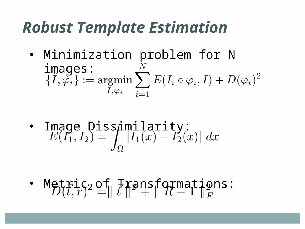
Robust Template Estimation
• Minimization problem for N images:
• Image Dissimilarity:
• Metric of Transformations:
Longitudinal Processing
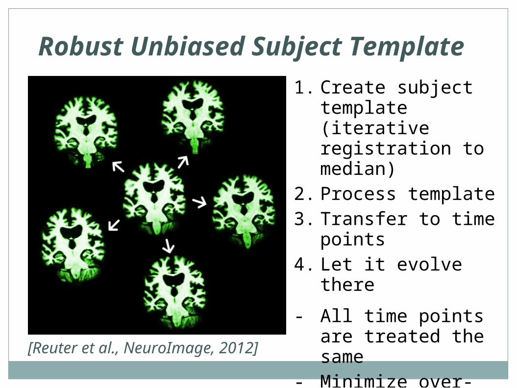
Robust Unbiased Subject Template
1. Create subject template (iterative registration to median)
2. Process template3. Transfer to time points4. Let it evolve there
- All time points are treated the same
- Minimize over-regularization by letting tps evolve freely
[Reuter et al., NeuroImage, 2012]
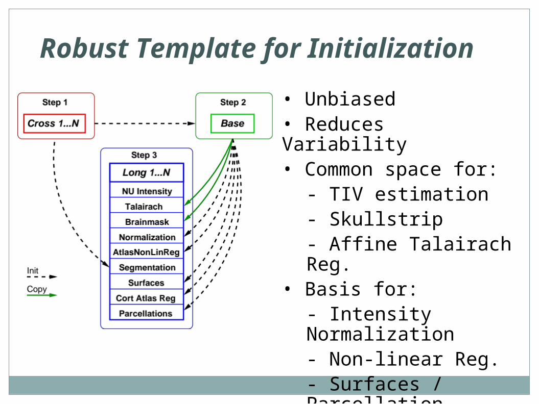
Robust Template for Initialization
• Unbiased • Reduces Variability• Common space for:
- TIV estimation- Skullstrip- Affine Talairach Reg.
• Basis for:- Intensity Normalization- Non-linear Reg.- Surfaces / Parcellation
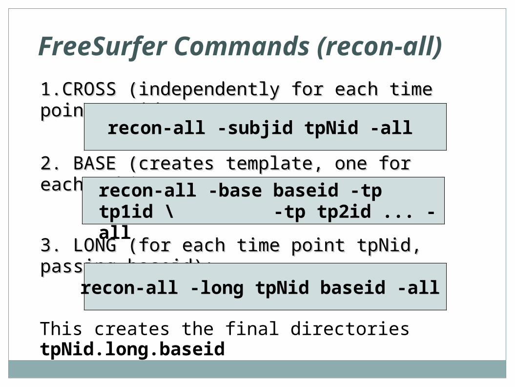
FreeSurfer Commands (recon-all)
1.CROSS (independently for each time point 1.CROSS (independently for each time point tpNid):tpNid):
This creates the final directories tpNid.long.baseid
3. LONG (for each time point tpNid, passing 3. LONG (for each time point tpNid, passing baseid):baseid):
recon-all -long tpNid baseid -all
recon-all -subjid tpNid -all
2. BASE (creates template, one for each 2. BASE (creates template, one for each subject):subject):recon-all -base baseid -tp tp1id \
-tp tp2id ... -all
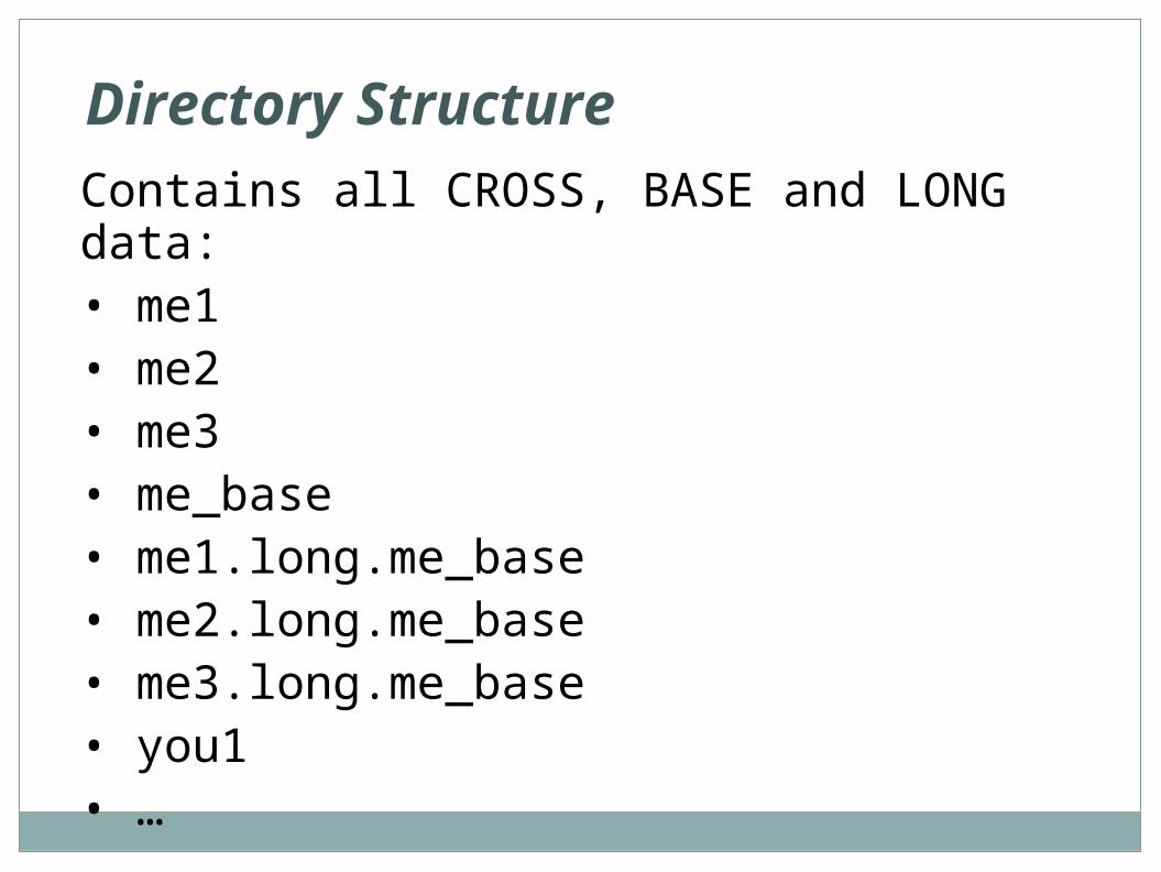
Directory Structure
Contains all CROSS, BASE and LONG data:• me1• me2• me3• me_base• me1.long.me_base• me2.long.me_base• me3.long.me_base• you1• …
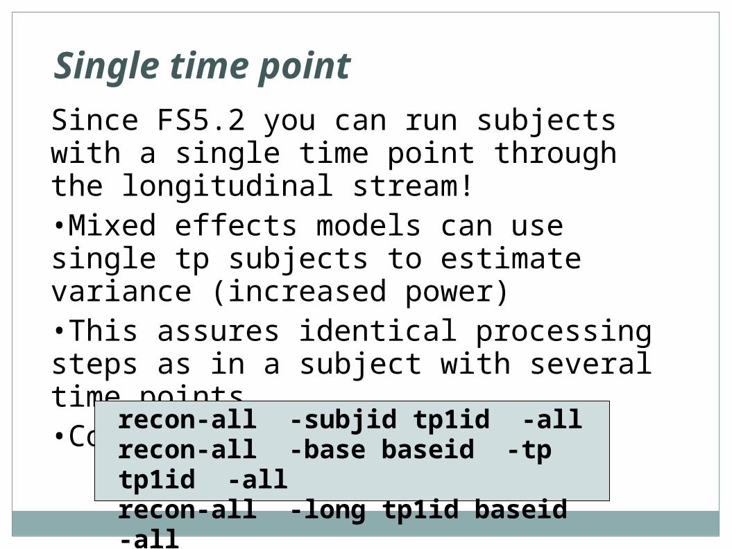
Single time point
Since FS5.2 you can run subjects with a single time point through the longitudinal stream!•Mixed effects models can use single tp subjects to estimate variance (increased power)•This assures identical processing steps as in a subject with several time points•Commands same as above:
recon-all -subjid tp1id -allrecon-all -base baseid -tp tp1id -allrecon-all -long tp1id baseid -all
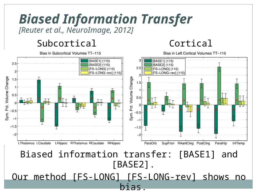
Biased Information Transfer[Reuter et al., NeuroImage, 2012]
Biased information transfer: [BASE1] and [BASE2].Our method [FS-LONG] [FS-LONG-rev] shows no bias.
Subcortical Cortical
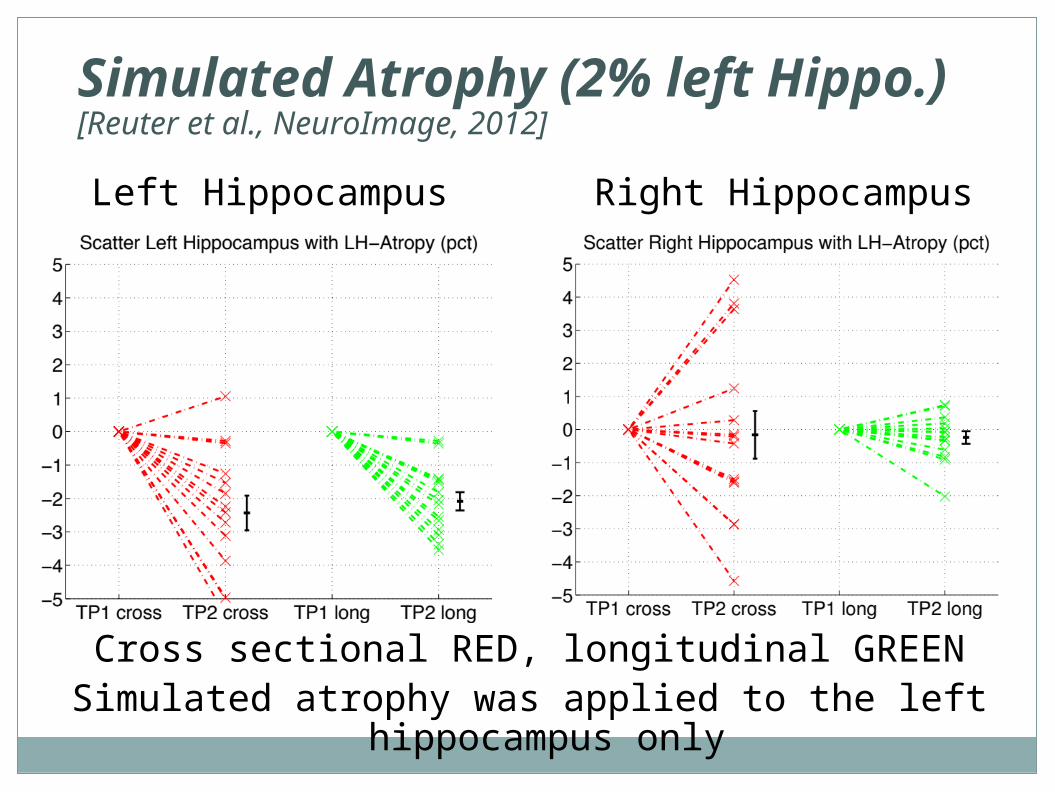
Simulated Atrophy (2% left Hippo.)[Reuter et al., NeuroImage, 2012]
Cross sectional RED, longitudinal GREENSimulated atrophy was applied to the left hippocampus only
Left Hippocampus Right Hippocampus
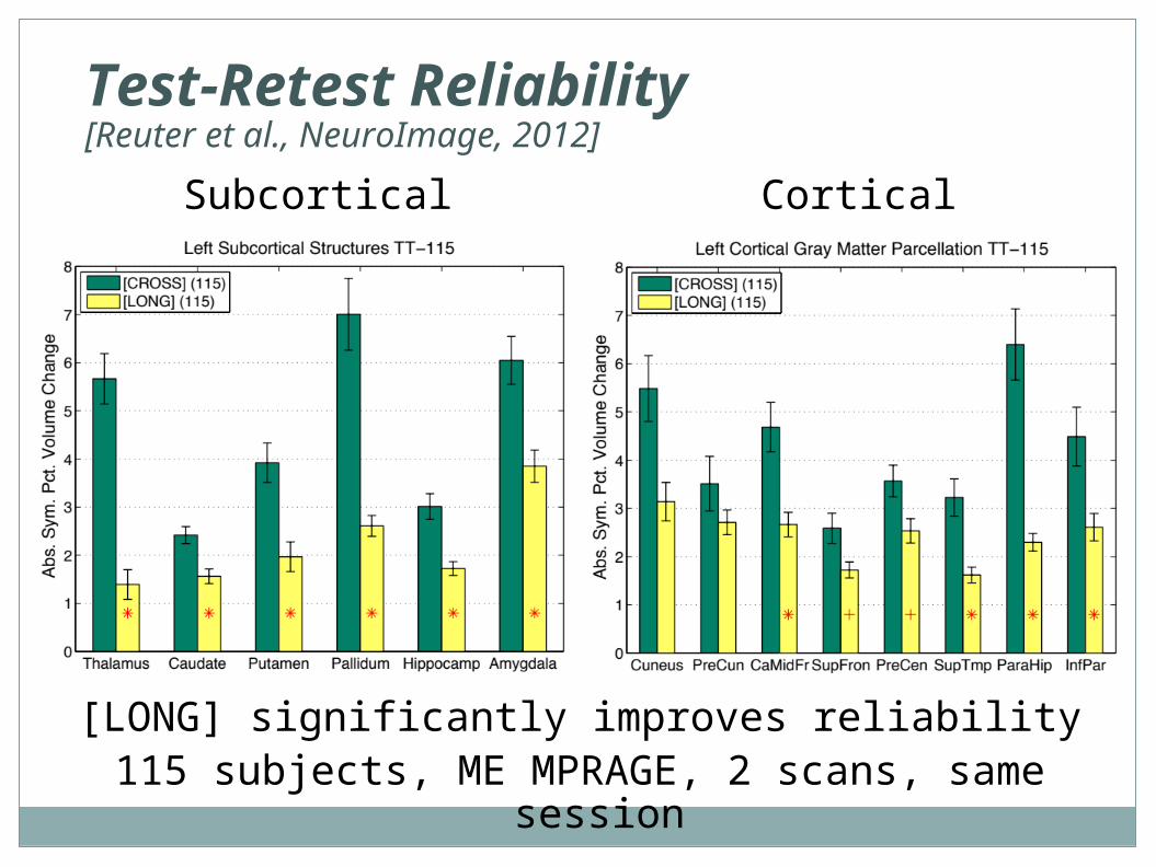
Test-Retest Reliability[Reuter et al., NeuroImage, 2012]
[LONG] significantly improves reliability115 subjects, ME MPRAGE, 2 scans, same session
Subcortical Cortical
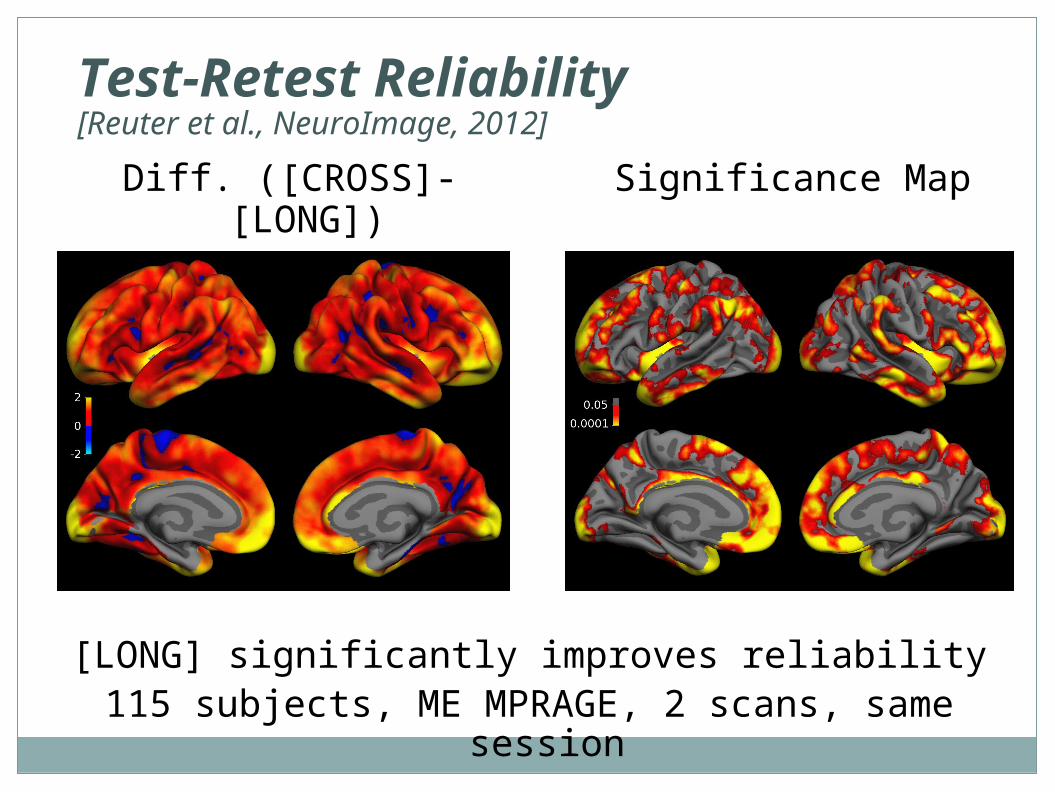
Test-Retest Reliability[Reuter et al., NeuroImage, 2012]
[LONG] significantly improves reliability115 subjects, ME MPRAGE, 2 scans, same session
Diff. ([CROSS]-[LONG])of Abs. Thick. Change:
Significance Map
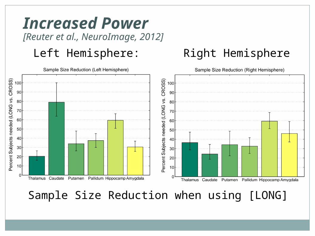
Increased Power[Reuter et al., NeuroImage, 2012]
Sample Size Reduction when using [LONG]
Left Hemisphere: Right Hemisphere
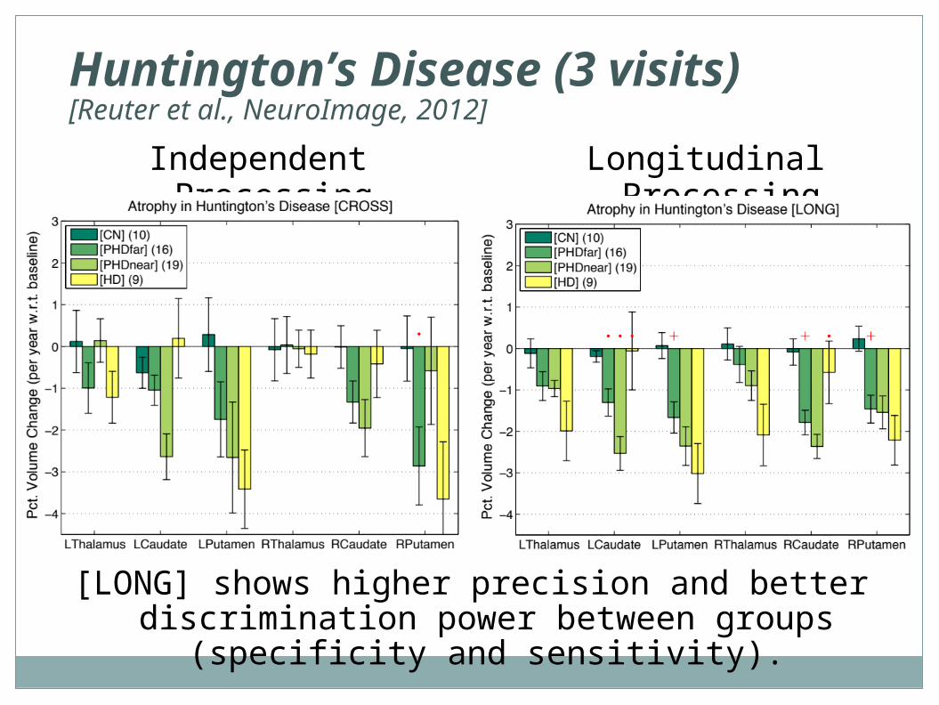
Huntington’s Disease (3 visits)[Reuter et al., NeuroImage, 2012]
[LONG] shows higher precision and better discrimination power between groups (specificity and sensitivity).
Independent Processing Longitudinal Processing
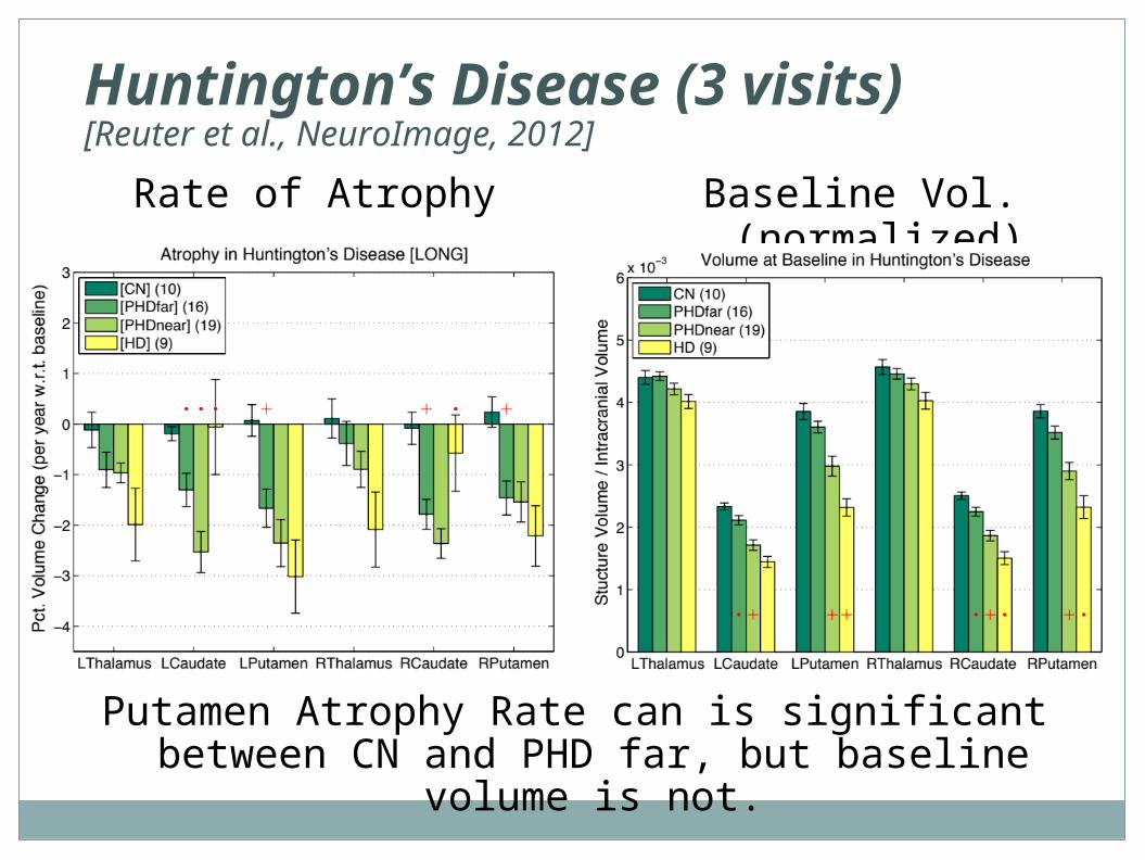
Huntington’s Disease (3 visits)[Reuter et al., NeuroImage, 2012]
Putamen Atrophy Rate can is significant between CN and PHD far, but baseline volume is not.
Rate of Atrophy Baseline Vol. (normalized)

Final Remarks …
Sources of Bias during Acquisition
BAD: these influence the images directly and cannot be easily removed!
• Different Scanner Hardware (Headcoil, Pillow?)
• Different Scanner Software (Shimming Algorithm)
• Scanner Drift and Calibration
• Different Motion Levels Across Groups
• Different Hydration Levels (season, time of day)
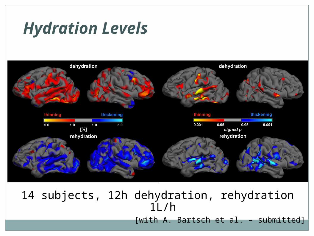
Hydration Levels
14 subjects, 12h dehydration, rehydration 1L/h[with A. Bartsch et al. – submitted]
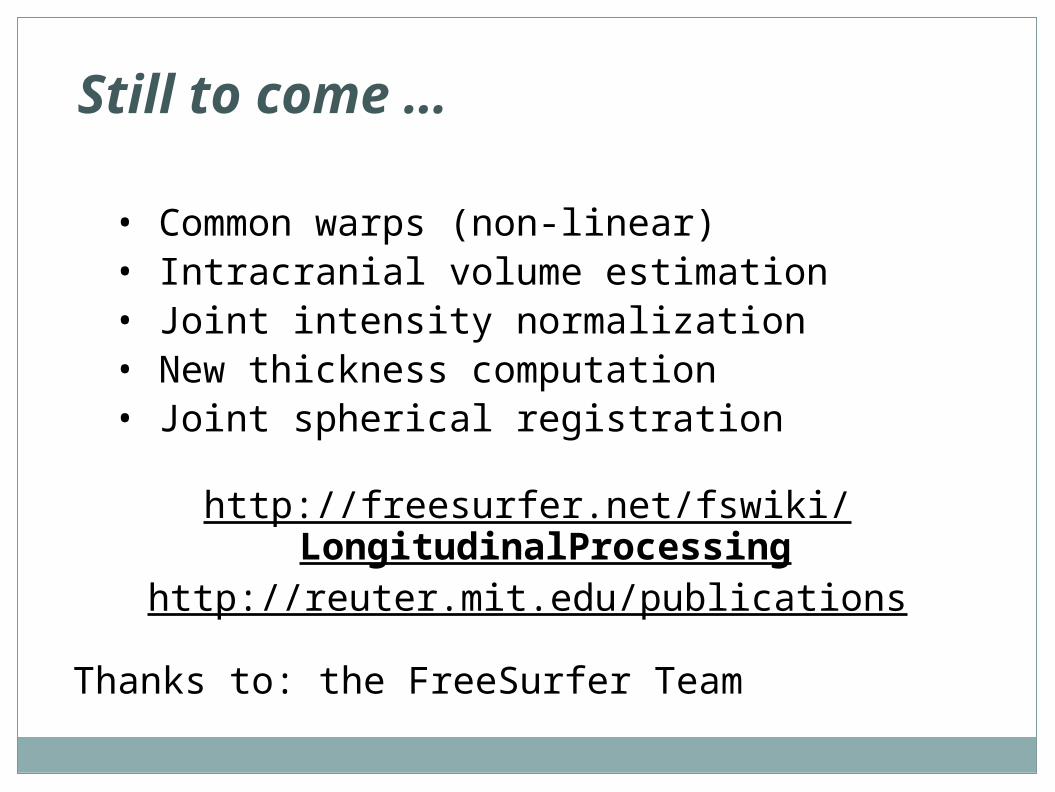
Still to come …
• Common warps (non-linear)• Intracranial volume estimation• Joint intensity normalization• New thickness computation• Joint spherical registration
http://freesurfer.net/fswiki/LongitudinalProcessinghttp://reuter.mit.edu/publications
Thanks to: the FreeSurfer Team

Longitudinal Tutorial
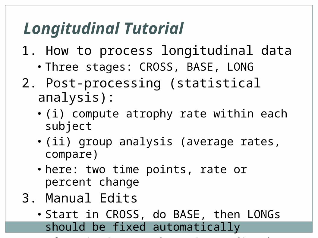
Longitudinal Tutorial1. How to process longitudinal data
• Three stages: CROSS, BASE, LONG
2. Post-processing (statistical analysis):• (i) compute atrophy rate within each subject• (ii) group analysis (average rates, compare)• here: two time points, rate or percent change
3. Manual Edits• Start in CROSS, do BASE, then LONGs should be
fixed automatically• Often it is enough to just edit the BASE• See http://freesurfer.net/fswiki/LongitudinalEdits
Longitudinal Tutorial
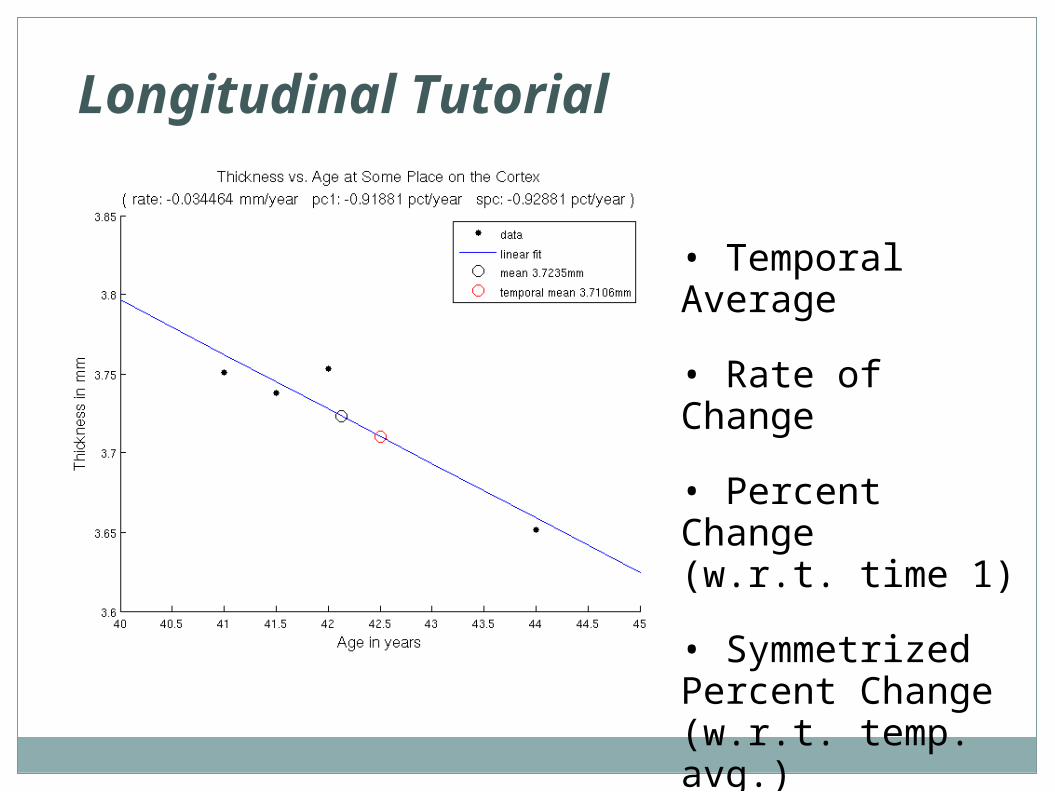
• Temporal Average
• Rate of Change
• Percent Change (w.r.t. time 1)
• Symmetrized Percent Change (w.r.t. temp. avg.)