88
Low Back Pain Adam Shuster DO Pain Management Consultants of SWFL

Low Back Pain
Adam Shuster DO Pain Management Consultants of SWFL

Low Back Pain
Radicular pain DDD/HNP Spondylosis Scoliosis Stenosis Infection
Spondylolisthesis SIJ pain Cancer Trauma/fractures Failed Back Syndrome Referred pain

Lumbar Pain
Axial Inferior to T12 Superior to S1

Sacral Pain
S1 Sacrococcygeal joint Lumbosacral pain includes pain of lumbar and/or
sacral pain and typically constitutes “low back pain.”

Pain Definition
An unpleasant SENSORY and EMOTIONAL experience associated with actual or potential tissue damage or that is described in terms of damage
Physiologic process Tend to be well localized and associated with
sensitivity in the injured region Nociceptive pain

Neuropathic Pain
Persistent pain following injury to the nervous system
Spontaneous (no stimulus) Hyperalgesia Allodynia

Back Pain Workup/Exam
Back pain vs leg pain vs back and leg pain Assess for:
Gait disturbance Numbness Weakness Paresthesias Diminished reflexes

Workup/Exam
Radicular pain travels along specific dermatome Lancinating Shooting Sharp Stabbing

Workup/Exam
L2 and L3 nerve roots (NR) Symptoms involving groin/inner thigh
L4 NR Buttock, anterior thigh, knee, medial calf, may have
weakness of knee extension and decreased patellar reflex
L5 NR Buttock, around hip, lateral leg, dorsal foot, great toe,
may have difficulty walking on heels

Workup/Exam
S1 NR Buttock, posterior thigh and leg, plantar surface of
foot, may have decreased achilles reflex, weakness of plantar flexion, difficulty walking on toes
Sacral NRs Decreased sensation buttock perineum (saddle
anesthesia), bowel/bladder dysfunction, autonomic dysfunction (loss of erection/vaginal anesthesia)

Red Flags
Majority of patients have musculoskeletal origin to their pain and it will resolve in 4-6 weeks.
“Red flag” conditions (Agency for Health Care Policy and Research) that may be life-threatening or compromise neurologic function Infection Tumors Cauda equina syndrome Fractures
Red Flags
Age younger than 20 Higher incidence of congenital and developmental
abnormalities Age older than 50
Prone to neoplasms, pathologic fractures, infections
Red Flags
Duration Acute and subacute back pain is less than 3 months Pain greater than 3 months is usually considered to be
of less serious etiology Trauma

Red Flags
Constitutional symptoms Fever Chills Night sweats Unexplained weight loss Malaise

Red Flags
Systemic illness Hx cancer Serious bacterial infection IVDA Immunosupression

Red Flags
Unrelenting pain Usually worse at night Not relieved with rest or analgesics

Cauda Equina Syndrome
Caused by acute compression of the nerve roots comprising the cauda equina (horse's tail)
Prevalence about 4/10,000 Most common cause is a large disc herniation, or
disc herniation in a stenotic spine 70% of patients with cauda equina have a history
of LBP

Cauda Equina Syndrome
Other causes of CES METS Hematoma Epidural abscess Traumatic fracture Acute transverse myelitis
• Inflammation of the spinal cord

Cauda Equina Syndrome
Present within 24 hrs Radicular pain Back pain Gait disturbance Weakness Abdominal discomfort
Motor/sensory deficits Saddle anesthesia Diminished sphincter
tone Evidence of urinary
retention

Cauda Equina Syndrome
MRI examination is the “gold standard” Once confirmed Tx includes Neurosurgical
consultation/IV steroids

Imaging
MRI Gold standard at determining etiology of lumbar
radicular symptoms Best resolution of spinal canal, spinal cord, neural
foramina, NR, disc spaces
Contrast is used in patients with previous back surgery to differentiate scar tissue and recurrent herniation

Imaging
MRI limitations Claustrophobia Overweight Most pacemakers SCS Retained metallic objects
Mechanical heart valves Aneurysm clips Cost (insurance?)

Imaging
CT scan is superior to MRI at evaluating the bony structures of the spine
May be combined with myelography

Imaging
Plain radiographs may detect fractures or deformities
Can help identify spondylolisthesis Not helpful in evaluating for disc displacement Flexion and Extension films may assess spinal
instability
EMG
EMG and NCS helpful in the diagnosis of establishing radicular pain vs some other type of neuropathy
May also be used if MRI does not necessarily correlate with symptoms and pt continues to complain of extremity pain/weakness/paresthesias

Normal Disc

Disc Structure

Nucleus Pulposus
Nucleus pulposus: located in the center of the disc, has a chondroid matrix of proteoglycans and collagen.
The proteoglycans of the nucleus has the ability of attracting and retaining water and can absorb and disperse forces.
Annulus Fibrosus
Annulus fibrosus composed of a 3‐dimensional network of collagen fibers surrounds the internal gelatinous nucleus pulposus.
The concentric lamellae of fibrocartilage in the annulus fibrosus run obliquely from 1 vertebra to another, inserting Sharpey fiber onto the articular surface of the vertebral end plates.

Vascular Supply
There is no blood vessel in the nucleus. The nucleus pulposus obtains its nutrition from the adjacent vertebral body surfaces and blood vessels in the annulus fibrosus by diffusion and possibly in conjunction with compressive loading.

Innervation
Ventral Rami/gray rami communicans supply anterior and lateral annulus and ALL
Sinuvertebral (recurrent branch of the ventral rami/gray rami communicans) supply posterior annulus and PLL

Innervation
Most of the afferent fibers from the low lumbar discs are believed to travel in the sinuvertebral nerve, pass through the ramus communicans and lumbar sympathetic chain, and finally enter the spinal cord through L2 ramus communicans and L2 spinal nerve roots.

Age-related Changes
Number of blood vessels disappear by the third decade of life.
The number of viable cells in the inner regions of the disc diminishes
The ratio of type I to type II collagen changes, with an increase in type I collagen.
Collagen cross‐links decrease with age.
Alterations in load‐bearing capability, leading to the development of localized tissue damage, such as IDD or annular tears.

Discogenic Pain
Disc inflammation causes an increase in NGF‐dependent neurons in the DRG, suggesting that NGF‐dependent neurons are possibly responsible for discogenic pain.
Nerve endings are positive for substance P

Discogenic Pain
Degenerative human disc tissue spontaneously secrete a number of proinflammatory mediators
These agents include interleukin (IL)‐1[beta], IL‐6, IL‐8, prostaglandin E2, nitric oxide, monocyte chemotactic protein 1, basic fibroblast growth factor, and transforming growth factor‐[beta].
It has been demonstrated that human nucleus pulposus can synthesize increased amounts of IL‐6, IL‐8, prostaglandin E2, and nitric oxide in response to stimulation.
Epidural inflammation due to annular tear can also contribute to the pathogenesis of pain. Human discs contain high levels of phospholipase A2.

IDD

IDD
IDD was first coined by Crock in 1970
He described IDD as a condition marked by alteration in the internal structure and metabolic functions of the intervertebral disc, usually proceeded by injuries.
Annular tears (including radial tear and circumferential tear) are the major forms of IDD

Clinical Symptoms
No specific history or findings in physical examination has high diagnostic value
Sitting intolerance is often a primary complaint. Pain usually gets worse when they sit without support, especially when sitting forward.
Discogenic pain is usually located in the low back area, with frequent radiation to bilateral lower extremities.

Annular Tear

Disc displacement
Displacement of the disc material beyond the IVD space
Disc bulging happens when the nucleus pulposus loses its turgor and the annulus loses its elasticity allowing the disc to bulge out beyond the IVD space

Disc Displacement
Herniated material may contain bone, annular tissue, and cartilage
Protruted disc Extrusion Sequestration
No continuity between herniated material and disc Most common level is L4-L5 Second most common L5-S1

Natural History
Majority of patients (60%) experience significant resolution of symptoms within the first few months
Clinical improvement may be accompanied by normalized imaging
Larger extrusions have a higher tendency to decrease in size then smaller protrusions

Natural History
Spontaneous regression is thought to be carried out by phagocytic processes, predominantly involving macrophages

Disc Bulge

Nerve Compression

Discogenic Pain

Protrusion

Extrusion

Acute Radicular Pain
Typically caused by HNP Can also be caused by narrowing if the foramina
secondary to age/degenerative changes May need surgical consult if conservative
treatment not effective or patient has neurologic deficit
90% of patients realize symptomatic relief without specific treatment (six weeks)

Lumbosacral Sprain
No radicular symptoms No obvious abnormalities on exam Can have traumatic sprain of muscles and
ligaments Typically see improved function in 3-4 weeks with
modification of daily activities and symptomatic management

Spondylosis/Facet Syndrome
Similar presentation to discogenic pain Deep, aching, sitting and standing intolerance Worse with extension and rotation of the L/S
spine Pts may also complain of “morning stiffness” Referred pain to buttocks, groin, hip, proximal
thighs (anterior or posterior) No neurologic deficits

Spondylosis/Facet Syndrome
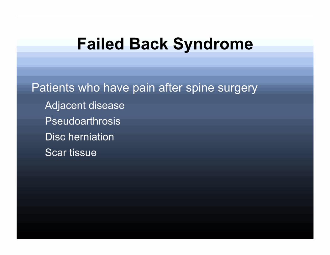
Failed Back Syndrome
Patients who have pain after spine surgery Adjacent disease Pseudoarthrosis Disc herniation Scar tissue

Non-invasive Interventions

Non-invasive Interventions
Bed Rest – Prolonged bed rest is no longer recommend
Bracing Traction
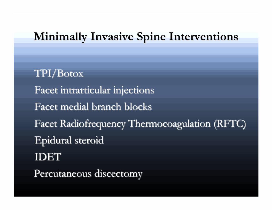

LESI

ESI

Facet Injection

Facet Medial Branch Block

Facet Radiofrequency Ablation

Facet RFTC

The linked image cannot be displayed. The file may have been moved, renamed, or deleted. Verify that the link points to the correct file and location.

Neuromodulation

Neuromodulation
• Consists of peripheral and spinal cord stimulators
• Intrathecal drug delivery systems • Deep brain stimulation • Gastric pacemakers etc

SCS
• Also known as dorsal column stimulator • Uses pulsed energy near the spinal cord • First placed in epidural space 1967 • Three companies currently produce SCS – Medtronic – Boston Scientific – St. Jude (ANS)

SCS

Spinal Cord Stimulation

Gate Theory
Melzack and Wall Foundation for SCS The notion that stimulation of A-beta fibers
“closes” the dorsal horn (gate) reducing nociceptive input from the periphery

Neuromodulation
Activation of descending and spinal pathways by serotonin and norepinephrine
Increased dorsal horn activity of GABA Suppression of CGRP

Advantages
• Analgesia on demand • Option when other treatments fail • Pt in control • Improved morale/quality of life • Avoids medication side effects

Advantages
• No restriction of daily activity • Reduction in pain medications • Reversible

Disadvantages
• Not effective in all cases (50-70%) • Invasive • Cost • Disconnection or equipment failure
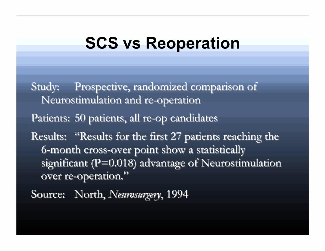
SCS vs Reoperation

SCS

SCS Indications Failed Back Syndrome
Most common in the US CRPS Extremity pain (neuropathic or vascular) Phantom Limb pain PVD Ischemic heart disease Abdominal pain Pelvic pain

SCS Contraindications
Infection Drug Abuse Uncontrolled psychiatric disease

SCS

The linked image cannot be displayed. The file may have been moved, renamed, or deleted. Verify that the link points to the correct file and location.

Intrathecal Drug Delivery System

IDDS
• IDD therapy involves the delivery of pain medicine in the intrathecal space
• The pump is connected to a thin, flexible catheter; both are implanted under the skin
• Smaller doses of medication are needed for effective pain relief because drug is delivered directly to the pain receptors

Potential Advantages

Efficacy

Non-Malignant Indications
• FBSS • Spinal stenosis • Spondylosis • Compression fx • Radiculitis • Post-thoracotomy pain • Postmastectomy
syndrome
• Peripheral neuropathy • Interstitial cystits • Chronic abdominal pain • Postherpetic neuralgia • RA

Important Considerations
• Is life expectancy greater than 3 months • Are pain complaints related to a physiologic
diagnosis • Is function limited by the pain • Is patient psychologically stable • Are there appropriate expectations and
understanding of risks • Has conservative treatment failed


MRI
• Synchromed II performance has not been established for greater than 3.0 Tesla horizontal, closed-bore MRI scanners
• Can cause motor stall • Pump can detect motor stall and motor stall
recovery • Pump should be interrogated after MRI to
confirm proper functioning

The linked image cannot be displayed. The file may have been moved, renamed, or deleted. Verify that the link points to the correct file and location.
The linked image cannot be displayed. The file may have been moved, renamed, or deleted. Verify that the link points to the correct file and location.


