Low Temperature Treatment at the YoungMicrospore Stage Induces ProteinChanges in Rice Anthers*□S
Nijat Imin‡, Tursun Kerim, Jeremy J. Weinman, and Barry G. Rolfe
Male reproductive development in rice is very sensitive tovarious forms of environmental stresses including lowtemperature. A few days of cold treatment (<20 °C) at theyoung microspore stage induce severe pollen sterility andthus large grain yield reductions. To investigate this phe-nomenon, anther proteins at the early stages of micros-pore development, with or without cold treatment at12 °C, were extracted, separated by two-dimensional gelelectrophoresis, and compared. The cold-sensitive culti-var Doongara and the relatively cold-tolerant cultivarHSC55 were used. The abundance of 37 anther proteinswas changed more than 2-fold after 1, 2, and 4 days ofcold treatment in cv. Doongara. Among them, one proteinwas newly induced, 32 protein spots were up-regulated,and four protein spots were down-regulated. Of these 37protein spots, we identified two anther-specific proteins(putative lipid transfer protein and Osg6B) and a calreti-culin that were down-regulated and a cystine synthase, a�-6 subunit of the 20 S proteasome, an H protein of theglycine cleavage system, cytochrome c oxidase subunitVB, an osmotin protein homologue, a putative 6-phospho-gluconolactonase, a putative adenylate kinase, a putativecysteine proteinase inhibitor, ribosomal protein S12E, acaffeoyl-CoA O-methyltransferase, and a monodehy-droascorbate reductase that were up-regulated. Identifi-cation of these proteins is available upon request. Accu-mulation of these proteins did not vary greatly after coldtreatment in panicles of cv. Doongara or in the anthers ofthe cv. HSC55. The newly induced protein named Oryzasativa cold-induced anther protein (OsCIA) was identifiedas an unknown protein. The OsCIA protein was detectedin panicles, leaves, and seedling tissues under normalgrowth conditions. Quantitative real time RT-PCR analy-sis of OsCIA mRNA expression showed no significantchange between low temperature-treated and untreatedplants. A possible regulatory role for the newly inducedprotein is proposed. Molecular & Cellular Proteomics 5:274–292, 2006.
Rice (Oryza sativa L.) evolved in tropical and subtropicalareas, and as a result it has the characteristic of being vul-nerable to cold weather. In particular, the combination of thelow temperature sensitivity and a maximal sensitivity to thesetemperatures at precise stages of pollen microspore devel-opment (1) makes cool midseason temperatures the majorenvironmental limitation (other than soil and water) for grow-ing rice in temperate regions and at high elevations in thetropics (2). During the life cycle of rice, pollen microsporedevelopment at the booting stage is known to be the mostsusceptible to cool weather damage (3). Cold damage at thisreproductive stage of rice causes limitation to the yields oftemperate-grown rice around the world and can reduce yieldsto 40% (3, 4). The main cause of this damage is pollen sterilityresulting from low temperature damage at the early stage ofmicrospore development (1). The developmental stage mostsensitive to the various forms of environmental stress, includ-ing cold damage, is just after meiosis. That is tetrad to earlymicrospore phase, or the young microspore stage (5). Incold-treated rice plants, both cytological and histological ab-normalities are greater in the anthers than in the pistil or otherorgans of the flower, and the cold damage can be rescued byartificial pollination with healthy pollen (5). In this process ofcold damage, anthers become smaller, and cells in the tapetallayer in the anthers that surrounds and is responsible fortransferring nutrients to the developing pollen undergo hyper-trophy and eventual breakdown. As a result, the normal de-velopment of pollen grains does not occur, and the pollengrains contain little or no starch and are functionally sterile (3,6). In anthers from cold-treated rice plants, anther respirationdecreases, sucrose accumulates in anthers, protein levelsdrop, and amino acid composition also changes including alarge decrease in proline and increase in asparagine (7). Re-cently it was found that a novel tetrasaccharide Ara2Gal2(where Ara is arabinose), which closely resembles the glycanchain of arabinogalactan protein, decreases after cold treat-ment in rice anthers (8). As �-1,3-glucanase, or callase, hy-drolyzes the callose wall and releases tetrads (9), Tsuchiya etal. (10) demonstrated that premature dissolution of the callosewall causes male sterility in transgenic tobacco. A decrease ininvertase activity in water-stressed rice anthers at the meiosisstage was also found, suggesting that a disturbance in car-bohydrate metabolism may be involved in male sterility whenanthers are subjected to water stress (11). Kitashiba et al. (12)
From the Australian Research Council Centre of Excellence forIntegrative Legume Research, Genomic Interactions Group, ResearchSchool of Biological Sciences, Australian National University, Can-berra, Australian Capital Territory 0200, Australia
Received, August 1, 2005, and in revised form, October 25, 2005Published, MCP Papers in Press, October 31, 2005, DOI 10.1074/
demonstrated partial male sterility in transgenic tobacco car-rying an antisense gene for an alternative oxidase under thecontrol of a tapetum-specific promoter, suggesting an impor-tant role of alternative oxidase in pollen development. It wasfound that a Petunia mutant that abolished pollen flavonolaccumulation and induced male sterility was complementedby a chalcone synthase transgene, indicating an involvementof flavonoid biogenesis in male sterility (13). It is also knownthat for many rice varieties, top dressing with nitrogen fertilizerat the booting stage exacerbates the problem of cold-inducedsterility (14). Again the reasons for these observations areunknown. A recent cDNA microarray analysis of rice anthergenes under chilling stress at the microsporogenesis stagerevealed as many as 160 cDNAs (of 8987 cDNAs) were up- ordown-regulated by chilling during the microspore releasestage, including two genes with the DNA transposon Cast-away in the 5�-flanking region (15). In that study the authorsused Hayayuki, a relatively cold-tolerant japonica rice varietyfrom Hokkaido (16) to identify genes involved in coldtolerance.
In general, a primary effect of chilling is considered to be thephase transition of membrane lipids at critical temperatures(17, 18). It has also been reported that, in rice plants, growthrate and metabolism are already markedly inhibited at tem-peratures above the chilling temperature in the range of 15–20 °C (19, 20). It is generally recognized that the proteinsynthesis patterns and mRNA levels change when plants areexposed to cold temperatures (21, 22). In addition, the mech-anism for the effects of low temperature stress on growth andthe accompanying metabolic changes and the relationship ofgeneral cold stress with cold-induced male sterility remainlargely unclear. Despite much recent research on the subject,the underlying mechanisms that cause cold-induced malesterility are poorly understood. In particular, it is not knownwhether the tapetal abnormalities, the tapetal hypertrophy, orthe biochemical changes seen are the causes or the conse-quences of the processes that lead to pollen sterility.
Proteomic analysis using two-dimensional gel electro-phoresis (2-DE)1 has been applied previously to investigatestress-related proteins (23–27) and developmentally regulatedproteins (28–31) in plants. To identify the earliest processes ofcold-induced male sterility, we focused on the identification ofthe anther proteins that are affected by cold temperatures at
the early stages of microspore development in rice using highresolution 2-DE and mass spectrometry analysis.
EXPERIMENTAL PROCEDURES
Chemicals—All chemicals used were of the highest obtainablegrade and are outlined in Refs. 32 and 33 unless stated. Deionizedwater (Millipore, Bedford, MA) with resistance of greater than 18megaohm-cm was used throughout.
Plant Materials, Growth, Cold Treatment, and Sampling—Cold-sensitive rice cv. Doongara was extensively studied in this project.The semidwarf, indica type Doongara was derived from a crossbetween tall, long grain cultivar Dawn (Texas) and IR8, a semidwarfcultivar carrying semidwarf-1 that was developed at the InternationalRice Research Institute in the 1960s (34). Relatively cold-tolerantHungarian cv. HSC55 was subsequently studied to examine thedifference between the responses of the two cultivars to cold tem-perature treatment. Seeds of rice (O. sativa L. cvs. Doongara andHSC55) were supplied by R. L. Williams, Yanco Agricultural Institute,New South Wales, Australia. Soil used in all the experiments was fromthe Yanco Agricultural Institute, New South Wales, Australia. Con-trolled environment facilities were used for rice growth and anthersampling. Rice (cvs. Doongara and HSC55) seeds were germinatedand grown in individual pots (height, 15 cm; diameter, 8.3 cm) in tubs(width, 36 cm; length, 52 cm; height, 30 cm). Nitrogen (15 g/m2) wasapplied as urea to 4-week-old seedlings, and the tubs were flooded tocover 10–20 cm of the shoots in a glasshouse under the followingconditions until panicle initiation: 30/20 °C (day/night), 70% relativehumidity, and natural day length. Plants under this growth conditionhad around three to four tillers each. Just after the time of panicleinitiation, the plants were transferred into a growth chamber andgrown under the same conditions except a day/night cycle of 12/12 hwith an average photon flux density of 330 �mol�m�2�s�1 was used.
Auricle distance (AD), the distance between the auricle of the flagleaf (last leaf) and that of the penultimate leaf, were used as anon-destructive measurement to gauge the stage of development ofthe rice panicle and pollen microspore. Correlation of the AD with thedevelopmental stages of the microspore was done by measuring ADof the panicle investigated and classifying the microspores in theanthers of the spikelets with the microscopic techniques describedbelow. We determined that the AD of �18 to �10 mm for cv. Doon-gara and �100 to �90 mm for cv. HSC55 corresponds with the tetradto early stage of microspore development of the top five spikeletsfrom the top three panicle branches as seen in Supplemental Table I.This measurement was used to determine the most temperature-sensitive stage for the start of cold treatment. However, it is importantto mention that the correlation of AD with the developmental stagesshould be used cautiously as many other factors such as growingseason, tiller number, and light and temperature conditions can alterthe correlation, and this needs to be checked in each batch of grownplants. Cold treatment was performed by moving individual plants inwhich the AD was about �15 mm for cv. Doongara and �100 to �90mm for cv. HSC55 into an identical chamber kept at 12 °C, 70%relative humidity with the same illumination and day/night cycle of thecontrol condition. To be able to compare anthers from cold-treatedplants with the anthers at the same stage of development of normallygrown plants, we used anthers from normally grown plants that hadthe same AD and developmental stage as the treated plants after thecompletion of their cold treatment (Supplemental Table I).
Microscopic Methods for Anther Analysis—Anthers were removedfrom the upper 5 spikelets from each of the top three branches ofimmature panicles with precision forceps and scalpels under a dis-section microscope. Then they were stained either by heating inacetocarmine solution (4% carmine in 45% acetic acid, boiled for 1 hunder reflux, and then filtered when cooled) or incubated with DAPI (1
1 The abbreviations used are: 2-DE, two-dimensional gel electro-phoresis; AD, auricle distance; CCoAOMT, caffeoyl-coenzyme A O-methyltransferase; COX, cytochrome c oxidase; COXVb, cytochromec oxidase subunit VB; cv., cultivar; HSC55, rice Hungarian cultivarHSC55; LTP, lipid transfer protein; ns-LTP, nonspecific lipid transferprotein; NCBI, National Center for Biotechnology Information; OsCIA,O. sativa cold-induced anther protein; PMF, peptide mass fingerprint;HSP, heat shock protein; sHSP, small heat shock protein; TIGR, TheInstitute for Genomic Research; %Vol, relative spot volume (the vol-ume divided by the total volume over the whole gel image); EST,expressed sequence tag.
Identification of Cold-responsive Proteins in Rice Anthers
�g/ml 4,6-diamidino-2-phenylindole in 50% ethanol) or with toluidineblue solution (pH 4.4) without heating (35). They were either prefixedin fixation solution (50% ethanol, 5% acetic acid) if they were to bestored prior to staining and microscopy, or they were stained directlyif microscopy proceeded immediately. DAPI-stained samples werevisualized under UV light. Alternatively for embedding and sectioning,anthers were fixed at least overnight in fixation solution, dehydrated at4 °C in increasing concentrations of ethanol (70, 80, 90, and 96% for1 h each), and infiltrated with Historesin (Leica Instruments, Heidel-berg, Germany) at 4 °C on a rotating wheel overnight. Sections of 10�m were cut on an Ultracut microtome (Reichert-Jung, Vienna, Aus-tria) with a glass knife. Sections were transferred to glass slides,stretched onto the slide surface, and then stained with a drop oftoluidine blue on top of each section. The slides were placed on awarming plate at 40 °C for 15 min after rinsing with water severaltimes. Color micrographs of the stained anthers were taken with aNikon FX35 camera mounted onto a Nikon SMZ-10 stereo micro-scope using Kodak Ektachrome 100 ASA, EPY 64T or AgfachromeRSX100 color reversal films.
Extraction of Rice Anther Proteins—Anthers were removed fromthe upper 15 spikelets of the top three branches of immature panicleswith precision forceps and scalpels under a dissection microscope.Anther protein extraction was preformed as described previously (36).The protein concentration of the supernatant was determined in amicrotiter plate by the Bradford assay (Bio-Rad) with bovine serumalbumin fraction V as the standard. The supernatants were stored inaliquots at �80 °C or directly loaded for isoelectric focusing. Eachindependent experiment was done at least three times. Anthers forthe binucleate and trinucleate stages were harvested 4 and 10 days,respectively, after AD reached �15 mm. Panicle samples were har-vested at the AD of �15 mm with and without cold treatment, and theproteins were extracted in the same way proportionally increasing thevolume of the extraction solutions.
Two-dimensional Gel Electrophoresis—2-DE was carried out in ahorizontal electrophoresis system, Multiphor II (Amersham Bio-sciences), as described previously (33, 36). For the first dimension,IPG gel strips (18 cm � 3 mm, pH 4–7 linear gradient and pH 6–11linear gradient; Amersham Biosciences) were used. 100 �g of totalproteins for silver stain and 500 �g for Coomassie stain were cup-loaded onto overnight-rehydrated IPG strips and focused with theImmobiline DryStrip kit in a Multiphor II electrophoresis unit (Amer-sham Biosciences) for 200 kV-h (for pH 4–7 linear) and 280 kV-h (forpH 6–11 linear) at 20 °C. After the first dimensional run, the IPG gelstrips were either sealed in plastic wrap and frozen at �80 °C orincubated at room temperature to equilibrate the strips in loadingbuffer prior to separation in the second dimension. The equilibratedIPG gel strips were then placed onto Amersham Bioscience ExcelGelSDS gels (12–14% T) and run on a Multiphor II unit at 5 °C.
Silver Staining, Colloidal Coomassie Staining, and Image Analysis—Proteins on analytical and micropreparative 2-DE gels were visualizedby silver staining and Coomassie staining, respectively, as describedpreviously (33). Detection, quantification, and matching of differen-tially displayed protein spots were performed with Melanie 3(GeneBio, Geneva, Switzerland; www.genebio.com/Melanie.html).Spot detection was carried out after reducing original images to 12-bitimages, and parameters were optimized by checking different proteinspots (big, intense, small, and faint) in certain regions of the gels. Theoptimized parameters used are as follows: number of smooths, 4;Laplacian threshold, 4; partials threshold, 2; saturation, 90; peaked-ness increase, 100; minimum perimeter, 30. To perform automaticdetermination of the observed pI and Mr values with the Melanieprogram, the digitized images were first calibrated. The apparentmolecular masses of the proteins were assigned after calibrating the2-DE gels by co-electrophoresis of protein standards (Amersham
Biosciences), and the apparent pI values of the proteins were as-signed after calculating the pI of selected landmark spots on the 2-DEgels using a ruler. This calculation was based on the informationprovided by the manufacturer that IPG (linear) strips have a linear pHgradient between a given pH range. Gel comparison was done visu-ally examining the spot intensity and subsequently calculating relativespot volume (%Vol, the volume divided by the total volume over thewhole gel image, whereas the volume of the spot is the integration ofthe highest optical density over the area of the spot) of the selectedprotein spots was calculated by the Melanie program. A protein spotwas classified as being differentially displayed if the %Vol varied morethan 2-fold between untreated and cold-treated plants in at leastthree experiments. Thus, we defined these proteins as cold-respon-sive proteins.
Western Blotting and Amino-terminal Sequencing—Semidry elec-trophoretic blotting onto PVDF membranes (Bio-Rad) was performedas described previously (33). Selected spots were excised from driedmembranes for amino-terminal Edman sequencing. This sequencingwas carried out at the Biomolecular Resource Facility of the AustralianNational University using Edman degradation chemistry on a Procise494-01 sequencer system (Perkin-Elmer Life Sciences).
MALDI-TOF MS and MS/MS Analysis—Proteins in preparative2-DE gels were visualized by colloidal Coomassie staining, and se-lected spots were excised from polyacrylamide gels. Selected spotswere in-gel digested and analyzed by MALDI-TOF as described pre-viously (33). Briefly the spots were destained and underwent an in-gel16-h tryptic digest at 37 °C. The resulting peptides were extractedfrom the gel with a 50% (v/v) acetonitrile, 1% (v/v) TFA solution. A 1-�laliquot was spotted onto a sample plate with 1 �l of matrix (�-cyano-4-hydroxcinnamic acid, 8 mg/ml in 50% (v/v) ACN, 1% (v/v) TFA) andallowed to air dry. MALDI mass spectrometry was performed with aMicromass TofSpec 2E time of flight mass spectrometer. A nitrogenlaser (337 nm) was used to irradiate the sample. The spectra wereacquired in reflectron mode in the mass range 600–3500 Da. Aninternal calibration was applied using two trypsin autodigestion peaksat 842.51 and 2211.1 Da.
For tandem mass spectrometry analysis, the in-gel digested spotswere analyzed either on a PE SCIEX (Foster City, CA) QSTAR hybridLC/MS/MS quadrupole TOF system or on a Micromass (Manchester,UK) LC/MS/MS quadrupole TOF system at the Australian ProteomeAnalysis Facility, Sydney, Australia. Data was acquired over the m/zrange 400–1800 Da to select peptides for MS/MS analysis. Afterpeptides were selected, the MS was switched to MS/MS mode, anddata were collected over the m/z range 50–2000 Da with variablecollision energy settings.
Sequence Analysis, Database Searching, and Peptide Mass Finger-print (PMF) Analysis—Signal peptides and the other targeting se-quences were predicted with the SignalP (37), MITOPROT (38), andTargetP (39) programs. Molecular mass and pI of the theoreticalproteins were calculated after deleting the signal peptides. For aminoacid sequence homology comparisons, amino-terminal protein se-quences were used to search non-redundant protein databases(Swiss-Prot, TrEMBL, and NCBInr) using the FASTA (40) and BLAST(41) programs or to search translated rice gene index (OsGI) se-quences (www.tigr.org) using WU-BLAST. For protein identificationfollowing MS/MS analysis, full or partial amino acid sequences gen-erated by decomposition analysis were used to search non-redun-dant protein databases as mentioned above. Masses of the full pep-tide were also considered for the confidence of the matches. In thesearches, methionine residues were assumed to be modified to me-thionine sulfoxide, and cysteine residues were assumed to be re-duced and alkylated by iodoacetamide to carboxyamidomethylcys-teine wherever necessary. Protein identification by PMF was done asdescribed previously (33). Briefly, the list of masses were used to
Identification of Cold-responsive Proteins in Rice Anthers
search against Swiss-Prot and NCBInr protein databases available at129.85.19.192/prowl-cgi/ProFound.exe using the search engine Pro-Found (42). All peptide masses were assumed as monoisotopic and[M � H]� (protonated molecular ions). In the searches, methionineresidues were assumed to be modified to methionine sulfoxide, cys-teine residues were assumed to be reduced and alkylated by iodoac-etamide to carboxyamidomethylcysteine wherever necessary, andone missed cleavage site was allowed. For PMF matches, the numberof peptides matched (at least four), sequence coverage (at least 20%),mass accuracy (lower than 50 ppm), number of missed cleavages (nomore than two single cleavages), number of chemical modifications(no more than two modifications), and Z score (above 1.6) (42) wereconsidered as confident identifications. Z score is the distance to thepopulation mean in unit of standard deviation. For example, a Z scoreof 1.65 for a search means there are about 5% of random matchesthat could yield higher Z scores than this search (see Ref. 42 fordetails). The multiple sequence alignment and the construction ofphylogenetic trees were done by using the ClustalW program (43)available at www.ebi.ac.uk/clustalw/.
RT-PCR and Real Time PCR—Total RNA from rice panicles andanthers was extracted using the RNeasy plant minikit (Qiagen) ac-cording to the manufacturer’s instructions. cDNA synthesis was doneusing 5 �g of total RNA for each sample. RNA was treated in 1�buffer with 2 units of DNase I (Ambion, Austin, TX) added to thereaction and incubated for 30 min at 37 °C. The reaction was stoppedby adding 1 �l of 25 mM EDTA followed by a 15-min incubation at65 °C. One microliter of 5 �M oligo(dT)18 was added to the reaction,and it was incubated for 10 min at 70 °C and then chilled on ice. Firststrand cDNA was synthesized using SuperScript II reverse tran-scriptase (Invitrogen) or water (for the non-reverse transcriptase con-trol). PCR for cloning was done using the primers OsCIA_F (5�-GTTGAC ATG TCG ACG GTG ACC TC-3�) and OsCIA_R (5�-GAC TCGAGT CGA CAT CGA-3�).
The following real time PCR primers were designed using PrimerExpress software (Applied Biosystems): forward 5�-AGA GAG GGTGTC CAG GCA AA-3� and reverse 5�-TCC TTG ACC TTG CCA ATGGT-3� for OsCIA and forward 5�-TGC GAT AAT GGA ACT GGT ATGG-3� and reverse 5�-ACA GCC CTG GGC GCA T-3� for the actin gene(GenBankTM accession number X15865 was used for normalizationas a control). PCR was carried out in a total volume of 20 �l containinga 0.2 �M concentration of each primer and 1� SYBR green PCRMaster Mix (PE Applied Biosystems). Reactions were amplified in anABI PRISM 7700 thermocycler as follows: 95 °C for 10 min, then 40cycles of 95 °C for 15 s, and 60 °C for 1.5 min. Normalization wasdone as described previously (44) against the rice actin gene. Twobiological and two technical repeats were done for each treatment.For each sample, a no RT enzyme reaction was performed also in tworeplicates.
Southern Blotting—Genomic DNA was isolated using the PURE-GENE DNA isolation kit (Gentra). Ten micrograms of genomic DNAwere digested with restriction enzymes electrophoresed through a0.8% agarose gel and then transferred to Hybond-N� membrane(Amersham Biosciences) using an alkaline method. Radiolabeledprobes were generated using PCR-amplified template by a Prime-a-Gene� labeling system (Promega). Hybridization was done at 60 °Cusing ExpressHyb (Clontech) solution according to the manufactur-er’s instructions.
Protein Expression, Antibody Production, and Immunodetection—PCR-amplified fragments were ligated into pET28�(a) vector (Nova-gen), transformed into BL21(DE3)pLysS competent cells (Invitrogen),and induced with 0.5 mM isopropyl 1-thio-�-D-galactopyranoside(Sigma). Recombinant protein with a carboxyl-terminal His6 tag waspurified with HIS-Select nickel affinity gels (Sigma) according to themanufacturer’s instructions. Polyclonal antibody was produced in
New Zealand White rabbits using purified protein as antigen, andimmunodetection was done as described previously (45).
RESULTS
Cold-treated Doongara Plants Showed High Levels of Ste-rility—To ensure that the cold treatments used were causingsterility, we tested periods of cold temperature initiated at theyoung microspore stage after which plants were grown nor-mally, and the effect of the treatments on eventual grains filledwas assessed. We focused on the analysis of the upper 15spikelets because they were reported as the most sensitivespikelets and synchronous in microspore development (6).Anthers at the young microspore stage were chosen as themain tissue and stage of analysis (5). A temperature conditionof 12/12 °C day/night was used to maximize the effect of coldstress. By growing rice plants under this temperature condi-tion, considerable sterility was observed in cv. Doongaraplants that had a cold treatment that commenced at theyoung microspore stage. Fig. 1 shows the sterility of plantsdue to varying lengths of cold treatment initiated at the earlystage of microspore development. The upper 15 spikelets fromthe top three branches of the cold-sensitive cv. Doongara plantsshowed 4% sterility without cold treatment. One-day cold treat-ment at a constant 12 °C initiated at the young microsporestage did not have much effect on the plant, only increasing themeasured sterility by 2%. However, 2 and 4 days of cold treat-ment caused 33 and 86% sterility, respectively. In contrast,the cv. HSC55 showed considerably greater tolerance to coldtreatment, having a sterility of 24% after 4 days of coldtreatment and 1% sterility without cold treatment (Fig. 1).
FIG. 1. Effect of cold treatment initiated at the young micros-pore stage on sterility of the rice cvs. Doongara and HSC55.Sterility was determined by counting the number of filled grains of thespikelets from the top three branches. Columns 1–4 represent controland 1, 2, and 4 days of cold treatment of cv. Doongara at 12 °C,respectively. Columns 5 and 6 represent control and 4 days of coldtreatment of cv. HSC55 at 12 °C, respectively. Error bars representstandard deviation (n � 40).
Identification of Cold-responsive Proteins in Rice Anthers
Cold-treated Doongara Plants Showed Cytological Abnor-malities in Anthers—As previous research has demonstratedcytological abnormalities in the tapetal cells due to cold treat-ment (3, 6), we also examined the effect of the cold treatmentwe were using on the anthers harvested from the upper 15spikelets. Under the microscope, cross-sections of cold-treated anthers showed abnormal tapetum and microsporedevelopment in the cold-sensitive cv. Doongara. There wassevere swelling of tapetal cells known as tapetal hypertrophyobserved after cold treatment. Microspores also showed ab-normal development (Fig. 2).
Cold Treatment Induced Protein Changes in Doongara An-thers—To study the effects of cold treatment on the proteinexpression level, we used 2-DE to display and compare allextractable rice anther proteins from cold-treated and un-treated plants. To do this, we have performed 1, 2, and 4 daysof cold treatment at 12 °C for cv. Doongara and 4 days of coldtreatment for cv. HSC55 plants. Anther proteins with andwithout cold treatment were extracted from microdissectedanthers separated by 2-DE. The 2-DE anther protein maps of
anther proteins from 4-day cold-treated cv. Doongara plantsare shown in Fig. 3 (pH 4–7) and in Fig. 4 (pH 6–11). Melanie3 image analysis of at least three replicate gels revealed thatover 4000 anther proteins were reproducibly resolved in sil-ver-stained gels over a combined pH range of 4–11 and sizerange of 6–120 kDa (Figs. 3 and 4). Silver-stained gels (pH4–7) were used for most of the comparisons and quantifica-tion, although narrow pH range gels were used for referenceand further separation of crowded protein spots (data notshown). Coomassie-stained gels were also generated (exceptfor 2-day cold treatment and its control due to insufficientmaterials for those typical analyses) and used for compari-sons and quantification of the proteins that do not stain wellwith the silver nitrate method. Silver-stained gels (pH 6–11)were generated for only 4-day cold-treated cv. Doongaraplants and their controls. Although the global pattern of antherproteins was largely unaltered (more than 99% of the totalprotein spots remained unchanged), 37 protein spots werereproducibly detected as changed in their protein levels after1, 2, and/or 4 days of cold temperature treatment at 12 °C in
FIG. 2. Effect of cold treatment on cv. Doongara anthers. The cross-sections of the early and middle stages of microspore developmentwere stained with toluidine blue. A, B, and C are the cross-sections of the anthers from plants that were untreated or that underwent 2 and4 days of cold treatment at 12/12 °C beginning at the young microspore stage, respectively. D, E, and F show enlarged loculi of A, B, and C,respectively. Swollen tapetal cells are indicated with arrows. Scale bars represent 100 �m for the top panels and 20 �m for the bottom panels.
Identification of Cold-responsive Proteins in Rice Anthers
the cold-sensitive cv. Doongara when compared with theirrelevant untreated plants as shown in Figs. 3 and 4. All the up-and down-regulated protein spots are shown, although some(spots 25, 34, and 36) were not detected as protein spots on
the silver-stained gel (Fig. 3). Selected parts of the gels arehighlighted in Fig. 5A to show comparisons of cold-respon-sive protein spots of rice anthers in cold-treated and un-treated plants. Relative protein levels (shown as %Vol) of
FIG. 3. 2-DE protein map of Doon-gara anther proteins cold-treated for4 days at the young microspore stage.Protein spots were assigned arbitraryidentifiers. Up- and down-regulated pro-tein spots are marked with rectanglesand circles, respectively. Isoelectric fo-cusing in the first dimension was on18-cm IPG strips with a linear gradientranging from pH 4 to 7 and loaded with100 �g of extracted anther protein. Forthe second dimension 12–14% T SDS-PAGE gels were used. Apparent molec-ular masses of protein standards (�103)are indicated on the left. Proteins werevisualized by silver staining. Spots 25and 34, which stained poorly with silver,are shown after Coomassie staining inFig. 5.
FIG. 4. 2-DE basic protein map of Doongara basic anther proteins cold-treated for 4 days at the young microspore stage. Proteinspots were assigned arbitrary numbers. Up- and down-regulated protein spots are marked with rectangles and circles, respectively. Isoelectricfocusing in the first dimension was on 18-cm IPG strips with a linear gradient ranging from pH 6 to 11 and loaded with 100 �g of extractedanther protein. For the second dimension 12–14% T SDS-PAGE gels were used. Apparent molecular masses of protein standards (�103) areindicated on the left. Proteins were visualized by silver staining.
Identification of Cold-responsive Proteins in Rice Anthers
these 37 cold-responsive proteins and their changes afterdifferent lengths of cold treatment are given in Table I. Exam-ples of protein spot comparison and their correlation withrelative expression levels are given in Fig. 5B. One proteinspot (spot 37) was newly induced in all cold treatments com-pared with controls. We named this protein O. sativa cold-induced anther protein (OsCIA). Three protein spots (spots 12,34, and 36) were down-regulated, and 34 protein spots wereup-regulated after different days of cold treatment (Figs. 3 and4 and Table I). For spots 25 and 34, %Vol was measured fromCoomassie-stained gels as these spots did not stain well withthe silver nitrate method (Figs. 5 and 6).
The Hungarian cv. HSC55 was reported as a relativelycold-tolerant rice cultivar,2 and this was also proved in ourexperiments (Fig. 1). We compared 2-DE anther protein maps(pH 4–7, silver-stained and Coomassie-stained) of controland 4-day cold-treated cv. HSC55 plants. Seven proteinspots were differentially displayed after cold treatment. Sixprotein spots (h1, h2, h3, h4, h5, and h7) were up-regulated,and one spot (h6) was down-regulated as shown in Table Iand Supplemental Fig. 1. By gel matching, we found that spoth3 was the same as spot 36 detected in cv. Doongara anthersamples. Except for protein spot 36, we detected no changesin cv. HSC55 that we also detected in cv. Doongara.
We also compared 2-DE panicle protein maps (pH 4–7,silver-stained and Coomassie-stained) of control and 4-daycold-treated cv. Doongara plants. Five protein spots weredetected as differentially displayed including one spot (p5)
that was up-regulated and four spots (p1, p2 p3, and p4) thatwere down-regulated. By gel matching, we found that spot p1was same as spot 11 detected in cv. Doongara anther sam-ples. Except for protein spot 11, we did not see any of thechanges in protein abundance that occurred in cv. Doongaraanthers upon cold treatment. However, spot 26 can be seenbut at a much reduced level in the Coomassie-stained gels ofpanicle samples run for the same pI range and running con-ditions, indicating an enrichment in the anther tissues (Sup-plemental Fig. 2).
Protein Spot 34 Is Detected in a Spatially and TemporallyExpressed Manner in Rice Anthers—Spot 34 could not bedetected in the silver-stained or Coomassie-stained gels ofpanicle samples of the same developmental stage run overthe same pI range under identical conditions, indicating an-ther-specific expression of the protein (Fig. 6A). In both cvs.Doongara and HSC55, spot 34 could be detected in 2-DE gels(in Coomassie-stained gels only) of anthers at the youngmicrospore stage. To gain insight into the expression patternof this spot, cv. Doongara anthers at the young microspore(uninucleate), binucleate, and trinucleate stages were ex-tracted, and their 2-DE protein patterns were compared (Fig.6A). We observed a decrease in spot 34 protein levels at thebinucleate stage, and it became completely undetectable atthe trinucleate stage. This stage contains mature pollen, indi-cating specific accumulation of protein spot 34 in the earlierstages of microspore development in rice anthers.
Identification of Cold-responsive Proteins—Amino-terminalsequences were obtained by Edman degradation analysis forsix of 11 cold-responsive protein spots blotted onto PVDF2 R. L. Williams and T. C. Farrel, personal communication.
FIG. 5. Examples of protein changes and their correlation with %Vol in cv. Doongara anthers in response to cold treatment. A, proteinspot numbers correlate to those in Table II. Con, control; 4D, 4 days of cold treatment. All enlargements are from silver-stained gels exceptfor the two at the bottom, which are from the colloidal Coomassie-stained gels. B, examples of protein differential display and their correlationwith relative protein levels in cvs. Doongara and HSC55 anthers in response to cold treatment. Spots 11 and 12 are from the silver-stained gels;spot 34 is from the colloidal Coomassie-stained gels. Numbers 1–4 represent the Doongara control, Doongara cold-treated, HSC55 control,and HSC55 4-day cold-treated at 12 °C samples, respectively. The gel images correspond to the %Vol shown below. %Vol was calculatedas described under “Experimental Procedures.”
Identification of Cold-responsive Proteins in Rice Anthers
membranes as shown in Table II. No sequence data could beobtained for protein spots 6, 9, 10, and 11 due to either theamino terminal being blocked or not enough protein being
available. Two amino-terminal sequences were generatedfrom spot 25, each matching to two mitochondrial proteins,the H protein of glycine decarboxylase complex and cyto-
TABLE IRelative protein levels in response to cold treatment for different days at 12 °C in cv. Doongara
a Cold-responsive protein spots were listed with the same identifiers given in Figs. 3 and 4.b Experimental molecular mass (�103) and pI.c 1, 2, and 4 represents 1, 2, and 4 days of cold treatment at 12 °C, respectively.d Percent spot volume ratio for a protein from cold-treated and untreated plants.e Newly induced, up-regulated, and down-regulated protein spots were marked with11,1, and2 symbols, respectively, and the relative
spot volume ratio changes greater (�) or less (�) than 2-fold or more are given.f %Vol measured from Coomassie-stained gels as these spots did not stain well with the silver nitrate method.
Identification of Cold-responsive Proteins in Rice Anthers
chrome c oxidase subunit VB (COXVb), respectively (Table II).For these two proteins, matches to database proteins oc-curred from the amino acids 35 and 55 of the databaseproteins, respectively. Putative mitochondrial targeting se-quences and the cleavage sites were detected for both pro-teins by using the MITOPROT program (38). High probabilityscores of 0.99 and 0.91 for export to mitochondria and puta-tive cleavage sites at amino acids 42 and 51, respectively,were given by the program. Amino-terminal sequences ofspots 12 and 34 were matched to the amino acid sequencesof rice calreticulin and a translation product of the t42 genefrom the amino acids 26 and 29, respectively. For both pro-teins, signal peptides of 25 and 28 amino acids, respectively,were predicted by the SignalP program (37).
Both BLASTP and FASTA searches against the non-redun-
dant NCBInr and Swiss-Prot protein databases showedstrong homology (about 40% identity) of the translated se-quence of the t42 gene (we refer to this protein as T42) tomany other plant nonspecific lipid transfer proteins (ns-LTPs).This match was validated by a search against the conserveddomain database at NCBI that showed the strongly con-served LTP domain in the sequence (E value, 3e�12). The LTPdomain of T42 can be written as CX9CX15CCX19CXCX22C-X13C. A further search of T42 against the translated Gen-BankTM dbEST using the TBLASTN algorithm gave �80%identity (with expected value lower than e�36) to anther-spe-cific ESTs from sorghum, goat grass, rye, and wheat withsimilar molecular weight and pI. Alignment and a phylogenetictree of T42 with two anther-specific EST translations andsome selected (on the basis of close homology from different
FIG. 6. Temporal and spatial expression of the T42 protein and its alignment and phylogenetic tree with other plant ns-LTPs. A,temporal and spatial expression of T42 protein. 2-DE gel (pH 4–7, Coomassie-stained) regions containing T42 protein (spot 34) of Doongarapanicle at the auricle distance of �15 mm (panel 1) and anther at the uninucleate (panel 2), binucleate (panel 3), and trinucleate stages (panel4) are shown. B, alignment of T42 protein with other plant LTPs and with translated amino acid sequence of anther ESTs from goat grass(BQ840880) and rye (BF429311) are shown. This was done using the ClustalW program. Signal peptide is shown at the amino-terminal end ofthe T42 protein sequence. Eight conserved cysteines are indicated with asterisks; phenylalanine 115 that replaced tyrosine of which the sidechain divides the hydrophobic cavity into two parts in other ns-LTPs is indicated with an arrow. C, a phylogenetic tree representing alignmentof T42 protein from selected sources is given. Lipid transfer proteins from different plant species and translated amino acid sequence of antherESTs from goat grass and rye are shown. The neighbor-joining tree using percent identities was displayed by the program Jalview based ona multiple sequence alignment generated with the program ClustalW. The scale represents percentage of substitution per site.
Identification of Cold-responsive Proteins in Rice Anthers
species) ns-LPTs based on percentage identity are given inFig. 6, B and C.
One to three sequence tags were obtained for five of theeight protein spots analyzed by MS/MS analysis (Table II).Seventeen cold-responsive protein spots were analyzed byMALDI-TOF MS, and eight of them could be identified by PMFanalysis (Table III). Seven other protein spots (spots 2, 6, 9,10, 18, 26, 31, and 35) could not be identified due to either anincomplete genome sequence being available, a poor massspectrum being generated, or post-translational modificationsof the mature proteins in vivo. The matches obtained fromMS/MS and amino-terminal sequencing analysis for proteinspots 8 and 25 were confirmed with PMF analysis.
OsCIA, a Novel Protein, Was Suppressed in Rice Anthersunder Normal Conditions but Induced by Cold Treatment—Asdescribed in Table I, spot 37 (OsCIA) was the only cold-induced protein spot detected after all cold treatment. Weexamined the panicle proteome of cv. Doongara and theanther proteome of the relatively cold-tolerant cv. HSC55 by2-DE protein differential display. In the cv. Doongara panicleproteome, a protein spot (p8) was detected at the sameposition as spot 37 from the cv. Doongara anther proteome bymatching 2-DE gels for the pH ranges of 4–7 and 5–6 (datanot shown). Interestingly this panicle protein spot did notchange in its protein level upon cold treatment (Fig. 7). Furtherevidence of the shared identity of these two protein spots wasobtained by amino-terminal Edman sequencing (for the pan-icle protein, the sequence was AISSNKPGDPKI) and MALDI-TOF analysis. The protonated masses of the peaks were1777.02, 2239.14, and 3100.40 for spot 37 and 1777.02,
2239.11, and 3100.31 for spot p8, respectively. At least it isevident that these two protein spots (37 and p8) share similarmolecular mass, pI, PMF pattern, and amino-terminal se-quence. OsCIA was detected in cv. Doongara anthers at ahigh level after as little as 1 day of cold treatment at 12 °C.However, upon restoring normal growth conditions after coldstress that lasted for 4 days from the young microspore stage,the level of OsCIA started to decrease, and it had disappearedby the trinucleate stage (data not shown). We detected aprotein spot (h8) in cv. HSC55 in the same position as spot 37by gel comparison and gel matching that did not change uponcold treatment (data not shown). However, we identified h8 asa putative small heat shock protein (sHSP) by MS/MS analysis(partial sequence obtained was PGAYAFVVDM, whichmatched to a rice putative 18-kDa heat shock protein, Gen-BankTM accession number XP_550428). Considering any pos-sible mismatching, we also analyzed other cv. HSC55 antherprotein spots close to spot h8 from the narrow range (pH 5–6)2-DE proteome map of cv. HSC55 anthers by MALDI-TOFwithout finding any PMF pattern similar to that of spot 37 (datanot shown).
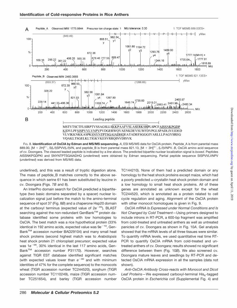
OsCIA proteins (spot 37) from five gel pieces and PVDFblots of rice anther 2-DE from cold-treated plants were pooledtogether for MS/MS analysis and Edman sequencing, respec-tively. MS/MS analysis generated two partial sequences fromtwo different peptides with overlapping sequences and over-lapping masses (Table II and Fig. 8A). Subtraction of themasses of the amino acids valine and proline from the mass ofthe carboxyl-terminal fragment of peptide_A is equal to themass of the carboxyl-terminal fragment of peptide_B (Fig. 8A).
TABLE IICold-responsive rice anther proteins identified by amino-terminal and MS/MS sequencing in cv. Doongara
Protein spots are listed with the same identifiers given in Figs. 3 and 4.
Spot no.Molecular mass
(kDa)/pIaSequences Matching identityb Accession
no.cMolecular mass
(kDa) /pId
8 26.5/6.39 DVFASATER,e SPSPLLLPAR,e
EYIDLRKDe�-6 subunit of 20 S proteasome O64464 24.3/6.43
12 35.8/4.36 EVFFQEKFEDGWESR Calreticulinf Q9SLY8 45.6/4.4824 15.4/5.41 ALCAEHNVHLVTVPSAKe 40 S ribosomal protein S12E Q8H2J8 14.8./5.3325a 14.7/4.38 STVLDGLKYSSSHE 15-kDa H protein of glycine
decarboxylase complex (fragment)fQ9FW99 7.1/8.98
25b 14.7/4.38 SGDKPATVEDVMPIA Cytochrome c oxidase subunit Vbf P92683 12.7/4.4431 31.4/5.68 SGTTTTXLTLHHHRT Osmotin protein homologuef AAX95344 21.3/5.3934 9.7/4.49 AVQCGQVMQLMAPCM,
DAAAGFPAVDFSReTranslated product of t42 genef CAA01672 9.4/4.49
a Experimental molecular mass (kDa) and pI. See “Experimental Procedures” for details.b All the peptides showed 100% identity to the matching sequences except for spot 31, which was 70%.c Swiss-Prot, TrEMBL, or GenBankTM accession numbers are given. All matches are in O. sativa.d Theoretical molecular mass (kDa) and pI of the matched proteins were calculated after removing experimentally identified leading
sequences.e Sequences from MS/MS analysis otherwise from N-terminal Edman sequencing. X represents an unknown amino acid. Low energy CID
cannot differentiate between the isomeric Leu/Ile pair; as such both leucine and isoleucine are shown as L.f Sequence alignment inferred the proteolytic cleavage of a signal peptide from these proteins.
Identification of Cold-responsive Proteins in Rice Anthers
Edman sequencing was done for the first 12 amino acids ofthe protein and for the 15 amino acids of the major peptideafter tryptic digestion. Searches for the Edman sequences orMS/MS sequences of the OsCIA against the non-redundantprotein database matched the protein to an unknown proteinin rice cv. Nipponbare (Table II). The match for the amino-terminal sequence was from amino acid 47 suggesting themature protein has a leader (or target) peptide cleaved offfrom the precursor protein. The corresponding gene for theunknown protein in cv. Nipponbare is located on chromo-some 10. We cloned the corresponding gene (both genomicDNA and cDNA) in cv. Doongara (cDNA was deposited inGenBankTM under accession number DQ116755), and thisidentified a 738-bp-long intron interrupting two exons. Whencompared with cv. Nipponbare, there were three nucleotidesubstitutions in two exons (Fig. 8). Interestingly there was nosubstitution in the intron. As a result of single nucleotidesubstitutions, serine 61 is replaced by leucine, lysine 118 isreplaced by asparagine, and lysine 189 is replaced by gluta-mine at the protein level in cv. Doongara (Fig. 8).
Southern hybridization was performed under standard con-ditions (65 °C) to estimate OsCIA gene copy number in ricecultivars Doongara and Nipponbare. Single bands were de-tected with four different restriction enzymes (BamHI, EcoRI,HindIII, and PstI). These results suggest that OsCIA is en-coded by single gene copy in the genome of Doongara andNipponbare (Supplemental Fig. 3). However, there were somedifferences in the hybridizing fragment sizes between Doon-gara and Nipponbare digested with BamHI and PstI (Fig. 10)indicating some differences in the flanking sequences of thesetwo cultivars.
Analysis of the OsCIA amino acid sequence using Peptide-Mass (au.expasy.org/tools/peptide-mass.html) and MS/MSand MALDI-TOF data indicated that the major peptide1777.02 [M � H]� (from MALDI-TOF) is the same as pep-tide_A, 889.00 [M � 2H]2� (from MS/MS), and the amino acidsequence derived from this peptide is SISPVVLVN-PVPVDGER. This peptide was a result of a chymotrypticcleavage by trypsin. The amino acid sequence of peptide_B,821.13 [M � 3H]3� (from MS/MS), is IDVLPFSISPVVLVN-PVPVDGER (the same sequence obtained from peptide_A is
FIG. 7. Comparison of OsCIA protein spot containing regions of2-DE gels from cv. Doongara anthers and panicles. 4D, 4 days ofcold treatment; Con, control (untreated). Note that protein spot p8was identified as OsCIA (see text).
a Experimental molecular mass (kDa) and pI.b Swiss-Prot or TrEMBL accession numbers are given.c Number of peptides matched (PM) and number of peptides identified (PI)/percent sequence coverage.d Z score is the distance to the population mean in unit of standard deviation. For example, a Z score of 1.65 for a search means there are
about 5% of random matches that could yield higher Z scores than this search (see Ref. 42 for details).e Theoretical molecular mass (kDa) and pI of the matched proteins.
Identification of Cold-responsive Proteins in Rice Anthers
underlined), and this was a result of tryptic digestion alone.The mass of peptide_B matches correctly to the above se-quence in which serine 61 has been substituted by leucine incv. Doongara (Figs. 7B and 8).
An InterPro domain search for OsCIA predicted a bipartite-type (two basic domains separated by a spacer) nuclear lo-calization signal just before the match to the amino-terminalsequence of spot 37 (Fig. 8B) and a chaperone Hsp20 domainat the carboxyl terminus (expected value of 2e�05). BLASTsearching against the non-redundant GenBankTM protein da-tabase identified some proteins with low homologies toOsCIA. The best match was a rice hypothetical protein (33%identical in 192 amino acids, expected value was 9e�10, Gen-BankTM accession number BAD29104) and many small heatshock proteins (second highest match was to Arabidopsisheat shock protein 21 chloroplast precursor; expected valuewas 1e�06, 30% identical in the last 117 amino acids, Gen-BankTM accession number P31170). However, searchesagainst TIGR EST database identified significant matches(with expected values lower than e�30 and with minimumidentities of 47% for the complete sequence) to the monocotswheat (TIGR accession number TC244520), sorghum (TIGRaccession number TC110249), maize (TIGR accession num-ber TC251654), and barley (TIGR accession number
TC144210). None of them had a predicted domain or anyhomology to the heat shock proteins except maize, which hada poorly predicted �-crystallin heat shock protein domain anda low homology to small heat shock proteins. All of thesegenes are annotated as unknown except for the wheatTC244520, which is annotated as a protein related to cellcycle regulation and aging. Alignment of the OsCIA proteinwith other monocot homologues is given in Fig. 9.
OsCIA mRNA Is Expressed under Normal Conditions and IsNot Changed by Cold Treatment—Using primers designed toinclude introns in RT-PCR, a 600-bp fragment was amplifiedfrom cold-treated and untreated anthers as well as untreatedpanicles of cv. Doongara as shown in Fig. 10A. Gel analysisshowed that the mRNA levels of all three tissues were similar.To quantify mRNA levels, we used quantitative real time RT-PCR to quantify OsCIA mRNA from cold-treated and un-treated anthers of cv. Doongara; results showed no significantdifference between them (Fig. 10B). We also screened cv.Doongara mature leaves and seedlings by RT-PCR and de-tected OsCIA mRNA expression in all the samples (data notshown).
Anti-OsCIA Antibody Cross-reacts with Monocot and DicotLeaf Proteins—We expressed carboxyl-terminal His6-taggedOsCIA protein in Escherichia coli (Supplemental Fig. 4) and
FIG. 8. Identification of OsCIA by Edman and MS/MS sequencing. A, ESI MS/MS data for OsCIA protein. Peptide_A is from parental mass889.00, [M � 2H]2�, S(L/I)SPVV(L/I)VN, and peptide_B is from parental mass 821.13, [M � 3H]3�, (L/I)VNPV. B, OsCIA amino acid sequenceof cv. Doongara. The cleaved leaded peptide is indicated by a line above. The predicted bipartite nuclear localization signal is boxed. PeptidesAISSNKPGDPKI and SNYNTPTSGAIADHQ (underlined) were obtained by Edman sequencing. Partial peptide sequence SISPVVLVNPV(underlined) was derived from MS/MS data.
Identification of Cold-responsive Proteins in Rice Anthers
purified the recombinant protein using immobilized-metal af-finity chromatography. The recombinant protein was identi-fied as OsCIA by Edman sequencing (data not shown). Poly-clonal antibodies were raised in rabbits. Using the anti-OsCIAantibody, we screened seedlings of several monocots (riceand maize) and dicots (Arabidopsis thaliana and Medicagotruncatula) and detected strong single 18- or 22-kDa bands inWestern blots (Fig. 11).
DISCUSSION
Previous studies identified some physiological and cytolog-ical characteristics of cold damage at the reproductive stageof plant development, leading to male sterility in rice (1, 3, 5).This research indicated the most cold-susceptible organ(which is the anther), the most sensitive stage (which is fromtetrad to early microspore stage), and the effect of differentdays of cold treatment on the sterility of the plant (1, 5, 6). Inour experimental system, we extended these observations to
the cold-sensitive cultivar Doongara. This enabled us to focuson anthers of the upper spikelets from the top three branchesof the panicle at the young microspore stage and use a 12 °Ccold treatment as it gives very high sterility to the spikelets ofinterest.
Most of the Anther Proteins Were Unaltered by Cold Treat-ment—Greater than 4000 rice anther proteins were resolved,
FIG. 10. RT-PCR analysis of OsCIAmRNA. Left panel, RT-PCR amplifica-tion of OsCIA mRNA from cv. Doongaraanther and panicle samples. To avoidgenomic DNA contamination, total RNAwas treated with DNase I and verified bya primer pair designed to include theintron. ACon, anther control (untreated);A1d, anther cold-treated for 1 day; Pc,panicle control (untreated). Right panel,quantitative real time RT-PCR analysis ofOsCIA mRNA levels in cv. Doongara an-ther with 1-day cold treatment (1D cold)or without cold treatment.
FIG. 11. Immunodetection of anti-OsCIA antibody-reacting pro-teins in monocots and dicots. Do, rice cv. Doongara; Mt, M. trun-catula; Zm, Z. mays; At, A. thaliana. 20 �g of total proteins wereloaded in each lane and probed with anti-OsCIA antibody.
FIG. 9. Alignment of OsCIA with its close homologues. Sorghum (TC110249), maize (TC251654), and wheat (TC244520) ESTs weretranslated and aligned using the ClustalW program.
Identification of Cold-responsive Proteins in Rice Anthers
and cold temperature treatments of 1, 2, and 4 days wereextensively studied to reveal a small subset of proteins thatare cold-responsive. Although the global expression patternof anther proteins was largely unaltered after cold treatment,35 proteins were observed as altered after comparison ofmany independent experiments. Eleven of these cold-respon-sive proteins have been identified previously, and they can beaccessed at the ANU-2DPAGE site (semele.anu.edu.au/2d/2d.html). Previously we have identified 75 protein spots fromrice anthers at the young microspore stage (33), and they canbe accessed at the ANU-2DPAGE site. We did not detectchanges in protein levels of most of these protein spots uponcold treatment except for protein spots 17 (a putative adeny-late kinase) and 23 (a cysteine proteinase inhibitor). For in-stance, ascorbate peroxidase, which was reported to be in-volved in the tolerance of the cell to low temperature stress inrice (46), did not vary two-fold or more in its protein level uponcold treatment (data not shown). This was also true for someproteins such as heat shock protein 70, heat shock protein 60,and enolase, which are required for survival under stressconditions in plants (47). Furthermore these were some of themost abundant proteins in the rice anther proteome regard-less of cold treatment, indicating either a high level of pre-formed stress defenses in the rice anther cells or a separaterole for these proteins in microspore development.
Cold-responsive Anther Proteins Were Detected and Iden-tified—Thirty-seven anther proteins were detected as cold-responsive proteins by 2-DE after 1, 2, and/or 4 days of coldtreatment at the young microspore stage in cv. Doongara. Weanalyzed 20 of the cold-responsive proteins by amino-termi-nal sequencing, MALDI-TOF analysis, and MS/MS analysisand were able to identify 13 proteins. An anther-specific pro-tein similar to lipid transfer proteins (spot 34); a known anther-specific protein, Osg6B (spot 36); and a calreticulin (spot 12)were down-regulated. A cystine synthase (spot 7), a �-6 sub-unit of the 20 S proteasome (spot 8), a monodehydroascor-bate reductase (spot 11), a putative adenylate kinase (spot17), a putative 6-phosphogluconolactonase (20), a putativecysteine proteinase inhibitor (spot 23), ribosomal protein S12E(spot 24), a 15-kDa H protein (spot 25) of the glycine cleavagesystem, cytochrome c oxidase subunit VB (spot 25), an os-motin protein homologue (spot 31), and a caffeoyl-CoA O-methyltransferase (spot 32) were up-regulated. There weretwo proteins in one spot (spot 25); both were up-regulated.
We observed only the �-6 subunit of the 20 S proteasomeas differentially displayed due to 2 and 4 days of cold treat-ment. In eukaryotes, the proteasome is the major non-lyso-somal protease in the energy-dependent degradation of in-tracellular proteins, such as abnormal proteins and proteinsthat turn over rapidly (48). They have a role to play in control-ling cellular processes, such as metabolism and the cell cycle,through signal-mediated proteolysis of key enzymes and reg-ulatory proteins. They also operate in the stress response byremoving abnormal proteins (49). From the up-regulation of a
subunit of 20 S proteasome, it could be inferred that anincreased rate of protein turnover is occurring in response tothe cold stress.
Calreticulin is a calcium-binding protein that is located inthe endoplasmic reticulum. Calreticulin is proposed to haveseveral functions including a role in Ca2� binding and storage,Ca2� signaling, cell adhesion, gene expression, and chaper-one function (50). In A. thaliana a calreticulin mRNA is accu-mulated abundantly in a set of subepidermal cells near theabaxial surface of the anther prior to dehiscence, and the au-thors suggested that calreticulin may be involved in the de-hiscence process (51). Cold-induced male sterility is associ-ated with poor anther dehiscence (52, 53). We observed thatcalreticulin is down-regulated in cv. Doongara anthers follow-ing 2 and 4 days of cold treatment at 12 °C. Although there isno evidence about the accumulation of rice calreticulin in thespecific cells of the anther that are involved in dehiscence, wehypothesize that the down-regulation of calreticulin may berelated to increased indehiscence, which contributes to in-creased sterility.
It is reported that caffeoyl-coenzyme A O-methyltrans-ferase (CCoAOMT) is the key element of an alternative path-way for lignin biosynthesis in zinnia cells during trachearyelement formation (54), and more recently it was proposed tobe involved in phenylpropanoid metabolism in tobacco (55).We observed a CCoAOMT that is up-regulated after 4 days ofcold treatment but not after 1 or 2 days of cold treatment incold-sensitive rice cultivar. CCoAOMT may be involved inexine formation during pollen maturation, a process that isclosely related to phenylpropanoid and lignin metabolism (56).Further study will be needed to determine whether up-regu-lation of CCoAOMT expression by cold treatment plays a rolein the cold-induced male sterility.
The amino-terminal sequence match for the COXVb in thisstudy supports the prediction that the N-terminal region of(�50 amino acids) is a presequence for mitochondrial target-ing proposed by aligning the rice COXVb amino acid se-quence with that of human COXVb (57). COXVb is a smallsubunit of cytochrome c oxidase also located in the mito-chondria. COX catalyzes the oxidization of ferrocytochromeinto ferricytochrome c (58). Chilling temperatures (above 0 °C)have been reported to lower expression and activity of COX inthe mitochondrial inner membranes of a chilling-susceptiblegenotype of maize (59). However, we saw an opposite effectof 4 days of cold treatment (12 °C) on rice anthers in cold-sensitive cv. Doongara. The timing of the changes of theproteins discussed above suggests that these changes arelater consequences of the earlier lowered temperature, whichultimately leads to sterility of the pollen in the rice anthers.
It was proposed that expression of sHSPs correlates withprotection against chilling injury in tomato (60). Recent workon maize small HSP transcripts has shown that sHSPs areexpressed and/or accumulate in a stage-specific manner dur-ing microsporogenesis, suggesting their gene-specific regu-
Identification of Cold-responsive Proteins in Rice Anthers
lation during microsporogenesis rather than regulation froman overall activation of the heat shock or stress response (61).We identified a protein belonging to class II HSPs in anthers ofthe relatively cold-tolerant cv. HSC55 at the young micros-pore stage. Accumulation of this protein did not change uponcold treatment, and we were unable to detect the same pro-tein in the cold-sensitive cv. Doongara by gel matching, se-quencing, and mass spectrometry. Expression of sHSP pro-teins may be one of the prerequisites for the cold tolerance inrice and thus could be used as a marker for cold tolerance atthe reproductive stage.
T42 Is an Anther-specific Plant ns-LTP with a Potential Rolein Cold-induced Male Sterility—Spot 34 was down-regulatedby cold treatment in the cold-sensitive cv. Doongara but not inthe relatively cold-tolerant cv. HSC55. In cv. Doongara, spot34 was identified as T42 by amino-terminal sequencing andMS/MS analysis. T42 shares moderate homology with othernonspecific LTPs; therefore, we propose that the t42 geneencodes a protein similar to the nonspecific lipid transferproteins. ns-LTPs are small (molecular mass close to 9 kDa)and basic (most of the LTPs have an isoelectric point near 9)with typical amino acid residues such as cysteines conserved,and they are abundant proteins in higher plants (62). T42 andthe products of rye, sorghum, and goat grass anther-specificESTs have molecular masses around 9 kDa and contain eightcysteines in positions strongly conserved in all plant LTPfamilies (Fig. 6B). Although T42 and the anther-specific ESTproducts share many similar properties with other plant ns-LTPs, there are some distinct dissimilarities between them.Although most of the LTPs are basic proteins (pI above 9.0),the rice T42 and the other anther-specific LTPs were revealedto be acidic proteins (pI below 4.6). Furthermore the criticalamino acid tyrosine 115, the side chain of which divides thehydrophobic cavity into two parts in the normal rice ns-LTP(63), is replaced with phenylalanine in T42 and its anther-specific homologues (Fig. 6B). Interestingly T42 and its an-ther-specific homologues all have strongly predicted signalpeptides that are extremely alkaline (pI above 9.5). Thesedifferences in the sequences indicate the existence of a newclass of LTPs: anther-specific acidic LTPs. A lipid transfergene is up-regulated by low temperature in the winter cultivarof barley but not in the spring cultivar (64), suggesting a rolefor LTPs in cold tolerance in plants. In general, LTPs arecapable of binding fatty acids and of transferring phospholip-ids between membranes in vitro. LTPs from this family containa signal peptide and are secreted into the wall. A proposedfunction of ns-LTPs is to transfer phospholipids and galacto-lipids across membranes, and they may be involved in cutinbiosynthesis, surface wax formation, pathogen defense reac-tions, or the adaptation of plants to environmental changes(62). A few anther-specific ns-LTPs (different from the anther-specific acidic LTPs discussed above) have been identified asexpressed in tapetal cells of Brassica napus (65), Lilium henryi(66), and Zea mays (67). Pollen grains contain several lipidic
structures, which play a key role in their development as malegametophytes. The elaborate extracellular pollen wall, theexine, is largely formed from acyl lipid and phenylpropanoidprecursors, which together form the exceptionally stablebiopolymer sporopollenin. It has been demonstrated thatLTPs represent the most abundant protein associated withthe extracellular wax of sporopollenin (68, 69). Sporopolleninis built from extracellular lipids originating from the tapetum(56, 70), and LTP function has been suggested to be involvedin the transfer of these lipids to the microspore (71). Thedeposition of the exine begins soon after the completion ofmeiosis II (56) at the time the rice plants become most sus-ceptible to cold damage. The presence of a putative signalpeptide indicates that T42 enters into the secretory pathwayto then be released into the anther loculus. It is possible thatT42 participates in the transport of fatty acids and/or othersporopollenin precursors from the tapetum to the microsporeduring the early stages of exine formation. Consequently areduction in the amount of this protein in response to coldwould reduce the degree to which the pollen grain can laydown sporopollenin, and this may play a role in the subse-quent pollen sterility.
OsCIA Is a Novel Protein That Is Differentially Regulated atthe Post-transcription Level—We observed a very interestingexpression pattern for OsCIA. First, OsCIA was induced in theanthers of a cold-sensitive rice cultivar by a cold treatmentthat produced a high level of cold-induced sterility (Fig. 1).Second, the OsCIA protein was present in panicle, leaves,and seedlings of the same cultivar under normal growth con-ditions (Figs. 3, 7A, and 11). Third, OsCIA mRNA was presentin anthers, panicles, leaves, and seedlings of cv. Doongaraunder normal conditions, and cold treatment did not changeits accumulation. These findings strongly indicate a post-transcriptional regulation of OsCIA expression, although it isnot certain whether the protein product is constantly de-graded or its translation is repressed in anther tissues undernormal conditions. The results also indicate differential regu-lation of OsCIA expression in different tissues and specificallyanthers versus other tissues.
We propose two potential roles for the OsCIA during antherdevelopment. In the first model, we propose that the OsCIAprotein may function as part of a negative regulatory pathwayfor anther development. The OsCIA protein is expressed inother tissues under normal conditions but not in anthers.Environmental stresses such as cold temperature can inducethe accumulation of the OsCIA protein in rice anther tissue atthe young microspore stage (the most sensitive stage ofmicrospore development to various stresses) of cold-sensi-tive cv. Doongara. This temporal and spatial expression ofOsCIA coincides precisely with the disruption of male game-togenesis by such stresses, suggesting that the OsCIA pro-tein may play a role in the negative regulation of pollen de-velopment. A role in regulation is further supported by thepresence of predicted bipartite NLS in the leader peptide.
Identification of Cold-responsive Proteins in Rice Anthers
Negative regulation of specific developmental pathways inplants has been reported previously. In Arabidopsis, seedlingsof a mutant called pickle continue to express embryonicidentity after germination (72). The PICKLE gene encodes aCHD3 chromatin remodeling factor (73, 74), and PICKLE isnecessary for repression of embryonic traits in Arabidopsisseedlings (75).
The second model is that the OsCIA protein may functionas a chaperone to protect cells during normal plant develop-ment as well as under environmental stresses such as colddamage. sHSPs and the structurally related eye lens �-crys-tallins are ubiquitous stress proteins that exhibit ATP-inde-pendent molecular chaperone activity (76). sHSPs are definedby a conserved carboxyl-terminal domain of 90 amino acids,called the �-crystallin domain, which is flanked by a shortcarboxyl-terminal extension and a variable length, non-con-served amino-terminal arm (77). sHSPs are very efficient atbinding denatured proteins, and current models propose thatthey function to prevent irreversible protein aggregation andsolubilization, thereby increasing the stress resistance of cells(76).
A possibility is that the OsCIA protein and/or its yet to beidentified associating partners could be engineered to inducedevelopmental regulation in anther tissues of plants and inparticular to induce nuclear male sterility. The generation ofmale-sterile lines has been a major goal of plant molecularbiology and has huge potential for application in commercialcrop systems where hybrid lines are used.
In conclusion, we detected and identified cold-responsiveproteins that are potentially involved in protein synthesis andfolding, stress responses, lipid biogenesis, cell wall formation,protein breakdown, and energy metabolism. These functionsare all potentially involved in processes that if perturbed maygive rise to the effects seen with cold temperature treatment.These would affect mitochondria (and its inner membrane),endoplasmic reticulum, ribosomes, and cell walls (exine for-mation), all of which have been observed to be affected bycold treatment. In the past, it was suggested that cold-in-duced male sterility might be caused by disruption of sugarmetabolism (3, 8, 11). However, our results indicate that thereare a number of additional cell functions that are being variedby cold. We demonstrated that other pathways are affectedby low temperature treatment. Furthermore we postulate thatdown-regulation of T42, which may transfer lipids or othersubstances from tapetal cells to developing microspores dur-ing the early stages of exine formation, could be a contributingfactor to cold-induced male sterility in rice.
Finally this research validates the proteomic approach. Itscapacity to handle very small amounts of tissue that arepooled to be synchronous at the specific developmentalstages and yet deliver identification of a few elements that arevarying in a complex population of proteins is most valuable.At a more practical level, the identification of a small set ofspecific proteins that are cold treatment-responsive in a sen-
sitive cultivar and yet are not (or less) cold-responsive in arelatively cold-tolerant cultivar indicates that these proteinscould be used as molecular markers in marker-assistedbreeding for improved cold tolerance in rice and possiblyother crops. It also gives insight into microspore developmentand its response to environmental stresses at the proteinlevel, a result that transcription studies could not haveachieved.
Acknowledgments—We acknowledge the Biomolecular ResourceFacility at the Australian National University and the Australian Pro-teome Analysis Facility at Macquarie University for N-terminal se-quencing and peptide mass fingerprinting, respectively.
* This work was funded in part by the Rural Industries Research andDevelopment Corporation and the Cooperative Research Centre forSustainable Rice Production, Australia. The costs of publication ofthis article were defrayed in part by the payment of page charges.This article must therefore be hereby marked “advertisement” inaccordance with 18 U.S.C. Section 1734 solely to indicate this fact.
□S The on-line version of this article (available at http://www.mcponline.org) contains supplemental material.
The nucleotide sequence(s) reported in this paper has been sub-mitted to the GenBankTMEBI Data Bank with accession number(s)DQ116755.
‡ To whom correspondence should be addressed: Australian Re-search Council Centre of Excellence for Integrative Legume Re-search, Genomic Interactions Group, Research School of BiologicalSciences, Australian National University, P. O. Box 475, CanberraCity, Australian Capital Territory 2601, Australia. Tel.: 61-2-6125-5099; Fax: 61-2-6125-0754; E-mail: [email protected].
REFERENCES
1. Satake, T. (1976) Determination of the most sensitive stage to steriletypecool injury in rice plants. Res. Bull. Hokkaido Natl. Agric. Exp. Stn. 113,1–43
2. Andaya, V. C., and Mackill, D. J. (2003) QTLs conferring cold tolerance atthe booting stage of rice using recombinant inbred lines from a japonicax indica cross. Theor. Appl. Genet. 106, 1084–1090
3. Nishiyama, I. (1995) in Science of the Rice Plant (Matsuo, T., Kumazawa, K.,Ishii, R., Ishihara, H., and Hirata, H., eds) pp. 769–793, Food and Agri-culture Policy Research Center, Tokyo
4. Angus, J. F., and Lewin, L. G. (1991) Forecasting Australian rice yields, inClimatic Variation and Change: Implication for the Pacific Rim (Geng, S.,and Cady, C. W., eds) pp. 1–8, University of California, Davis, CA
5. Satake, T., and Hayase, H. (1970) Male sterility caused by cooling treatmentat the young microspore state in rice plants. V. Estimations of pollendevelopmental stage and the most sensitive stage to coolness. Proc.Crop Sci. Soc. Jpn. 39, 468–473
6. Nishiyama, I. (1984) Climatic influence on pollen formation and fertilization,in Developments in Crop Science: Biology of Rice (Shigesaburo, T., andTakahashi, N., eds), pp. 153–171, Japan Scientific Societies Press,Tokyo
7. Ito, N. (1972) Male sterility caused by cooling treatment at the youngmicrospore stage in rice plant VIII. Free amino acids in anthers. Proc.Crop Sci. Soc. Jpn. 41, 32–37
8. Kawaguchi, K., Shibuya, N., and Ishii, T. (1996) A novel tetrasaccharide,with a structure similar to the terminal sequence of an arabinogalactan-protein, accumulates in rice anthers in a stage-specific manner. Plant J.9, 777–785
9. Stieglitz, H., and Stern, H. (1973) Regulation of �(1,3)-glucanase activity indeveloping anthers of Lilium. Dev. Biol. 34, 169–192
10. Tsuchiya, T., Toriyama, K., Yoshikawa, M., Ejiri, S., and Hinata, K. (1995)Tapetum-specific expression of the gene for an endo-�-1,3-glucanasecauses male sterility in transgenic tobacco. Plant Cell Physiol. 36,487–494
Identification of Cold-responsive Proteins in Rice Anthers
11. Sheoran, I. S., and Saini, H. S. (1996) Drought-induced male sterility inrice—changes in carbohydrate levels and enzyme activities associatedwith the inhibition of starch accumulation in pollen. Sex. Plant Reprod. 9,161–169
12. Kitashiba, H., Kitazawa, E., Kishitani, S., and Toriyama, K. (1999) Partialmale sterility in transgenic tobacco carrying an antisense gene for alter-native oxidase under the control of a tapetum-specific promoter. Mol.Breed. 5, 209–218
13. Napoli, C. A., Fahy, D., Wang, H. Y., and Taylor, L. P. (1999) White anther:a petunia mutant that abolishes pollen flavonol accumulation, inducesmale sterility, and is complemented by a chalcone synthase transgene.Plant Physiol. 120, 615–622
14. Williams, R. L., and Angus, J. F. (1994) Deep floodwater protects high-nitrogen rice crops from low-temperature damage. Aust. J. Exp. Agric.34, 927–932
15. Yamaguchi, T., Nakayama, K., Hayashi, T., Yazaki, J., Kishimoto, N., Kiku-chi, S., and Koike, S. (2004) cDNA microarray analysis of rice anthergenes under chilling stress at the microsporogenesis stage revealed twogenes with DNA transposon Castaway in the 5�-flanking region. Biosci.Biotechnol. Biochem. 68, 1315–1323
16. Abe, N., Kotaka, S., Toriyama, K., and Kobayashi, M. (1989) Developmentof the rice Norin-PL8 with high tolerance to cool temperature at thebooting stage. Res. Bull. Hokkaido Natl. Agric. Exp. Stn. 152, 9–17
17. Raison, J. K., Lyons, J. M., and Keith, A. D. (1971) Temperature-inducedphase changes in mitochondrial membranes detected by spin labeling.J. Biol. Chem. 246, 4036–4040
18. Lyons, J. M. (1973) Chilling injury in plants. Annu. Rev. Plant Physiol. 24,445–446
19. Kabaki, N., Yoneyama, T., and Tajima, K. (1982) Physiological mechanismof growth retardation in rice seedlings as affected by low temperature.Jpn. J. Crop Sci. 51, 82–88
20. Takanashi, J., Maruyama, S., Kabaki, N., and Tajima, K. (1987) Temperaturedependence of protein synthesis by cell-free system constructed withpolysomes from rice radicle. Jpn. J. Crop Sci. 56, 44–50
21. Thomashow, M. F. (1990) Molecular genetics of cold acclimation in higherplants. Adv. Genet. 28, 99–131
22. Koga-ban, Y., Abe, M., and Kitagawa, Y. (1991) Alteration in gene expres-sion during cold treatment of rice plant. Plant Cell Physiol. 32, 901–905
23. Riccardi, F., Gazeau, P., Devienne, D., and Zivy, M. (1998) Protein changesin response to progressive water deficit in maize—quantitative variationand polypeptide identification. Plant Physiol. 117, 1253–1263
24. Chang, W. W. P., Huang, L., Shen, M., Webster, C., Burlingame, A. L., andRoberts, J. K. M. (2000) Patterns of protein synthesis and tolerance ofanoxia in root tips of maize seedlings acclimated to a low-oxygen envi-ronment, and identification of proteins by mass spectrometry. PlantPhysiol. 122, 295–317
25. Zivy, M., and de Vienne, D. (2000) Proteomics: a link between genomics,genetics and physiology. Plant Mol. Biol. 44, 575–580
26. Salekdeh, G. H., Siopongco, J., Wade, L. J., Ghareyazie, B., and Bennett,J. (2002) Proteomic analysis of rice leaves during drought stress andrecovery. Proteomics 2, 1131–1145
27. Yan, S., Tang, Z., Su, W., and Sun, W. (2005) Proteomic analysis of saltstress-responsive proteins in rice root. Proteomics 5, 235–244
28. Imin, N., De Jong, F., Mathesius, U., van Noorden, G., Saeed, N. A., Wang,X. D., Rose, R. J., and Rolfe, B. G. (2004) Proteome reference maps ofMedicago truncatula embryogenic cell cultures generated from singleprotoplasts. Proteomics 4, 1883–1896
29. Imin, N., Nizamidin, M., Daniher, D., Nolan, K. E., Rose, R. J., and Rolfe,B. G. (2005) Proteomic analysis of somatic embryogenesis in Medicagotruncatula. Explant cultures grown under 6-benzylaminopurine and1-naphthaleneacetic acid treatments. Plant Physiol. 137, 1250–1260
30. Bahrman, N., Le Gouis, J., Negroni, L., Amilhat, L., Leroy, P., Laine, A. L.,and Jaminon, O. (2004) Differential protein expression assessed bytwo-dimensional gel electrophoresis for two wheat varieties grown atfour nitrogen levels. Proteomics 4, 709–719
31. Tanaka, K., S. (2005) Expression and function of proteins during develop-ment of the basal region in rice seedlings. Mol. Cell. Proteomics 4,796–808
32. Guerreiro, N., Redmond, J. W., Rolfe, B. G., and Djordjevic, M. A. (1997)New Rhizobium leguminosarum flavonoid-induced proteins revealed byproteome analysis of differentially displayed proteins. Mol. Plant-Microbe
Interact. 10, 506–51633. Imin, N., Kerim, T., Weinman, J. J., and Rolfe, B. G. (2001) Characterisation
of rice anther proteins expressed at the young microspore stage. Pro-teomics 1, 1149–1161
34. International Rice Research Institute (1967) Annual Report for 1966, pp.59–82, International Rice Research Institute, Los Banos, Philippines
35. O’Brien, T. A., and Gennis, R. B. (1979) Studies on the interaction betweenEscherichia coli pyruvate oxidase and a detergent activator by utilizationof the fluorescence probe bis(8-p-toluidino-1-naphthalenesulfonate).Biochemistry 18, 804–809
36. Imin, N., Kerim, T., Rolfe, B. G., and Weinman, J. J. (2004) Effect of earlycold stress on the maturation of rice anthers. Proteomics 4, 1873–1882
37. Nielsen, H., Engelbrecht, J., Brunak, S., and Vonheijne, G. (1997) Identifi-cation of prokaryotic and eukaryotic signal peptides and prediction oftheir cleavage sites. Protein Eng. 10, 1–6
38. Claros, M. G., and Vincens, P. (1996) Computational method to predictmitochondrially imported proteins and their targeting sequences. Eur.J. Biochem. 241, 779–786
39. Emanuelsson, O., Nielsen, H., Brunak, S., and von Heijne, G. (2000) Pre-dicting subcellular localization of proteins based on their N-terminalamino acid sequence. J. Mol. Biol. 300, 1005–1016
40. Pearson, W. R., and Lipman, D. J. (1988) Improved tools for biologicalsequence comparison. Proc. Natl. Acad. Sci. U. S. A. 85, 2444–2448
41. Altschul, S. F., Madden, T. L., Schaffer, A. A., Zhang, J. H., Zhang, Z., Miller,W., and Lipman, D. J. (1997) Gapped blast and psi-blast—a new gen-eration of protein database search programs. Nucleic Acids Res. 25,3389–3402
42. Zhang, W., and Chait, B. T. (2000) ProFound: an expert system for proteinidentification using mass spectrometric peptide mapping information.Anal. Chem. 72, 2482–2489
43. Thompson, J. D., Higgins, D. G., and Gibson, T. J. (1994) CLUSTALW—improving the sensitivity of progressive multiple sequence alignmentthrough sequence weighting, position-specific gap penalties and weightmatrix choice. Nucleic Acids Res. 22, 4673–4680
44. Searle, I. R., Men, A. E., Laniya, T. S., Buzas, D. M., Iturbe-Ormaetxe, I.,Carroll, B. J., and Gresshoff, P. M. (2003) Long-distance signaling innodulation directed by a CLAVATA1-like receptor kinase. Science 299,109–112
45. Kerim, T., Imin, N., Weinman, J. J., and Rolfe, B. G. (2003) Proteomicanalysis reveals developmentally expressed rice homologues of grassgroup II pollen allergens. Funct. Plant Biol. 30, 843–852
46. Saruyama, H., and Tanida, M. (1995) Effect of chilling on activated oxygen-scavenging enzymes in low temperature-sensitive and -tolerant cultivarsof rice (Oryza sativa L.). Plant Sci. 109, 105–113
47. Boston, R. S., Viitanen, P. V., and Vierling, E. (1996) Molecular chaperonesand protein folding in plants. Plant Mol. Biol. 32, 191–222
48. Coux, O., Tanaka, K., and Goldberg, A. L. (1996) Structure and functions ofthe 20S and 26S proteasomes. Annu. Rev. Biochem. 65, 801–847
49. Hilt, W., and Wolf, D. H. (1996) Proteasomes—destruction as a programme.Trends Biochem. Sci. 21, 96–102
50. Crofts, A. J., and Denecke, J. (1998) Calreticulin and calnexin in plants.Trends Plant Sci. 3, 396–399
51. Nelson, D. E., Glaunsinger, B., and Bohnert, H. J. (1997) Abundant accu-mulation of the calcium-binding molecular chaperone calreticulin in spe-cific floral tissues of Arabidopsis thaliana. Plant Physiol. 114, 29–37
52. Ito, N., Hayase, H., Satake, T., and Nishiyama, I. (1970) Male sterility causedby cooling treatment at the meiotic stage in rice plants. III. Male abnor-malities at anthesis. Proc. Crop Sci. Soc. Jpn. 39, 60–64
53. Satake, T., and Yoshida, S. (1978) High temperature-induced sterility inindica rice at flowering. Jpn. J. Crop Sci. 47, 6–17
54. Ye, Z. H., Kneusel, R. E., Matern, U., and Varner, J. E. (1994) An alternativemethylation pathway in lignin biosynthesis in zinnia. Plant Cell 6,1427–1439
55. Maury, S., Geoffroy, P., and Legrand, M. (1999) Tobacco O-methyltrans-ferases involved in phenylpropanoid metabolism. The different caffeoyl-coenzyme A/5-hydroxyferuloyl-coenzyme A 3/5-O-methyltransferaseand caffeic acid/5-hydroxyferulic acid 3/5-O-methyltransferase classeshave distinct substrate specificities and expression patterns. PlantPhysiol. 121, 215–223
56. Piffanelli, P., Ross, J. H. E., and Murphy, D. J. (1998) Biogenesis andfunction of the lipidic structures of pollen grains. Sex. Plant Reprod. 11,
Identification of Cold-responsive Proteins in Rice Anthers
65–8057. Kadowaki, K. I., Kubo, N., Ozawa, K., and Hirai, A. (1996) Targeting prese-
quence acquisition after mitochondrial gene transfer to the nucleusoccurs by duplication of existing targeting signals. EMBO J. 15,6652–6661
58. Malatesta, F., Antonini, G., Sarti, P., and Brunori, M. (1995) Structure andfunction of a molecular machine—cytochrome c oxidase. Biophys.Chem. 54, 1–33
59. Prasad, T. K., Anderson, M. D., and Stewart, C. R. (1994) Acclimation,hydrogen peroxide, and abscisic acid protect mitochondria against irre-versible chilling injury in maize seedlings. Plant Physiol. 105, 619–627
60. Sabehat, A., Lurie, S., and Weiss, D. (1998) Expression of small heat-shockproteins at low temperatures—a possible role in protecting against chill-ing injuries. Plant Physiol. 117, 651–658
61. Atkinson, B. G., Raizada, M., Bouchard, R. A., Frappier, R. H., and Walden,D. B. (1993) The independent stage-specific expression of the 18-kDaheat shock protein genes during microsporogenesis in Zea mays L. Dev.Genet. 14, 15–26
62. Kader, J. C. (1997) Lipid-transfer proteins—a puzzling family of plant pro-teins. Trends Plant Sci. 2, 66–70
63. Lee, J. Y., Min, K., Cha, H., Shin, D. H., Hwang, K. Y., and Suh, S. W. (1998)Rice non-specific lipid transfer protein—the 1.6-angstrom crystal struc-ture in the unliganded state reveals a small hydrophobic cavity. J. Mol.Biol. 276, 437–448
64. White, A. J., Dunn, M. A., Brown, K., and Hughes, M. A. (1994) Comparativeanalysis of genomic sequence and expression of a lipid transfer proteingene family in winter barley. J. Exp. Bot. 45, 1885–1892
65. Koltunow, A. M. (1990) Different temporal and spatial gene expressionpatterns occur during anther development. Plant Cell 2, 1201–1224
66. Crossley, S. J., Greenland, A. J., and Dickinson, H. G. (1995) The charac-terisation of tapetum-specific cDNAs isolated from a Lilium henryi L.meiocyte subtractive cDNA library. Planta 196, 523–529
67. Lauga, B., Charbonnel-Campaa, L., and Combes, D. (2000) Characteriza-tion of MZm3-3, a Zea mays tapetum-specific transcript. Plant Sci. 157,65–75
68. Pyee, J., Yu, H. S., and Kolattukudy, P. E. (1994) Identification of a lipidtransfer protein as the major protein in the surface wax of broccoli(Brassica oleracea) leaves. Arch. Biochem. Biophys. 311, 460–468
69. Pyee, J., and Kolattukudy, P. E. (1995) The gene for the major cuticularwax-associated protein and three homologous genes from broccoli(Brassica oleracea) and their expression patterns. Plant J. 7, 49–59
70. Evans, D. E., Taylor, P. E., Singh, M. B., and Knox, R. B. (1992) Theinterrelationship between the accumulation of lipids protein and the levelof acyl carrier protein during the development of Brassica napus L.pollen. Planta 186, 343–354
71. Foster, G. D., Robinson, S. W., Blundell, R. P., Roberts, M. R., Hodge, R.,Draper, J., and Scott, R. J. (1992) A Brassica napus mRNA encoding aprotein homologous to phospholipid transfer proteins is expressed spe-cifically in the tapetum and developing microspores. Plant Sci. 84,187–192
72. Ogas, J., Cheng, J. C., Sung, Z. R., and Somerville, C. (1997) Cellulardifferentiation regulated by gibberellin in the Arabidopsis thaliana picklemutant. Science 277, 91–94
73. Ogas, J., Kaufmann, S., Henderson, J., and Somerville, C. (1999) PICKLE isa CHD3 chromatin-remodeling factor that regulates the transition fromembryonic to vegetative development in Arabidopsis. Proc. Natl. Acad.Sci. U. S. A. 96, 13839–13844
74. Eshed, Y., Baum, S. F., and Bowman, J. L. (1999) Distinct mechanismspromote polarity establishment in carpels of Arabidopsis. Cell 99,199–209
75. Henderson, J. T., Li, H. C., Rider, S. D., Mordhorst, A. P., Romero-Sever-son, J., Cheng, J. C., Robey, J., Sung, Z. R., de Vries, S. C., and Ogas,J. (2004) PICKLE acts throughout the plant to repress expression ofembryonic traits and may play a role in gibberellin-dependent responses.Plant Physiol. 134, 995–1005
76. van Montfort, R. L. M., Slingsby, C., and Vierling, E. (2002) in ProteinFolding in the Cell (Horwich, A. L., ed) pp. 105–156, Academic Press,New York
77. deJong, W. W., Leunissen, J. A., and Vooter, C. E. (1993) Evolution of the�-crystallin/small heat-shock protein family. Mol. Biol. Evol. 10, 103–126
Identification of Cold-responsive Proteins in Rice Anthers