Lower eyelid lengthening surgery targeting the posterior layer of the lower eyelid retractors via a transcutaneous approach
Hirohiko KakizakiMasahiro ZakoMasayoshi Iwaki
Department of Ophthalmology, Aichi Medical University, Nagakute-cho, Aichi-gun, Aichi-ken, Japan
Correspondence: Hirohiko Kakizaki Department of Ophthalmology, Aichi Medical University, Nagakute-cho, Aichi-gun, Aichi-ken,480-1195, Japan Tel +81 561 62 3311 Fax +81 561 63 7255Email [email protected]
Abstract: The lower eyelid retractors consist of double layers, the posterior layer of which is
the main tractional component. Therefore, shortening of the posterior layer of the lower eyelid
retractors causes lower eyelid retraction or cicatricial entropion. Based on this concept, we report
a modifi ed lower eyelid lengthening surgery involving complete recession of the posterior layer
of the lower eyelid retractors by way of a transcutaneous approach that leaves the palpebral
conjunctiva intact and inserts ear cartilage as a rigid spacer between the lower edge of the tarsal
plate and the recessed anterior layer of the lower eyelid retractors. This procedure completely
extirpated the preoperative maladjusted states of lower eyelid retraction and cicatricial entropion.
Our procedure also prevented postoperative discomfort of the ocular surface due to the intact
palpebral conjunctiva. As well, lower eyelid mobility and contour were good and within their
respective permissible ranges. The lower eyelid lengthening surgery focusing on the posterior
layer of the lower eyelid retractors using auricular cartilage via a transcutaneous approach is a
useful procedure for lower eyelid retraction or cicatricial entropion.
Keywords: posterior layer of the lower eyelid retractors, lower eyelid retraction, cicatricial
entropion, ear cartilage, transcutaneous approach
IntroductionShortening of the posterior lamella of the lower eyelid causes lower eyelid retraction
(Henderson 1965; Harvey and Anderson 1981; Baylis et al 1985; Bartley and Kay
1989; Kerstern et al 1990; Cohen and Shorr 1992; Gardner et al 1992; Olver et al 1998;
Fay et al 2001; Wearne et al 2001; Moon et al 2005; Patel et al 2005) or entropion
(Bartley and Kay 1989; Cohen and Shorr 1992; Goldberg et al 1999), for which the
shortened posterior lamella needs to be lengthened for treatment (Henderson 1965;
Harvey and Anderson 1981; Baylis et al 1985; Bartley and Kay 1989; Kerstern et al
1990; Cohen and Shorr 1992; Gardner et al 1992; Olver et al 1998; Goldberg et al 1999;
Fay et al 2001; Wearne et al 2001; Moon et al 2005; Patel et al 2005). The posterior
layer of the lower eyelid retractors has to be targeted then because it represents the site
of the pathology of such diseases (Kakizaki et al 2006a). Although the lower eyelid
retractors were originally thought to consist of a complicated single layer (Hawes and
Dortzbach 1982), they have now been revealed to be composed of a defi nite double
layer (Kakizaki et al 2006a), consisting of anterior and posterior layers. The posterior
layer, which is comprised of dense fi bers of the capsulopalpebral fascia with scattered
smooth muscle fi bers and continues to the tarsus, is the main tractional component of
the lower eyelid retractors (Kakizaki et al 2006a). Surgical operations targeting the
posterior layer of the lower eyelid retractors for involutional lower eyelid entropion
(Kakizaki et al 2007a) or reverse ptosis (Kakizaki et al 2007b), the pathologies of
which are in the lower eyelid retractors, have been reported with good results.
Clinical Ophthalmology 2007:1(2)142
Kakizaki et al
The transconjunctival approach has been always taken
in lower eyelid lengthening surgeries (Henderson 1965;
Harvey and Anderson 1981; Baylis et al 1985; Bartley and
Kay 1989; Kerstern et al 1990; Cohen and Shorr 1992;
Gardner et al 1992; Olver et al 1998; Goldberg et al 1999;
Fay et al 2001; Wearne et al 2001; Moon et al 2005; Patel
et al 2005; Ben Simon et al 2006). The shortened posterior
lamellae of the lower eyelid retractors can be lengthened by
spacer techniques (Baylis et al 1985; Bartley and Kay 1989;
Kerstern et al 1990; Cohen and Shorr 1992; Gardner et al
1992; Goldberg et al 1999; Fay et al 2001; Wearne et al 2001;
Moon et al 2005; Patel et al 2005; Ben Simon et al 2006) or
nonspacer techniques, such as recession, tenotomy or extirpa-
tion of the lower eyelid retractors (Henderson 1965; Harvey
and Anderson 1981; Olver et al 1998). Since the latter tech-
niques have limited indications (Olver et al 1998; Wearne
et al 2001) because of their lack of rigidity or postoperative
fi brous contracture, spacer techniques are usually selected
(Cohen and Shorr 1992; Olver et al 1998; Wearne et al
2001). The hard palate mucosa and nasal turbinate mucosa
are often used as autologous spacers (Bartley and Kay 1989;
Kersten et al 1990; Cohen and Shorr 1992; Wearne et al
2001; Patel et al 2005; Ben Simon et al 2006), since they
have a mucosal surface of appropriate rigidity (Cohen and
Shorr 1992; Wearne et al 2001). Although keratinization
can occur, it only irritates the ocular surface (Kersten et al
1990; Ben Simon et al 2006; Cohen and Shorr 1992). As
an alternative material, ear cartilage is sometimes used
via a transconjunctival approach (Baylis et al 1985; Moon
et al 2005), and some of this remains exposed and requires
removal (Kersten et al 1990). Although acellular human
dermis is sometimes used as a substitution for autologous
materials, evaluations of its use are not consistent because
of a tendency to contract (Sullivan and Dailey 2003; Li
et al 2005). In many cases, the transconjunctival approach
for lower eyelid lengthening is not suitable for maintaining
a stable environment of the ocular surface. Successfully
keeping the palpebral conjunctiva intact, namely a trans-
cutaneous approach, is essential for maintaining a sound
ocular surface environment.
In the present study, we report a modifi ed lower eyelid
lengthening surgery that completely recesses the posterior
layer of the lower eyelid retractors by way of a transcutane-
ous approach to keep the palpebral conjunctiva intact. Ear
cartilage is inserted between the lower edge of the tarsal plate
and the recessed anterior layer of the lower eyelid retractors.
We used ear cartilage as a rigid spacer (Olver et al 1998)
since it does not produce any exudates or keratin, whereas
hard palate mucosa does (Cohen and Shorr 1992) and so is
not suitable for confi ned spaces.
Patients and methodsThe outcomes of patients receiving lower eyelid lengthening
surgery between 2005 and 2006 were reviewed, as a ret-
rospective case series, from clinical records held at the
Department of Ophthalmology, Aichi Medical University.
A total of 6 lower eyelids in 5 patients were then included
in the study. The average age of the patients was 41.5 years
(range: 24–79 years).
Indications for the surgery are thyroid-related lower
eyelid retraction and cicatricial lower eyelid entropion, the
pathological foci of which are in the lower eyelid retractors.
Cases without pathological foci in the lower eyelid retrac-
tors, such as marginal entropion or compromised orbicularis
action, were not included in this study.
Preoperative data of lower eyelid retraction patients
are shown in Table 1. The position of the lower eyelid was
measured relative to the lower corneal limbus in the primary
position of gaze. Overall, 3 eyelids in 2 patients with thyroid-
without proptosis (16 mm OU) showed a simple lower eyelid
retraction after an earlier surgery for a squint (inferior rectus
muscle recession) (Wearne et al 2001) due to an eye move-
ment disorder caused by TAO. The other 2 right eyelids
each had cicatricial entropion after repeated modifi ed Hotz
procedures for lower eyelid entropion (Duke-Elder SS and
MacFaul PA 1976).
The lower eyelid mobility during a downward gaze and
the contour of the lower eyelids were also estimated. Clinical
data of the lower eyelid retraction cases were analyzed by
Wilcoxon signed ranks test using the SPSS software 8.0
(SPSS, Chicago, Illinois). Statistical signifi cance was defi ned
as P � 0.05.
Surgical techniqueDetails of our operative methods for the exposure of the
sheet-like lower lid retractors have been reported elsewhere
(Kakizaki et al 2005, 2007a); actually as described for
involutional entropion repair. For a retracted lower eyelid
(Figure 1.A), fi rst, local anaesthesia was performed with 2 ml
of 2% lidocaine and epinephrine (1:100,000 dilution). After
exposing the retractors as much as possible (Figure 1B),
the double layers of the retractors were clearly discerned
(Figure 1C). In the cases here, the posterior layer of the
lower eyelid retractors was always shrunken toward the
orbit. Next, the lateral and medial horns of the lower eyelid
retractors were incised at a width of 17 mm to disconnect the
traction via the horns (Figure 1D). The harvested auricular
cartilage was then interposed between the lower edge of
the tarsus and the distal edge of the anterior layer of the
lower eyelid retractors, while the posterior layer remained
unfi xed to any structures (Figure 1E). The cartilage was
fi xed with 5 sutures on each of the distal and proximal sides
(Figure 1F). The volume of harvested auricular cartilage for
the retraction patients was twice that of the retraction from
the lower corneal limbus (Kersten et al 1990) with 1 mm of
Figure 1A A 3-mm left lower eyelid retraction caused by previous surgery for a squint (inferior rectus muscle recession) is shown.Figure 1B The anterior surface of the lower eyelid retractors is shown. Part of the Lockwood ligament can be seen.Figure 1C The posterior surface of the lower eyelid retractors is shown.The double layers of the retractors can be clearly discerned.The posterior layer of the lower eyelid retractors is always shorter than the anterior layer.Figure 1D The lateral and medial horns of the lower eyelid retractors are incised at the width of 17 mm.
Clinical Ophthalmology 2007:1(2)144
Kakizaki et al
extra volume in the proximal and distal margins, respectively,
for sutures, while the height of the harvested auricular carti-
lage for the entropion patients was 4 mm (Cohen and Shorr
1992) (horizontal length in both groups was always constant
at 17 mm). At the end of the procedure, the pretarsal orbi-
cularis oculi muscle and the lower edge of the tarsus were
secured at three points, which were permanently confi ned so
that they did not touch the cilia on the ocular surface. The skin
was sutured with interrupted 6–0 nylon sutures (Figure 1G);
after which two tarsorrhaphy sutures were placed (Feldman
et al 1992; McInnes et al 2006).
ResultsThe retracted lower eyelids were all elevated successfully to
around the lower corneal limbi, with an average elevation
of 2.88 mm (Table 2). However, outcomes were not
statistically signifi cant (P = 0.066). The entropion eyelids
were improved in all cases. Although the minimum observa-
tion period was 3 months (ranging up to 23 months, mean
follow-up time: 11.6 months), no exacerbation of the lower
eyelid retraction or recurrence of entropion occurred. The
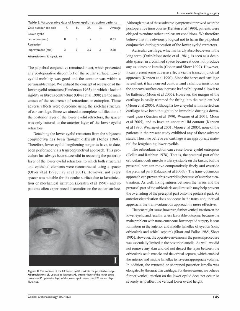
lower eyelid moved down suffi ciently during a downward
gaze (Figure 1H) and the contour was within a permissible
range (Figure 1I). Examples of preoperative and postopera-
tive case photographs are shown in Figure 2.
DiscussionThe lower eyelid lengthening procedure described here
was clearly shown to recess the main tractional component
of the posterior layer of the lower eyelid retractors.
Figure 1E The harvested auricular cartilage is interposed between the lower edge of the tarsus and the distal edge of the anterior layer of the lower eyelid retractors while the posterior layer remains unfi xed to any structure.The volume of the harvested cartilage for the retraction patients is twice the retraction from the lower corneal limbus with 1 mm of extra volume in the proximal and distal margins, respectively, for sutures.The height of the harvested cartilage for the entropion patients is 4 mm (horizontal length in both groups is always constant at 17 mm).Figure 1F The cartilage is fi xed with 5 sutures on each of the distal and proximal sides.The suture on the right upper edge is masked by the recessed anterior layer of the lower eyelid retractors in this image.Figure 1G Suffi cient elevation of the lower eyelid is shown.The skin is sutured with interrupted 6–0 nylon sutures.Figure 1H The rehabilitated left lower eyelid moves down suffi ciently during a downward gaze.
Clinical Ophthalmology 2007:1(2) 145
Lower eyelid lengthening surgery
The palpebral conjunctiva remained intact, which prevented
any postoperative discomfort of the ocular surface. Lower
eyelid mobility was good and the contour was within a
permissible range. We utilised the concept of recession of the
lower eyelid retractors (Henderson 1965), in which a lack of
rigidity or fi brous contracture (Olver et al 1998) are the main
causes of the recurrence of retractions or entropion. These
adverse effects were overcome using the skeletal structure
of ear cartilage. Since we aimed at complete nullifi cation of
the posterior layer of the lower eyelid retractors, the spacer
was only sutured to the anterior layer of the lower eyelid
retractors.
Detaching the lower eyelid retractors from the subjacent
conjunctiva has been thought difficult (Jones 1968).
Therefore, lower eyelid lengthening surgeries have, to date,
been performed via a transconjunctival approach. This pro-
cedure has always been successful in recessing the posterior
layer of the lower eyelid retractors, to which both structural
and epithelial elements were reconstructed using a spacer
(Olver et al 1998; Fay et al 2001). However, not every
spacer was suitable for the ocular surface due to keratiniza-
tion or mechanical irritation (Kersten et al 1990), and so
patients often experienced discomfort on the ocular surface.
Although most of these adverse symptoms improved over the
postoperative time course (Kersten et al 1990), patients were
obliged to endure rather unpleasant conditions. We therefore
believe that it is obviously logical not to harm the palpebral
conjunctiva during recession of the lower eyelid retractors.
Auricular cartilage, which is hardly absorbed even in the
long term (Ortiz-Monasterio et al 1981), is seen as a desir-
able spacer in a confi ned space because it does not produce
any exudates or keratin (Cohen and Shorr 1992). However,
it can present some adverse effects via the transconjunctival
approach (Kersten et al 1990). Since the harvested cartilage
is resilient, it has a curved contour, and a shallow incision on
the concave surface can increase its fl exibility and allow it to
be fl attened (Moon et al 2005). However, the margin of the
cartilage is easily trimmed for fi tting into the recipient bed
(Moon et al 2005). Although a lower eyelid with inserted ear
cartilage have been thought to be immobile during a down-
ward gaze (Kersten et al 1990; Wearne et al 2001; Moon
et al 2005), and to have an unnatural lid contour (Kersten
et al 1990; Wearne et al 2001; Moon et al 2005), none of the
patients in the present study exhibited any of these adverse
states. Thus, we believe ear cartilage is an appropriate mate-
rial for lengthening lower eyelids.
The orbicularis action can cause lower eyelid entropion
(Collin and Rathbun 1978). That is, the pretarsal part of the
orbicularis oculi muscle is always stable on the tarsus, but the
preseptal part can move comparatively freely and override
the pretarsal part (Kakizaki et al 2006b). The trans-cutaneous
approach can prevent this overriding because of anterior cica-
trisation. As well, fi xing sutures between the tarsus and the
pretarsal part of the orbicularis oculi muscle may help prevent
the overriding of the preseptal part onto the pretarsal part. As
anterior cicatrisation does not occur in the trans-conjunctival
approach, the trans-cutaneous approach is more effective.
The scar might cause, however, further vertical traction on the
lower eyelid and result in a less favorable outcome, because the
main problem with trans-cutaneous lower eyelid surgery is scar
formation in the anterior and middle lamellae of eyelids (skin,
orbicularis and orbital septum) (Shorr and Fallor 1985; Shorr
1995). However, the operative invasion in the present procedure
was essentially limited in the posterior lamella. As well, we did
not remove any skin and did not dissect the layer between the
orbicularis oculi muscle and the orbital septum, which enabled
the anterior and middle lamellae to have an appropriate volume.
In addition, the retracted or shortened posterior lamella was
elongated by the auricular cartilage. For these reasons, we believe
further vertical traction on the lower eyelid does not occur so
severely as to affect the vertical lower eyelid height.
Table 2 Postoperative data of lower eyelid retraction patients
Case number and side 1R 1L 2R 3L Average
Lower eyelid
retraction (mm) 0 0 1.5 1 0.63
Retraction
improvement (mm) 3 3 3.5 2 2.88
Abbreviations: R, right; L, left
Figure 1I The contour of the left lower eyelid is within the permissible range.Abbreviations: LL, Lockwood ligament; AL, anterior layer of the lower eyelid retractors; PL, posterior layer of the lower eyelid retractors; EC, ear cartilage; Ta, tarsus.
Clinical Ophthalmology 2007:1(2)146
Kakizaki et al
A midface lift with lateral canthal resuspension has
recently been applied to lower eyelid retractions, such as in
post-blepharoplasty, midface descent, and thyroid-related
orbitopathy (Ben Simon et al 2006). It is important to note
in the present procedure whether the anterior or middle
lamella is targeted or not for therapy. Thyroid-related
lower eyelid retraction, the main pathology for which is
in the posterior lamella, is well treated by just our present
procedure. As well, for entities including anterior and
middle lamellae as well as the posterior lamella (Shorr
1995; Li et al 2005), such as in post-blepharoplasty lower
eyelid retraction, our procedure also can help improve
the pathology, simultaneously with rehabilitation of
the anterior and middle lamellae. However, the present
procedure cannot be separately applied to the cases with
just anterior or middle lamellar cicatrix (Shorr and Fallor
1985; Shorr 1995) because our method only targets the
posterior lamellar pathology.
Because of the small sample size, statistically signifi cant
outcomes could not be demonstrated in this study. However,
the lower eyelid lengthening procedure clearly elevated
retracted or cicatricial lower eyelids, and improved cicatricial
entropion. Lower eyelid lengthening surgery focusing on the
posterior layer of the lower eyelid retractors using auricular
cartilage insertion via a transcutaneous approach was shown
to be a useful procedure for lower eyelid retraction or cica-
tricial entropion.
DisclosureThere is no financial support or interest related to this
manuscript.
ReferencesBartley GB, Kay PP. 1989. Posterior lamellar eyelid reconstruction with a
hard palate mucosal graft. Am J Ophthalmol, 107:609–12.Baylis HI, Perman KI, Fett DR, et al. 1985. Autogenous auricular cartilage
grafting for lower eyelid retraction. Ophthal Plast Reconstr Surg, 1:23–7.
Figure 2A The patient shows 5-mm right and 4-mm left retractions from the lower corneal limbi with proptosis of 26 mm OU. Just the right lower eyelid took two Hotz procedures for the lower eyelid entropion.Figure 2B After lower eyelid lengthening surgery, the right lower eyelid retraction and cicatricial entropion are suffi ciently improved.
Clinical Ophthalmology 2007:1(2) 147
Lower eyelid lengthening surgery
Ben Simon GJ, Lee S, Schwarcz RM, et al. 2006. Subperiosteal midface lift with or without a hard palate mucosal graft for correction of lower eyelid retraction. Ophthalmology, 113:1869–73.
Cohen MS, Shorr N. 1992. Eyelid reconstruction with hard palate mucosa grafts. Ophthal Plast Reconstr Surg, 8:183–95.
Collin JRO, Rathbun JE. 1978. Involutional entropion. A review with evalu-ation of a procesure. Arch Ophthalmol, 96:1058–64.
Duke-Elder SS, MacFaul PA. 1976. Abnormalities of the palpebral aperture. In: System of Ophthalmology vol. XIII– The Ocular adnexa. London, Henry Kimpton, pp 573–81.
Fay AM, Pieroth L, Rubin PA. 2001. An animal model of lower eyelid spacer grafting with a cellular dermis. Ophthal Plast Reconstr Surg, 17:270–5.
Feldman KA, Putterman AM, Farber MD. 1992. Surgical treatment of thyroid-related lower eyelid retraction: a modifi ed approach. Ophthal Plast Reconstr Surg, 8:278–86.
Gardner TA, Kennerdell JS, Buerger GF. 1992. Treatment of dysthyroid lower lid retraction with autogenous tarsus transplants. Ophthal Plast Reconstr Surg, 8:26–31.
Goldberg RA, Joshi AR, McCann JD, et al. 1999. Management of severe cicatricial entropion using shared mucosal grafts. Arch Ophthalmol, 117:1255–9.
Harvey JT, Anderson RL. 1981. The aponeurotic approach to eyelid retrac-tion. Ophthalmology, 88:513–24.
Hawes MJ, Dortzbach RK. 1982. The microscopic anatomy of the lower eyelid retractors. Arch Ophthalmol, 100:1313–8.
Henderson JW. 1965. Relief of eyelid retraction: A surgical procedure. Arch Ophthalmol, 74:205–16.
Jones LT. 1968. A new concept of the orbital fascia and rectus muscle sheaths and its surgical implications. Trans Am Acad Ophthalmol, 72:755–64.
Kakizaki H, Zako M, Mito H, et al. 2005. Modifi ed operation to correctly detect and fi x the lower eyelid retractor in involutional entropion. Jpn J Ophthalmol, 49:330–2.
Kakizaki H, Zhao J, Nakano T, et al. 2006a. The lower eyelid retractor consists of defi nite double layers. Ophthalmology, 113:2346–50.
Kakizaki H, Zhao J, Zako M, et al. 2006b. Microscopic anatomy of asian lower eyelids. Ophthal Plast Reconstr Surg, 22:430–3.
Kakizaki H, Zako M, Kinoshita S, et al. 2007a. Posterior layer advancement of the lower eyelid retractor in involutional entropion repair. Ophthal Plast Reconstr Surg, In press.
Kakizaki H, Zako M, Iwaki M. 2007b. Reverse ptosis repair targeting the posterior layer of the lower lid retractor. Ophthal Plast Reconstr Surg, In press.
Kersten RC, Kulwin DR, Levartovsky S, et al. 1990. Management of lower-lid retraction with hard-palate mucosa grafting. Arch Ophthalmol, 108:1339–43.
Li TG, Shorr N, Goldberg RA. 2005. Comparison of the effi cacy of hard palate grafts with acellular human dermis grafts in lower eyelid surgery. Plast Reconstr Surg, 116:873–8.
McInnes AW, Burroughs JR, Anderson RL, et al. 2006. Temporary suture tarsorrhaphy. Am J Ophthalmol, 142:344–6.
Moon JW, Choung HK, Khwarg SI. 2005. Correction of lower lid retraction combined with entropion using an ear cartilage graft in the anophthalmic socket. Korean J Ophthalmol, 19:161–7.
Olver JM, Rose GE, Khaw PT, et al. 1998. Correction of lower eyelid retraction in thyroid eye disease: a randomised controlled trial of retractor tenotomy with adjuvant antimetabolite versus scleral graft. Br J Ophthalmol, 82:174–80.
Ortiz-Monasterio F, Olmedo A, Oscoy LO. 1981. The use of cartilage grafts in primary aesthetic rhinoplasty. Plast Reconstr Surg, 67:597–605.
Patel MP, Shapiro MD, Spinelli HM. 2005. Combined hard palate spacer graft, midface suspension, and lateral canthoplasty for lower eyelid retraction: a tripartite approach. Plast Reconstr Surg, 115:2105–14.
Shorr N, Fallor MK. 1985. “Madame Butterfl y” procedure: combined cheek and lateral canthal suspension procedure for post-blepharoplasty, “round eye,” and lower eyelid retraction. Ophthal Plast Reconstr Surg, 1:229–35.
Shorr N. 1995. “Madame Butterfl y” procedure: Total lower eyelid recon-struction in three layers utilizing a hard palate graft: Management of the unhappy post-blepharoplasty patient with round eye and scleral show. Int J Aesthetic Restorative Surg, 3:3–26.
Sullivan SA, Dailey RA. 2003. Graft contraction: a comparison of acellular dermis versus hard palate mucosa in lower eyelid surgery. Ophthal Plast Reconstr Surg, 19:14–24.
Wearne MJ, Sandy C, Rose GE, et al. 2001. Autogenous hard palate mucosa: the ideal lower eyelid spacer? Br J Ophthalmol, 85:1183–7.