38
Lumbar Puncture Bucky Boaz, ARNP-C
| Date post: | 23-Dec-2015 |
| Category: |
Documents |
| Upload: | heather-stevens |
| View: | 227 times |
| Download: | 0 times |

Lumbar Puncture
Bucky Boaz, ARNP-C

CSF Formation
140 ml spinal and cranial CSF
30 ml in the spinal cord
Production is approx. 0.35 ml/min
Net flow out of ventricles 50 – 100 ml/day
Reduces brain weight from 1400 to 50g.

Indications for Lumbar Puncture
Primary indication for emergent spinal tap is possibility of CNS infection
The second indication for an emergent spinal puncture is a suspected spontaneous subarachnoid hemorrhage.
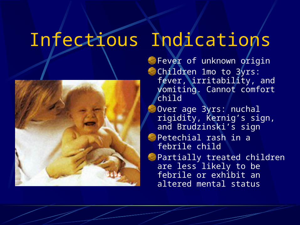
Infectious IndicationsFever of unknown originChildren 1mo to 3yrs: fever, irritability, and vomiting. Cannot comfort childOver age 3yrs: nuchal rigidity, Kernig’s sign, and Brudzinski’s signPetechial rash in a febrile childPartially treated children are less likely to be febrile or exhibit an altered mental status

Subarachnoid Hemorrhage
Diagnosis usually made by CT scan or by blood in CSF.
Initial presentation: CT 92-98% accurate
Later than 24 hr presentation: 76% accurate
20-60% of aneurismal subarachnoid hemorrhage will have “sentinel thunderclap” or “warning clap”
After initial leak, CT is usually negative

Contraindications for LP
Absolutely contraindicated in the presence of infection in the tissues near the puncture site.
Relatively contraindicated in presence of SOL or increased ICP
Caution advised when lateralizing signs or signs of uncal herniation.
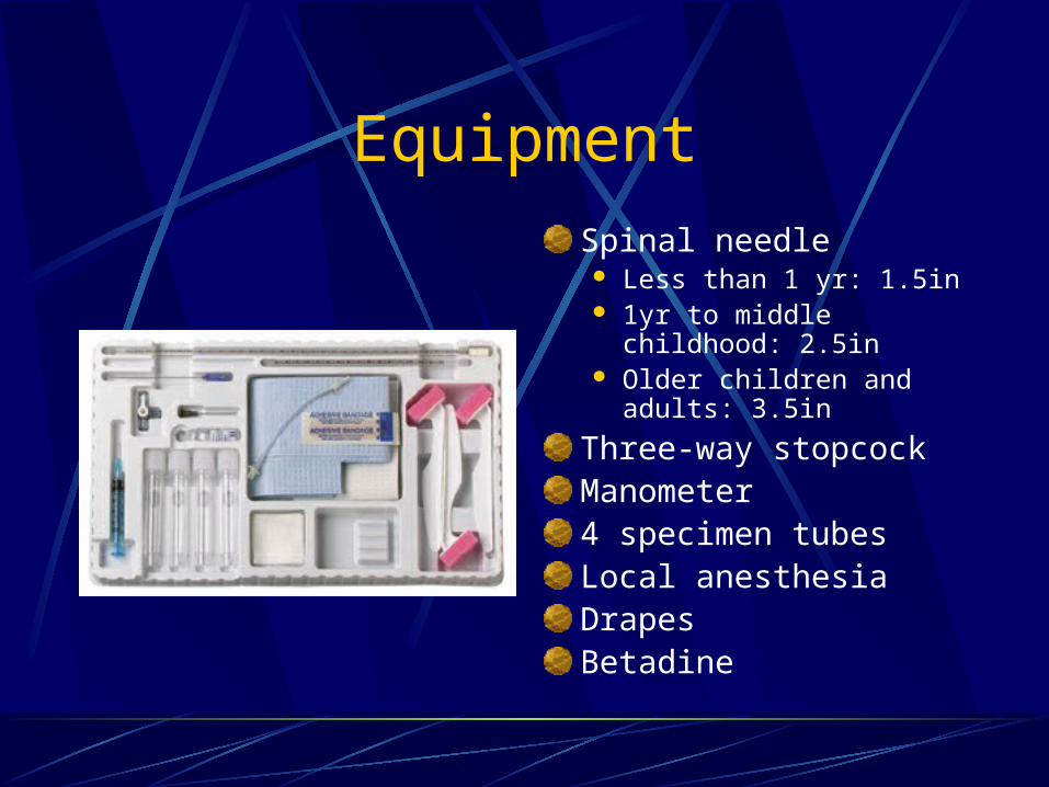
EquipmentSpinal needle Less than 1 yr: 1.5in 1yr to middle childhood:
2.5in Older children and adults:
3.5in
Three-way stopcockManometer4 specimen tubesLocal anesthesiaDrapesBetadine

Interpretations
PressureOpening pressure is taken promptly,
avoiding falsely low values due to leakage through and around the needle
Normal pressure is between 70 and 180 mm H20

Interpretation
Appearance If CSF is not crystal clear, a pathologic
condition of the CNS should be suspectedCompare fluid to waterFluid may be clear with as many as 400
RBCs/mm3 and 200 WBCs/mm3

Interpretation
Cells WBC counts over 5 cells/mm3 should be taken to
indicate the presence of pathologic condition Polymorphonuclear leukocytes are never seen in
normal adults Neutrophilic pleocytosis is commonly associated
with bacterial infections or early stages of viral infections, tuberculosis, meningitis, hematogenous meningitis, and chemical meningitis due to foreign bodies.

Interpretation
Cells Eosinophils are always abnormal and most
commonly represent a parasite infestation. Eosinophils have also been reported in cases of
subarachnoid hemorrhage, lymphoma, Hodgkin’s disease, brucellosis, fungal meningitis, mycoplasma pneumonia infection, measles, lymphocytic choriomeningitis, rickettsial infections, leukemia, demyelinating disease, sarcoiodosis, acute inflammatory demyelinating polyneuropathy, allergic reactions, and idiopathic eosinophilic meningitis.
Interpretation
CellsNormal CSF RBCs are less than 10/mm3.Counts that are otherwise unexplained
may be due to a traumatic tap.Herpes simplex virus encephalitis may
elevate the CSF RBC count in many patients.
Interpretation
GlucoseLow CSF glucose concentration indicates
increased glucose use in the brain and the spinal cord.
The normal range of CSF glucose is between 50 and 80 mg/dl
60-70% of serum glucose concentrationOnly low concentrations of glucose are
significance

Interpretation
Low CSF Glucose SyndromesBacterial meningitis Syphilis
Tuberculous meningitis Chemical meningitis
Fungal meningitis Subarachnoid meningitis
Sarcoidosis Mumps meningitis
Meningeal carcinomatosis
Herpes simplex encephalitis
Amebic meningitis Hypoglycemia
Cysticercosis Trichinosis

Interpretation
Protein Increase in CSF total protein levels are a
nonspecific abnormality associated with many disease states.
Levels > 500mg/dl are uncommon and are seen mainly in meningitis, in subarachnoid bleeding, and with spinal tumors.
The Traumatic Tap
It should not be difficult to distinguish between subarachnoid hemorrhage and a traumatic tap.
In traumatic taps, the fluid generally clears between 1st and 3rd tubes.

CSF Analysis with Infections
Bacterial Infections The Gram stain is of great importance, because
this often dictates the initial choice of antibiotic. Gram-negative intracellular or extracellular
diplococci are indicative of Neisseria meningitidis Small Gram-negative bacilli may include
Haemophilus influenza, especially in children. Gram-positive cocci indicates Streptococcus
pneumoniae, other Streptococcus species, or Staphylococcus.
20% of Gram stains may be falsely negative.

CSF Analysis with Infections
Bacterial Infections While the culture is pending, one may suspect a
bacterial infection in the presence of an elevated opening pressure and a marked pleocytosis ranging between 500 and 20,000 WBCs/mm3.
The differential count is usually chiefly neutrophils. A count above 1000 cells/mm3 seldom occurs in
viral infections.

CSF Analysis with Infections
Bacterial InfectionsCSF glucose levels less than 40 mg/dl or
less than 50% of a simultaneous blood glucose level should raise the question of bacterial meningitis.
The CSF protein content in bacterial meningitis ranges from 500 to 1500 mg/dl.

CSF Analysis with Infections
Viral StudiesThe organisms most commonly isolated in
viral meningitis are enteroviruses and mumps.
Enteroviruses: summer and fallMumps: winter and spring

CSF Analysis with Infections
Viral StudiesWBC count in viral meningitis and
encephalitis usually: 10 to 1000 cells/mm3.The differential count is predominantly
lymphocytic and mononuclear in type.Protein levels are usually mildly elevatedAntibiotic coverage pending culture results
may be reasonably initiated pending culture results if in doubt.

Complications
Headache After Lumbar Puncture Most common
complication Occurs 5-30% of all
spinal taps Usually starts up to
48 hours after to procedure.
Usually lasts 1-2 days (occas 14 days)

Complications
Headache After Lumbar PunctureUsually begins within minutes after arising
and resolves with recumbent position.Pain is mild to incapacitating and is usually
cervical and sub-occipital, but may involve the shoulders and the entire cranium.
Caused by leaking of fluid through dural puncture site.

Complications
Headache After Lumbar Puncture Incidence is higher in younger patients and
females, and those with headache history.Treatment: barbiturates, fluids, caffeine
(500mg in 2 ml NS IV push) more common 500mg in 2 L over 1 hr.
Blood patch by anesthesia if no improvement.

Procedure
Performed with the patient in the lateral recumbent position.A line connecting the posterior superior iliac crest will intersect the midline at approx. the L4 spinous process.Spinal needles entering the subarachnoid space at this point are well below the termination of the spinal cord.
Procedure
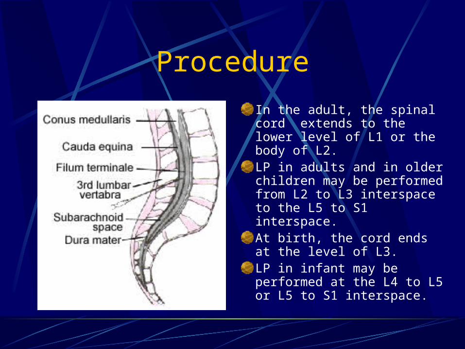
In the adult, the spinal cord extends to the lower level of L1 or the body of L2.LP in adults and in older children may be performed from L2 to L3 interspace to the L5 to S1 interspace.At birth, the cord ends at the level of L3.LP in infant may be performed at the L4 to L5 or L5 to S1 interspace.
Procedure
Almost all patients are afraid of an LP. Explaining the procedure in advance and discussing each step aids in reducing anxiety.
Inquire about allergies to anesthetics.
Informed consent.
Procedure
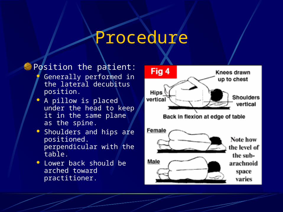
Position the patient: Generally performed in
the lateral decubitus position.
A pillow is placed under the head to keep it in the same plane as the spine.
Shoulders and hips are positioned. perpendicular with the table.
Lower back should be arched toward practitioner.

Procedure
Sterile gloves MUST be used.Wash back with antiseptic solution.Sterile towel under hips.The skin and deeper subcutaneous tissue are infiltrated with local anesthetic.Warn patient of transient discomfort of anesthetic.
Procedure
Anesthetizing the deeper subcutaneous tissue significantly reduces the procedure discomfort.
Some operators not only anesthetize the interspinous ligament but also apply local anesthesia in a vertical fanning distribution on both sides of the spinous processes near the lamina.

Procedure
The patient should be told to report any pain and should be informed that he or she will feel some pressure.
The needle is placed into the skin in the midline parallel to the bed.
The needle is held with both thumbs and index fingers.
Procedure
After the subcutaneous tissue has been penetrated, the needle is angled toward the umbilicus.
The bevel of the needle should be facing laterally (toward patients side).

Procedure
a. Ligament flavum is a strong, elastic, yellow membrane covering the interlaminar space between the vertebrae.
b. Interspinal ligaments join the inferior and superior borders of adjacent spinous processes.
c. Supraspinal ligament connects the spinous processes

Procedure
The ligaments offer resistance to the needle, and a “pop” is often felt as they are penetrated.
Clear fluid will flow from the needle when the subarachnoid space has been penetrated.

Procedure
If bone is encountered, withdrawal into subcutaneous tissue and redirect.Attach a manometer and record opening pressure.Turn stopcock and collect fluid.Withdrawal needle and place a dressing.
Procedure
Tube 1 is used for determining protein and glucose
Tube 2 is used for microbiologic and cytologic studies
Tube 3 is for cell counts and serologic tests for syphilis


Questions?


