Page 1
1
Lumps and bumps of the gingiva: a pathological miscellany
Daniel J Brierley1, Hannah Crane1 and Keith D Hunter1,2
1Academic Unit of Oral and Maxillofacial Medicine and Pathology, School of Clinical Dentistry, University of Sheffield, UK.
2Oral Pathology and Biology, University of Pretoria, Pretoria, South Africa.
Corresponding Author:
Professor Keith D Hunter Academic Unit of Oral and Maxillofacial Medicine and Pathology School of Clinical Dentistry University of Sheffield Claremont Crescent Sheffield, S10 2TA United Kingdom [email protected] Tel: +44 (0)114 215 9405 ORCID: 0000-0002-7873-0877
Abstract
Lesions of the gingivae are amongst the commonest lesions seen in patients and the vast
majority are reactive hyperplasias, related to a number of chronic irritant stimuli. However,
there are a number of entities that have a predilection for the gingivae, which are much less
common in other parts of the oral cavity. The purpose of this paper is to discuss the clinical
and histological differential diagnoses when presented with a lump on the gingivae, including
the approach to diagnosis and diagnostic pitfalls.
Keywords
Epulis, gingiva, fibrous hyperplasia, giant cell granuloma, pyogenic granuloma,
ligneous gingivitis, Granulomatosis with polyangiitis, benign tumor, malignant tumor.
Page 2
2
Introduction
Lesions of the gingiva are very common and provide a significant proportion of the diagnostic
workload of any oral pathology practice. The majority of these lesions are reactive (with varied
appearance), but other developmental and neoplastic conditions can also present in the gingiva,
giving rise to areas of clinical and histological uncertainty in diagnosis. In this review, we aim
to address the main entities which may present as “lumps and bumps” in the gingiva. As with
all common diagnoses, there is variability in the clinical presentation of such lesions of the
gingivae and to some extent, in the histological appearances. As such, it is not possible to
describe every possible presentation. However, the descriptions below will cover the most
salient features of a range of different pathologies of the gingival tissues.
A word on terminology …
As in many areas of histopathology, terminology is variably used and those used in the
description of clinical and histological lesions of the gingivae are no different. Some use of
terminology is, strictly, inaccurate, but has, for a number of reasons, become well established
as common usage. This includes, but is not limited to, the terms ‘polyp’ and ‘epulis’ as
histological diagnoses, the use of the term ‘fibroma’ in the context of these lesions, and the
common usage in some parts of the world of the term ‘peripheral ossifying fibroma’, which
causes confusion in others(1,2). Whilst it is not helpful to be dogmatic about which terms
should or should not be used, it is important that histopathologists and referring clinicians have
a common understanding of the terminology, to ensure effective communication of diagnosis
and resulting treatment strategies. Where appropriate, synonyms will be indicated in the text.
Reactive lesions
Fibrous hyperplasia of the gingiva
Page 3
3
Epidemiology
Nodules of inflammatory fibrous hyperplasia (syn. fibroepithelial polyp, fibroma; on gingivae,
fibrous epulis) are very common, with fibrous hyperplasia accounting for up to 40% of mucosal
pathology in large series(3). Lesions occur over a wide age range and are more common in
females.
Clinical presentation and differential diagnosis
In dentate patients, these lesions most commonly occur on the interdental papilla, but may also
include the facial surface of the tooth (Figure 1A). When large, lesions may extend through
the contact point to appear in the papilla on both sides of arch, with a rather “dumb-bell”
appearance, although this appearance is more commonly seen in peripheral giant cell
granuloma (see below). The lesions are most commonly mucosal colored, but may be focally
ulcerated. More extensive lesions occur in patients taking certain medications, for example:
phenytoin, nifedipine (and other calcium channel blockers) or cyclosporine (4). This drug-
induced gingival hyperplasia is an exaggerated form of the more focal reactive lesions
described above (Figure 1B). In edentulous/partially dentate patients, similar lesions can occur
on the alveolus in relation to the presence of ill-fitting prostheses, often termed denture
irritation hyperplasia or denture hyperplasia(5). These lesions are most common in the mucosa
in contact with the periphery of a denture and are usually broad based leaf-like folds of mucosa.
The clinical differential diagnosis most often includes other reactive lesions of the gingiva
including pyogenic granuloma and peripheral giant cell granuloma. Considerations in more
generalised lesions include hereditary gingival fibromatosis, which may occur as an isolated
lesion or as part of a syndrome. A number of genetic lesions, including mutations of the Son-
of-Sevenless-1 (SOS1) gene have been associated with the isolated from of this condition(6).
Page 4
4
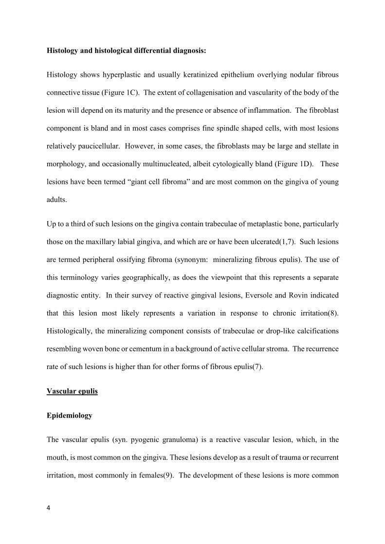
Histology and histological differential diagnosis:
Histology shows hyperplastic and usually keratinized epithelium overlying nodular fibrous
connective tissue (Figure 1C). The extent of collagenisation and vascularity of the body of the
lesion will depend on its maturity and the presence or absence of inflammation. The fibroblast
component is bland and in most cases comprises fine spindle shaped cells, with most lesions
relatively paucicellular. However, in some cases, the fibroblasts may be large and stellate in
morphology, and occasionally multinucleated, albeit cytologically bland (Figure 1D). These
lesions have been termed “giant cell fibroma” and are most common on the gingiva of young
adults.
Up to a third of such lesions on the gingiva contain trabeculae of metaplastic bone, particularly
those on the maxillary labial gingiva, and which are or have been ulcerated(1,7). Such lesions
are termed peripheral ossifying fibroma (synonym: mineralizing fibrous epulis). The use of
this terminology varies geographically, as does the viewpoint that this represents a separate
diagnostic entity. In their survey of reactive gingival lesions, Eversole and Rovin indicated
that this lesion most likely represents a variation in response to chronic irritation(8).
Histologically, the mineralizing component consists of trabeculae or drop-like calcifications
resembling woven bone or cementum in a background of active cellular stroma. The recurrence
rate of such lesions is higher than for other forms of fibrous epulis(7).
Vascular epulis
Epidemiology
The vascular epulis (syn. pyogenic granuloma) is a reactive vascular lesion, which, in the
mouth, is most common on the gingiva. These lesions develop as a result of trauma or recurrent
irritation, most commonly in females(9). The development of these lesions is more common
Page 5
5
in circumstances where alterations in sex hormones levels are present, for example; in puberty,
pregnancy (syn. pregnancy epulis; granuloma gravidarum) and effects of the oral contraceptive
pill or hormone replacement therapy(10).
Clinical presentation and differential diagnosis
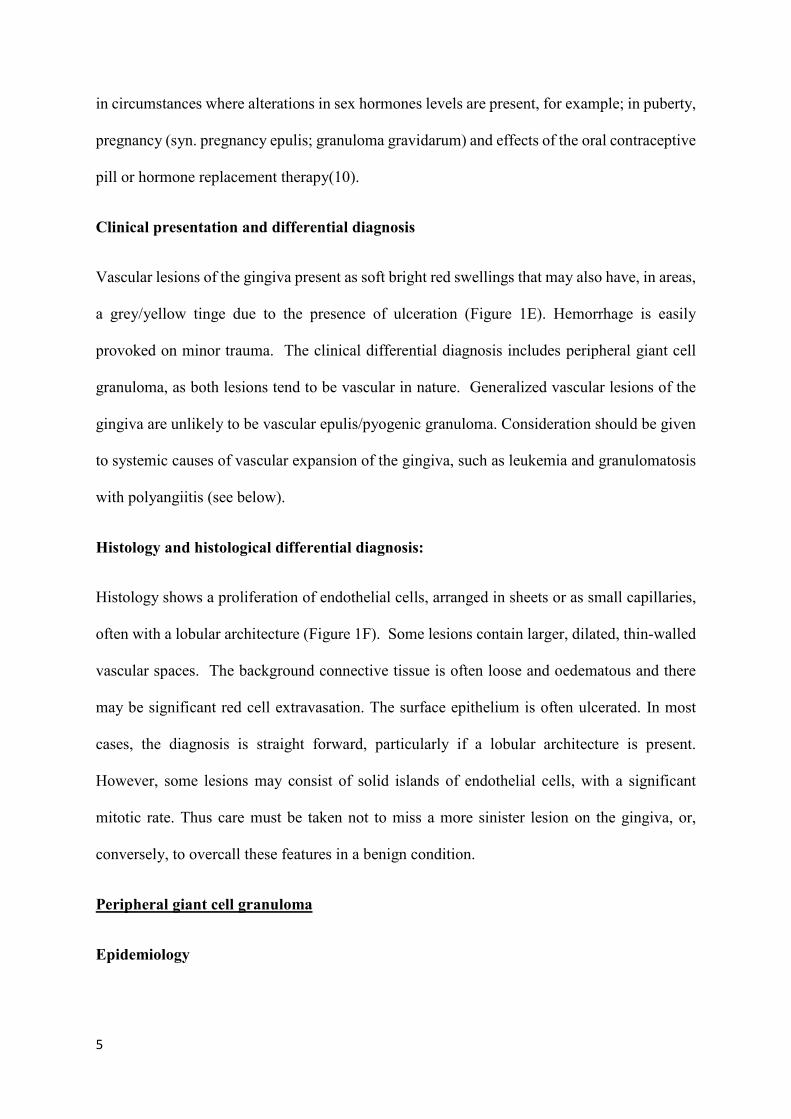
Vascular lesions of the gingiva present as soft bright red swellings that may also have, in areas,
a grey/yellow tinge due to the presence of ulceration (Figure 1E). Hemorrhage is easily
provoked on minor trauma. The clinical differential diagnosis includes peripheral giant cell
granuloma, as both lesions tend to be vascular in nature. Generalized vascular lesions of the
gingiva are unlikely to be vascular epulis/pyogenic granuloma. Consideration should be given
to systemic causes of vascular expansion of the gingiva, such as leukemia and granulomatosis
with polyangiitis (see below).
Histology and histological differential diagnosis:
Histology shows a proliferation of endothelial cells, arranged in sheets or as small capillaries,
often with a lobular architecture (Figure 1F). Some lesions contain larger, dilated, thin-walled
vascular spaces. The background connective tissue is often loose and oedematous and there
may be significant red cell extravasation. The surface epithelium is often ulcerated. In most
cases, the diagnosis is straight forward, particularly if a lobular architecture is present.
However, some lesions may consist of solid islands of endothelial cells, with a significant
mitotic rate. Thus care must be taken not to miss a more sinister lesion on the gingiva, or,
conversely, to overcall these features in a benign condition.
Peripheral giant cell granuloma
Epidemiology
Page 6
6
The peripheral giant cell granuloma (PGCG) (syn. giant cell epulis) accounts for
approximately 10% of epulides(11). They occur over a wide age range with a lower age peak
incidence for males than females, and a female predilection. These lesions can occur in any
part of the gingiva in dentate patients or on the alveolar ridge in edentulous patients, but most
occur anterior to the molar region and are slightly more common in the mandible(12).
Clinical presentation and differential diagnosis
PGCG are most often deep red/purple colored sessile swellings that may reach an appreciable
size (Figure 2A). They may extend through the contact point between teeth in a dumb-bell
type pattern. The clinical differential diagnosis includes ulcerated fibrous epulis and vascular
epulis. The comments on generalized swelling in the fibrous hyperplasia section also apply.
Histology and histological differential diagnosis
The main body of the lesion comprises a vascular and cellular stroma of mononuclear cells
(fibroblasts, macrophages and endothelial cells), in which are scattered numerous
multinucleated giant cells (Figure 2B). The giant cells vary in size and number of nuclei. In
some cases, fibrous septae are present and the lesional tissue is separated from the overlying
epithelium by a band of fibrous tissue. Extravasated red blood cells and haemosiderin pigment
are commonly identified.
PGCG are indistinguishable histologically from central giant cell granuloma and lesions of the
jaws seen in hyperparathyroidism(13). In many lesions, the initial excision is incomplete and
the reporting pathologist should note this, together with a comment on the need to ensure that
further investigations, such as radiological examination and, if deemed necessary,
measurement of serum calcium levels are undertaken to exclude the aforementioned entities.
The histological differential diagnosis of cherubism is rarely an issue due to the distinctive
Page 7
7
clinical and radiological features of that condition(14). These include a family history,
appearance of swellings early in life, and multiple radiolucent lesions of the jaws evident on
radiological examination.
Ligneous gingivitis
Epidemiology
This rare lesion, caused by an inherited plasminogen deficiency, is seen in up to a third of
such patients(15).
Clinical presentation and differential diagnosis
The presentation is variable, from focal lesions to generalized nodular gingival enlargement.
Patients may complain of bleeding and soreness. These lesions have an irregular surface and
may ulcerate (Figure 2C). An association with a form of aggressive periodontal disease has
been reported.
Histology and histological differential diagnosis
Fibrin accumulates in the superficial lamina propria, often associated with the vasculature in
that area. If extensive, the fibrin can form large sheets, raising the suspicion of amyloid. The
surface epithelium may be attenuated or mildly hyperplastic. There is often an associated
scattered inflammatory infiltrate. The main histological differential diagnosis is that of
amyloidosis. Histochemical stains to rule out amyloid (Congo or Sirius Red) and, on occasion,
a trichrome stain (for example martius scarlet blue) may be useful in identification of the
fibrinous material(16).
Granulomatosis with polyangiitis (GPA)
Epidemiology
Page 8
8
GPA is a rare condition with a wide range of clinical presentations. Most are associated with
the respiratory tract but any body system may be affected. The condition occurs over a wide
age range with slight female predilection and almost all cases occur in Caucasians(17).
Clinical presentation and differential diagnosis
Around 2% of patients present with oral lesions(18). The classic oral presentation is that of
“strawberry gingivitis’ (Figure 2E). These lesions are nodular and erythematous with an
irregular surface and a tendency to bleed easily. Lesions may be focal or widespread and may
be asymptomatic. On occasion, there may be associated ulceration and destructive lesions have
been reported(19). The clinical appearance is characteristic, but the differential diagnosis may
include some of the vascular lesions described above, particularly if the lesion is focal.
Histology and histological differential diagnosis
The characteristic feature is of a destructive vasculitis (leukocytoclastic vasculitis: Figure 2F).
Affected vessels show inflammatory cells throughout the wall and these may be associated
with vessel obliteration creating areas of necrosis. Gingival lesions also may show prominent
red cell extravasation. Non-caseating epitheloid granulomas are also identified and these may
contain multinucleated giant cells. Diagnosis can be challenging in small gingival biopsies due
to the lack of granulomas and limited vasculitis. Other granulomatous conditions must be
excluded, including tuberculosis, certain fungal infections, Crohn’s and sarcoidosis.
Depending on the histological features, appropriate histochemical stains (Ziehl Neelsen,
Grocott etc.) can be used to refine the differential diagnosis. Serological testing for PR3-ANCA
should be suggested if GPA is in the differential diagnosis.
Verruciform Xanthoma
Page 9
9
This lesion is most common on the gingiva and the palate. As it often has a yellow coloration,
the reader is referred to “Yellow lesions of the oral mucosa” and “Non-HPV Papillary lesions
of the oral mucosa” in this special edition of the journal.
Developmental lesions
Congenital epulis
Epidemiology
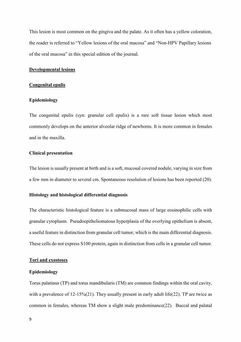
The congenital epulis (syn: granular cell epulis) is a rare soft tissue lesion which most
commonly develops on the anterior alveolar ridge of newborns. It is more common in females
and in the maxilla.
Clinical presentation
The lesion is usually present at birth and is a soft, mucosal covered nodule, varying in size from
a few mm in diameter to several cm. Spontaneous resolution of lesions has been reported (20).
Histology and histological differential diagnosis
The characteristic histological feature is a submucosal mass of large eosinophilic cells with
granular cytoplasm. Pseudoepitheliomatous hyperplasia of the overlying epithelium is absent,
a useful feature in distinction from granular cell tumor, which is the main differential diagnosis.
These cells do not express S100 protein, again in distinction from cells in a granular cell tumor.
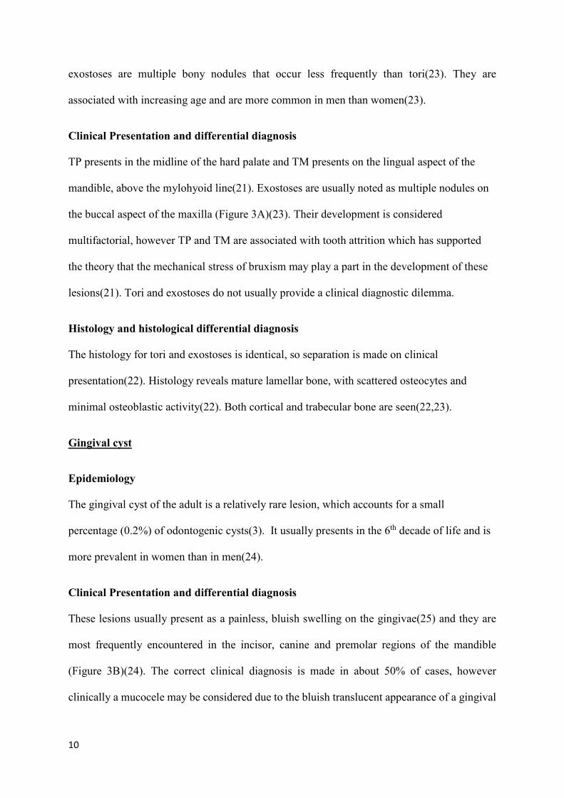
Tori and exostoses
Epidemiology
Torus palatinus (TP) and torus mandibularis (TM) are common findings within the oral cavity,
with a prevalence of 12-15%(21). They usually present in early adult life(22). TP are twice as
common in females, whereas TM show a slight male predominance(22). Buccal and palatal
Page 10
10
exostoses are multiple bony nodules that occur less frequently than tori(23). They are
associated with increasing age and are more common in men than women(23).
Clinical Presentation and differential diagnosis
TP presents in the midline of the hard palate and TM presents on the lingual aspect of the
mandible, above the mylohyoid line(21). Exostoses are usually noted as multiple nodules on
the buccal aspect of the maxilla (Figure 3A)(23). Their development is considered
multifactorial, however TP and TM are associated with tooth attrition which has supported
the theory that the mechanical stress of bruxism may play a part in the development of these
lesions(21). Tori and exostoses do not usually provide a clinical diagnostic dilemma.
Histology and histological differential diagnosis
The histology for tori and exostoses is identical, so separation is made on clinical
presentation(22). Histology reveals mature lamellar bone, with scattered osteocytes and
minimal osteoblastic activity(22). Both cortical and trabecular bone are seen(22,23).
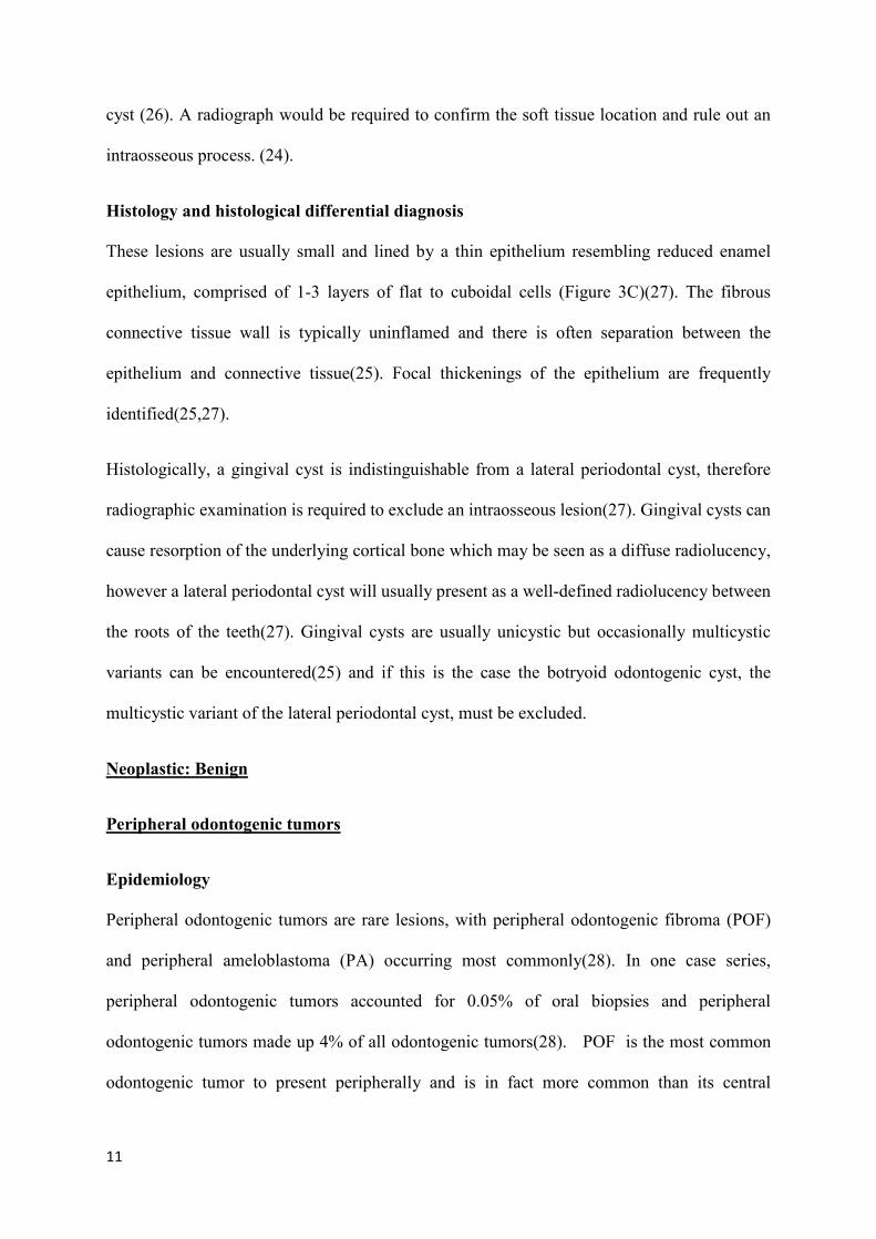
Gingival cyst
Epidemiology
The gingival cyst of the adult is a relatively rare lesion, which accounts for a small
percentage (0.2%) of odontogenic cysts(3). It usually presents in the 6th decade of life and is
more prevalent in women than in men(24).
Clinical Presentation and differential diagnosis
These lesions usually present as a painless, bluish swelling on the gingivae(25) and they are
most frequently encountered in the incisor, canine and premolar regions of the mandible
(Figure 3B)(24). The correct clinical diagnosis is made in about 50% of cases, however
clinically a mucocele may be considered due to the bluish translucent appearance of a gingival
Page 11
11
cyst (26). A radiograph would be required to confirm the soft tissue location and rule out an
intraosseous process. (24).
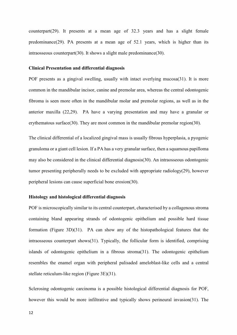
Histology and histological differential diagnosis
These lesions are usually small and lined by a thin epithelium resembling reduced enamel
epithelium, comprised of 1-3 layers of flat to cuboidal cells (Figure 3C)(27). The fibrous
connective tissue wall is typically uninflamed and there is often separation between the
epithelium and connective tissue(25). Focal thickenings of the epithelium are frequently
identified(25,27).
Histologically, a gingival cyst is indistinguishable from a lateral periodontal cyst, therefore
radiographic examination is required to exclude an intraosseous lesion(27). Gingival cysts can
cause resorption of the underlying cortical bone which may be seen as a diffuse radiolucency,
however a lateral periodontal cyst will usually present as a well-defined radiolucency between
the roots of the teeth(27). Gingival cysts are usually unicystic but occasionally multicystic
variants can be encountered(25) and if this is the case the botryoid odontogenic cyst, the
multicystic variant of the lateral periodontal cyst, must be excluded.
Neoplastic: Benign
Peripheral odontogenic tumors
Epidemiology
Peripheral odontogenic tumors are rare lesions, with peripheral odontogenic fibroma (POF)
and peripheral ameloblastoma (PA) occurring most commonly(28). In one case series,
peripheral odontogenic tumors accounted for 0.05% of oral biopsies and peripheral
odontogenic tumors made up 4% of all odontogenic tumors(28). POF is the most common
odontogenic tumor to present peripherally and is in fact more common than its central
Page 12
12
counterpart(29). It presents at a mean age of 32.3 years and has a slight female
predominance(29). PA presents at a mean age of 52.1 years, which is higher than its
intraosseous counterpart(30). It shows a slight male predominance(30).
Clinical Presentation and differential diagnosis
POF presents as a gingival swelling, usually with intact overlying mucosa(31). It is more
common in the mandibular incisor, canine and premolar area, whereas the central odontogenic
fibroma is seen more often in the mandibular molar and premolar regions, as well as in the
anterior maxilla (22,29). PA have a varying presentation and may have a granular or
erythematous surface(30). They are most common in the mandibular premolar region(30).
The clinical differential of a localized gingival mass is usually fibrous hyperplasia, a pyogenic
granuloma or a giant cell lesion. If a PA has a very granular surface, then a squamous papilloma
may also be considered in the clinical differential diagnosis(30). An intraosseous odontogenic
tumor presenting peripherally needs to be excluded with appropriate radiology(29), however
peripheral lesions can cause superficial bone erosion(30).
Histology and histological differential diagnosis
POF is microscopically similar to its central counterpart, characterised by a collagenous stroma
containing bland appearing strands of odontogenic epithelium and possible hard tissue
formation (Figure 3D)(31). PA can show any of the histopathological features that the
intraosseous counterpart shows(31). Typically, the follicular form is identified, comprising
islands of odontogenic epithelium in a fibrous stroma(31). The odontogenic epithelium
resembles the enamel organ with peripheral palisaded ameloblast-like cells and a central
stellate reticulum-like region (Figure 3E)(31).
Sclerosing odontogenic carcinoma is a possible histological differential diagnosis for POF,
however this would be more infiltrative and typically shows perineural invasion(31). The
Page 13
13
differential diagnosis for a PA includes salivary gland tumors with similar histology and basal
cell carcinomas (31), however oral mucosal involvement of basal cell carcinomas is rare.
Immunohistochemistry is useful in cases where there is uncertainty.
Neoplastic: Malignant
Verrucous carcinoma (VC)
Epidemiology
Within the oral cavity, the gingiva (12%) and buccal mucosa (10%) are the most common sites
for VC(32) unlike conventional squamous cell carcinoma which is often found on the lateral
border of the tongue and floor of mouth.
Clinical presentation and differential diagnosis
VC presents as a thickened, white lesion with a roughened or papillary surface. Often, the
degree of whiteness and hyperplasia varies within the same lesion. VC tends to grow slowly
and does not metastasize. Clinically, VC is indistinguishable from verrucous hyperplasia(33)
and often a spectrum of disease is present within the same lesion. Papillary squamous cell
carcinoma may look similar. More subtle, small lesions may be confused with verruciform
xanthoma, traumatic keratosis or papilloma.
Histology and histological differential diagnosis
VC is characterized by extensive hyperplasia of rete processes that push deeply into the
connective tissue, creating a buttress between the carcinoma and adjacent epithelium. There
are varying degrees of hyperkeratosis and keratin plugging giving a verrucous surface
morphology. Cellular and nuclear pleomorphism are minimal.
Page 14
14
The classic histological conundrum is distinguishing VC from verrucous hyperplasia,
especially on small or superficial biopsies. VC requires rete pegs to extend beneath the level
of the adjacent epithelium, and without this buttress, it can be very difficult to make the
distinction. A conventional squamous cell carcinoma may also have exo-endophytic qualities
but contains significantly more cellular atypia and pleomorphism with foci of conventional
tumor islands invading the superficial connective tissue.
Metastases
Epidemiology
Metastatic disease to the oral cavity is often secondary spread from other metastatic lesions,
most commonly the lungs(34,35). Metastases to the gingiva represent one of the most common
sites for metastases to the oral cavity(34,36–38). Those aged between the 5th and 7th decades
are most commonly affected with a male predominance (2:1 ratio) (39). Metastases to the
gingivae are most commonly from the lung, kidney and skin in men and the breast, genital
organs and lung in women(39). Metastases from the gastrointestinal tract and prostate are also
possible.
Clinical presentation and differential diagnosis
The clinical presentation is usually non-specific but the presence of swelling, ulceration and/or
adjacent tooth mobility is common (Figure 3A). Other malignant tumors, such as squamous
cell carcinoma, lymphoma and melanoma, are often considered. When the patient has a history
of a previous malignancy elsewhere in the body, the possibility of a metastasis should always
be considered.
Histology and histological differential diagnosis
Page 15
15
It is not possible to discuss every type of tumor that may metastasize to the gingivae but the
key is not to discount the possibility of metastases, especially when the morphology is unusual
for those tumors that are more often seen in the head and neck region. Immunohistochemistry
is a very useful adjunct for confirming the site of origin. The appearance of metastases varies
greatly depending on the site of origin of the metastatic tumor. For instance, a renal cell
carcinoma metastasis consists of sheets of bland clear cells in a vascular stroma (typically RCC,
CD10, PAX8 positive). In the gingiva, clear cell carcinoma and clear cell odontogenic
carcinoma would need excluding (CD10, PAX8, RCC negative). Lobular carcinoma of the
breast tends to have cords of cells similar to polymorphous adenocarcinoma, however the
former is usually ER and PR positive unlike the latter (Figure 3F).
Kaposi’s sarcoma (KS)
Epidemiology
KS is always associated with HHV8 infection and has four epidemiological categories, but the
AIDS-related type is the only one associated with oral manifestations(40). Oral KS is most
common in the 4th and 5th decades affecting the palate and gingiva the most commonly.
Clinical presentation and differential diagnosis
Lesions vary from being subtle areas of discolouration (often red/purple macules and papules)
to more extensive nodular lesions that have a more sinister appearance (Figure 4A). The
differential diagnosis often includes vascular lesions, such as haemangioma, pyogenic
granuloma and giant cell epulis, especially if the lesion is nodular in appearance. Advanced
lesions become large and ulcerated where other malignant differentials may be considered such
as squamous cell carcinoma.
Histology and histological differential diagnosis
Page 16
16
The early lesion (patch stage) is fairly subtle and composed of thin, slit-like vessels within the
lamina propria that transect collagen fibers and are seen in association with extravasated red
blood cells and lymphocytes. In the plaque stage, the spindle-cell proliferation is more
obvious and associated with hyaline globules (Kamino bodies). The nodular stage comprises
sheets of spindle cells, which appear atypical with increased mitoses (Figure 4B). The tumor
cells are positive for endothelial markers such as CD31 and CD34, but are characteristically
positive for HHV8.
The early features of KS can be easily missed. For instance, the increase in vessels may be
mistaken for granulation tissue in relation to recent ulceration or trauma. When the tumor
enters the more cellular phases, the histological differential diagnoses to consider are other
spindle cell neoplasms with vasoformative qualities such as angiosarcoma (HHV8 negative).
Lymphoma/leukemia
Epidemiology
The head and neck is the second most common extra-nodal site for lymphoma (11-33%),
typically affecting patients over 50 years old(41). Intraorally, the most common sites affected
are the vestibule, gingiva, mandible, palate, maxilla and tongue(42–46). The majority are Non-
Hodgkin B cell lymphomas (mostly diffuse large B-cell lymphoma) but T-cell lymphomas
account for 12% of oral lymphomas in the Japanese population(44,47). The most common
type of leukemia to affect the gingivae is acute myeloid leukemia (AML) of monocytic
derivation(48).
Clinical presentation and differential diagnosis
Lymphoma and leukemia have a non-specific clinical presentation, but often present with
swelling and reddening of the gingival tissues (Figure 4C and 4D). Advanced cases are likely
Page 17
17
to be accompanied by bone loss and tooth mobility. Unless the patient has a known history of
lymphoma or leukemia, the differential diagnosis is likely to include a range of non-neoplastic
and neoplastic conditions depending on the extent of disease at presentation(49). When lesions
are diffuse and present with reddening and swelling of the gingivae, conditions such as GPA,
periodontitis and hyperplastic gingivitis may be considered. More localized swelling may be
mistaken for pyogenic granuloma or giant cell epulis.
Histology and histological differential diagnosis
The range of morphological features present in lymphomas and leukemia is beyond the scope
of this article, but the key feature is that the normal connective tissue is effaced by atypical
lymphoid/myeloid cells (Figure 4E). The atypical cells are often arranged in sheets with high-
grade lesions exhibiting obvious mitoses, nuclear and cellular pleomorphism and necrosis.
Indolent B-cell lymphomas may be more monotonous in appearance but are less common in
the gingiva. A basic immunohistochemical panel of CD20 and CD3 will highlight the lack of
a mixed population in lymphomas. Referral to a haematopathologist is necessary for definitive
subtyping.
If the lymphoma/leukemia is high grade, a reactive process is unlikely to be considered.
Sometimes, there can be surface ulceration or co-existing periodontal disease which obscures
the neoplastic infiltrate meaning careful attention must be paid to the clinical presentation and
cytology.
Osteosarcoma and Chondrosarcoma
Epidemiology
Osteosarcoma accounts for approximately 1% of all head and neck cancers(50) and the jaw
bone are the 4th most common site for osteosarcoma(31). Head and neck osteosarcoma occurs
Page 18
18
at a later age than it’s peripheral counterparts, with a median age of 36 years(31,50).
Chondrosarcomas are rare, accounting for 0.1% of all head and neck neoplasms(51) and 3-4%
of all chondrosarcomas(31). They usually occur in middle age and are more common in males
than in females(22,31).
Clinical Presentation and differential diagnosis
The presentation of head and neck osteosarcoma depends on the location of the tumor, with
most patients presenting with a mass alongside pain,possible paresthesia and loosening of teeth
(31,50). Radiographically, an ill-defined mixed radiolucent and radiopaque lesion is seen,
occasionally with the classical “sunburst” appearance (52). Chondrosarcoma shows a similar
clinical picture, with swelling being the primary presentation and other symptoms are specific
to the location of the tumor, such as cranial nerve dysfunction, loose teeth and pain(31,51).
Based on the radiology, the clinical differential for an osteosarcoma may include
osteomyelitis(52), and in some cases without significant bony destruction, the possibility of a
benign cemento-osseous lesion may be raised(53). The clinical differential for a
chondrosarcoma may include osteosarcoma or another more common malignant tumor such as
a squamous cell carcinoma(22).
Histology and histological differential diagnosis
Essential to the diagnosis of osteosarcoma is the presence of neoplastic bone, which is
characteristically lace-like, woven in nature and closely associated with the tumor cells(54).
The tumor cells show significant pleomorphism and may be epithelioid, plasmacytoid, spindled
or fusiform(54). Histological subtypes of osteosarcoma include chondroblastic, fibroblastic
and osteoblastic(54).
Chondrosarcoma of the head and neck shows similar histology to those found elsewhere in the
body, with lobules of blue-grey cartilaginous matrix which may be separated by fibrous bands
Page 19
19
and can show calcification(54) (Figure 4F). They are graded from I-III based on their level of
cellularity, mitoses and cellular atypia(54).
The main histological differential diagnoses for osteosarcomas include osteoblastoma and
chondrosarcoma(22). Correlation with the radiology is useful in the case of an osteoblastoma,
which should be circumscribed with a sclerotic margin(22). It is also important to differentiate
between chondrosarcoma and chondroblastic osteosarcoma, due to the improved prognosis of
a chondrosarcoma(55) and because of the differing treatments of these two entities.
Chondroblastic osteosarcomas can have an abundant chondroid component, however
production of malignant osteoid by mesenchymal cells is diagnostic of osteosarcoma.(55).
Conclusion
The clinical and histological features of lumps and bumps are remarkably varied, encompassing
much of any standard textbook “surgical sieve”. We have sought to outline these lesions,
highlighting areas of difficulty and diagnostic pitfalls. As most of these lesions are reactive,
close communication with the referring clinicians is required to ensure that appropriate
management plans are enacted, including removal of the initiating factors. Nevertheless, it is
important to keep rarer diagnoses in mind, as some represent disease processes with
significance well beyond the gingiva.
Compliance with Ethical Standards:
Funding: No funding was received for this study.
Conflict of Interest: Daniel J Brierley declares that he has no conflict of interest. Hannah
Crane declares that she has no conflict of interest. Keith D Hunter declares that he has no
conflict of interest.
Page 20
20
Ethical approval: This article does not contain any studies with human participants or
animals performed by any of the authors.
Page 21
21
References
1. Zain RB, Janakarajah N. Peripheral ossifying fibroma/ossifying fibrous epulis. Dent J
Malays. 1988 May;10(1):17–9.
2. Anneroth G, Sigurdson A. Hyperplastic lesions of the gingiva and alveolar mucosa. A
study of 175 cases. Acta Odontol Scand. 1983;41(2):75–86.
3. Jones A V., Franklin CD. An analysis of oral and maxillofacial pathology found in
adults over a 30-year period. J Oral Pathol Med. 2006 Aug;35(7):392–401.
4. Trackman PC, Kantarci A. Molecular and Clinical Aspects of Drug-induced Gingival
Overgrowth. J Dent Res. 2015 Apr 13;94(4):540–6.
5. Jainkittivong A, Aneksuk V, Langlais RP. Oral mucosal lesions in denture wearers.
Gerodontology. 2010 Mar;27(1):26–32.
6. Gawron K, Łazarz-Bartyzel K, Potempa J, Chomyszyn-Gajewska M. Gingival
fibromatosis: clinical, molecular and therapeutic issues. Orphanet J Rare Dis. 2016 Jan
27;11(1):9.
7. Buchner A, Hansen LS. The histomorphologic spectrum of peripheral ossifying
fibroma. Oral Surg Oral Med Oral Pathol. 1987 Apr;63(4):452–61.
8. Eversole LR, Rovin S. Reactive lesions of the gingiva. J Oral Pathol. 1972;1(1):30–8.
9. Gomes S, Shakir Q, Thaker P, Tavadia J. Pyogenic granuloma of the gingiva: A
misnomer? - A case report and review of literature. J Indian Soc Periodontol. 2013
Jul;17(4):514.
10. Johnson TM, Demsar WJ, Herold RW, Bisch FC, Gerlach RC, Swiec GD. Pyogenic
granuloma occurring in a postmenopausal woman on hormone replacement therapy.
Page 22
22
US Army Med Dep J. 2011 Jan-Mar, 86–90.
11. Truschnegg A, Acham S, Kiefer BA, Jakse N, Beham A. Epulis: a study of 92 cases
with special emphasis on histopathological diagnosis and associated clinical data. Clin
Oral Investig. 2016 Sep 18;20(7):1757–64.
12. Lester SR, Cordell KG, Rosebush MS, Palaiologou AA, Maney P. Peripheral giant cell
granulomas: a series of 279 cases. Oral Surg Oral Med Oral Pathol Oral Radiol. 2014
Oct;118(4):475–82.
13. Smith BR, Fowler CB, Svane TJ. Primary hyperparathyroidism presenting as a
“peripheral” giant cell granuloma. J Oral Maxillofac Surg. 1988 Jan;46(1):65–9.
14. Pinheiro LR, Pinheiro JJ V, Júnior SA, Guerreiro N, Cavalcanti MGP. Clinical and
imagiological findings of central giant cell lesion and cherubism. Braz Dent J. 2013;
24(1):74–9.
15. Tefs K, Gueorguieva M, Klammt J, Allen CM, Aktas D, Anlar FY, et al. Molecular
and clinical spectrum of type I plasminogen deficiency: A series of 50 patients. Blood.
2006 Nov 1;108(9):3021–6.
16. Naudi KB, Hunter K, MacDonald D, Felix D. Ligneous alveolar gingivitis in the
absence of plasminogen deficiency. J oral Pathol Med. Wiley Online Library;
2006;35(10):636–638.
17. Panupattanapong S, Stwalley DL, White AJ, Olsen MA, French AR, Hartman ME.
Epidemiology and Outcomes of Granulomatosis with Polyangiitis (GPA) in Pediatric
and Working-age Adults Populations in the United States: Analysis of a Large
National Claims Database. Arthritis Rheumatol. 2018 May 27; doi: 10.1002/art.40577.
[Epub ahead of print].
Page 23
23
18. Trimarchi M, Sinico RA, Teggi R, Bussi M, Specks U, Meroni PL.
Otorhinolaryngological manifestations in granulomatosis with polyangiitis
(Wegener’s). Autoimmun Rev. 2013 Feb;12(4):501–5.
19. Genuis K, Pewarchuk J. Granulomatosis with polyangiitis (Wegener’s) as a
necrotizing gingivitis mimic: a case report. J Med Case Rep. 2014 Sep 7;8(1):297.
20. Ruschel HC, Beilke LP, Beilke RP, Kramer PF. Congential epulis of newborn: report
of a spontaneous regression case. J Clin Pediatr Dent. 2008;33(2):167–9.
21. Morrison MD, Tamimi F. Oral Tori Are Associated With Local Mechanical and
Systemic Factors: A Case-Control Study. J Oral Maxillofac Surg. 2013 Jan;71(1):14–
22.
22. Thompson L, Wenig B, Muller S, Nelson B. Diagnostic Pathology: Head and Neck.
Second Edi. Philadelphia: Elsevier; 2016.
23. Jainkittivong A, Langlais RP. Buccal and palatal exostoses: Prevalence and
concurrence with tori. Oral Surgery, Oral Med Oral Pathol Oral Radiol
Endodontology. 2000 Jul;90(1):48–53.
24. Chrcanovic B, Gomez R. Gingival cyst of the adult, lateral periodontal cyst, and
botryoid odontogenic cyst: An updated systematic review. Oral Dis. 2018 Feb;
25. Shear M, Speight PM. Cysts of the Oral and Maxillofacial Regions. Fourth Edi.
Oxford: Blackwell Munksgaard; 2007. 79-84 p.
26. Giunta JL. Gingival Cysts in the Adult. J Periodontol. 2002 Jul;73(7):827–31.
27. Wagner VP, Martins MD, Curra M, Martins MAT, Munerato MC. Gingival Cysts of
Adults: Retrospective Analysis from Two Centers in South Brazil and a Review of the
Page 24
24
Literature. J Int Acad Periodontol. 2015 Jan;17(1):14–9.
28. Ide F, Obara K, Mishima K, Saito I, Horie N, Shimoyama T, et al. Peripheral
odontogenic tumor: a clinicopathologic study of 30 cases. General features and
hamartomatous lesions. J Oral Pathol Med. 2005 Oct;34(9):552–7.
29. Buchner A, Merrell PW, Carpenter WM. Relative frequency of peripheral odontogenic
tumors: a study of 45 new cases and comparison with studies from the literature. J Oral
Pathol Med. 2006 Aug;35(7):385–91.
30. Philipsen HP, Reichart PA, Nikai H, Takata T, Kudo Y. Peripheral ameloblastoma:
biological profile based on 160 cases from the literature. Oral Oncol. 2001
Jan;37(1):17–27.
31. World Health Organisation. WHO classification of head and neck tumours. 4th Editio.
El-Naggar A, Chan J, Grandis J, Takata T, Slootweg P, editors. Lyon: International
Agency for Research on Cancer (IARC); 2017. 1-347 p.
32. Koch BB, Trask DK, Hoffman HT, Karnell LH, Robinson RA, Zhen W, et al. National
survey of head and neck verrucous carcinoma: Patterns of presentation, care, and
outcome. Cancer. 2001;92(1):110–20.
33. Murrah V.A., Batsakis J.G. Proliferative verrucous leukoplakia and verrucous
hyperplasia. Ann Otol Rhinol Laryngol. 1994;103(8 I):660–3.
34. Hirshberg A, Shnaiderman-Shapiro A, Kaplan I, Berger R. Metastatic tumours to the
oral cavity - Pathogenesis and analysis of 673 cases. Oral Oncol. 2008;44(8):743–52.
35. Hirshberg A, Leibovich P, Buchner A. Metastases to the oral mucosa: analysis of 157
cases. Vol. 22, Journal of Oral Pathology & Medicine. 1993. p. 385–90.
Page 25
25
36. Seoane J, Van Der Waal I, Van Der Waal RIF, Cameselle-Teijeiro J, Antón I, Tardio
A, et al. Metastatic tumours to the oral cavity: A survival study with a special focus on
gingival metastases. J Clin Periodontol. 2009;36(6):488–92.
37. Elkhoury J, Cacchillo DA, Tatakis DN, Kalmar JR, Allen CM, Sedghizadeh PP.
Undifferentiated Malignant Neoplasm Involving the Interdental Gingiva: A Case
Report. J Periodontol. 2004;75(9):1295–9.
38. Munakata R, Sawair FA, Cheng J, Saku T. Gingival metastasis of ovarian carcinoma:
report of a case and review of the literature. Int J Oral Maxillofac Surg.
2009;38(10):1123–6.
39. Allon I, Pessing A, Kaplan I, Allon DM, Hirshberg A. Metastatic Tumors to the
Gingiva and the Presence of Teeth as a Contributing Factor: A Literature Analysis. J
Periodontol. 2014;85(1):132–9.
40. Pantanowitz L, Khammissa RAG, Lemmer J, Feller L. Oral HIV-associated Kaposi
sarcoma. Vol. 42, Journal of Oral Pathology and Medicine. 2013. p. 201–7.
41. Wulfrank D, Speelman T, Pauwels C, Roels H, De Schryver A. Extranodal non-
Hodgkin’s lymphoma of the head and neck. Radiother Oncol. 1987;8(3):199–207.
42. Wilson TG, Wright JM. Non-Hodgkin’s lymphoma of the gingiva: review of the
literature. Report of a case. J Periodontol. 1986;57(3):155–8.
43. Sirois DA, Miller a. ]A. S, Harwick RD, Vonderheid EC. Oral manifestations of
cutaneous T-cell lymphoma. A report of eight cases. Oral Surgery, Oral Med Oral
Pathol. 1993;75(6).
44. Takahashi H, Fujita S, Okabe H, Tsuda N, Tezuka F. Immunophenotypic Analysis of
Page 26
26
Extranodal Non-Hodgkins-Lymphomas in the Oral Cavity. Pathol Res Pract.
1993;189(3):300–11.
45. Handlers JP, Howell RE, Abrams AM, Melrose RJ. Extranodal oral lymphoma. Part I.
A morphologic and immunoperoxidase study of 34 cases. Oral Surgery, Oral Med Oral
Pathol. 1986;61(4):362–7.
46. Epstein JB, Epstein JD, Le ND, Gorsky M. Characteristics of oral and paraoral
malignant lymphoma: A population-based review of 361 cases. Vol. 92, Oral Surgery,
Oral Medicine, Oral Pathology, Oral Radiology, and Endodontics. 2001. p. 519–25.
47. Silva TDB, Ferreira CBT, Leite GB, De Menezes Pontes JR, Antunes HS. Oral
manifestations of lymphoma: A systematic review. Vol. 10, ecancermedicalscience.
2016.
48. Wu J, Fantasia JE, Kaplan R. Oral Manifestations of Acute Myelomonocytic
Leukemia: A Case Report and Review of the Classification of Leukemias. J
Periodontol. 2002;73(6):664–8.
49. Richards A, Costelloe MA, Eveson JW, Scully C, Irvine GH, Rooney N. Oral mucosal
non-Hodgkin’s lymphoma - A dangerous mimic. Oral Oncol. 2000;36(6):556–8.
50. Mendenhall WM, Fernandes R, Werning JW, Vaysberg M, Malyapa RS, Mendenhall
NP. Head and neck osteosarcoma. Am J Otolaryngol. 2011 Nov;32(6):597–600.
51. Coca-Pelaz A, Rodrigo JP, Triantafyllou A, Hunt JL, Fernández-Miranda JC, Strojan
P, et al. Chondrosarcomas of the head and neck. Eur Arch Oto-Rhino-Laryngology.
2013 Nov;271(10):2601–9.
52. Arora P, Rehman F, Girish KL, Kalra M. Osteosarcoma of mandible: Detailed
Page 27
27
radiographic assessment of a case. Contemp Clin Dent. Wolters Kluwer -- Medknow
Publications; 2013 Jul;4(3):382–5.
53. Nakayama E, Sugiura K, Ishibashi H, Oobu K, Kobayashi I, Yoshiura K. The clinical
and diagnostic imaging findings of osteosarcoma of the jaw. Dentomaxillofacial
Radiol. 2005 May;34(3):182–8.
54. Fletcher C, Bridge J, Hogendoorn P, Mertens F. WHO Classification of Tumours of
Soft Tissue and Bone. 4th Editio. Lyon: International Agency for Research on Cancer
(IARC); 2013.
55. Inwards CY. Update on cartilage forming tumors of the head and neck. Head Neck
Pathol. Springer; 2007 Sep;1(1):67–74.
Page 28
28
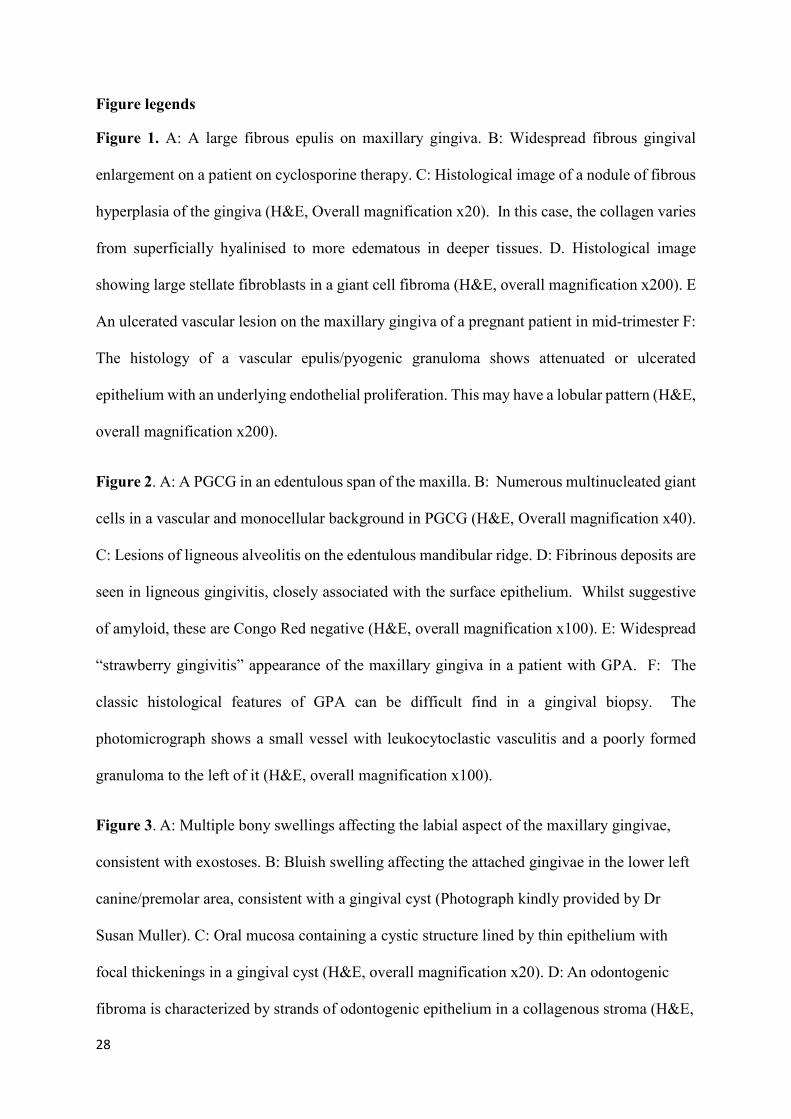
Figure legends
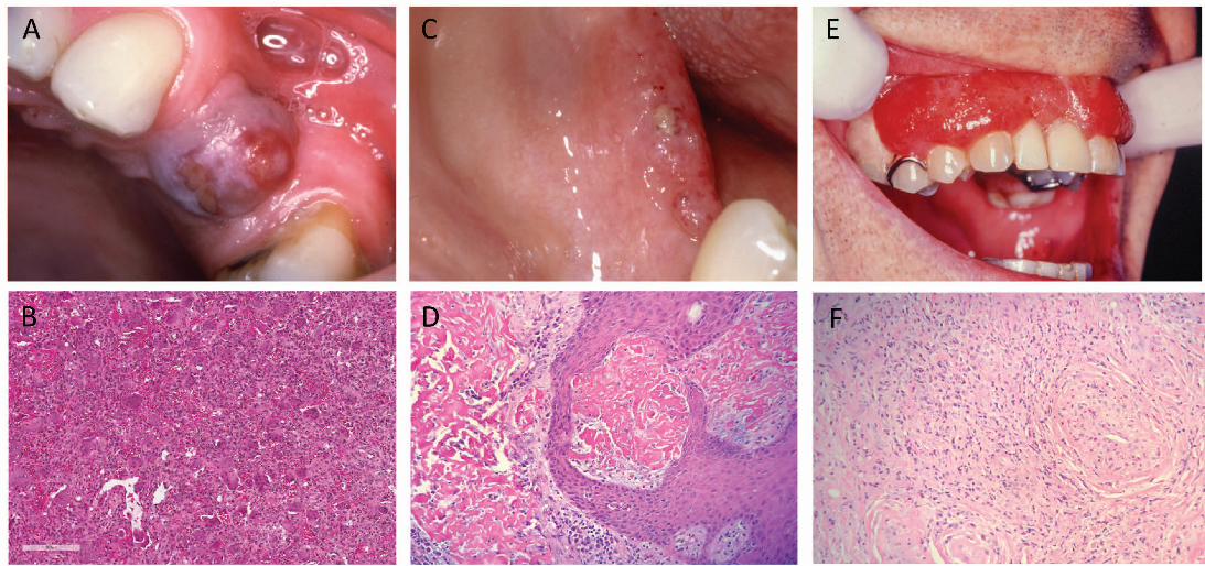
Figure 1. A: A large fibrous epulis on maxillary gingiva. B: Widespread fibrous gingival
enlargement on a patient on cyclosporine therapy. C: Histological image of a nodule of fibrous
hyperplasia of the gingiva (H&E, Overall magnification x20). In this case, the collagen varies
from superficially hyalinised to more edematous in deeper tissues. D. Histological image
showing large stellate fibroblasts in a giant cell fibroma (H&E, overall magnification x200). E
An ulcerated vascular lesion on the maxillary gingiva of a pregnant patient in mid-trimester F:
The histology of a vascular epulis/pyogenic granuloma shows attenuated or ulcerated
epithelium with an underlying endothelial proliferation. This may have a lobular pattern (H&E,
overall magnification x200).
Figure 2. A: A PGCG in an edentulous span of the maxilla. B: Numerous multinucleated giant
cells in a vascular and monocellular background in PGCG (H&E, Overall magnification x40).
C: Lesions of ligneous alveolitis on the edentulous mandibular ridge. D: Fibrinous deposits are
seen in ligneous gingivitis, closely associated with the surface epithelium. Whilst suggestive
of amyloid, these are Congo Red negative (H&E, overall magnification x100). E: Widespread
“strawberry gingivitis” appearance of the maxillary gingiva in a patient with GPA. F: The
classic histological features of GPA can be difficult find in a gingival biopsy. The
photomicrograph shows a small vessel with leukocytoclastic vasculitis and a poorly formed
granuloma to the left of it (H&E, overall magnification x100).
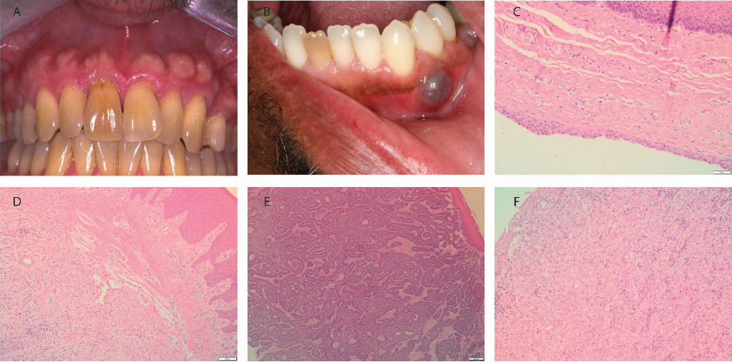
Figure 3. A: Multiple bony swellings affecting the labial aspect of the maxillary gingivae,
consistent with exostoses. B: Bluish swelling affecting the attached gingivae in the lower left
canine/premolar area, consistent with a gingival cyst (Photograph kindly provided by Dr
Susan Muller). C: Oral mucosa containing a cystic structure lined by thin epithelium with
focal thickenings in a gingival cyst (H&E, overall magnification x20). D: An odontogenic
fibroma is characterized by strands of odontogenic epithelium in a collagenous stroma (H&E,
Page 29
29
overall magnification x10). E: Peripheral ameloblastoma showing islands of odontogenic
epithelium with characteristic peripheral palisading (H&E, overall magnification x4). F.
Cords of atypical epithelial cells in fibrous stroma in a metastatic lobular carcinoma of breast
(H&E, overall magnification x20).
Figure 4. A. Red, nodular swelling affecting the facial gingiva above the left maxillary
canine and lateral incisor in Kaposi Sarcoma. B. Streams of spindled cells with slit-like
vessels and lymphangiomatous pattern superficially in Kaposi’s sarcoma (H&E, overall
magnification x 4). C: Non-Hodgkin lymphoma presenting as an ulcerated swelling affecting
the posterior left retromolar region. D. Generalized erythema and swelling affecting the
gingiva in a case of AML. E. Connective tissue effaced by sheets of atypical myeloid cells in
AML (H&E, overall magnification x20). F: Chondrosarcoma classically has a lobular
architecture, with blue-grey cartilaginous matrix (H&E, overall magnification x4).